nonalcoholic fatty liver disease: a pathological...
TRANSCRIPT
Chapter 8
Nonalcoholic Fatty Liver Disease: A Pathological View
Joaquín Cabezas, Marta Mayorga and Javier Crespo
Additional information is available at the end of the chapter
http://dx.doi.org/10.5772/52622
1. Introduction
Nonalcoholic fatty liver disease (NAFLD) is the most common liver disorder of our times.The spectrum of this disease goes from steatosis to non-alcoholic steatohepatitis (NASH),cirrhosis and hepatocellular carcinoma. NAFLD can appear in the context of many condi‐tions. Probably, NAFLD could be a component of metabolic syndrome, with its completephenotypic expression: insulin resistance, obesity, type 2 diabetes, hypertension, hypercho‐lesterolemia, and hypertriglyceridemia.
The pathogenesis involves insulin resistance, hepatic fat deposition, increased oxidantstress, apoptosis, inflammation and fibrosis. At present day, a new hormone has been dis‐covered. Muscle cells products this new hormone, called irisin. Irisin can induce changes inadipose tissue.
Diagnosis of NAFLD cannot be performed with a single test and it should be one of exclu‐sion, as well.
Nowadays, there is not a single therapeutic intervention. The focus of management shouldbe treatment of the risk factors for NASH (insulin resistance, obesity…). Principal methodsused for weight management are dietary modifications and life style changes. Then, phar‐macotherapy may include insulin sensitizers, cholesterol-lowering agents, anti-obesity andanti-oxidant agents. Morbid obese patients may benefit from surgical weight loss, reducingthe progression of NASH.
2. Definition
NAFLD definition [1] requires that there is evidence of hepatic steatosis, either by imagingor by histology and there are no causes for secondary hepatic fat accumulation (Table 1).
© 2012 Cabezas et al.; licensee InTech. This is an open access article distributed under the terms of theCreative Commons Attribution License (http://creativecommons.org/licenses/by/3.0), which permitsunrestricted use, distribution, and reproduction in any medium, provided the original work is properly cited.
NAFLD is usually associated with metabolic risk factors such as metabolic syndrome, obesi‐ty, diabetes mellitus, and dyslipidaemia.
COMMON CAUSES OF SECONDARY HEPATIC STEATOSIS
Macrovesicularsteatosis
Excessive alcohol consumption.
Hepatitis C (genotype 3)
Wilson’s disease.
Lipodistrophy
Starvation
Parenteral nutrition.
Abetalipoproteinemia.
Medication (amiodarone, methotrexate, tamoxifen, corticosteroids)
Microvesicularstatosis
Reye’s syndrome.
Medications (valproate, anti-retroviral medicines)
Acutte fatty liver of pregnancy
HELLP syndrome
Inborn errors of metabolism (LCAT deficiency, cholesterol ester storage disease, Wolman disease)
Table 1. Causes of secondary fat accumulation.
NAFLD includes a constellation of histological findings that goes from steatosis, to necroin‐flammation, called NASH and progression to advanced fibrosis and cirrhosis.
3. Epidemiology
NAFLD is becoming the leading cause of liver disease. One of the causes is the increasing ofobesity[2].
The incidence of NAFLD has been evaluated in a few number of studies, it ranges from31-86 cases/1000 person-year in Japan to 29 cases per 100000 person-year in England [3, 4].
The prevalence of NAFLD is increasing. Recent studies presented in the Digestive DiseasesWeek 2012 summarizes this increased prevalence over the last 20 years [5, 6]. Investigatorsreport an increasing in obesity. This increase is followed by a rising in steatosis and NASH,the presence of steatosis among obese people has increased from 23% in the 80s, 43% in the
Liver Biopsy – Indications, Procedures, Results162
90s and finally to 60% nowadays [4]. Even in non-obese patients, the prevalence of steatosisincreased from 12%, to 27% and 36%, respectively [5].
In children/adolescents, over the last 20 years, obesity has increased from 11% to 21%, sus‐pected NAFLD from 4% to 10, and the prevalence of altered aminotransferases among obeseadolescents has increased from 17% to 37% [6].
4. NAFLD and liver biopsy
Liver biopsy remains the gold standard for characterizing liver histology in patients withNAFLD. However, it is expensive and carries some morbidity and very rare mortality risk.Thus, it should be performed in those who would benefit the most from diagnostic, thera‐peutic guidance, and prognostic perspectives.
The last guideline for NAFLD management recommends liver biopsy [1]: in patients whoare at risk to have steatohepatitis and advance fibrosis; theses patients could be identified bythe presence of metabolic syndrome and NAFLD fibrosis score; and a liver biopsy should beconsidered in patients in whom other etiologies are suspected and cannot be excluded with‐out a liver biopsy.
Liver biopsy allows confirming the diagnosis, evaluation and semiquantitation of necroin‐flammatory lesions and fibrosis.
On the other hand, liver biopsy suffers from challenges. An adequate biopsy represents only1/50000-1/65000 of the organ. Sampled area should be carefully chosen and sample lengthmust be enough, a least 15mm. This size can reduce sample error. Finally, experienced path‐ologist is important to haver a greater yield of findings.
5. Histology of NAFLD
NAFLD represents a histopathologic spectrum ranging from steatosis alone, to necroinflam‐mation, summarized as NASH; and progression to advanced fibrosis and cirrhosis.
The histologic characterization of NAFLD and NASH may include description of steatosisand cell injury in addition to inflammation and fibrosis. Kleiner and Brunt [7] propose cate‐gorizing the histologic changes when studying NAFLD as follows in table 2.
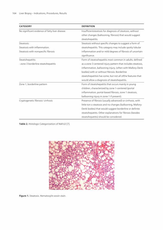
The main histological characteristic of NAFLD is the accumulation of fat in the form of tri‐glyicerides within hepatocytes, lesion termed steatosis (Figure 1 and 2); this term is definedby the guideline [1] as NAFL – non-alcoholic fatty liver, where the risk of progression to cir‐rhosis and liver failure is minimal. The presence of >5% steatoic hepatocytes in a liver biopsyis accepted as the minimum criterion for thehistological diagnosis of NAFLD [8].
Nonalcoholic Fatty Liver Disease: A Pathological Viewhttp://dx.doi.org/10.5772/52622
163
CATEGORY DEFINITION
No significant evidence of fatty liver disease. Insufficientsteatosis for diagnosis of steatosis, without
other changes (ballooning, fibrosis) that would suggest
steatohepatitis.
Steatosis:
Steatosis with inflammation.
Steatosis with nonspecific fibrosis
Steatosis without specific changes to suggest a form of
steatohepatitis. This category may include spotty lobular
inflammation and/or mild degrees of fibrosis of uncertain
significance.
Steatohepatitis:
- zone 3 borderline steatohepatitis
Form of steatoshepatitis most common in adults; defined
as a zone 3 centered injury pattern that includes steatosis,
inflammation, ballooning injury, (often with Mallory-Denk
bodies) with or without fibrosis. Borderline
steatohepatitsis has some, but not all ofthe features that
would allow a diagnosis of steatohepatitis.
Zone 1, borderline pattern Form of steatohepatitis that occurs mainly in young
children, characterized by zone 1-centered (portal
inflammation, portal-based fibrosis, zone 1 steatosis,
ballooning injury in zone 1 if present).
Cryptogenetic fibrosis/ cirrhosis Presence of fibrosis (usually advanced) or cirrhosis, with
little ton o steatosis and no changes (ballooning, Mallory-
Denk bodies) that would suggest borderline or definite
steatohepatitis. Other explanations for fibrosis (besides
steatohepatitis) should be considered.
Table 2. Histologic Categorization of NAFLD [7].
Figure 1. Steatosis. Hematoxylin-eosin stain.
Liver Biopsy – Indications, Procedures, Results164
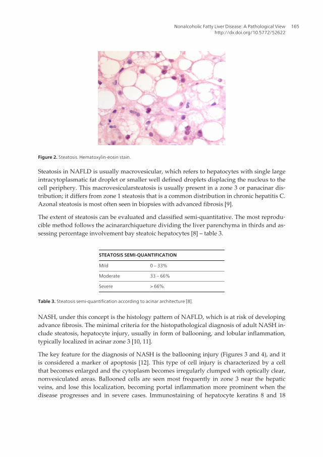
Figure 2. Steatosis. Hematoxylin-eosin stain.
Steatosis in NAFLD is usually macrovesicular, which refers to hepatocytes with single largeintracytoplasmatic fat droplet or smaller well defined droplets displacing the nucleus to thecell periphery. This macrovesicularsteatosis is usually present in a zone 3 or panacinar dis‐tribution; it differs from zone 1 steatosis that is a common distribution in chronic hepatitis C.Azonal steatosis is most often seen in biopsies with advanced fibrosis [9].
The extent of steatosis can be evaluated and classified semi-quantitative. The most reprodu‐cible method follows the acinararchiqueture dividing the liver parenchyma in thirds and as‐sessing percentage involvement bay steatoic hepatocytes [8] – table 3.
STEATOSIS SEMI-QUANTIFICATION
Mild 0 – 33%
Moderate 33 – 66%
Severe > 66%.
Table 3. Steatosis semi-quantification according to acinar architecture [8].
NASH, under this concept is the histology pattern of NAFLD, which is at risk of developingadvance fibrosis. The minimal criteria for the histopathological diagnosis of adult NASH in‐clude steatosis, hepatocyte injury, usually in form of ballooning, and lobular inflammation,typically localized in acinar zone 3 [10, 11].
The key feature for the diagnosis of NASH is the ballooning injury (Figures 3 and 4), and itis considered a marker of apoptosis [12]. This type of cell injury is characterized by a cellthat becomes enlarged and the cytoplasm becomes irregularly clumped with optically clear,nonvesiculated areas. Ballooned cells are seen most frequently in zone 3 near the hepaticveins, and lose this localization, becoming portal inflammation more prominent when thedisease progresses and in severe cases. Immunostaining of hepatocyte keratins 8 and 18
Nonalcoholic Fatty Liver Disease: A Pathological Viewhttp://dx.doi.org/10.5772/52622
165
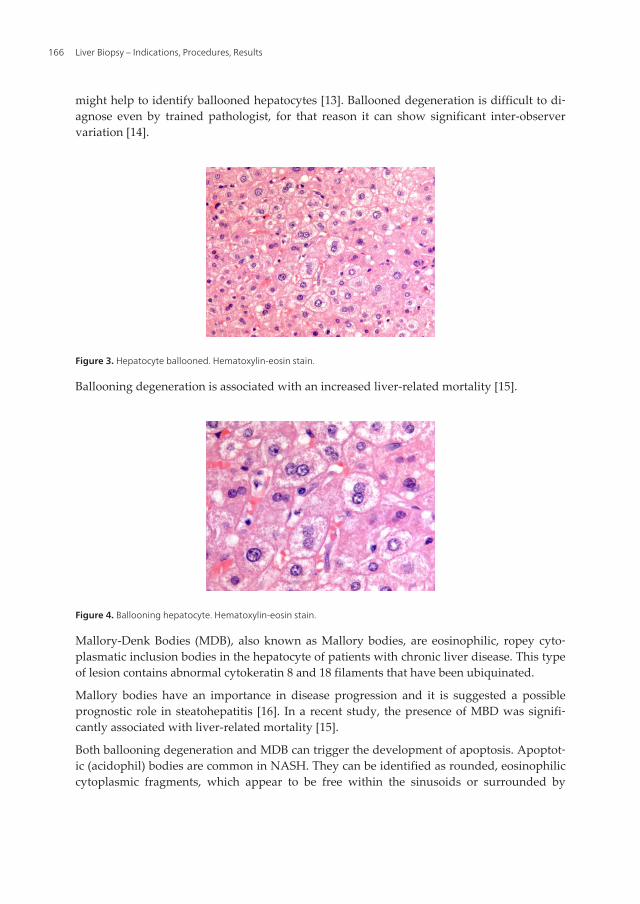
might help to identify ballooned hepatocytes [13]. Ballooned degeneration is difficult to di‐agnose even by trained pathologist, for that reason it can show significant inter-observervariation [14].
Figure 3. Hepatocyte ballooned. Hematoxylin-eosin stain.
Ballooning degeneration is associated with an increased liver-related mortality [15].
Figure 4. Ballooning hepatocyte. Hematoxylin-eosin stain.
Mallory-Denk Bodies (MDB), also known as Mallory bodies, are eosinophilic, ropey cyto‐plasmatic inclusion bodies in the hepatocyte of patients with chronic liver disease. This typeof lesion contains abnormal cytokeratin 8 and 18 filaments that have been ubiquinated.
Mallory bodies have an importance in disease progression and it is suggested a possibleprognostic role in steatohepatitis [16]. In a recent study, the presence of MBD was signifi‐cantly associated with liver-related mortality [15].
Both ballooning degeneration and MDB can trigger the development of apoptosis. Apoptot‐ic (acidophil) bodies are common in NASH. They can be identified as rounded, eosinophiliccytoplasmic fragments, which appear to be free within the sinusoids or surrounded by
Liver Biopsy – Indications, Procedures, Results166
Kupffer or other inflammatory cells. Apoptosis has been validated as an accurate marker fordiagnosis of NASH based on immunochemistry in liver tissue [17].
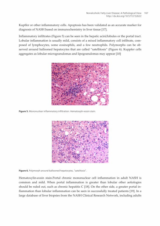
Inflammatory infiltrates (Figure 5) can be seen in the hepatic acini/lobules or the portal tract.Lobular inflammation is usually mild, consists of a mixed inflammatory cell infiltrate, com‐posed of lymphocytes, some eosinophils, and a few neutrophils. Polymorphs can be ob‐served around ballooned hepatocytes that are called “satellitosis” (Figure 6). Kuppfer cellsaggregates as lobular microgranulomas and lipogranulomas may appear [10]
Figure 5. Mononuclear inflammatory infiltration. Hematoxylin-eosin stain.
Figure 6. Polymorph around ballooned hepatocytes, “satelitosis”.
Hematoxylin-eosin stain.Portal chronic mononuclear cell inflammation in adult NASH iscommon and mild. When portal inflammation is greater than lobular other aetiologiesshould be ruled out, such as chronic hepatitis C [18]. On the other side, a greater portal in‐flammation than lobular inflammation can be seen in successfully treated patients [19]. In alarge database of liver biopsies from the NASH Clinical Research Network, including adults
Nonalcoholic Fatty Liver Disease: A Pathological Viewhttp://dx.doi.org/10.5772/52622
167
and children, portal chronic inflammation was associated with clinical and histologic fea‐tures of severity and advance disease [20].
Vascular alterations in NAFLD. Recent paper has focused the study of NASH in microves‐sels of the liver [21]. This work has found an intraacinar branch of the hepatic artery in theperivenular region in active steatohepatitis. This finding is important because it can lead toconfusion for a portal tract resulting in an equivocal diagnosis. Likewise, the presence of thisvessel correlates with higher stage of fibrosis.
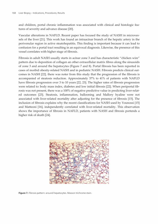
Fibrosis in adult NASH usually starts in acinar zone 3 and has characteristic “chicken wire”pattern due to deposition of collagen an other extracellular matrix fibres along the sinusoidsof zone 3 and around the hepatocytes (Figure 7 and 8). Portal fibrosis has been reported incases of morbid obesity-related NASH and in pediatric NASH. Fibrosis predicts clinical out‐comes in NASH [22]. there was noter from this study that the progression of the fibrosis isaccompanied of steatosis reduction. Approximately 37% to 41% of patients with NAFLDhave fibrosis progression over 3 to 10 years [22, 23]. The higher rates of fibrosis progressionwere related to: body mass index, diabetes and low initial fibrosis [22]. When periportal fib‐rosis was not present, there was a 100% of negative predictive value in predicting liver-relat‐ed outcomes [23]. Steatosis, inflammation, ballooning and Mallory hyaline were notassociated with liver-related mortality after adjusting for the presence of fibrosis [15]. Theinclusion of fibrosis explains why the recent classifications for NASH used by Younossi [15]and Matteoni [16], independently correlated with liver-related mortality. This observationshows the importance of fibrosis in NAFLD, patients with NASH and fibrosis portends ahigher risk of death [24].
Figure 7. Fibrosis pattern: around hepatocytes. Masson trichrome stain.
Liver Biopsy – Indications, Procedures, Results168
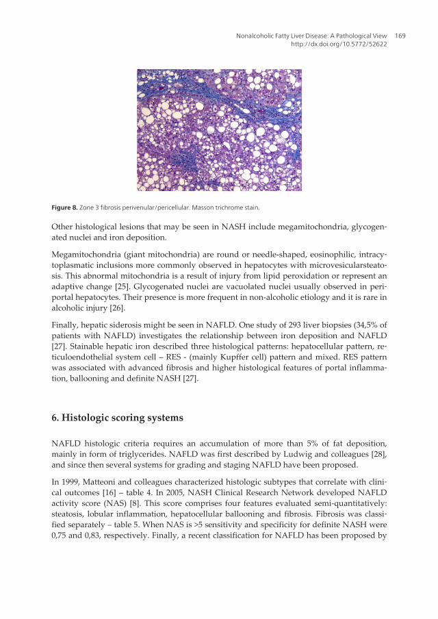
Figure 8. Zone 3 fibrosis perivenular/pericellular. Masson trichrome stain.
Other histological lesions that may be seen in NASH include megamitochondria, glycogen‐ated nuclei and iron deposition.
Megamitochondria (giant mitochondria) are round or needle-shaped, eosinophilic, intracy‐toplasmatic inclusions more commonly observed in hepatocytes with microvesicularsteato‐sis. This abnormal mitochondria is a result of injury from lipid peroxidation or represent anadaptive change [25]. Glycogenated nuclei are vacuolated nuclei usually observed in peri‐portal hepatocytes. Their presence is more frequent in non-alcoholic etiology and it is rare inalcoholic injury [26].
Finally, hepatic siderosis might be seen in NAFLD. One study of 293 liver biopsies (34,5% ofpatients with NAFLD) investigates the relationship between iron deposition and NAFLD[27]. Stainable hepatic iron described three histological patterns: hepatocellular pattern, re‐ticuloendothelial system cell – RES - (mainly Kupffer cell) pattern and mixed. RES patternwas associated with advanced fibrosis and higher histological features of portal inflamma‐tion, ballooning and definite NASH [27].
6. Histologic scoring systems
NAFLD histologic criteria requires an accumulation of more than 5% of fat deposition,mainly in form of triglycerides. NAFLD was first described by Ludwig and colleagues [28],and since then several systems for grading and staging NAFLD have been proposed.
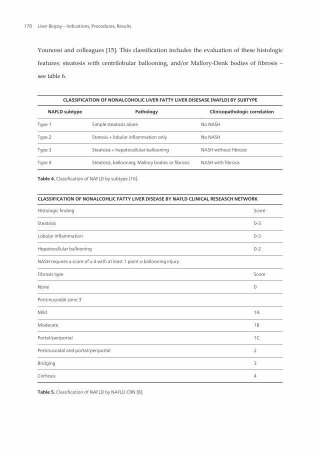
In 1999, Matteoni and colleagues characterized histologic subtypes that correlate with clini‐cal outcomes [16] – table 4. In 2005, NASH Clinical Research Network developed NAFLDactivity score (NAS) [8]. This score comprises four features evaluated semi-quantitatively:steatosis, lobular inflammation, hepatocellular ballooning and fibrosis. Fibrosis was classi‐fied separately – table 5. When NAS is >5 sensitivity and specificity for definite NASH were0,75 and 0,83, respectively. Finally, a recent classification for NAFLD has been proposed by
Nonalcoholic Fatty Liver Disease: A Pathological Viewhttp://dx.doi.org/10.5772/52622
169
Younossi and colleagues [15]. This classification includes the evaluation of these histologic
features: steatosis with centrilobular ballooning, and/or Mallory-Denk bodies of fibrosis –
see table 6.
CLASSIFICATION OF NONALCOHOLIC LIVER FATTY LIVER DISESASE (NAFLD) BY SUBTYPE
NAFLD subtype Pathology Clinicopathologic correlation
Type 1 Simple steatosis alone No NASH
Type 2 Statosis + lobular inflammation only No NASH
Type 3 Steatosis + hepatocellular ballooning NASH without fibrosis
Type 4 Steatosis, ballooning, Mallory bodies or fibrosis NASH with fibrosis
Table 4. Classification of NAFLD by subtype [16].
CLASSIFICATION OF NONALCOHLIC FATTY LIVER DISEASE BY NAFLD CLINICAL RESEASCH NETWORK
Histologic finding Score
Steatosis 0-3
Lobular inflammation 0-3
Hepatocellular ballooning 0-2
NASH requires a score of ≥ 4 with at least 1 point o ballooning injury.
Fibrosis type Score
None 0
Perisinusoidal zone 3
Mild 1A
Moderate 1B
Portal/periportal 1C
Persinusoidal and portal/periportal 2
Bridging 3
Cirrhosis 4
Table 5. Classification of NAFLD by NAFLD CRN [8].
Liver Biopsy – Indications, Procedures, Results170
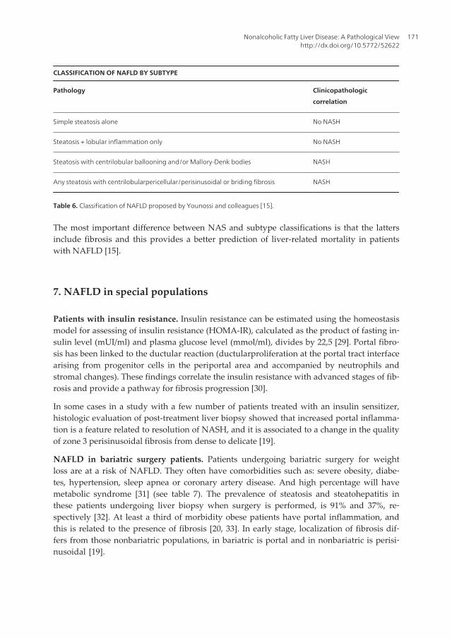
CLASSIFICATION OF NAFLD BY SUBTYPE
Pathology Clinicopathologic
correlation
Simple steatosis alone No NASH
Steatosis + lobular inflammation only No NASH
Steatosis with centrilobular ballooning and/or Mallory-Denk bodies NASH
Any steatosis with centrilobularpericellular/perisinusoidal or briding fibrosis NASH
Table 6. Classification of NAFLD proposed by Younossi and colleagues [15].
The most important difference between NAS and subtype classifications is that the lattersinclude fibrosis and this provides a better prediction of liver-related mortality in patientswith NAFLD [15].
7. NAFLD in special populations
Patients with insulin resistance. Insulin resistance can be estimated using the homeostasismodel for assessing of insulin resistance (HOMA-IR), calculated as the product of fasting in‐sulin level (mUI/ml) and plasma glucose level (mmol/ml), divides by 22,5 [29]. Portal fibro‐sis has been linked to the ductular reaction (ductularproliferation at the portal tract interfacearising from progenitor cells in the periportal area and accompanied by neutrophils andstromal changes). These findings correlate the insulin resistance with advanced stages of fib‐rosis and provide a pathway for fibrosis progression [30].
In some cases in a study with a few number of patients treated with an insulin sensitizer,histologic evaluation of post-treatment liver biopsy showed that increased portal inflamma‐tion is a feature related to resolution of NASH, and it is associated to a change in the qualityof zone 3 perisinusoidal fibrosis from dense to delicate [19].
NAFLD in bariatric surgery patients. Patients undergoing bariatric surgery for weightloss are at a risk of NAFLD. They often have comorbidities such as: severe obesity, diabe‐tes, hypertension, sleep apnea or coronary artery disease. And high percentage will havemetabolic syndrome [31] (see table 7). The prevalence of steatosis and steatohepatitis inthese patients undergoing liver biopsy when surgery is performed, is 91% and 37%, re‐spectively [32]. At least a third of morbidity obese patients have portal inflammation, andthis is related to the presence of fibrosis [20, 33]. In early stage, localization of fibrosis dif‐fers from those nonbariatric populations, in bariatric is portal and in nonbariatric is perisi‐nusoidal [19].
Nonalcoholic Fatty Liver Disease: A Pathological Viewhttp://dx.doi.org/10.5772/52622
171
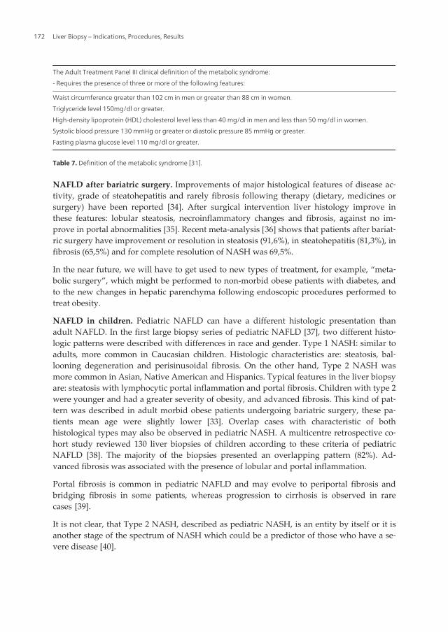
The Adult Treatment Panel III clinical definition of the metabolic syndrome:
- Requires the presence of three or more of the following features:
Waist circumference greater than 102 cm in men or greater than 88 cm in women.
Triglyceride level 150mg/dl or greater.
High-density lipoprotein (HDL) cholesterol level less than 40 mg/dl in men and less than 50 mg/dl in women.
Systolic blood pressure 130 mmHg or greater or diastolic pressure 85 mmHg or greater.
Fasting plasma glucose level 110 mg/dl or greater.
Table 7. Definition of the metabolic syndrome [31].
NAFLD after bariatric surgery. Improvements of major histological features of disease ac‐tivity, grade of steatohepatitis and rarely fibrosis following therapy (dietary, medicines orsurgery) have been reported [34]. After surgical intervention liver histology improve inthese features: lobular steatosis, necroinflammatory changes and fibrosis, against no im‐prove in portal abnormalities [35]. Recent meta-analysis [36] shows that patients after bariat‐ric surgery have improvement or resolution in steatosis (91,6%), in steatohepatitis (81,3%), infibrosis (65,5%) and for complete resolution of NASH was 69,5%.
In the near future, we will have to get used to new types of treatment, for example, “meta‐bolic surgery”, which might be performed to non-morbid obese patients with diabetes, andto the new changes in hepatic parenchyma following endoscopic procedures performed totreat obesity.
NAFLD in children. Pediatric NAFLD can have a different histologic presentation thanadult NAFLD. In the first large biopsy series of pediatric NAFLD [37], two different histo‐logic patterns were described with differences in race and gender. Type 1 NASH: similar toadults, more common in Caucasian children. Histologic characteristics are: steatosis, bal‐looning degeneration and perisinusoidal fibrosis. On the other hand, Type 2 NASH wasmore common in Asian, Native American and Hispanics. Typical features in the liver biopsyare: steatosis with lymphocytic portal inflammation and portal fibrosis. Children with type 2were younger and had a greater severity of obesity, and advanced fibrosis. This kind of pat‐tern was described in adult morbid obese patients undergoing bariatric surgery, these pa‐tients mean age were slightly lower [33]. Overlap cases with characteristic of bothhistological types may also be observed in pediatric NASH. A multicentre retrospective co‐hort study reviewed 130 liver biopsies of children according to these criteria of pediatricNAFLD [38]. The majority of the biopsies presented an overlapping pattern (82%). Ad‐vanced fibrosis was associated with the presence of lobular and portal inflammation.
Portal fibrosis is common in pediatric NAFLD and may evolve to periportal fibrosis andbridging fibrosis in some patients, whereas progression to cirrhosis is observed in rarecases [39].
It is not clear, that Type 2 NASH, described as pediatric NASH, is an entity by itself or it isanother stage of the spectrum of NASH which could be a predictor of those who have a se‐vere disease [40].
Liver Biopsy – Indications, Procedures, Results172
8. Imaging tecnology in NAFLD
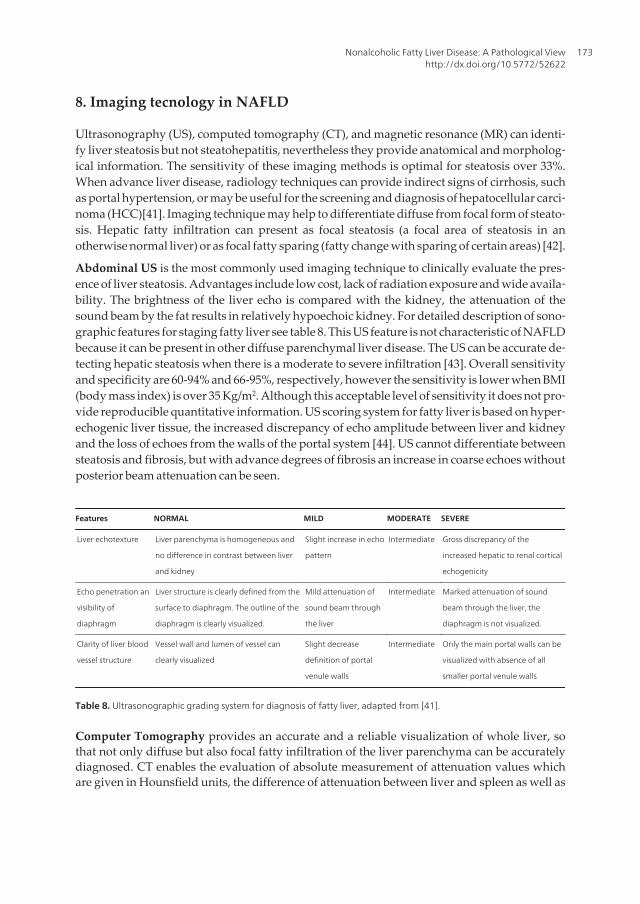
Ultrasonography (US), computed tomography (CT), and magnetic resonance (MR) can identi‐fy liver steatosis but not steatohepatitis, nevertheless they provide anatomical and morpholog‐ical information. The sensitivity of these imaging methods is optimal for steatosis over 33%.When advance liver disease, radiology techniques can provide indirect signs of cirrhosis, suchas portal hypertension, or may be useful for the screening and diagnosis of hepatocellular carci‐noma (HCC)[41]. Imaging technique may help to differentiate diffuse from focal form of steato‐sis. Hepatic fatty infiltration can present as focal steatosis (a focal area of steatosis in anotherwise normal liver) or as focal fatty sparing (fatty change with sparing of certain areas) [42].
Abdominal US is the most commonly used imaging technique to clinically evaluate the pres‐ence of liver steatosis. Advantages include low cost, lack of radiation exposure and wide availa‐bility. The brightness of the liver echo is compared with the kidney, the attenuation of thesound beam by the fat results in relatively hypoechoic kidney. For detailed description of sono‐graphic features for staging fatty liver see table 8. This US feature is not characteristic of NAFLDbecause it can be present in other diffuse parenchymal liver disease. The US can be accurate de‐tecting hepatic steatosis when there is a moderate to severe infiltration [43]. Overall sensitivityand specificity are 60-94% and 66-95%, respectively, however the sensitivity is lower when BMI(body mass index) is over 35 Kg/m2. Although this acceptable level of sensitivity it does not pro‐vide reproducible quantitative information. US scoring system for fatty liver is based on hyper‐echogenic liver tissue, the increased discrepancy of echo amplitude between liver and kidneyand the loss of echoes from the walls of the portal system [44]. US cannot differentiate betweensteatosis and fibrosis, but with advance degrees of fibrosis an increase in coarse echoes withoutposterior beam attenuation can be seen.
Features NORMAL MILD MODERATE SEVERE
Liver echotexture Liver parenchyma is homogeneous and
no difference in contrast between liver
and kidney
Slight increase in echo
pattern
Intermediate Gross discrepancy of the
increased hepatic to renal cortical
echogenicity
Echo penetration an
visibility of
diaphragm
Liver structure is clearly defined from the
surface to diaphragm. The outline of the
diaphragm is clearly visualized.
Mild attenuation of
sound beam through
the liver
Intermediate Marked attenuation of sound
beam through the liver, the
diaphragm is not visualized.
Clarity of liver blood
vessel structure
Vessel wall and lumen of vessel can
clearly visualized
Slight decrease
definition of portal
venule walls
Intermediate Only the main portal walls can be
visualized with absence of all
smaller portal venule walls
Table 8. Ultrasonographic grading system for diagnosis of fatty liver, adapted from [41].
Computer Tomography provides an accurate and a reliable visualization of whole liver, sothat not only diffuse but also focal fatty infiltration of the liver parenchyma can be accuratelydiagnosed. CT enables the evaluation of absolute measurement of attenuation values whichare given in Hounsfield units, the difference of attenuation between liver and spleen as well as
Nonalcoholic Fatty Liver Disease: A Pathological Viewhttp://dx.doi.org/10.5772/52622
173
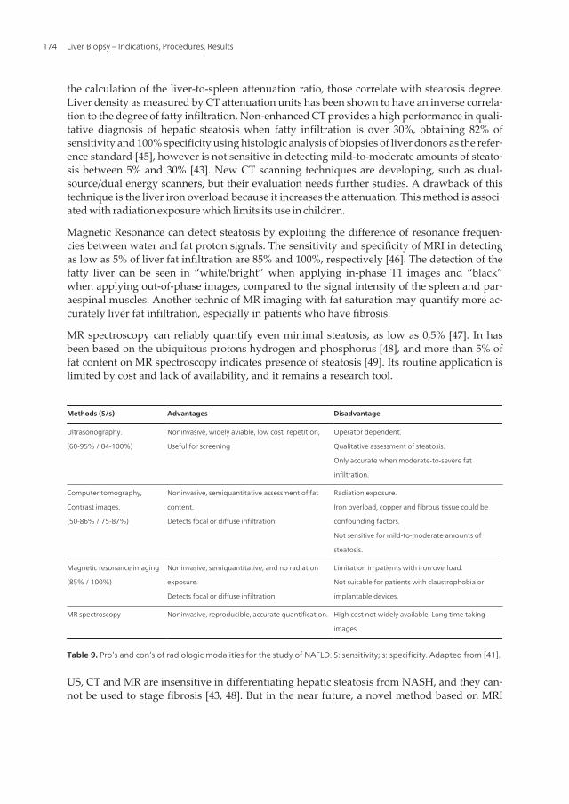
the calculation of the liver-to-spleen attenuation ratio, those correlate with steatosis degree.Liver density as measured by CT attenuation units has been shown to have an inverse correla‐tion to the degree of fatty infiltration. Non-enhanced CT provides a high performance in quali‐tative diagnosis of hepatic steatosis when fatty infiltration is over 30%, obtaining 82% ofsensitivity and 100% specificity using histologic analysis of biopsies of liver donors as the refer‐ence standard [45], however is not sensitive in detecting mild-to-moderate amounts of steato‐sis between 5% and 30% [43]. New CT scanning techniques are developing, such as dual-source/dual energy scanners, but their evaluation needs further studies. A drawback of thistechnique is the liver iron overload because it increases the attenuation. This method is associ‐ated with radiation exposure which limits its use in children.
Magnetic Resonance can detect steatosis by exploiting the difference of resonance frequen‐cies between water and fat proton signals. The sensitivity and specificity of MRI in detectingas low as 5% of liver fat infiltration are 85% and 100%, respectively [46]. The detection of thefatty liver can be seen in “white/bright” when applying in-phase T1 images and “black”when applying out-of-phase images, compared to the signal intensity of the spleen and par‐aespinal muscles. Another technic of MR imaging with fat saturation may quantify more ac‐curately liver fat infiltration, especially in patients who have fibrosis.
MR spectroscopy can reliably quantify even minimal steatosis, as low as 0,5% [47]. In hasbeen based on the ubiquitous protons hydrogen and phosphorus [48], and more than 5% offat content on MR spectroscopy indicates presence of steatosis [49]. Its routine application islimited by cost and lack of availability, and it remains a research tool.
Methods (S/s) Advantages Disadvantage
Ultrasonography.
(60-95% / 84-100%)
Noninvasive, widely aviable, low cost, repetition,
Useful for screening
Operator dependent.
Qualitative assessment of steatosis.
Only accurate when moderate-to-severe fat
infiltration.
Computer tomography,
Contrast images.
(50-86% / 75-87%)
Noninvasive, semiquantitative assessment of fat
content.
Detects focal or diffuse infiltration.
Radiation exposure.
Iron overload, copper and fibrous tissue could be
confounding factors.
Not sensitive for mild-to-moderate amounts of
steatosis.
Magnetic resonance imaging
(85% / 100%)
Noninvasive, semiquantitative, and no radiation
exposure.
Detects focal or diffuse infiltration.
Limitation in patients with iron overload.
Not suitable for patients with claustrophobia or
implantable devices.
MR spectroscopy Noninvasive, reproducible, accurate quantification. High cost not widely available. Long time taking
images.
Table 9. Pro’s and con’s of radiologic modalities for the study of NAFLD. S: sensitivity; s: specificity. Adapted from [41].
US, CT and MR are insensitive in differentiating hepatic steatosis from NASH, and they can‐not be used to stage fibrosis [43, 48]. But in the near future, a novel method based on MRI
Liver Biopsy – Indications, Procedures, Results174
imaging and a new software will be able to stage fibrosis and to distinguish NASH from no-NASH. Professor Romero-Gomez conducts this study and it will be soon published.
Table 9 summarizes advantages and disadvantages of these radiologic methods.
9. Non-invasive assesment in NAFLD
Liver biopsy remains a useful tool to confirm the diagnosis and exclude other disease orhelps to discover concomitant chronic liver disease. It provides prognostic information bystaging and grading this disease. At present non-invasive diagnostic markers could providea new tool for differentiating fatty liver from NASH as well as for grading /staging NAFLD.
The investigation of these new diagnostic methods comes from the well known drawbacksof liver biopsy. These include sampling error, inadequate biopsy size, variability in patholo‐gist interpretation, cost and associated morbidity (complications 0,3%, mortality rate 0,01%).
An ideal non-invasive test should be simple, reproducible, readily available, less expensivethan liver biopsy, able to predict the full spectrum of liver fibrosis stages, and reflectchanges occurring with therapy [48].
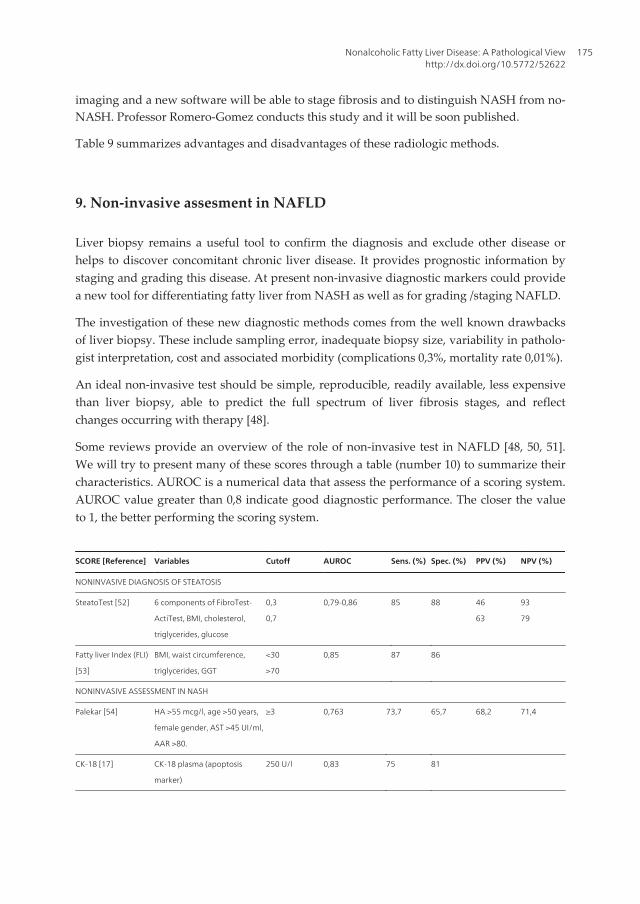
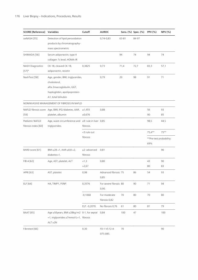
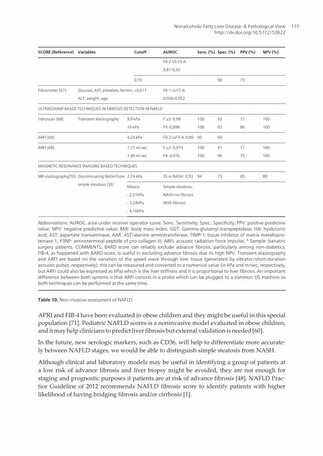
Some reviews provide an overview of the role of non-invasive test in NAFLD [48, 50, 51].We will try to present many of these scores through a table (number 10) to summarize theircharacteristics. AUROC is a numerical data that assess the performance of a scoring system.AUROC value greater than 0,8 indicate good diagnostic performance. The closer the valueto 1, the better performing the scoring system.
SCORE [Reference] Variables Cutoff AUROC Sens. (%) Spec. (%) PPV (%) NPV (%)
NONINVASIVE DIAGNOSIS OF STEATOSIS
SteatoTest [52] 6 components of FibroTest-
ActiTest, BMI, cholesterol,
triglycerides, glucose
0,3
0,7
0,79-0,86 85 88 46
63
93
79
Fatty liver Index (FLI)
[53]
BMI, waist circumference,
triglycerides, GGT
<30
>70
0,85 87 86
NONINVASIVE ASSESSMENT IN NASH
Palekar [54] HA >55 mcg/l, age >50 years,
female gender, AST >45 UI/ml,
AAR >80.
≥3 0,763 73,7 65,7 68,2 71,4
CK-18 [17] CK-18 plasma (apoptosis
marker)
250 U/l 0,83 75 81
Nonalcoholic Fatty Liver Disease: A Pathological Viewhttp://dx.doi.org/10.5772/52622
175
SCORE [Reference] Variables Cutoff AUROC Sens. (%) Spec. (%) PPV (%) NPV (%)
oxNASH [55] Detection of lipid peroxidation
products by chromatography-
mass spectrometric
0,74-0,83 63-81 84-97
SHIMADA [56] Serum adiponectin, type 4
collagen 7s level, HOMA-IR
94 74 94 74
NASH Diagnostics
[57]*
CK-18, cleaved CK-18,
adiponectin, resistin
0,3825 0,73 71,4 72,7 83,3 57,1
NashTest [58] Age, gender, BMI, triglycerides,
cholesterol,
alfa-2macroglobulin, GGT,
haptoglobin, apolipoprotein-
A1, total bilirubin
0,79 29 98 91 71
NONINVASIVE MANAGEMENT OF FIBROSIS IN NAFLD
NAFLD fibrosis score
[59]
Age, BMI, IFG/diabetes, AAR,
platelet, albumin
≤1,455
≥0,676
0,88 56
90
93
85
Pediatric NAFLD
fibrosis index [60]
Age, waist circumference and
triglycerides.
≥9: rule in liver
fibrosis.
0,85 98,5 44,5
<3:rule out
fibrosis
75,4** 75**
**Pre-test probability:
69%
BARD score [61] BMI ≥28 =1, AAR ≥0,8 =2,
diabetes=1,
≥2: advanced
fibrosis
0,81 96
FIB-4 [62] Age, AST, platelet, ALT <1,3
>2,67
0,80 43
80
90
83
APRI [63] AST, platelet 0,98 Advanced fibrosis:
0,85
75 86 54 93
ELF [64] HA, TIMP1, P3NP. 0,3576 For severe fibrosis
0,90.
80 90 71 94
-0,1068 For moderate
fibrosis 0,82
70 80 70 80
ELF: -0,2070. No fibrosis 0,76 61 80 81 79
BAAT [65] Age ≥50years, BMI ≥28kg/m2
=1, triglycerides ≥7mmol/L=1,
ALT ≥2N
0-1, for septal
fibrosis
0,84 100 47 100
Fibrotest [66] 0,30 F0-1 VS F2-4:
075-085.
70 90
Liver Biopsy – Indications, Procedures, Results176
SCORE [Reference] Variables Cutoff AUROC Sens. (%) Spec. (%) PPV (%) NPV (%)
F0-2 VS F3-4:
0,81-0,92
0,70 98 73
Fibrometer [67] Glucose, AST, platelets, ferritin,
ALT, weight, age.
<0,611 F0-1 vs F2-4:
0,936-0,952
ULTRASOUND BASED TECHNIQUES IN FIBROSIS DETECTION IN NAFLD
Fibroscan [68] Transient elastography 9,9 kPa
16 kPa
F ≥3: 0,99
F4: 0,998
100
100
93
93
77
86
100
100
ARFI [69] 4,24 kPa F0-2 vsF3-4: 0,90 90 90
ARFI [68] 1,77 m/sec
1,90 m/sec
F ≥3: 0,973
F4: 0,976
100
100
91
96
71
75
100
100
MAGNETIC RESONANCE IMAGING BASED TECHNIQUES
MR elastography[70] Discriminating NASH from
simple steatosis (SS)
2,74 kPa SS vs NASH: 0,93 94 73 85 89
Means:
- 2,51kPa.
- 3,24kPa.
- 4,16kPa.
Simple steatosis.
NASH no fibrosis.
With fibrosis
Abbreviations: AUROC: area under receiver operator curve. Sens.: Sensitivity; Spec.: Specificity; PPV: positive predictivevalue; NPV: negative predictive value; BMI: body mass index; GGT: Gamma-glutamyl-transpeptidase; HA: hyaluronicacid; AST: aspartate transaminase; AAR: AST/alanine aminotransferase, TIMP-1: tissue inhibitor of matrix metallopro‐teinase 1; P3NP: aminoterminal peptide of pro-collagen III; ARFI: acoustic radiation force impulse; * Sample: bariatricsurgery patients. COMMENTS: BARD score can reliably exclude advance fibrosis, particularly among non-diabetics.FIB-4, as happened with BARD score, is useful in excluding advance fibrosis due its high NPV. Transient elastographyand ARFI are based on the variation of the speed wave through liver tissue (generated by vibrator/short-durationacoustic pulses, respectively), this can be measured and converted to a numerical value (in kPa and m/sec, respectively,but ARFI could also be expressed as kPa) which is the liver stiffness and it is proportional to liver fibrosis. An importantdifference between both systems is that ARFI consists in a probe which can be plugged to a common US machine soboth techniques can be performed at the same time.
Table 10. Non-invasive assessment of NAFLD.
APRI and FIB-4 have been evaluated in obese children and they might be useful in this specialpopulation [71]. Pediatric NAFLD scores is a noninvasive model evaluated in obese children,and it may help clinicians to predict liver fibrosis but external validation is needed [60].
In the future, new serologic markers, such as CD36, will help to differentiate more accurate‐ly between NAFLD stages, we would be able to distinguish simple steatosis from NASH.
Although clinical and laboratory models may be useful in identifying a group of patients ata low risk of advance fibrosis and liver biopsy might be avoided, they are not enough forstaging and prognostic purposes if patients are at risk of advance fibrosis [48]. NAFLD Prac‐tice Guideline of 2012 recommends NAFLD fibrosis score to identify patients with higherlikelihood of having bridging fibrosis and/or cirrhosis [1].
Nonalcoholic Fatty Liver Disease: A Pathological Viewhttp://dx.doi.org/10.5772/52622
177
10. New diagnostic platforms in nafld
NAFLD is a disease with wide spectrum: from steatosis through inflammation to fibrosisand finally cirrhosis and hepatocellular carcinoma, even in absence of cirrhosis [72]. Thestrongest predictor of fibrosis progression in NAFLD is steatohepatitis. The most importantfeatures are hepatocellular degeneration (ballooning) and inflammatory cell infiltration.
These new techniquesinclude genomics, metabolomics and proteomics.
Genomics. Gene expression studies provide an insight into possible mechanism of patho‐genesis as well as potential biomarkers of disease. One method for studying gene expressionis micro-arrays of DNA. A study using this test found 34 gens with different expression inNASH vs controls, these genes where implicated in lipid metabolism and extracellular ma‐trix remodelling [73]. Another study compared gene expression in NASH-related cirrhosiswith other causes of cirrhosis. In NASH cirrhosis group genes involved in anti-oxidantstress were underexpressed, along with genes involved in fatty and glucose metabolism[74]. In our centre we used micro-arrays to study gene expression in obese patients withNAFLD [75]. Obese patients with NASH without fibrosis show an overexpression of proin‐flammatory and proapoptotic genes; and those with fibrosis show an overexpression of fi‐brogenic genes, including the leptin receptor Ob-Rb.
Most recent genomic tests, such GWAS (Genome-wide association studies) provide a methodfor evaluating a large number of single nucleotide polymorphisms (SNP) with the same experi‐ment. A study performed with a GWAS study found a SNP in farnesyldiphosfatasefarnesyl‐transferase 1 (FDFT1) which was associated with different histological parameters (a SNP withportal inflammation and another different SNP with fibrosis stage) and the total NAFLD activ‐ity score [76]. In an earlier GWAS study [77], an SNP in PNPA3 (adiponutrin/patatin-like phos‐pholipase-3) was strongly associated with both hepatic fat content and hepatic inflammation.The prevalence of this mutation may explain the difference in susceptibility to NAFLD seen indifferent ethnicities [77]. A subsequent study [78], confirmed the relationship between thisSNP and histological score, the no association with metabolic syndrome.
These studies are incredibly interesting and they could help the development of new nonin‐vasive markers, nevertheless all of them share limitations, mainly concerning to sample size.It is easily understandable given the fact that they use expensive and complex tools [51].
Proteomics. Proteomic tools look specifically at protein expression patterns and profiles.There are several approaches to proteomic studies depending on the used tool. These toolsare complex; they are based on diverse types of mass-spectrometry. For more detailed infor‐mation refer to [79, 80]. The different proteomic platforms support the use of either liver tis‐sue or blood. This platform allows identifying, quantifying and comparing proteins in thestudy groups of interest. That novel approach has been applied for the study of NAFLD[81-84]. These studies have found several proteins related to disease progression: alfa andbeta-hemoglobin [84], lumican and FABP1 (fatty acid binding protein-1) [82]; and finally fi‐brinogen beta-chain, retinol binding protein-4, serum amiloyd p-component, lumican, trans‐grelin-2 and CD5-like antigen, in 6-panel model and complement component 7, transgrelin-2
Liver Biopsy – Indications, Procedures, Results178
and insulin grow factor acid labile subunit, in a 3-panel model. These panels performed inthe diagnosis of the diverse NAFLD stages get an area under the receiver operator curve(AUROC) ranging from 0,83 to 0,91 [83].
Metabolomics. In the natural history of NAFLD the progression to hepatic fibrosis occursonly in 10 to 25% of cases, leading to cirrhosis, end-stage liver disease or hepatocellular car‐cinoma. The strongest predictor of fibrotic progression, apart from pre-existing fibrosis, issteatohepatisis. A two-hit model has been proposed as an explanation for why some pa‐tients progress to NASH. In a first step, because of insulin resistance, adipose tissue has en‐hanced triglyceride lipolysis, which leads to increased serum free fatty acids, and impairedhepatic triglyceride export. In this model, hepaticsteatosis (hit 1) exposes the liver parenchy‐ma to environmental and extracellular hepatic insults (hit 2), leading to inflammation, stea‐tonecrosis and fibrosis. Impaired mitochondrial oxidation and lipid export may alsocontribute to hepatic fat deposition.
Leptin system is also implicated, and its receptor expression is related to fibrosis degree [85].
As it was explained in the introduction, irisin is a newly identified hormone. Irisin is pro‐duced in muscle cells induced in exercise [86-88]. Irisin activates changes in adipose tissue,and make its change from white adipose tissue to brown adipose tissue, and this causes asignificant increase in total body energy expenditure and resistance to obesity-linked insulinresistance. So this advance opens new pathogenic pathways in NAFLD.
Inflammation is considered to be the central clue for the progression of NAFLD, the originsand components are considered in this review [89]. Hepatocytes injured by toxic lipid mole‐cules play a central role in the recruitment of innate immunity involving Toll-like receptors(TLR), Kuppfer cells, lymphocytes and neutrophils and possibly inflammasome. On thisway, a study was carried to determine the lipidomic signature in NAFLD [90]. Using proteo‐mic tools (mass spectrometry) the investigators found metabolites from nonenzymatic oxi‐dation product of arachidonic acid and from impaired peroxisomal polyunsaturated fattyacid (PUFA). This study links to another, where investigators characterize metabolic profileto distinguish steatosis and NASH [91], they also found arachidonic acid, among other sub‐stances, relation to NASH and fibrosis. Metabolomics analysis was performed to NAFLDpatients showing a lower concentrations of glutathione, an antioxidant substance, in thisgroup [92].
The key pro-inflammatory signalling pathways in NASH are nuclear factor-kappa B (NF-kB) and c-Jun N-terminal kinase (JNK). It could be possible that inflammation in NASHcould originate outside the liver. Gut microbiota, the related Kupffer/TLR response, in‐flamed adipose tissue and circulating inflammatory cell can contribute or act as co-factorsthat triggers or maintain hepatic injury. In a study conducted in our centre to study the rela‐tionship between endotoxemia and NAFLD, we found higher levels of LBP (Lipopolysac‐charide-binding protein) in patients with NASH when compared to patients with simplesteatosis [93]. The LBP increase correlates with the level of tumor necrosis factor alfa (TNF-alfa) which is overexpressed in patient with NASH and significant fibrosis. [94] Detailed in‐
Nonalcoholic Fatty Liver Disease: A Pathological Viewhttp://dx.doi.org/10.5772/52622
179
formation in pathophysiology of NAFLD and NASH is not the aim of this paper, if you areinterested refer to this review [89].
11. Conclusion
NAFLD is an emerging problem. The study of pathology is ever evolving which is allowingthe development of new therapeutic targets, and the emergence of new diagnostic techni‐ques allow better identification of patients who will benefit from new treatments.
Author details
Joaquín Cabezas1, Marta Mayorga2 and Javier Crespo1
1 Gastroenterology and Hepatology Unit, University Hospital “Marqués de Valdecilla”,Santander, Spain
2 Pathology Department, University Hospital “Marqués de Valdecilla”, Santander, Spain
References
[1] Chalasani N, Younossi Z, Lavine JE, Diehl AM, Brunt EM, Cusi K, et al. The diagno‐sis and management of non-alcoholic fatty liver disease: practice Guideline by theAmerican Association for the Study of Liver Diseases, American College of Gastroen‐terology, and the American Gastroenterological Association. Hepatology. 2012;55(6):2005-23. Epub 2012/04/11.
[2] Younossi ZM, Stepanova M, Afendy M, Fang Y, Younossi Y, Mir H, et al. Changes inthe prevalence of the most common causes of chronic liver diseases in the UnitedStates from 1988 to 2008. Clin Gastroenterol Hepatol. 2011;9(6):524-30 e1; quiz e60.Epub 2011/03/29.
[3] Whalley S, Puvanachandra P, Desai A, Kennedy H. Hepatology outpatient serviceprovision in secondary care: a study of liver disease incidence and resource costs.Clin Med. 2007;7(2):119-24. Epub 2007/05/12.
[4] Suzuki A, Angulo P, Lymp J, St Sauver J, Muto A, Okada T, et al. Chronological de‐velopment of elevated aminotransferases in a nonalcoholic population. Hepatology.2005;41(1):64-71. Epub 2005/02/04.
[5] Lee FY, Evans A, Kim D. Prevalence of fatty liver disease: a community-based autop‐sy study. Program and abstracts of Digestive Disease Week 2012; May 19-22, 2012;San Diego, California; Abstract 1054.
Liver Biopsy – Indications, Procedures, Results180
[6] Vos MB, Welsh J. Prevalence of suspected NAFLD is increasing among U.S. adoles‐cents. Digestive Disease Week 2012; May 19-22, 2012; San Diego, California. Abstract705.
[7] Kleiner DE, Brunt EM. Nonalcoholic fatty liver disease: pathologic patterns and biop‐sy evaluation in clinical research. Semin Liver Dis. 2012;32(1):3-13. Epub 2012/03/16.
[8] Kleiner DE, Brunt EM, Van Natta M, Behling C, Contos MJ, Cummings OW, et al.Design and validation of a histological scoring system for nonalcoholic fatty liver dis‐ease. Hepatology. 2005;41(6):1313-21. Epub 2005/05/26.
[9] Chalasani N, Wilson L, Kleiner DE, Cummings OW, Brunt EM, Unalp A. Relation‐ship of steatosis grade and zonal location to histological features of steatohepatitis inadult patients with non-alcoholic fatty liver disease. J Hepatol. 2008;48(5):829-34.Epub 2008/03/07.
[10] Brunt EM, Tiniakos DG. Histopathology of nonalcoholic fatty liver disease. World JGastroenterol. 2010;16(42):5286-96. Epub 2010/11/13.
[11] Tiniakos DG. Nonalcoholic fatty liver disease/nonalcoholic steatohepatitis: histologi‐cal diagnostic criteria and scoring systems. Eur J Gastroenterol Hepatol. 2010;22(6):643-50. Epub 2009/05/30.
[12] Malhi H, Gores GJ, Lemasters JJ. Apoptosis and necrosis in the liver: a tale of twodeaths? Hepatology. 2006;43(2 Suppl 1):S31-44. Epub 2006/02/01.
[13] Lackner C, Gogg-Kamerer M, Zatloukal K, Stumptner C, Brunt EM, Denk H. Bal‐looned hepatocytes in steatohepatitis: the value of keratin immunohistochemistry fordiagnosis. J Hepatol. 2008;48(5):821-8. Epub 2008/03/11.
[14] Ratziu V, Charlotte F, Heurtier A, Gombert S, Giral P, Bruckert E, et al. Sampling var‐iability of liver biopsy in nonalcoholic fatty liver disease. Gastroenterology.2005;128(7):1898-906. Epub 2005/06/09.
[15] Younossi ZM, Stepanova M, Rafiq N, Makhlouf H, Younoszai Z, Agrawal R, et al.Pathologic criteria for nonalcoholic steatohepatitis: interprotocol agreement and abil‐ity to predict liver-related mortality. Hepatology. 2011;53(6):1874-82. Epub2011/03/02.
[16] Matteoni CA, Younossi ZM, Gramlich T, Boparai N, Liu YC, McCullough AJ. Nonal‐coholic fatty liver disease: a spectrum of clinical and pathological severity. Gastroen‐terology. 1999;116(6):1413-9. Epub 1999/05/29.
[17] Feldstein AE, Wieckowska A, Lopez AR, Liu YC, Zein NN, McCullough AJ. Cytoker‐atin-18 fragment levels as noninvasive biomarkers for nonalcoholic steatohepatitis: amulticenter validation study. Hepatology. 2009;50(4):1072-8. Epub 2009/07/09.
[18] Brunt EM. Nonalcoholic steatohepatitis: pathologic features and differential diagno‐sis. Semin Diagn Pathol. 2005;22(4):330-8. Epub 2006/08/31.
Nonalcoholic Fatty Liver Disease: A Pathological Viewhttp://dx.doi.org/10.5772/52622
181
[19] Neuschwander-Tetri BA, Brunt EM, Wehmeier KR, Oliver D, Bacon BR. Improvednonalcoholic steatohepatitis after 48 weeks of treatment with the PPAR-gamma li‐gand rosiglitazone. Hepatology. 2003;38(4):1008-17. Epub 2003/09/27.
[20] Brunt EM, Kleiner DE, Wilson LA, Unalp A, Behling CE, Lavine JE, et al. Portalchronic inflammation in nonalcoholic fatty liver disease (NAFLD): a histologic mark‐er of advanced NAFLD-Clinicopathologic correlations from the nonalcoholic steato‐hepatitis clinical research network. Hepatology. 2009;49(3):809-20. Epub 2009/01/15.
[21] Gill RM, Belt P, Wilson L, Bass NM, Ferrell LD. Centrizonal arteries and microvesselsin nonalcoholic steatohepatitis. Am J Surg Pathol. 2011;35(9):1400-4. Epub 2011/08/13.
[22] Adams LA, Sanderson S, Lindor KD, Angulo P. The histological course of nonalco‐holic fatty liver disease: a longitudinal study of 103 patients with sequential liver bi‐opsies. J Hepatol. 2005;42(1):132-8. Epub 2005/01/05.
[23] Ekstedt M, Franzen LE, Mathiesen UL, Thorelius L, Holmqvist M, Bodemar G, et al.Long-term follow-up of patients with NAFLD and elevated liver enzymes. Hepatolo‐gy. 2006;44(4):865-73. Epub 2006/09/29.
[24] Pagadala MR, McCullough AJ. The relevance of liver histology to predicting clinical‐ly meaningful outcomes in nonalcoholic steatohepatitis. Clin Liver Dis. 2012;16(3):487-504. Epub 2012/07/25.
[25] Caldwell SH, Chang CY, Nakamoto RK, Krugner-Higby L. Mitochondria in nonalco‐holic fatty liver disease. Clin Liver Dis. 2004;8(3):595-617, x. Epub 2004/08/28.
[26] Pinto HC, Baptista A, Camilo ME, Valente A, Saragoca A, de Moura MC. Nonalco‐holic steatohepatitis. Clinicopathological comparison with alcoholic hepatitis in am‐bulatory and hospitalized patients. Dig Dis Sci. 1996;41(1):172-9. Epub 1996/01/01.
[27] Nelson JE, Wilson L, Brunt EM, Yeh MM, Kleiner DE, Unalp-Arida A, et al. Relation‐ship between the pattern of hepatic iron deposition and histological severity in non‐alcoholic fatty liver disease. Hepatology. 2011;53(2):448-57. Epub 2011/01/29.
[28] Ludwig J, Viggiano TR, McGill DB, Oh BJ. Nonalcoholic steatohepatitis: Mayo Clinicexperiences with a hitherto unnamed disease. Mayo Clin Proc. 1980;55(7):434-8. Epub1980/07/01.
[29] Levy JC, Matthews DR, Hermans MP. Correct homeostasis model assessment (HO‐MA) evaluation uses the computer program. Diabetes Care. 1998;21(12):2191-2. Epub1998/12/05.
[30] Richardson MM, Jonsson JR, Powell EE, Brunt EM, Neuschwander-Tetri BA, BhathalPS, et al. Progressive fibrosis in nonalcoholic steatohepatitis: association with alteredregeneration and a ductular reaction. Gastroenterology. 2007;133(1):80-90. Epub2007/07/17.
[31] Grundy SM, Cleeman JI, Daniels SR, Donato KA, Eckel RH, Franklin BA, et al. Diag‐nosis and management of the metabolic syndrome: an American Heart Association/
Liver Biopsy – Indications, Procedures, Results182
National Heart, Lung, and Blood Institute Scientific Statement. Circulation.2005;112(17):2735-52. Epub 2005/09/15.
[32] Machado M, Marques-Vidal P, Cortez-Pinto H. Hepatic histology in obese patientsundergoing bariatric surgery. J Hepatol. 2006;45(4):600-6. Epub 2006/08/11.
[33] Abrams GA, Kunde SS, Lazenby AJ, Clements RH. Portal fibrosis and hepatic steato‐sis in morbidly obese subjects: A spectrum of nonalcoholic fatty liver disease. Hepa‐tology. 2004;40(2):475-83. Epub 2004/09/16.
[34] Vuppalanchi R, Chalasani N. Nonalcoholic fatty liver disease and nonalcoholic stea‐tohepatitis: Selected practical issues in their evaluation and management. Hepatolo‐gy. 2009;49(1):306-17. Epub 2008/12/10.
[35] Dixon JB, Bhathal PS, Hughes NR, O'Brien PE. Nonalcoholic fatty liver disease: Im‐provement in liver histological analysis with weight loss. Hepatology. 2004;39(6):1647-54. Epub 2004/06/09.
[36] Mummadi RR, Kasturi KS, Chennareddygari S, Sood GK. Effect of bariatric surgeryon nonalcoholic fatty liver disease: systematic review and meta-analysis. Clin Gastro‐enterol Hepatol. 2008;6(12):1396-402. Epub 2008/11/07.
[37] Schwimmer JB, Behling C, Newbury R, Deutsch R, Nievergelt C, Schork NJ, et al.Histopathology of pediatric nonalcoholic fatty liver disease. Hepatology. 2005;42(3):641-9. Epub 2005/08/24.
[38] Carter-Kent C, Yerian LM, Brunt EM, Angulo P, Kohli R, Ling SC, et al. Nonalcoholicsteatohepatitis in children: a multicenter clinicopathological study. Hepatology.2009;50(4):1113-20. Epub 2009/07/29.
[39] Roberts EA. Non-alcoholic steatohepatitis in children. Clin Liver Dis. 2007;11(1):155-72, x. Epub 2007/06/05.
[40] Hsu E, Murray K. Is nonalcoholic Fatty liver disease in children the same disease asin adults? Clin Liver Dis. 2012;16(3):587-98. Epub 2012/07/25.
[41] Cuadrado A, Orive A, Garcia-Suarez C, Dominguez A, Fernandez-Escalante JC,Crespo J, et al. Non-alcoholic steatohepatitis (NASH) and hepatocellular carcinoma.Obes Surg. 2005;15(3):442-6. Epub 2005/04/14.
[42] Charatcharoenwitthaya P, Lindor KD. Role of radiologic modalities in the manage‐ment of non-alcoholic steatohepatitis. Clin Liver Dis. 2007;11(1):37-54, viii. Epub2007/06/05.
[43] Saadeh S, Younossi ZM, Remer EM, Gramlich T, Ong JP, Hurley M, et al. The utilityof radiological imaging in nonalcoholic fatty liver disease. Gastroenterology.2002;123(3):745-50. Epub 2002/08/29.
[44] Hamaguchi M, Kojima T, Itoh Y, Harano Y, Fujii K, Nakajima T, et al. The severity ofultrasonographic findings in nonalcoholic fatty liver disease reflects the metabolic
Nonalcoholic Fatty Liver Disease: A Pathological Viewhttp://dx.doi.org/10.5772/52622
183
syndrome and visceral fat accumulation. Am J Gastroenterol. 2007;102(12):2708-15.Epub 2007/09/27.
[45] Park SH, Kim PN, Kim KW, Lee SW, Yoon SE, Park SW, et al. Macrovesicular hepaticsteatosis in living liver donors: use of CT for quantitative and qualitative assessment.Radiology. 2006;239(1):105-12. Epub 2006/02/18.
[46] Mazhar SM, Shiehmorteza M, Sirlin CB. Noninvasive assessment of hepatic steatosis.Clin Gastroenterol Hepatol. 2009;7(2):135-40. Epub 2009/01/03.
[47] Cassidy FH, Yokoo T, Aganovic L, Hanna RF, Bydder M, Middleton MS, et al. Fattyliver disease: MR imaging techniques for the detection and quantification of liversteatosis. Radiographics. 2009;29(1):231-60. Epub 2009/01/27.
[48] Grandison GA, Angulo P. Can nash be diagnosed, graded, and staged noninvasive‐ly? Clin Liver Dis. 2012;16(3):567-85. Epub 2012/07/25.
[49] Reeder SB, Cruite I, Hamilton G, Sirlin CB. Quantitative assessment of liver fat withmagnetic resonance imaging and spectroscopy. J Magn Reson Imaging. 2011;34(4):729-49. Epub 2011/09/20.
[50] Adams LA, Feldstein AE. Non-invasive diagnosis of nonalcoholic fatty liver andnonalcoholic steatohepatitis. J Dig Dis. 2011;12(1):10-6. Epub 2010/11/26.
[51] Miller MH, Ferguson MA, Dillon JF. Systematic review of performance of non-inva‐sive biomarkers in the evaluation of non-alcoholic fatty liver disease. Liver Int.2011;31(4):461-73. Epub 2011/03/09.
[52] Poynard T, Ratziu V, Naveau S, Thabut D, Charlotte F, Messous D, et al. The diag‐nostic value of biomarkers (SteatoTest) for the prediction of liver steatosis. CompHepatol. 2005;4:10. Epub 2005/12/27.
[53] Bedogni G, Bellentani S, Miglioli L, Masutti F, Passalacqua M, Castiglione A, et al.The Fatty Liver Index: a simple and accurate predictor of hepatic steatosis in the gen‐eral population. BMC Gastroenterol. 2006;6:33. Epub 2006/11/04.
[54] Palekar NA, Naus R, Larson SP, Ward J, Harrison SA. Clinical model for distinguish‐ing nonalcoholic steatohepatitis from simple steatosis in patients with nonalcoholicfatty liver disease. Liver Int. 2006;26(2):151-6. Epub 2006/02/02.
[55] Feldstein AE, Lopez R, Tamimi TA, Yerian L, Chung YM, Berk M, et al. Mass spec‐trometric profiling of oxidized lipid products in human nonalcoholic fatty liver dis‐ease and nonalcoholic steatohepatitis. J Lipid Res. 2010;51(10):3046-54. Epub2010/07/16.
[56] Shimada M, Kawahara H, Ozaki K, Fukura M, Yano H, Tsuchishima M, et al. Useful‐ness of a combined evaluation of the serum adiponectin level, HOMA-IR, and serumtype IV collagen 7S level to predict the early stage of nonalcoholic steatohepatitis.Am J Gastroenterol. 2007;102(9):1931-8. Epub 2007/05/22.
Liver Biopsy – Indications, Procedures, Results184
[57] Younossi ZM, Jarrar M, Nugent C, Randhawa M, Afendy M, Stepanova M, et al. Anovel diagnostic biomarker panel for obesity-related nonalcoholic steatohepatitis(NASH). Obes Surg. 2008;18(11):1430-7. Epub 2008/05/27.
[58] Poynard T, Ratziu V, Charlotte F, Messous D, Munteanu M, Imbert-Bismut F, et al.Diagnostic value of biochemical markers (NashTest) for the prediction of non alco‐holo steato hepatitis in patients with non-alcoholic fatty liver disease. BMC Gastroen‐terol. 2006;6:34. Epub 2006/11/14.
[59] Angulo P, Hui JM, Marchesini G, Bugianesi E, George J, Farrell GC, et al. TheNAFLD fibrosis score: a noninvasive system that identifies liver fibrosis in patientswith NAFLD. Hepatology. 2007;45(4):846-54. Epub 2007/03/30.
[60] Nobili V, Alisi A, Vania A, Tiribelli C, Pietrobattista A, Bedogni G. The pediatricNAFLD fibrosis index: a predictor of liver fibrosis in children with non-alcoholic fat‐ty liver disease. BMC Med. 2009;7:21. Epub 2009/05/05.
[61] Harrison SA, Oliver D, Arnold HL, Gogia S, Neuschwander-Tetri BA. Developmentand validation of a simple NAFLD clinical scoring system for identifying patientswithout advanced disease. Gut. 2008;57(10):1441-7. Epub 2008/04/09.
[62] Shah AG, Lydecker A, Murray K, Tetri BN, Contos MJ, Sanyal AJ. Comparison ofnoninvasive markers of fibrosis in patients with nonalcoholic fatty liver disease. ClinGastroenterol Hepatol. 2009;7(10):1104-12. Epub 2009/06/16.
[63] Kruger FC, Daniels CR, Kidd M, Swart G, Brundyn K, van Rensburg C, et al. APRI: asimple bedside marker for advanced fibrosis that can avoid liver biopsy in patientswith NAFLD/NASH. S Afr Med J. 2011;101(7):477-80. Epub 2011/09/17.
[64] Guha IN, Parkes J, Roderick P, Chattopadhyay D, Cross R, Harris S, et al. Noninva‐sive markers of fibrosis in nonalcoholic fatty liver disease: Validating the EuropeanLiver Fibrosis Panel and exploring simple markers. Hepatology. 2008;47(2):455-60.Epub 2007/11/27.
[65] Ratziu V, Giral P, Charlotte F, Bruckert E, Thibault V, Theodorou I, et al. Liver fibro‐sis in overweight patients. Gastroenterology. 2000;118(6):1117-23. Epub 2000/06/02.
[66] Ratziu V, Massard J, Charlotte F, Messous D, Imbert-Bismut F, Bonyhay L, et al. Di‐agnostic value of biochemical markers (FibroTest-FibroSURE) for the prediction ofliver fibrosis in patients with non-alcoholic fatty liver disease. BMC Gastroenterol.2006;6:6. Epub 2006/03/01.
[67] Cales P, Laine F, Boursier J, Deugnier Y, Moal V, Oberti F, et al. Comparison of bloodtests for liver fibrosis specific or not to NAFLD. J Hepatol. 2009;50(1):165-73. Epub2008/11/04.
[68] Yoneda M, Yoneda M, Mawatari H, Fujita K, Endo H, Iida H, et al. Noninvasive as‐sessment of liver fibrosis by measurement of stiffness in patients with nonalcoholicfatty liver disease (NAFLD). Dig Liver Dis. 2008;40(5):371-8. Epub 2007/12/18.
Nonalcoholic Fatty Liver Disease: A Pathological Viewhttp://dx.doi.org/10.5772/52622
185
[69] Palmeri ML, Wang MH, Rouze NC, Abdelmalek MF, Guy CD, Moser B, et al. Nonin‐vasive evaluation of hepatic fibrosis using acoustic radiation force-based shear stiff‐ness in patients with nonalcoholic fatty liver disease. J Hepatol. 2011;55(3):666-72.Epub 2011/01/25.
[70] Chen J, Talwalkar JA, Yin M, Glaser KJ, Sanderson SO, Ehman RL. Early detection ofnonalcoholic steatohepatitis in patients with nonalcoholic fatty liver disease by usingMR elastography. Radiology. 2011;259(3):749-56. Epub 2011/04/05.
[71] Yang HR, Kim HR, Kim MJ, Ko JS, Seo JK. Noninvasive parameters and hepatic fib‐rosis scores in children with nonalcoholic fatty liver disease. World J Gastroenterol.2012;18(13):1525-30. Epub 2012/04/18.
[72] Torres DM, Harrison SA. Nonalcoholic steatohepatitis and noncirrhotic hepatocellu‐lar carcinoma: fertile soil. Semin Liver Dis. 2012;32(1):30-8. Epub 2012/03/16.
[73] Younossi ZM, Gorreta F, Ong JP, Schlauch K, Del Giacco L, Elariny H, et al. Hepaticgene expression in patients with obesity-related non-alcoholic steatohepatitis. LiverInt. 2005;25(4):760-71. Epub 2005/07/07.
[74] Sreekumar R, Rosado B, Rasmussen D, Charlton M. Hepatic gene expression in histo‐logically progressive nonalcoholic steatohepatitis. Hepatology. 2003;38(1):244-51.Epub 2003/06/28.
[75] Cayon A, Crespo J, Guerra AR, Pons-Romero F. (Gene expression in obese patientswith non-alcoholic steatohepatitis). Rev Esp Enferm Dig. 2008;100(4):212-8. Epub2008/06/20. Expresion genica en pacientes obesos con enfermedad hepatica por de‐posito de grasa.
[76] Chalasani N, Guo X, Loomba R, Goodarzi MO, Haritunians T, Kwon S, et al. Ge‐nome-wide association study identifies variants associated with histologic features ofnonalcoholic Fatty liver disease. Gastroenterology. 2010;139(5):1567-76, 76 e1-6. Epub2010/08/17.
[77] Romeo S, Kozlitina J, Xing C, Pertsemlidis A, Cox D, Pennacchio LA, et al. Geneticvariation in PNPLA3 confers susceptibility to nonalcoholic fatty liver disease. NatGenet. 2008;40(12):1461-5. Epub 2008/09/30.
[78] Speliotes EK, Butler JL, Palmer CD, Voight BF, Hirschhorn JN. PNPLA3 variants spe‐cifically confer increased risk for histologic nonalcoholic fatty liver disease but notmetabolic disease. Hepatology. 2010;52(3):904-12. Epub 2010/07/22.
[79] Griffin TJ, Aebersold R. Advances in proteome analysis by mass spectrometry. J BiolChem. 2001;276(49):45497-500. Epub 2001/10/05.
[80] Kito K, Ito T. Mass spectrometry-based approaches toward absolute quantitative pro‐teomics. Curr Genomics. 2008;9(4):263-74. Epub 2009/05/20.
Liver Biopsy – Indications, Procedures, Results186
[81] Younossi ZM, Baranova A, Ziegler K, Del Giacco L, Schlauch K, Born TL, et al. A ge‐nomic and proteomic study of the spectrum of nonalcoholic fatty liver disease. Hepa‐tology. 2005;42(3):665-74. Epub 2005/08/24.
[82] Charlton M, Viker K, Krishnan A, Sanderson S, Veldt B, Kaalsbeek AJ, et al. Differen‐tial expression of lumican and fatty acid binding protein-1: new insights into the his‐tologic spectrum of nonalcoholic fatty liver disease. Hepatology. 2009;49(4):1375-84.Epub 2009/03/31.
[83] Bell LN, Theodorakis JL, Vuppalanchi R, Saxena R, Bemis KG, Wang M, et al. Serumproteomics and biomarker discovery across the spectrum of nonalcoholic fatty liverdisease. Hepatology. 2010;51(1):111-20. Epub 2009/11/04.
[84] Trak-Smayra V, Dargere D, Noun R, Albuquerque M, Yaghi C, Gannage-Yared MH,et al. Serum proteomic profiling of obese patients: correlation with liver pathologyand evolution after bariatric surgery. Gut. 2009;58(6):825-32. Epub 2008/04/12.
[85] Cayon A, Crespo J, Mayorga M, Guerra A, Pons-Romero F. Increased expression ofOb-Rb and its relationship with the overexpression of TGF-beta1 and the stage of fib‐rosis in patients with nonalcoholic steatohepatitis. Liver Int. 2006;26(9):1065-71. Epub2006/10/13.
[86] Kelly DP. Medicine. Irisin, light my fire. Science. 2012;336(6077):42-3. Epub2012/04/12.
[87] Pedersen BK. A muscular twist on the fate of fat. N Engl J Med. 2012;366(16):1544-5.Epub 2012/04/20.
[88] Bostrom P, Wu J, Jedrychowski MP, Korde A, Ye L, Lo JC, et al. A PGC1-alpha-de‐pendent myokine that drives brown-fat-like development of white fat and thermo‐genesis. Nature. 2012;481(7382):463-8. Epub 2012/01/13.
[89] Farrell GC, van Rooyen D, Gan L, Chitturi S. NASH is an Inflammatory Disorder:Pathogenic, Prognostic and Therapeutic Implications. Gut Liver. 2012;6(2):149-71.Epub 2012/05/10.
[90] Puri P, Wiest MM, Cheung O, Mirshahi F, Sargeant C, Min HK, et al. The plasma lipi‐domic signature of nonalcoholic steatohepatitis. Hepatology. 2009;50(6):1827-38.Epub 2009/11/26.
[91] Barr J, Vazquez-Chantada M, Alonso C, Perez-Cormenzana M, Mayo R, Galan A, etal. Liquid chromatography-mass spectrometry-based parallel metabolic profiling ofhuman and mouse model serum reveals putative biomarkers associated with theprogression of nonalcoholic fatty liver disease. J Proteome Res. 2010;9(9):4501-12.Epub 2010/08/06.
[92] Kalhan SC, Guo L, Edmison J, Dasarathy S, McCullough AJ, Hanson RW, et al. Plas‐ma metabolomic profile in nonalcoholic fatty liver disease. Metabolism. 2011;60(3):404-13. Epub 2010/04/29.
Nonalcoholic Fatty Liver Disease: A Pathological Viewhttp://dx.doi.org/10.5772/52622
187
[93] Ruiz AG, Casafont F, Crespo J, Cayon A, Mayorga M, Estebanez A, et al. Lipopoly‐saccharide-binding protein plasma levels and liver TNF-alpha gene expression inobese patients: evidence for the potential role of endotoxin in the pathogenesis ofnon-alcoholic steatohepatitis. Obes Surg. 2007;17(10):1374-80. Epub 2007/11/15.
[94] Crespo J, Cayon A, Fernandez-Gil P, Hernandez-Guerra M, Mayorga M, Dominguez-Diez A, et al. Gene expression of tumor necrosis factor alpha and TNF-receptors, p55and p75, in nonalcoholic steatohepatitis patients. Hepatology. 2001;34(6):1158-63.Epub 2001/12/04.
Liver Biopsy – Indications, Procedures, Results188