development of collagen-based scaffolds for
TRANSCRIPT

Binghamton University Binghamton University
The Open Repository @ Binghamton (The ORB) The Open Repository @ Binghamton (The ORB)
Graduate Dissertations and Theses Dissertations, Theses and Capstones
7-2018
DEVELOPMENT OF COLLAGEN-BASED SCAFFOLDS FOR DEVELOPMENT OF COLLAGEN-BASED SCAFFOLDS FOR
DIFFERENTIATION OF INDUCED PLURIPOTENT STEM CELLS DIFFERENTIATION OF INDUCED PLURIPOTENT STEM CELLS
Siteng Fang Binghamton University--SUNY, [email protected]
Follow this and additional works at: https://orb.binghamton.edu/dissertation_and_theses
Part of the Biomedical Engineering and Bioengineering Commons
Recommended Citation Recommended Citation Fang, Siteng, "DEVELOPMENT OF COLLAGEN-BASED SCAFFOLDS FOR DIFFERENTIATION OF INDUCED PLURIPOTENT STEM CELLS" (2018). Graduate Dissertations and Theses. 87. https://orb.binghamton.edu/dissertation_and_theses/87
This Thesis is brought to you for free and open access by the Dissertations, Theses and Capstones at The Open Repository @ Binghamton (The ORB). It has been accepted for inclusion in Graduate Dissertations and Theses by an authorized administrator of The Open Repository @ Binghamton (The ORB). For more information, please contact [email protected].

DEVELOPMENT OF COLLAGEN-BASED SCAFFOLDS FOR
DIFFERENTIATION OF INDUCED PLURIPOTENT STEM CELLS
BY
SITENG FANG
BS, China Pharmaceutical University, 2016
THESIS
Submitted in partial fulfillment of the requirements for
the degree of Master of Science in Biomedical Engineering
in the Graduate School of
Binghamton University
State University of New York
2018

© Copyright by Siteng Fang 2018
All Rights Reserved

iii
Accepted in partial fulfillment of the requirements for
the degree of Master of Science in Biomedical
Engineering in the Graduate School of
Binghamton University
State University of New York
2018
July 25, 2018
Sha Jin, Chair
Department of Biomedical Engineering, Binghamton University
Ammar Abdo, Member
Department of Biomedical Engineering, Binghamton University
Tracy Hookway, Member
Department of Biomedical Engineering, Binghamton University

iv
Abstract
Collagen hydrogel has been broadly studied and applied in engineering
3D scaffold materials in tissue engineering. A collagen hydrogel can provide
cells with a porous and soft environment to proliferate and differentiate.
However, lacking mechanical stiffness and shrinkage resistance made it a
challenge to sustain shape and size during a long stem cell differentiation
process. In addition, a cytocompatible scaffold for human induced
pluripotent stem cell (iPSC)-laden culture has not been fully investigated.
The goal of this study is to develop stable and biocompatible collagen-based
scaffolds that are suitable for direct seeding and lineage progression of
iPSCs. In this work, three formulas of collagen-based scaffolds were
developed by fabricating poly(ethylene glycol) diacrylate (PEGDA) into the
collagen hydrogel to form an interpenetrating network (IPN). Stability test
showed significant improvement of shrinkage resistance compared to pure
collagen hydrogel. Assessment of biocompatibility showed high cell
viability throughout the stem cell differentiation period tested. Quantitative
real-time polymerase chain reaction (qRT-PCR) analysis indicated the
scaffolds developed preferentially support iPSCs to differentiate into

v
mesoderm. Taken together, the study has developed collagen-based scaffolds
that support iPSC seeding, proliferation, and differentiation in 3D cultures.

vi
Acknowledgement
First, I would like to express my gratitude to my supervisor, Dr. Sha Jin.
As my instructor, Dr. Jin not only trained me laboratory skills but also
assisted me in project design, data acquisition and data analysis. Dr. Jin also
showed great patience and responsibility teaching me information gathering
and scientific writing, I really appreciate it.
I want to thank my other two defense committee members: Dr. Abdo and
Dr. Hookway, for taking the responsibility of being in the committee and
giving me questions and suggestions of my thesis.
I also want to thank Dr. Ye, for providing laboratory space and
instruments for my research. Thank the two Ph.D. students in Dr. Ye’s
laboratory: Mr. Sebastian Freeman and Ms. Subhadra Jayaraman, for giving
me instructions and assistance using the devices.
Finally, thank the two Ph.D. students in my laboratory: Mr. Huanjing Bi
and Ms. Soujanya Sathyanarayana Karanth, for their tremendous support in
daily life and laboratory research. Thank them for always being patient to
my questions and giving me answers.

vii
Table of contents
Chapter 1. Introduction and Objectives .............................................................................. 1
Tissue engineering applied to diabetes treatment ........................................................ 1
Human induced pluripotent stem cell (iPSCs) in pancreatic tissue engineering ......... 3
Collagen scaffold ......................................................................................................... 5
Collagen-PEGDA interpenetrating network ................................................................ 7
Objectives of the study ................................................................................................ 8
Chapter 2. Materials and Methods .................................................................................... 10
IMR90 cells culture ................................................................................................... 10
IMR90 cells passaging............................................................................................... 10
Seeding IMR90 cells into 3D collagen gel scaffold .................................................. 11
Prepare of PEGDA precursor solution and photocrosslinking of PEGDA ............... 13
Spontaneous differentiation of IMR90 in the hydrogel ............................................. 15
RNA extraction and quantitative real-time polymerase chain reaction analysis ....... 16
Stability test for the collagen-PEGDA hydrogel scaffold ......................................... 16
Chapter 3. Results ............................................................................................................. 17
Optimization of LAP concentration in precursor solution ........................................ 17
Optimization of I2959 concentration in precursor solution ....................................... 19
UV irradiation duration affects cell viability ............................................................. 23
Optimization of PEGDA concentration in the precursor solution ............................. 26
Assessment of the stability of collagen/PEGDA IPN scaffolds ................................ 34
Assessment of the pluripotency of the cells cultured in the scaffolds ....................... 36
Chapter 4. Discussion and Conclusion ............................................................................. 40
References......................................................................................................................... 45

viii
List of figures
Figure 1. Schematic representation of the sequential Collagen-PEGDA hydrogel
fabricating process. ........................................................................................... 15
Figure 2. Live & dead staining of IMR90 cell spontaneous differentiation with
different LAP concentrations, day 1 and day 7. ................................................ 18
Figure 3. Cell morphologies of IMR90 cell spontaneous differentiation with
different precursor solution I2959 concentrations, at day 1, 4, and 7. .............. 22
Figure 4. Cell morphologies of IMR90 cell (treated with 0.3% w/v I2959 as
photoinitiator) spontaneous differentiation with different UV irradiation time, at
day 1 and day 4. ................................................................................................ 25
Figure 5. Cell morphologies of IMR90 cell (treated with 0.25% w/v LAP as
photoinitiator, 2 minutes of UV irradiation) spontaneous differentiation with
different PEGDA concentrations, at day 1, 4, and 7. ........................................ 29
Figure 6. Cell morphologies of IMR90 cell (treated with 0.03% w/v I2959 as
photoinitiator, 6 minutes of UV irradiation) spontaneous differentiation with
different PEGDA concentrations, at day 1, 4, and 7. ........................................ 33
Figure 7. Surface area change of each group on week 1, 2, 3 and 4 compared to
control group. .................................................................................................... 35
Figure 8. Gene expression analysis of RNA samples extracted from the 3 collagen-
PEGDA scaffolds, undifferentiated IMR90 as control. .................................... 38

1
Chapter 1. Introduction and Objectives
Tissue engineering applied to diabetes treatment
Diabetes mellitus, also known as diabetes, is a group of disease in which
high blood sugar level maintains for an abnormal prolonged period. Diabetes
can cause various compilations including acidosis, kidney dysfunction, heart
disease, eye damage, coma and even death.
There are three major types of diabetes: Gestational diabetes, which
usually occurs in pregnant women; Type 1 diabetes, which is caused by beta
cell apoptosis directed by multiple cytokines produced by invading immune
cells.; Type 2 diabetes is caused by beta cell apoptosis induced by chronic
exposure to elevated levels of glucose and free fatty acids (Cnop et al., 2005,
Donath et al., 2008). As of 2015, an estimated 415 million people had
diabetes worldwide, and type 2 diabetes made up about 90% of the cases.
This represents 8.3% of the adult population, with equal rates in both
women and men (Y. Shi, et.al., 2014).
Popular treatment on diabetes, especially type 1 diabetes, includes
insulin injection and pancreas transplantation. Another promising method is
building artificial pancreas by means of tissue engineering. To harvest
functional beta cells that produce insulin, stem cells are an excellent

2
resource to regenerate endocrine cells. They can be seeded into a 3D
scaffold and cultured in differentiation media containing signaling factors.
Those signaling factors induce stem cells to differentiate into multiple types
of somatic cells, and finally develop into a functional artificial pancreas.
Kroon, E. et al described a four-stage protocol for differentiating human
embryonic stem cells(hESCs)to pancreatic hormone–expressing
endocrine cells. Cells at stage 4 of this protocol were similar to fetal 6- to 9-
week pancreatic tissue in that they consist primarily of pancreatic epithelial
cells, but with few hormone-expressing cells.
During the four stages stepwise differentiation, hESCs turned into
endoderm, definitive endoderm (DE), primitive gut tube (PG), posterior
foregut (PF), and finally pancreatic endoderm (PE). The identification of
cells is based on the expression level of cell markers showed in Figure 1.
There are 3 essential parts in tissue engineering: cell, scaffold and
signaling molecules. Cells are the major component of desired artificial
tissue. A scaffold provides an agreeable environment for cells to attach to,
proliferate and differentiate. A proper 3D scaffold contributes to the
formation of some specific structures, mimicking microenvironments in the
body. Signaling factor, including protein molecules and hormones, interacts

3
with receptor of the cells, and eventually manipulate the differentiation
pathway of pluripotent cells into certain types of cells.
Human induced pluripotent stem cell (iPSCs) in pancreatic tissue
engineering
Three types of human pluripotent stem cells are popular cell sources in
tissue engineering: human embryotic stem cells (hESCs), umbilical cord
stem cells, and induced pluripotent stem cells (iPSCs). Embryotic stem cells
are harvested from the inner cell mass of a blastocyst, so the fetus is
destroyed during the harvest of hESCs. In that case, the usage of hESCs is
limited due to the ethical issues. Using umbilical stem cells does not face the
destruction of a fetus, but to take advantage of this kind of cells, a patient
needs to preserve the umbilical since birth. It is apparently not feasible for
all patients.
Takahashi and Yamanaka discovered induced pluripotent stem cell
technology in 2006. The first iPSC line was generated by co-transduction
with viral vectors expressing 24 different factors, and in further experiments
they narrowed the required factors down to only four: Oct3/4, Sox 2, Klf4,
and c-Myc. Oct-4 is a homeodomain transcription factor of the POU family
and this protein is critically involved in the self-renewal of undifferentiated
embryonic stem cells by being a marker for undifferentiated cells. Sox2 is a

4
transcription factor that is essential for maintaining self-renewal of
undifferentiated embryonic stem cells. As a member of the Sox family, Sox2
plays a critical role in the maintenance of undifferentiated embryonic and
neural stem cells and mammalian development. C-Myc is a regulator gene
that codes a transcription factor, which plays a role in cell cycle progression,
apoptosis and cellular transformation. KLF4 is a member of KLF family of
transcription factors and regulates proliferation, differentiation, apoptosis,
and somatic cell reprogramming. It is also an indicator for stem-like
capacity. Takahashi et.al. induced adult mouse fibroblast into iPSCs using
these four factors, and the generated iPSCs showed properties and cell
morphologies similar with those of embryonic stem cells (Takahashi et al.,
2016). Thus, iPSCs provide a source and has little or no ethical issues,
making it to be promising materials in pancreatic tissue engineering.
By using signaling factors, iPSCs can be induced to functional insulin-
secreting beta cells. For example, Wang et.al. reported a method of inducing
mouse iPSCs into modified embryoid bodies which are similar to the mouse
pancreatic beta cell line MIN6 (Wang et.al., 2014). Hoveizi et.al. used
multiple signaling factors and inducer of definitive endoderm 1 to make
human iPSCs differentiate into definitive endoderm in a 3D poly(lactic
acid)/gelatin (PLA/gelatin) nanofibrous scaffold (Hoveizi et.al., 2013).

5
Collagen scaffold
As mentioned above, a scaffold is another key part in tissue engineering.
A 3D culture scaffold represents more accurately the actual
microenvironment where cells reside in tissues. Thus, the behavior of 3D-
cultured cells is more reflective of in vivo cellular responses. In fact,
research have found that cells in 3D culture environments show different
morphology and physiology from cells in 2D culture environments. The 3D
structure of the culture can influence the spatial organization of the cell
surface receptors engaged in interactions with surrounding cells and induce
physical constraints to cells. In that way, the signal transduction in 3D
culture is closer to the real conditions in vivo.
Collagen is a main structural protein found in connective tissues, such as
bones, tendons, ligaments and skin. Collagen also exists in other tissues
including guts, blood vessels and muscles. Fibroblast cells are the most
common cells that produce collagen in the body. There are 28 types of
collagen discovered and identified so far. Collagen can be categorized into
two basic types: fibrillar and non-fibrillar. Among all types of collagen, type
I collagen, which is over 90% of all collagen, is the most abundant type in

6
human body (Thompson et.al., 2006). Therefore, collagen hydrogels are
good materials in building 3D scaffolds for tissue engineering.
As mentioned earlier, compared to 2D culture, a 3D scaffold can provide
the cells with a differentiation and proliferation environment that is closer to
in vivo physiological conditions, and thus induce the formation of specific
somatic cells and the artificial tissue. However, culturing cells in a 3D
scaffold also has drawback. For example, diffusional transport limitations
for oxygen and nutrients to the cells may exist.
Collagen hydrogel has good histocompatibility, degradability and cell
recognition signal. Its soft and porous structure allow cells to cluster and
elongate inside the collagen scaffold. Unlike other extracellular matrix
products that are harvested in tumor cells such as Matrigel, type I collagen
extracted from rat tail are better chemical-defined, and has lower possibility
of inducing tumor formation, especially when using cells with high
pluripotency such as iPSCs. Collagen hydrogel can also be used as material
of 3D printing. 3D printing technology can provide scaffolds with more
defined structure, thus assist stem cells differentiate into target cell types and
form the tissue.
Although collagen hydrogel can provide a good environment for cells to
spread and elongate inside, it faces shrinkage problems as cells interact with

7
surrounding extracellular matrix. The hydrogel cannot sustain its size and
structure throughout a long culture period such as iPSC differentiation into a
specialized cell or tissue type, which usually takes a month. Usually, it
requires multiple weeks for iPSCs to differentiate into functional insulin
secreting beta cells. Simply increasing collagen concentration alters
mechanical property of the gel and may negatively affect iPSC proliferation
and differentiation (Weinberg and Bell, 1983). One possible solution is to
develop scaffolds by blending another polymer fiber, such as poly(ethylene
glycol) diacrylate (PEGDA), into the original collagen fibers.
Collagen-PEGDA interpenetrating network
PEGDA is a popular polymer applied in tissue engineering recently.
Munoz-Pinto et al. reported a PEGDA-collagen interpenetrating network
(IPN) for vascular tissue engineering. They first formed a collagen hydrogel,
infiltrated with PEGDA solution, and subsequently crosslinked the PEGDA
by exposing to longwave UV light. The collagen-PEGDA IPN showed not
only improvement of mechanical stiffness, but also better thromboresistance
and resistance to contraction. They used human mesenchymal stem cells
(MSCs) to evaluate cell viability, the result showed a 90% survival rate after
the cell encapsulation and fabrication process. They claimed that collagen-

8
PEGDA IPN can support the initial stages of smooth muscle cell lineage
progression by elongated human mesenchymal stems cells (Munoz-Pinto et.
al., 2014).
Objectives of the study
The goal of this study is to develop a collagen-PEGDA hydrogel scaffold
for iPSC lineage specification. Studies in our laboratory suggested that 3D
scaffolds developed for culture and differentiation of MSCs may not
applicable to iPSCs, due to unique cell-cell and cell-ECM interactions
required for iPSCs to survive, proliferate, and differentiate. To date,
development of scaffold gels that support direct seeding and differentiation
of human iPSCs remains challenging. We hypothesized that a collagen-
PEGDA IPN may not only increase scaffold’s stiffness and stability, but also
allow for direct seeding and differentiation of iPSCs. The scaffold should
show enough stiffness and a significant improvement of shrinkage resistance
throughout a four-week culture process compared to a no-PEGDA/UV
treated collagen hydrogel scaffold. The collagen-PEGDA IPN scaffold
should also support iPSCs’ proliferation and spontaneous differentiation.
Thus, in this study, we developed collagen-based scaffolds. We assessed the
stability of the scaffolds, cell viability, and spontaneous differentiation

9
capacity of iPSCs seeded in the scaffolds. The research work helps build a
panel of scaffolds that are potential for artificial pancreatic development in
the future. The ultimate goal is to develop proper collagen-based 3D
scaffolds for human iPSCs differentiation into functioning pancreatic cells.

10
Chapter 2. Materials and Methods
IMR90 cells culture
In this project, the iPSC line, IMR90, was purchased from WiCell.
Maintenance and culture of IMR90 involved using mTeSR1 medium, a
proprietary cell culture medium developed for stem cell culture. To prepare
complete mTeSR1 medium, mTeSR1 basal medium (StemCell Technologies)
and mTeSR1 5X supplement (StemCell Technologies) were thawed at 4°C
overnight. Mix 100mL of mTeSR1 5X supplement with 400 mL of mTeSR1
basal medium together to prepare 500 mL mTeSR1 medium, then keep the
mixture under 4°C for further use. After passaging, IMR90 cells were cultured
in mTeSR1 medium under 37°C, 5% CO2 atmosphere for 3 to 4 days, before
colonies were big enough to touch nearby colonies. The medium should be
changed daily to provide enough nutrients and anti-differentiation signaling
molecules.
IMR90 cells passaging
When two IMR90 colonies get close enough, they may exchange signaling
factors and are more likely to differentiate and lose pluripotency. To avoid
such problem, IMR90 cells requires passaging every 3 to 4 days, depending

11
on the cell density. In this project, an enzyme-based passaging protocol
provided by StemCell Technologies was followed. Passaging protocol is
introduced briefly below: First, coat the vessel with 1:100 diluted Matrigel
(Corning) solution in DMEM/F12 (Hyclone) for 60 minutes before the
passaging. Then take out the cells to be passaged, remove the unhealthy
colonies, and incubate with Dispase (1 U/mL, StemCell Technologies) at 37°C
for 6 minutes. After the incubation, aspirate Dispase and rinse the cells with
DMEM/F12 medium twice. After that, add mTeSR1 medium and gently
scrape off the colonies, transfer the detached cells to a 15mL centrifuge tube.
Break up the cell colonies into proper size by gentle pipetting, and then
transfer the cells to the pre-coated vessels for further culture.
Seeding IMR90 cells into 3D collagen gel scaffold
After reaching proper density, cells can be harvested for seeding. To
prepare a collagen hydrogel seeded with single IMR90 cells, the following
ingredients were required: Type I collagen (from rat tail, 10.25 mg/mL,
Corning), 10x DMEM medium (Thermo Scientific), cell culture grade water
(Thermo Scientific), NaOH solution for pH adjustment, and cell suspension.
To prepare the cell suspension, incubate the cell colonies in mTeSR1
medium containing Rock Inhibitor (5 μM, StemCell technologies) at 37°C, 5%

12
CO2 atmosphere for 2 hours. Then remove medium and add 2 mL Accutase
(for a 100 mm dish) into the petri dish. Incubate at 37°C for 7 minutes to digest
the colonies, then add 10 mL DMEM/F12 medium (5 times the volume of
Accutase) to cease the reaction. Transfer the cell suspension into a centrifuge
tube and centrifuge at 200 rcf for 5 minutes. After centrifugation, remove the
supernatant and resuspend the cell pellet again with mTeSR1 medium to make
cell suspension. The volume of mTeSR1 medium depends on the cell density
required in the final hydrogel.
In this project, a final concentration of 2.5 mg/mL for collagen I was used.
In the beginning of the project, final cell density was set to 2 million cells per
microliter, however this number was adjusted and increased to 4 to 5 million
cells per microliter in further experiments to improve cell viability. To prepare
the hydrogel, collagen I, 10x DMEM medium, and cell culture grade water
were mixed properly, and pH was adjusted to 7.4 using 1 M NaOH. After the
pH was neutralized, add in cell suspension, and mix gently and thoroughly.
This whole procedure of preparing the hydrogel was carried out on ice to
prevent gelation.
After cell suspension was properly mixed with collagen, transfer the
hydrogel into a 24-well plate (Costar, Corning Inc.). In this project,
hydrogels were made 2 mm thick, so the volume of hydrogel solution in

13
each well of 24 well plate was 380 micro liters. The physical crosslinking of
collagen hydrogel requires incubation at 37°C for at least 30 minutes. After
the gelation, add 600 microliters of mTeSR1 medium with Rock Inhibitor to
each well and culture the cells at 37C CO2 incubator for 24 hours.
Prepare of PEGDA precursor solution and photocrosslinking of PEGDA
To make the inter-penetrating network, PEGDA fibers are fabricated into
the collagen hydrogel. This procedure includes two parts: infiltration of the
PEGDA precursor solution into the collagen gel and photocrosslinking under
UV light.
In this project, PEGDA was purchased from Sigma Aldrich with average
molecular weight of 2000 Da. Two different types of photocrosslinking
initiators (photoinitiator, PI) were selected in this project: 2-Hydroxy-4’-(2-
hydroxyethoxy)-2-methylpropiophenone (Irgacure D-2959, I2959, Aldrich)
and lithium phenyl-2,4,6-trimethylbenzoylphosphinate (LAP, Aldrich).
I2959 has been reported to be used in tissue engineering by multiple articles
(Montgomery et al., 2017, Ahadian et al., 2016, Assal et al., 2016). LAP is
another PI with greater solubility in water, and it is functional under both
UV and visible light (Singh et al., 2015, Fairbanks et al., 2009). In this
project, a UVL-56 Handheld UV Lamp (6 Watt, 365nm) was used as the

14
light source for photopolymerization.
To prepare the PEGDA precursor solution, PI and PEGDA powder are
dissolved in mTeSR1 medium. According to the results of the pre-
experiments, the concentration of I2959 in this project was 0.03% w/v and
LAP concentration was 0.25% w/v. The PEGDA concentration is set to 7%,
5%, 3%, 1%, and 0%. Collagen hydrogel with the same collagen I
concentration but without PEGDA treatment was used as a control.
After the hydrogel has rested for twenty-four hours, remove the medium in
each well, and add 190 micro liters of precursor solution in relevant well,
then let infiltrate for 45 minutes. After that, replace the precursor solution
with 600 microliters of fresh mTeSR1 medium containing Rock Inhibitor.
Put the plate under the UV lamp for photopolymerization for 6 minutes
(I2959 as PI) or 2 minutes (LAP as PI). During the photopolymerization,
PEGDA fibers would penetrated into the hydrogel. Schematic representation
of the sequential collagen-PEGDA hydrogel fabricating process is showed
below in figure 4.
After the photocrosslinking, put the plate back into the incubator for 24
hours at 37°C, 5% CO2 atmosphere.

15
Figure 1. Schematic representation of the sequential Collagen-PEGDA hydrogel fabricating process.
Spontaneous differentiation of IMR90 in the hydrogel
To let IMR90 cells differentiate spontaneously, culture medium is
switched from mTeSR1 to IMDM (Gibco) with 10% embryonic stem (ES)
cell fetal bovine serum (FBS, Gibco) 24 hours after the photocrosslinking is
finished. The new IMDM/10%FBS medium is changed every other day.
Rock inhibitor (10 μM) is added to the medium for the first two days in
order to improve cell viability. Photos of cell morphology is taken on day 1
(one day after spontaneous differentiation initiates), day 4, and day 7. The
differentiation period is set to 7 days, after which the collagen hydrogel
scaffold is used for either live & dead staining to evaluate cell viability, or
RNA extraction to get total RNA for evaluation of marker gene expression.
Photos were analyzed using NIS-Elements Analysis Software (Version 3.2).

16
RNA extraction and quantitative real-time polymerase chain reaction
analysis
After 7 days of differentiation, the scaffolds were harvested for RNA
extraction. Total RNA samples were extracted using RNeasy plus mini kit
(QIAGEN). To evaluate how IMR90 cells spontaneously differentiate into
endoderm, mesoderm, and ectoderm, collected RNA samples were analyzed
by quantitative qRT-PCR. Six genes were examined: Sox17 and Foxa2
(markers of endoderm cells), Hand1 and ABCG2 (markers of mesoderm
cells), and Nestin and CD44 (markers of ectoderm cells). Cyclophilin is used
as a housekeeping gene. In this project, QuantiTect Multiplex RT-PCR NR
Kit (QIAGEN) is used.
Stability test for the collagen-PEGDA hydrogel scaffold
Hydrogel solution was transferred into 96 well plate (Corning) to make
1.5 mm-thick gel. After treated with relevant PEGDA precursor solution and
photocrosslinked, hydrogels were cultured in IMDM containing 10%FBS for
28 days. Images of the gels were taken on day 7 (week 1), day 14 (week 2),
day 21 (week 3), and day 28 (week 4). Since the measurement of thickness
is not feasible after weeks of culture, in stability test gel thickness was set to
1.5 mm and the change of surface area of each group was recorded.

17
Chapter 3. Results
Optimization of LAP concentration in precursor solution
According to previous studies in the laboratory, pure PEGDA molecule
with a molecular weight of 2000 Da didn’t show toxic effect on IMR90
cells. A validation of photoinitiator concentration was required to reach the
optimal photocrosslinking and culture condition.
To test the tolerance of IMR90 cells to photoinitiator LAP molecule,
IMR90 cells were seeded in collagen hydrogel and treated with different
concentrations of LAP. No PEGDA were used to avoid PEGDA’s effect on
cell proliferation and differentiation. The concentrations of LAP were set to
0.5% w/v, 0.25% w/v, 0.1% w/v, and 0.05% w/v. A group with no LAP was
used as control.
The differentiation process lasted for 7 days. Live & dead staining was
carried out on day 1 and day 7 to examine cell viability. The result was
shown as below:

18
Figure 2. Live & dead staining of IMR90 cell spontaneous differentiation with different LAP
concentrations, day 1 and day 7. Magnification: 200 (day 1 results) and 100 (day 7 results), scale bar: 100
microns (day 1 results) and 50 microns (day 7 results). (a) 0.05% w/v and 0.1% w/v. (b) 0.25 % w/v and
0.5% w/v of LAP.

19
According to the results, cell can survive when treated with 0.25% w/v
LAP. To improve the photocrosslinking efficiency, a higher dose of catalyst
was preferred. Therefore, 0.25% w/v was decided to be the LAP
concentration in the precursor solution for further experiments.
Also, the staining result indicated that throughout the spontaneous
differentiation, cell aggregates showed better viability. Most of the single
cells was dead after 7 days of proliferation and differentiation.
Optimization of I2959 concentration in precursor solution
I2959 was another type of photoinitiator used in this project. Similarly,
to find the optimal I2959 concentration, concentrations of 0.01% w/v, 0.03%
w/v, 0.1% w/v, and 0.3% w/v were tested. The UV irradiation time was set
to 6 minutes. A I2959/UV un-treated group was used as a control. Photos of
cell morphologies were taken on day 1, 4, and 7. The results were shown as
below.

20

21

22
Figure 3. Cell morphologies of IMR90 cell spontaneous differentiation with different precursor solution
I2959 concentrations, at day 1, 4, and 7. Magnification: 200. Scale bar: 50 microns. (a) Control group and
0.01% w/v. (b) 0.03 % w/v and 0.1% w/v. (c) 0.3% w/v of I2959.
According the results, 0.01% w/v and 0.03% w/v groups produced large
and dense sphere-like cell aggregates (showed in green in live/dead staining
image) which are similar to those in I2959/UV un-treated control group. In
0.1% w/v and 0.3% w/v groups, most cells are dead. The results indicated
that IMR90 cell can tolerate at least 0.03% w/v of I2959. This may allow

23
reaching to a higher photo-polymerization efficiency. Thus, I2959
concentration in precursor solution was set to 0.03% w/v.
UV irradiation duration affects cell viability
Our experimental results indicated that the optimal I2959 concentration
was 10 times less than the concentration reported by a literature, where0.3%
w/v of I2959 was used for MSCs. (Munoz-Pinto et. al., 2014). In addition,
our optimal LAP concentration was half of reported concentration for mouse
3T3 fibroblast, where 0.5% w/v of LAP was used. (Xu et al., 2018). These
comparisons again indicated that human iPSCs require unique
microenvironments for cell survival and proliferation as mentioned earlier. It
may also due to different tolerance to PIs of different cell lines.
Another factor that affects cell viability can be UV irradiation dose, since
energy accumulation of UV may reduce cell viability. An experiment was
done to validate this assumption.
In this experiment, cells were seeded into the collagen hydrogel (2.5
mg/mL collagen concentration) with the same cell density. I2959 was chosen
as the photoinitiator. The concentration of I2959 was set to 0.3% w/v. The
UV irradiation time tested were 6 minutes, 2 minutes and 0 minutes (0 min

24
as control). Cell morphologies and viability were monitored throughout a 4-
day differentiation process. The images are shown below:

25
Figure 4. Cell morphologies of IMR90 cell (treated with 0.3% w/v I2959 as photoinitiator) spontaneous
differentiation with different UV irradiation time, at day 1 and day 4. Magnification: 40 (left panel

26
in each row), 200 (right panel in each row). Scale bar: 100 micrometers. (a) Control group, no UV
irradiation. (b) 2 minutes UV irradiation. (c) 6 minutes UV irradiation.
The result of 6 minutes UV irradiation group was consistent to previous
result, the cell viability was low. However, the 2 minutes UV irradiation
group and control group showed dense sphere-like cell aggregates after 4
days of spontaneous differentiation. However, 2 min of UV treatment might
be inadequate for an efficient photocrosslinking.
Optimization of PEGDA concentration in the precursor solution
After the optimal photoinitiator concentrations were decided, further
experiments were carried out to find optimal PEGDA concentration for a
collagen-PEGDA IPN scaffold.
Optimization of PEGDA concentration in PEGDA/LAP precursor
solution
To find the optimal PEGDA concentration in the precursor solution, LAP
concentration was set to 0.25% w/v and UV irradiation time is set to 2
minutes. Cell seeding and photocrosslinking protocols were the same as
mentioned in the Method part. PEGDA concentrations were set to 1% w/v,
3% w/v, 5% w/v, and 7% w/v. A PEGDA/UV un-treated group was set as a
control. Cell status and morphologies were monitored throughout the 7-day

27
spontaneous differentiation process. After 7 days of differentiation, RNA
was extracted from each group for further qRT-PCR analysis.

28

29
Figure 5. Cell morphologies of IMR90 cell (treated with 0.25% w/v LAP as photoinitiator, 2 minutes of UV
irradiation) spontaneous differentiation with different PEGDA concentrations, at day 1, 4, and 7.
Magnification: 200. Scale bar: 50 microns (a) and 100 microns (b and c). (a) Control group (no
PEGDA/UV treatment) and 1% w/v PEGDA group. (b) 3% w/v PEGDA group and 5% w/v PEGDA group
(c) 7% PEGDA group.
Large quantity of sphere-shape or plate-shape cell aggregates were found
in 1% w/v PEGDA group after 7 days (Figure 5a), and enough RNA sample
for further gene expression analysis was extracted from this group. In 3%
w/v group, cell aggregates appeared in the first 4 days, but on day 7 few
aggregates could be visualized, and the amount of extracted RNA was not
enough for qRT-PCR analysis. For the 5% w/v and 7% w/v PEGDA groups,

30
much smaller aggregates were formed in the first 4 days, which eventually
vanished on day 7 of differentiation (Figure 5b and c), leading to inadequate
amount of RNA sample extracted.
Amount of RNA sample extracted from the cells and cell morphologies
represented the cell viability after 7 days of differentiation. According to the
results, a 1% w/v PEGDA + 0.25% w/v LAP/2 minutes UV irradiation
treatment was a feasible photocrosslinking condition for iPSCs.
Optimization of PEGDA concentration in PEGDA/I2959 precursor
solution
To find the optimal PEGDA concentration in the precursor solution,
I2959 concentration was set to 0.03% w/v and UV irradiation time was set to
6 minutes. Cell seeding and photocrosslinking protocols were the same as
mentioned in method part. PEGDA concentrations tested were 1% w/v, 3%
w/v, 5% w/v, and 7% w/v. A PEGDA/UV un-treated group was used as a
control. Cell status and morphologies were monitored throughout the 7-day
spontaneous differentiation process. After 7 days of differentiation, RNA
samples were extracted from each group for further qRT-PCR analysis.

31

32

33
Figure 6. Cell morphologies of IMR90 cell (treated with 0.03% w/v I2959 as photoinitiator, 6 minutes of
UV irradiation) spontaneous differentiation with different PEGDA concentrations, at day 1, 4, and 7.
Magnification: 200. Scale bar: 100 microns. (a) Control group (no PEGDA/UV treatment) and 1% w/v
PEGDA group. (b) 3% w/v PEGDA group and 5% w/v PEGDA group. (c) 7% PEGDA group.
Large quantity of sphere-shape or plate-shape cell aggregates were found
in 1% w/v and 3% PEGDA group after 7 days (Figure 6a and b), and enough
RNA sample for further gene expression analysis was extracted from these
two groups. In 5% w/v group, not many large cell aggregates could be found
in the hydrogel (Figure 6b), and RNA extracted was not enough for qRT-
PCR analysis. For the 7% w/v group, cells failed to form aggregates

34
throughout the differentiation process (Figure 6c). Most cells were dead
after 7 days of IMDM/10% FBS treatment.
Amount of RNA sample extracted from the cells and the cell
morphologies represent the cell viability after 7 days of differentiation.
According to results above, two treatments: 1% w/v PEGDA + 0.03% w/v
I2959/6 minutes UV irradiation, and 3% w/v PEGDA + 0.03% w/v I2959/6
minutes UV irradiation, were considered to be optimal photocrosslinking
conditions for this project.
Assessment of the stability of collagen/PEGDA IPN scaffolds
The main propose of fabricating PEGDA fibers into collagen fibers is to
improve the shrinkage resistance of the scaffold, allowing the scaffold to
withstand up to several weeks during differentiation process. As described
above, we identified three formulas that are feasible to proliferate and
differentiate. They are: (1) 1% w/v PEGDA + 0.25% w/v LAP/2 minutes
UV irradiation; (2) 1% w/v PEGDA + 0.03% w/v I2959/6 minutes UV
irradiation; and (3) 3% w/v PEGDA + 0.03% w/v I2959/6 minutes UV
irradiation. This stability test aims to evaluate the abilities of formulas
mentioned above against scaffold shrinkage.
35
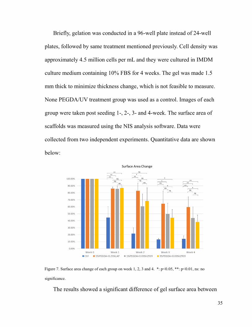
Briefly, gelation was conducted in a 96-well plate instead of 24-well
plates, followed by same treatment mentioned previously. Cell density was
approximately 4.5 million cells per mL and they were cultured in IMDM
culture medium containing 10% FBS for 4 weeks. The gel was made 1.5
mm thick to minimize thickness change, which is not feasible to measure.
None PEGDA/UV treatment group was used as a control. Images of each
group were taken post seeding 1-, 2-, 3- and 4-week. The surface area of
scaffolds was measured using the NIS analysis software. Data were
collected from two independent experiments. Quantitative data are shown
below:
Figure 7. Surface area change of each group on week 1, 2, 3 and 4. *: p<0.05, **: p<0.01, ns: no
significance.
The results showed a significant difference of gel surface area between

36
pure collagen control and three scaffolds after 4 weeks of culture. This
indicated all the three formulas can significantly improve the shrinkage
resistance of the scaffolds. There is a significant improvement of shrinkage
resistance between 1% PEGDA +0.25% LAP/2 min UV and 3% PEGDA +
0.03% I2959/6 min UV. No significance was found in other two
comparisons
Assessment of the pluripotency of the cells cultured in the scaffolds
To assess whether iPSCs cultured in the 3D scaffolds still maintain
pluripotency, the cells were cultured in spontaneous differentiation medium
for a week to allow the initiation of the development of three germ layers
from iPSCs. The differentiation status of each group was evaluated by qRT-
PCR to measure marker gene expression. Sox17, Foxa2, Hand1, ABCG2,
Nestin, and CD44 were chosen as expression markers for endoderm,
mesoderm, and ectoderm, respectively. Cyclophilin was chosen as the
housekeeping gene. Undifferentiated IMR90 cultured in 2D (4 days, in
mTeSR1) for 4 days and IMR90 cultured in pure collagen hydrogel (7 days,
in IMDM + 10% FBS) were used as control. The experiments were repeated
three times and RNA samples from three independent experiments were
subjected to the analysis.

37

38
Figure 8. Gene expression analysis of RNA samples extracted from the collagen-PEGDA scaffolds. RNA
extracted from undifferentiated IMR90 was applied as a control for normalization. Three independent
experiments were carried out. Y axis is fold change. (a) Relative expression of Sox17, Foxa2, ABCG2,
Nestin, and CD44 in the three types of scaffolds and pure collagen scaffold. (b) Relative expression of
Hand1 in the three types of scaffolds and pure collagen scaffold. Asterisks represent significant level of
relative expression difference compared with undifferentiated cells, *: p<0.05, **: p<0.01, ns: no
significance.
Figure 8 indicated that iPSCs seeded in all three types of scaffolds
differentiated spontaneously and preferentially towards mesoderm, as
marked by extremely high Hand1 expression after one week of culture
(Figure 8b). There was a certain degree of ectoderm progression as shown in
Figure 8a with Nestin and CD44. Endoderm differentiation that measured by

39
Sox17 and Foxa2 was not supported by the three types of gels tested. The
expression level of ABCG2 and Nestin in the three scaffolds is higher
compared to undifferentiated IMR90 but lower than cell seeded in pure
collagen hydrogel.

40
Chapter 4. Discussion and Conclusion
The elastic modulus of collagen hydrogel ranges from 1 to 100 Pa (Yang,
Kaufman, 2009). Also, as mentioned before, increasing collagen
concentration does not significantly change the stiffness (Weinberg, 1986).
The collagen hydrogel scaffolds provide a soft and porous environment to
cells to elongate and cluster. During the iPSC biocompatibility assessment of
this study, the control group with no PEGDA/UV treatment showed the
highest cell viability. Cells in control group clustered and formed larger
sphere-shaped and plate-shaped aggregates, and the nutrients were
exhausted more quickly than other experimental groups.
During the development of scaffolds for iPSC culture and differentiation,
several factors showed ability to affect cell viability. The first factor is cell
seeding density. In the beginning of this project, cell seeding density is set to
2.5 million cells per mL of hydrogel to remain consistent to former project
of the laboratory. This seeding density is raised to 4.5 million cells per mL
eventually to reach better cell viability and spontaneous differentiation.
Talukdar et. al. reported that an increase of initial seeding density of
chondrocytes in a 3D silk fibroin scaffold can significantly increase total wet
weight after 2 weeks of culture. Seeding iPSCs at a higher density may

41
enhance cell-cell interaction and thus improve cell proliferation and
differentiation.
The second factor is the toxicity of precursor solution, which in this
project is the toxicity of the photoinitiators. Our experimental results
revealed that when treat cells with UV irradiation, experimental groups
treated with higher concentration of photoinitiator showed lower cell
viability after the 7 days differentiation process. iPSCs’ tolerance to PIs are
lower than other types of stem cells such as MSCs. However, PIs act as the
catalyst in the PEGDA photocrosslinking reaction. Thus, a low PI
concentration may affect the photocrosslinking efficiency, influencing the
stiffnesses of the scaffolds.
UV irradiation is another factor. Figure 4 revealed that when treat cells
with precursor solution with the same I2959 concentration, a longer UV
irradiation time leads to a decrease of cell viability. Compared to I2959,
LAP requires 2 minutes of UV irradiation instead of 6 minutes. The mock
control (no PEGDA, only PIs and UV) results of LAP showed better cell
viability than I2959 at same concentrations.
LAP can also catalyze PEGDA photocrosslinking under visible light
(400 nm), thus avoid the harmful effect of UV. Also, the improved
polymerization kinetics enable cell encapsulation at reduced initiator

42
concentration, which has been shown to reduce initiator toxicity and
increase cell viability. This is consistent to the stability test results that LAP
treated scaffold showed greater stability at the same PEGDA concentration.
In general, LAP, the PI with a shorter UV irradiation time and a higher
catalysis efficiency, is superior to I2959 in this project. Compared to UV,
visible lights are more biocompatible to stem cells. Eosin Y photosensitizer
is another type of PI that can initiate photo polymerization of PEGDA under
visible light (Bahney et al., 2011, Wang et al., 2015).
In addition, 1% PEGDA + 0.25% LAP + 2 minutes UV group showed
greater shrinkage resistance than the other I2959 treated groups, but the
amount of RNA extracted (around 3000 ng per well) is less than I2959
treated groups (6000 to 8400 ng per well). These data imply that I2959-
treated collagen gels are more biocompatible to iPSCs, and perhaps they are
softer than LAP-treated gel. The softness of I2959 groups may result from
the lack of photocrosslinking efficiency of I2959 at 365 nm. Also, when
optimize the PEGDA concentration in LAP groups, 3%, 5% and 7% PEGDA
groups showed low cell survival rate. This may imply that when increasing
the PEGDA concentration in PEGDA/LAP precursor solutions, more
PEGDA fibers are polymerized and fabricated into the collagen hydrogel,

43
thus increase the stiffness of the scaffold, and limit the cluster formation and
aggregation of cells, and ultimately hinder the cell viability.
For the pluripotency assessment, the gene expression level of ABCG2,
Nestin, and CD44 are not particularly higher than control. This may due to
the lack of differentiation time, since cells may only differentiate into an
early stage of certain germ layers. Another drawback of this study is that the
stability assessment was done twice instead of three times due to insufficient
time, making the result not as reliable.
In further research, mechanical properties such as elastic modulus may
be measured to evaluate the effect of mechanical properties of these
collagen-PEGDA scaffolds on iPSC cultures. In human body, different
tissues have different stiffnesses. Take pancreas tissue as an example, the
overall mean shear stiffness of pancreas is 1150±170 Pa at 40Hz and
2090±330 Pa at 60 Hz (Shi, et.al, 2015). This project built a panel for
PEGDA-collagen hydrogel scaffold for tissue engineering. The stiffness of
the collagen-PEGDA hydrogel scaffolds can be adjusted to proper level to
support pancreas tissue engineering by altering several parameters, such as
PEGDA molecular weight and PEGDA concentration. In addition, iPSC
differentiation capacity in the scaffolds should be assessed by culture cells
for a few weeks to allow fully lineage progression.

44
As for conclusion, this project aimed to develop a collagen-PEGDA
hydrogel scaffold that can support direct seeding and differentiation of
iPSCs. By optimizing multiple factors, including photoinitiator type,
photoinitiator concentration, PEGDA concentration, and UV irradiation,
three formulas (1) 1% w/v PEGDA + 0.25% w/v LAP/2 minutes UV
irradiation, (2) 1% w/v PEGDA + 0.03% w/v I2959/6 minutes UV
irradiation, and (3) 3% w/v PEGDA + 0.03% w/v I2959/6 minutes UV
irradiation were developed. qRT-PCR results revealed that the scaffolds
could support IMR90 cells to spontaneously differentiate into mesoderm.
The stability test results showed significant improvement of shrinkage
resistance of the formulas compared to collagen hydrogel. This work
provides a method for developing 3D collagen scaffolds that can support
iPSC seeding, proliferation, and differentiation in long-term 3D culture.

45
References
Ahadian, S. et al. Moldable elastomeric polyester-carbon nanotube scaffolds
for cardiac tissue engineering. Acta Biomaterialia 52, 81–91 (2017).
Assal, R. E. et al. 3-D Microwell Array System for Culturing Virus Infected
Tumor Cells. Scientific Reports 6, (2016).
Bahney, C. et al. Visible light photoinitiation of mesenchymal stem cell-
laden bioresponsive hydrogels. European Cells and Materials 22, 43–55
(2011).
Cnop, M. et al. Mechanisms of Pancreatic -Cell Death in Type 1 and Type 2
Diabetes: Many Differences, Few Similarities. Diabetes 54, (2005).
Donath, M. Y. & Ehses, J. A. Mechanisms of Beta-Cell Death in Diabetes.
Pancreatic Beta Cell in Health and Disease 75–89, (2008)
doi:10.1007/978-4-431-75452-7_5
Fairbanks, B. D., Schwartz, M. P., Bowman, C. N. & Anseth, K. S.
Photoinitiated polymerization of PEG-diacrylate with lithium phenyl-
2,4,6-trimethylbenzoylphosphinate: polymerization rate and
cytocompatibility. Biomaterials 30, 6702–6707 (2009).
Hoveizi, E., Nabiuni, M., Parivar, K., Ai, J. & Massumi, M. Definitive
endoderm differentiation of human-induced pluripotent stem cells using

46
signaling molecules and IDE1 in three-dimensional polymer
scaffold. Journal of Biomedical Materials Research Part A 102, 4027–
4036 (2014).
Jin, S., Yao, H., Weber, J. L., Melkoumian, Z. K. & Ye, K. Correction: A
Synthetic, Xeno-Free Peptide Surface for Expansion and Directed
Differentiation of Human Induced Pluripotent Stem Cells. PLoS ONE 8,
(2013).
Kroon, E. et al. Pancreatic endoderm derived from human embryonic stem
cells generates glucose-responsive insulin-secreting cells in vivo. Nature
Biotechnology 26, 443–452 (2008).
Mcclure, M. J., Sell, S. A., Simpson, D. G., Walpoth, B. H. & Bowlin, G. L.
A three-layered electrospun matrix to mimic native arterial architecture
using polycaprolactone, elastin, and collagen: A preliminary study. Acta
Biomaterialia 6, 2422–2433 (2010).
Montgomery, M. et al. Flexible shape-memory scaffold for minimally
invasive delivery of functional tissues. Nature Materials 16, 1038–1046
(2017).
Munoz-Pinto, D. J., Jimenez-Vergara, A. C., Gharat, T. P. & Hahn, M. S.
Characterization of sequential collagen-poly(ethylene glycol) diacrylate

47
interpenetrating networks and initial assessment of their potential for
vascular tissue engineering. Biomaterials 40, 32–42 (2015).
Shi, Y. & Hu, F. B. The global implications of diabetes and cancer. The
Lancet 383, 1947–1948 (2014).
Singh, S. P. et al. A synthetic modular approach for modeling the role of the
3D microenvironment in tumor progression. Scientific Reports 5, (2015).
Takahashi, K. et al. Induction of Pluripotent Stem Cells from Adult Human
Fibroblasts by Defined Factors. Cell 131, 861–872 (2007).
Talukdar, S., Nguyen, Q. T., Chen, A. C., Sah, R. L. & Kundu, S. C. Effect
of initial cell seeding density on 3D-engineered silk fibroin scaffolds for
articular cartilage tissue engineering. Biomaterials 32, 8927–8937 (2011).
Thompson, I. J. Sabiston Textbook of Surgery, 17th Edition, Board
Review. ANZ Journal of Surgery 76, 13–13 (2006).
Wang, L. et al. Differentiation of iPSCs into insulin-producing cells via
adenoviral transfection of PDX-1, NeuroD1 and MafA. Diabetes
Research and Clinical Practice 104, 383–392 (2014).
Wang, Z. et al. A simple and high-resolution stereolithography-based 3D
bioprinting system using visible light crosslinkable
bioinks. Biofabrication 7, 045009 (2015).

48
Weinberg, C. & Bell, E. A blood vessel model constructed from collagen
and cultured vascular cells. Science 231, 397–400 (1986).
Xu, C., Lee, W., Dai, G. & Hong, Y. Highly Elastic Biodegradable Single-
Network Hydrogel for Cell Printing. ACS Applied Materials & Interfaces
10, 9969–9979 (2018).
Yang, T., Malkoch, M. & Hult, A. The influence of diffusion time on the
properties of sequential interpenetrating PEG hydrogels. Journal of
Polymer Science Part A: Polymer Chemistry 51, 1378–1386 (2012).
Yang, Y.-L. & Kaufman, L. J. Rheology and Confocal Reflectance
Microscopy as Probes of Mechanical Properties and Structure during
Collagen and Collagen/Hyaluronan Self-Assembly. Biophysical Journal
96, 1566–1585 (2009).