collagen-hyaluronic acid scaffolds for tissue … · 1 collagen-hyaluronic acid scaffolds for...
TRANSCRIPT

___________________________________________________________________________ 1
Collagen-Hyaluronic Acid Scaffolds for Adipose Tissue Engineering
N. Davidenko1,*, J.J. Campbell2, E.S. Thian1, 3, C.J. Watson2 and R.E. Cameron1
1Department of Materials Science and Metallurgy
University of Cambridge, Pembroke Street
Cambridge, CB2 3QZ, UK
2Department of Pathology
University of Cambridge, Tennis Court Road
Cambridge, CB2 1QP, UK
3Department of Mechanical Engineering
National University of Singapore, 9 Engineering Drive 1
Singapore 117 576, Singapore
*Corresponding Author.
Email: [email protected] Tel: +44 1223 334560. Fax: +44 1223 334567
Abstract
Three-dimensional (3D) in vitro models of the mammary gland require a scaffold
matrix that supports the development of adipose stroma within a robust freely permeable
matrix. 3D porous collagen-hyaluronic acid (HA: 7.5 and 15%) scaffolds were produced by
controlled freeze-drying technique and cross-linking with 1-ethyl-3-(3-
dimethylaminopropyl)-carbodiimide hydrochloride. All scaffolds displayed uniform,
interconnected pore structure (total porosity ∼ 85%). Physical and chemical analysis showed
no signs of collagen denaturation during the formation process. The values of thermal
characteristics indicated that cross-linking occurred and that its efficiency was enhanced by
the presence of HA. Although the cross-linking reduced the swelling of the strut material in
___________________________________________________________________________ 2
water, the collagen-HA matrix as a whole tended to swell more and show higher dissolution
resistance than pure collagen samples. The compressive modulus and elastic collapse stress
were higher for collagen-HA composites. All the scaffolds were shown to support the
proliferation and differentiation 3T3-L1 preadipocytes while collagen-HA samples
maintained a significantly increased proportion of cycling cells (Ki-67+). Furthermore,
collagen-HA composites displayed significantly raised Adipsin gene expression with
adipogenic culture supplementation for 8 days versus control conditions. These results
indicate that collagen-HA scaffolds may offer robust, freely permeable 3D matrices that
enhance mammary stromal tissue development in vitro.
Keywords: Collagen; crosslinking; freeze-drying; hyaluronic acid; scaffolds, adipose
tissue engineering
1. Introduction
The mammary gland comprises a complex branched epithelial network invested
within an adipocyte-rich stroma termed a fat-pad. Uniquely, the majority of mammary gland
development is post-natal, where the rudimentary organ penetrates the underlying pre-
developed fat-pad under hormonal control, however the influence of the stroma in guiding the
formation of this structure has been well documented [1,2]. Accurate 3-dimensional (3D)
models of the mammary gland should encourage stromal tissue development as a basis for
further epithelial coculture studies. A great deal of research has been carried out with the
hope of obtaining an effective means of achieving adipose tissue formation in-vitro [3,4]. The
most widely-used approach involves locating the relevant cells in a physical 3D scaffold
under ‘controlled’ culture conditions, so facilitating cell attachment, proliferation,
differentiation and, finally, formation of a tissue structure of suitable shape and size.
___________________________________________________________________________ 3
To date, the majority of in vitro mammary cell and tissue culture investigations utilize
reconstituted basement membrane gels (Matrigel) or pure collagen gels. While useful,
native whole tissue preparations such as Matrigel are undefined in composition and exhibit
batch variation [5]. Moreover they cannot be tuned to the specific requirements of an in vitro
model system. Alternatively, pure collagen gels, whilst defined, exhibit cell-mediated matrix
contraction [6], varying mechanical properties throughout long-term culture and limiting
oxygen and nutrient transport to cells within [7]. Furthermore, gel-based systems are random
with regard to cellular distribution and limit direct cell-contact, an important component of
coculture signaling mechanisms [8,9]. Currently, there is a great need for more defined in
vitro mammary gland models that may increase our understanding of basic biological
processes, aid drug discovery and minimize intensive animal use with in vivo studies.
Properly in-vitro engineered tissues may offer an excellent possibility of carrying out detailed
and continuous control and monitoring of cellular responses, with the further advantage of
“humanising” the system by using human cells instead of animal model, which would be
more appropriate for drug screening and cancer studies [10,11]. Towards our goal of
producing a novel tissue engineered mammary gland, we have developed a naturally derived
scaffold with controlled architecture and robust mechanical characteristics that encourages
adipose tissue development.
In order to successfully create an artificial 3-dimensional structure capable of
supporting in vitro tissue formation, several key attributes have to be taken into account. It
should be highly porous with an interconnected architecture [12-14]; predictable and
controllable material degradability, and should have biocompatibility with the seeded cells to
enhance cellular activity [15-17]. It should also possess an adequate mechanical stiffness
[18-20] to withstand stress incurred during cell culturing. Furthermore, the formation of a
new tissue in the 3D matrix structure is highly influenced by the chemical composition of the
___________________________________________________________________________ 4
bio-scaffold [21]. ollagen, being a major protein of the natural extracellular matrix (ECM),
has been commonly used in the form of a gel for adipose tissue engineering [22-25] in spite
of limitations principally related to cell-mediated contraction.
HA, a naturally occurring unsulfated glycosaminoglycan distributed in the ECM of
soft tissues [26] has been implicated in diverse biological processes such as angiogenesis and
migration [27,28] as well as the proliferation and differentiation of progenitor cells [29].
Recent studies have demonstrated the utility of HA-based scaffolds in enhancing adipose
tissue development in vivo [30,31] and in vitro [32,33].
The combination of collagen and HA has displayed advantages over the use of either
material alone for tissue engineering applications [34-40]. For example, the incorporation of
HA to the collagen matrix has shown positive effects in stimulating chondrocyte and
fibroblast expression in-vitro [34-36]. In addition to direct biological effects, the
incorporation of HA should enhance the strength of the collagen-based gels, thus inhibiting
the cell induced contraction. The properties of scaffolds made from collagen and HA
combinations are explored in the current paper.
Freeze-drying, also known as lyophilization, is commonly employed to produce
water-soluble polymer scaffolds such as collagen [12, 41-43]. In this technique, a suspension
of the water-soluble polymer is frozen, thereby forming an interpenetrating network of ice
crystals. Next, these ice crystals are removed by reducing the chamber pressure to induce
sublimation, thus leading to the formation of a porous scaffold. A controlled freezing process
during production normally leads to uniform nucleation and growth of ice crystals and hence
the formation of a homogeneous pore structure. With a well-defined pore structure, improved
biomechanical properties of the scaffold can be achieved [43, 44].
To enhance the structural stability of collagen-based sponges various chemical and/or
physical cross-linkers are frequently introduced in the production process [43, 45, 46]. EDC
___________________________________________________________________________ 5
(in the presence of N-hydroxysuccinimide, NHS) and glutaraldehyde (GTA) are two of the
most commonly used chemical agents which work in a distinctly different manner. EDC
forms “zero length” crosslinks, whereas GTA crosslinks take the form of long polymer
chains. This means that EDC is limited to crosslinking collagen/HA molecules that are
directly adjacent to each other (1nm) whereas GTA can crosslink molecules that are rather
more separated. However, the incorporation of GTA into scaffolds can have implications for
biocompatibility. EDC, on the other hand, is known as a non-toxic and biocompatible cross-
linker because it generates peptide-like bonds. Cross-linking EDC, in the presence of NHS,
produces substituted urea as a by-product which can be easily eliminated by washing.
In this study, the reaction with water soluble EDC in the presence of NHS was
selected. This crosslinking process is governed by certain complex mechanisms [45,46]. On
the basis of many results, Pieper et al. [47] have concluded that EDC/NHS can be utilised for
attaching glycosaminoglycans (GAGs), such as: dermatan sulphate, heparin and chondroitin
sulphates, to collagen. These findings suggest that similar reactions may occur between
collagen and HA because of the similarity of the HA chemical structure to the other GAGs .
In the current study, therefore, the formation of defined collagen-HA scaffolds
containing various amounts of HA and with uniform pore structure, and hence good
mechanical properties, suitable for tissue engineering purposes, using a freeze-drying
technique and non-toxic and biocompatible cross-linking system, is investigated. Some
relevant structural, physico-chemical, mechanical and biological characteristics of such
scaffolds are defined.

___________________________________________________________________________ 6
2. Materials and methods
2.1. Collagen and Collagen-Hyaluronic Acid Scaffold preparation
The highly porous scaffolds were produced from a collagen-HA suspension using a
freeze-drying technique. Firstly, the suspension of 1%-wt. of different component
compositions were prepared from an insoluble, type I microfibrillar collagen derived from
bovine Achilles tendon (Sigma-Aldrich Co. Ltd., UK) and HA derived from bovine vitreous
humor (Sigma-Aldrich Co. Ltd., UK) in 0.05 M acetic acid solution (Sigma-Aldrich Co. Ltd.,
UK), where the resultant pH value was adjusted to below 2.0 with 1 M hydrochloric acid
(VWR International Ltd., UK). Each suspension was then blended at 20,000 rpm using an
overhead homogenizer for 30 min at 40C. After mixing, the suspension was centrifuged at
2500 rpm for 5 min using a bench-top centrifuge to remove air bubbles formed during the
blending. Two different HA weight percentages were produced: 7.5 and 15 wt-% (collagen-
7.5HA and collagen 15HA, respectively), in addition to suspensions were made with pure
collagen.
The prepared collagen and collagen-HA suspensions were then frozen in 316L
stainless steel plates at a controlled rate (0.9°C min-1) to -30°C and held for 90 min, using a
computer-controlled freeze-dryer. The frozen suspensions were subsequently sublimed at 0°C
for 20 h under a vacuum of less than 100 mTorr.
2.2. Scaffold Crosslinking
Lyophilized collagen and collagen-HA scaffolds were crosslinked with a water-
soluble carbodiimide. Scaffolds were immersed in 95 % ethanol solution containing 33mM of
1-ethyl-3-(3-dimethylaminopropyl)- carbodiimide hydrochloride (EDC, Sigma-Aldrich Co.
Ltd., UK) and 6mM of N-hydroxysuccinimide (NHS, Sigma-Aldrich Co. Ltd., UK) for 4 h at
25°C. After the crosslinking process, the scaffolds were washed thoroughly in distilled water
(5 min x 5) and were subsequently re-frozen and re-lyophilized using the previous freeze-
___________________________________________________________________________ 7
drying cycle. Disc samples of diameter 10 mm were then cut from sheets of scaffolds using a
punch.
2.3. Scaffold morphology
X-ray microtomography ( Skyscan 1072 Micro-CT (µCT) system), with a 28 kV/ 164
μA X-ray source (using 0-360o range of rotation angle with a step size of 0.45o and 3.1 sec of
exposure time per step) and provided with 12-bit cooled CCD camera, was employed to
obtain a general view of the interior microstructure of the scaffolds. Cross-sections were
generated using a full cone beam Feldkamp reconstruction algorithm.
Analysis of the scaffold pore structure was carried out on a scanning electron
microscopy (SEM) (JEOL 5800). Samples were sputter-coated with a layer of platinum for
observation at 10kV at various levels of magnification.
2.4. Physicochemical Analysis of Scaffolds
X-ray study
Changes in the structural composition of the scaffolds were identified using a Philips
PW3020 X-ray Diffractometer (XRD) with CuKα radiation operating at 40 kV and 40 mA. θ-
2θ scans were performed and data were collected over a 2θ range of 5-35°, with a step size of
0.05° and a dwell time of 10 s.
FTIR analysis
A BRUKER Tensor 27 Fourier Transform Infrared Spectrometer (FTIR), utilizing the
attenuated total reflectance technique, was used to analyze the molecular structure of the
scaffolds. The spectra were collected over a range of 1000-4000 cm-1 to monitor any changes
in the amide groups. A resolution of 4 cm-1 was used, and the samples were scanned 16 times
to increase the signal-to-noise ratio.
Porosity

___________________________________________________________________________ 8
The porosity level of the scaffolds was determined using a computer-controlled
Quantachrome Poremaster 33 Mercury Intrusion Porosimeter (MIP by applying various
levels of pressure to a sample immersed in mercury (Hg). The pressure required to intrude
mercury into the sample pores is inversely proportional to the size of the pores. To perform
an analysis a known quantity of each scaffold (between 0.0036g and 0.0038g) was loaded
into a penetrometer. The penetrometer was sealed and placed in a low pressure chamber,
where the sample was evacuated for 5 min at 0.05 torr to remove air and moisture, and then
automatically backfilled with mercury (filling pressure 6.6kPa). Subsequently the
penetrometer was moved to a high-pressure chamber and subjected to pressure in steps from
0.69kPa to 206Mpa. After completing this process Hg was extruded from the sample pores
by stepwise pressure reduction and the extruded volume of Hg corresponding to each step
was recorded. The porosity level (%) was then automatically calculated from intrusion data
using software provided by this equipment.
2.5. Differential scanning calorimetry (DSC) and thermogravimetric analysis (TG)
The thermal stability of the collagen–HA matrix was assessed with DSC Q2000 and
TGA Q 500 analyzers scanning from 0 to 450°C. The samples were weighed and placed into
aluminium pans. The pans were heated at a constant rate of 10°C min-1 in a nitrogen
atmosphere. In the case of DSC testing, an empty aluminium pan was used as reference. The
sample mass was in the range of 1-3 mg. All samples were run in duplicate. The denaturation
temperatures were measured at the mid-point of the transition peaks and the temperatures at
the start of the process were measured at the onset of the peak. From the TG and DSC curves
obtained by means of the Universal Analysis software, the following parameters, which
describe the thermal behaviour of these scaffolds, were calculated: temperatures of thermal
denaturation (Td), maximum temperature of each decomposition stage (T1-T3); percentage of
the corresponding mass loss (∆m %), and total mass loss at 450oC in nitrogen (∆m total).
___________________________________________________________________________ 9
2.6. Water Uptake of Scaffolds
Dried samples were weighed prior to the swelling study (Wd). Next, the samples were
immersed in 5 ml distilled water at 37°C for different periods of time up to 7 days. At each
time point, they were removed from the distilled water and two different measurements of
their capacity to retain water were made.
The first measurement was aimed at assessing the ability of the scaffold structure as a
whole (the material itself together with the pore system) to absorb water. For this, at each
time point, the samples were removed from water, shaken gently, and then weighed without
dripping ( Wws).
The second measurement was carried out after pressing and “drying” the same soaked
samples between sheets of filter paper to remove the water retained in its porous structure
(Wwm). In this way the swelling ability of scaffold material itself was determined.
The percentage of water uptake, in both cases, was calculated as shown:
Water uptake (%) = [(Ww – Wd) / Wd] x 100
Where Ww represents Wws or Wwm.
2.7. Dissolution of Scaffolds
Dried samples were weighed prior to the dissolution study (Wb). The samples were
then immersed in 5 ml distilled water at 37°C in a humid atmosphere for various periods of
time, up to 7 days. At each time point, they were removed from the distilled water, dried for
48h (under reduced pressure to constant weight) and weighed (Wa). The percentage weight
loss was calculated as shown:
Weight loss (%) = [(Wb – Wa) / Wb] x 100
The pH value of the distilled water after each time interval was measured using a pH
meter. Each sample was measured in triplicate.
___________________________________________________________________________ 10
2.8. Mechanical Analysis of Scaffolds
Compressive stress-stain analysis of the scaffolds was performed using a Mechanical
Tester. Scaffolds were immersed in distilled water at 37°C in a humid atmosphere for 24 h
before testing. A cell load of 5 N and a crosshead speed of 5 mm min-1 were used.
Mechanical characteristics which describe stress–strain behaviour, namely: the linear elastic
(Young’s) modulus (E*), the elastic collapse stress and strain (σ*el, ε*el), and the slopes of
the collapse plateau (∆σ/∆ε) were determined from each curve for all scaffolds tested. E* was
obtained via linear regression of the initial linear region of the stress–strain curve; ∆σ/∆ε was
calculated via linear regression of the linear region following the ‘‘knee’’ corresponding to
the strut bending–buckling transition; σ*el and ε*el were determined from the intersection of
the E* and ∆σ/∆ε regression curves (see Fig.10b and explanation of stress-strain profiles in
Results and Discussion part). Five replica tests were made for each scaffold.
2.9. Preadipocyte cell culture within 3D scaffolds
A murine preadipocyte (3T3-L1) cell line (a generous gift from Prof. Kenneth Siddle,
University of Cambridge) was grown to passage 5 in Dulbecco’s modified eagles medium
(DMEM) + 10% (vol/vol) newborn calf serum (Invitrogen, UK). Pure collagen and collagen
mixed HA scaffolds were sterilized by UV exposure and rinsed thrice in both distilled H2O
and PBS before 5 scaffolds of each type were bathed in a 10ml suspension of 3T3-L1 cells at
a density of 1x10⁶ cells/ml in a 50mL Falcon tube. Scaffolds were compressed onto the wall
of each tube using a sterile pipette in order to remove air bubbles and encourage cell
penetration throughout the scaffold. Each scaffold/cell suspension was then gently agitated on
a rocker plate for 4 hours at 37°C to encourage further cell penetration. Cell seeded scaffolds
were then removed to an incubator for 3 days until confluence judged by live-cell fluorescent
visualization with 5µM Calcein-AM (Invitrogen, UK). Terminal differentiation of 3T3-L1

___________________________________________________________________________ 11
preadipocytes was induced by incubation for 48 h within DMEM, 10% fetal calf serum (FCS,
Hyclone, Thermofisher), 1µM dexamethasone, 0.5mM 3-isobutyl-1-methylxanthine and
0.01mg/ml insulin (all Sigma-Aldrich Co. Ltd., UK) followed by a further 6 days incubation
in DMEM, 10% FCS and insulin. Adipogenic control specimens were incubated for 8 days
in DMEM + FCS.
2.10. Oil-Red O and Immunohistochemical staining
Following 8 days adipogenic induction, cell-seeded scaffolds were fixed in 4%
paraformaldehyde/PBS embedded and frozen within O.C.T and cryosectioned at 10µm. Lipid
was stained with Oil-Red O and counter stained with haematoxylin using routine methods.
For immunohistochemistry, sections were blocked at room temperature for 1hr with 10%
normal goat serum (Sigma-Aldrich Co. Ltd., UK), incubated with an anti-Ki67 primary
antibody (Abcam, UK) overnight at 4°C and detected with an Alexafluor 488 conjugated
secondary antibody (Invitrogen, UK). Nuclei were stained with Hoerscht 33258. Proliferation
rate was measured by scoring Ki-67 positive nuclei in a x200 field of view.
2.11. Gene expression analysis
Cell-seeded scaffolds exposed to adipogenic or control culture conditions were snap
frozen in liquid nitrogen. Total RNA was extracted using Trizol reagent (Invitrogen) and an
RNeasy mini kit (Qiagen) following the manufacturers instructions. cDNA was reverse
transcribed from 1µg of total RNA using 0.5U/µl reverse transcriptase (Roche) and random
primers (Promega). Adipogenic gene expression was analysed by real-time quantitative PCR
(Biorad Icycler Biorad Laboratories Inc, USA) using SYBR green chemistry (Sigma).
Sequences for real-time PCR were obtained using the PrimerBank [48] website
(http://pga.mgh.harvard.edu/primerbank/) for peroxisome proliferator-activated receptor
gamma (PPARγ), Fw: TCGCTGATGCACTGCCTATG, Rv:
___________________________________________________________________________ 12
GAGAGGTCCACAGAGCTGATT and adipsin, Fw: CATGCTCGGCCCTACATGG, Rv:
CACAGAGTCGTCATCCGTCAC. Relative mRNA abundance for each gene of interest was
measured against a pooled sample standard and expressed as a ratio of cyclophillin A Fwd,
CCTTGGGCCGCGTCTCCTT, Rev, CACCCTGGCACATGAATCCTG for each sample.
3. Results and discussion
3.1. Scaffold preparation
It is well known that polymer scaffolds for use in cell culture must be highly porous
with large surface/volume ratios to provide sufficient space for cell growth and proliferation
[43]. The pore structure determines the matrix permeability for diffusion of nutrients and
waste products within the scaffolds and its size must be large enough to allow infiltration of
the cells towards the centre of the scaffolds. At the same time, it should be sufficiently small
so as to provide adequate ligand density for cellular attachment. Development of highly
porous scaffolds by a freeze-drying approach was used in this work due to the advantage of
this method in employing only water upon freezing as a porogen. Freeze-drying conditions
consisting of a relatively high freezing temperature (-30°C) and a rather slow rate of cooling
(less that 1°C min-1) were chosen to achieve the formation of highly connected ice crystals of
rather large size (100 or more micrometers). After sublimation of these ice crystals under
vacuum at 0°C, sponges with the inner morphology (pore size, shape, distribution,
interconnectivity, etc.) that mirror the structure of these ice crystals were obtained.
3.2. Structure Morphology
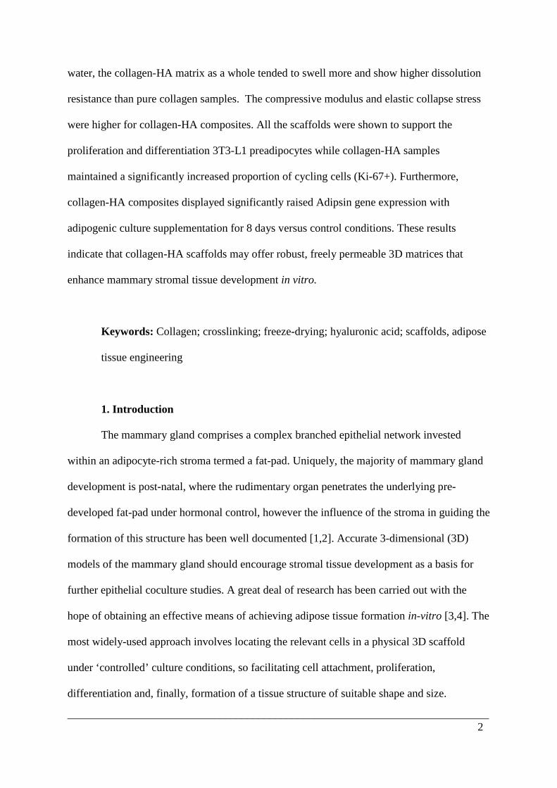
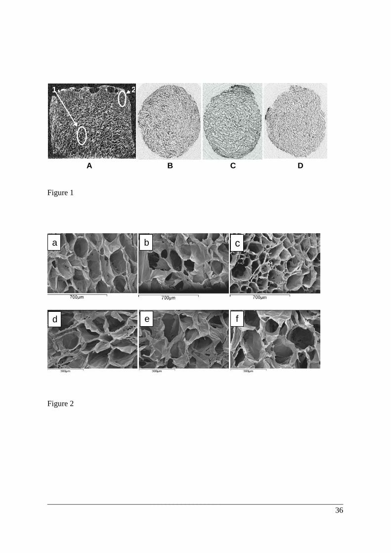
Fig. 1 shows a series of µCT images taken of sections of various scaffolds formed
using the freeze-drying technique. Generally, all scaffolds were highly porous,
interconnected, and appeared to be relatively homogenous throughout the bulk of the
scaffold. A fairly uniform pore structure characterized by thin collagen and collagen-HA

___________________________________________________________________________ 13
struts was obtained. However, several zones of larger pore sizes (Fig. 1A, area 1) and of
material densification, especially at the edges of the scaffold sheets, were detected (Fig. 1A,
area 2).
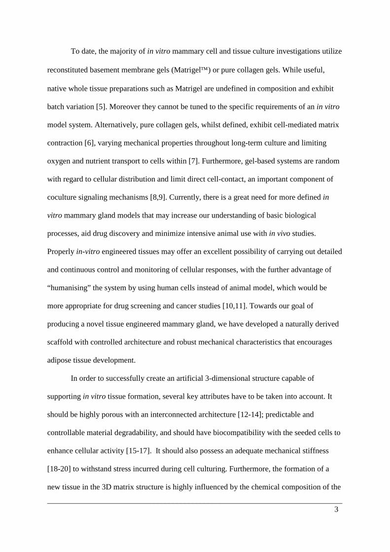
Scanning electron microscope images (Fig. 2) of the different types of cross-section
of all scaffolds showed a continuous structure of irregular but rather uniformly distributed
interconnected pores. The pore dimensions estimated from SEM microphotographs by the
manual measurement of pore zones with arbitrary shapes were mostly in the range of 100 to
220 μm, and were similar for all scaffold compositions. It was logical to expect that the inner
morphology of all scaffolds would be similar given that the same freezing conditions were
employed for their production. This in turn suggests that the presence of HA does not alter
the characteristics of water dispersion in the collagen-HA mixture and leads to the formation
of a similar ice structure during the freezing process.
Heterogeneity in the scaffold microstructures (the presence of large pores, local
regions of higher density, etc.) may be partially the result of the relatively high concentration
of collagen or collagen-HA suspension (1%-wt) used for scaffold preparation. Difficulties in
achieving completely uniform collagen-type sponges from rather highly concentrated
suspensions (≥ 1%-wt) were reported in other works [49]. It was also reported that scaffolds
obtained from less concentrated suspensions (0.5% -wt) displayed a more uniform pore
microstructure but their mechanical properties were fairly poor in comparison with those
exhibited by sponges achieved from more concentrated suspensions. Structural non-
uniformities within the microstructure could cause some variation in the values of several
physico-chemical and mechanical characteristics of these systems, but it is necessary to
compromise when selecting experimental conditions so as to achieve desirable resultant
properties when considering the practical application of scaffolds.

___________________________________________________________________________ 14
3.3. Porosity Level and Pore Size
All the samples had almost identical porosity (of about 85 %) (data not shown)
regardless of the HA level incorporated into the collagen scaffolds.
3.4. Structural and molecular Compositions
X-ray study
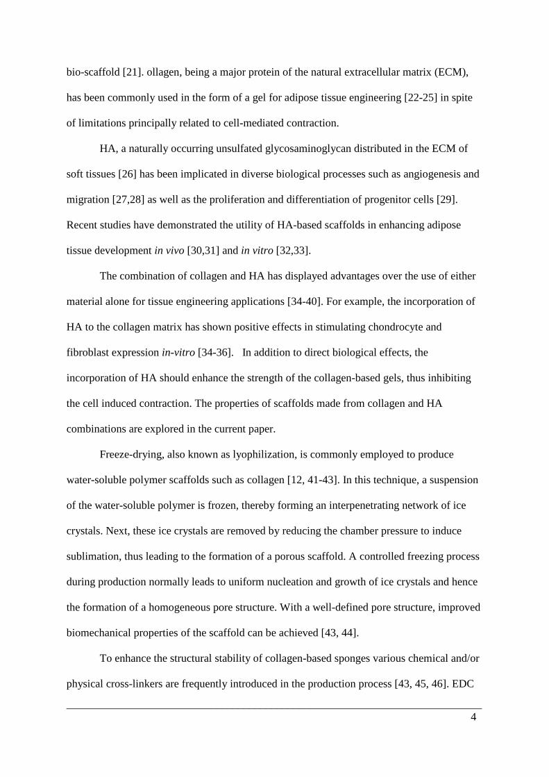
Fig. 3 shows the XRD patterns obtained for various crosslinked scaffolds. All patterns
showed no sign of collagen denaturation, with the emergence of a distinct peak centered at 2θ
= 7° which is indicative of the position of the characteristic equatorial peak of collagen.
Furthermore, a broad peak between 2θ = 15-18° indicated the position of the characteristic
interchain spacing of the collagen triple helix.
FTIR-analysis
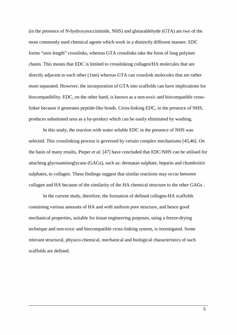
The FTIR spectra of all the scaffolds exhibited several features characteristic of the
molecular organization (Fig. 4). Peptide bonds give rise to several infrared active vibration
modes and there was no significant evidence of change in the spectra with HA addition.
The presence of amide I band at 1780 cm-1 arose due to the stretching vibration of C=O
groups in the peptide groups. In addition, the presence of amide II bands at 1623 and 1535
cm-1 arose due to the motion combining both the amide N-H deformation and C-N stretching
vibrations. Besides these bands, amide N-H stretching vibration modes could be obtained at
both 3290 and 3058 cm-1 [50,51]
XRD and FTIR analyses confirmed that no other chemical entities were introduced
into the crosslinked scaffolds, during the crosslinking reaction. As such, the ability to custom-
make collagen-HA scaffolds with appropriate ratios can be easily controlled by altering the
initial ratios of the two starting biopolymers.
3.5 Differential scanning calorimetry and thermogravimetric analysis

___________________________________________________________________________ 15
Differential scanning calorimetry and thermogravimetric measurements were used in
order to detect the temperature of denaturation, Td, of pure collagen and collagen–HA
scaffolds and to assess their thermal stability. It is expected, that modification (by
crosslinking) in the molecular organization of scaffolds should induce favourable variations
in their thermal behaviour. It is worth mentioning that the effect of temperature on the
conformational properties of these systems is still not clearly elucidated.
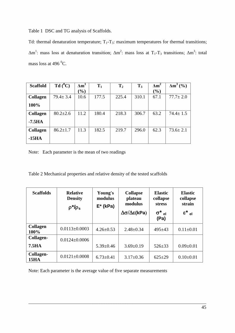
Examples of typical DSC and TG thermograms of cross-linked scaffold matrices are
shown in Fig. 5. The profiles show four different endothermic peaks: the first, within the
temperature range from 30°C to about 130°C, relates to the temperature of thermal
denaturation of the scaffold matrix, and the other three are connected with a complex
phenomenon of thermal modification, finally leading to the destruction of materials.
Parameters describing the thermal behaviour of these scaffolds, calculated from TG and DSC
curves, are displayed in Table 1.
It is known [52], that the temperature of thermal denaturation is related to the
transition from the triple helix to a randomly coiled conformation, this taking place in the
domains between the crosslinks of collagen macromolecules. The stability of this
conformation should depend upon the presence of intra- and inter-molecular hydrogen bonds
as well as hydrogen-bound water and, as reported [52,53], is strongly influenced by the water
content in the matrix and its degree of crosslinking between chains. The values of Td found
in this work were very similar for all scaffolds (Table 1), being slightly shifted to the region
of higher temperatures in the case of collagen-15HA samples (from ∼ 80°C for collagen and
collagen-7.5HA to 86.2°C for collagen-15HA). The comparison of these values with that
reported for a non-crosslinked collagen matrix (69.5°C [52]) may indicate that the
crosslinking process occurred in all the scaffolds and that its efficiency was enhanced by the
presence of HA, with the consequent increase of the thermal stability of samples. The
___________________________________________________________________________ 16
denaturation parameters obtained for collagen and collagen-HA samples are in the range of
data reported in the literature for analogous systems [43, 52, 54]. This thermal transition was
accompanied by a gradual mass decrease (∼ 10%), as observed in the TG curve (Fig. 5) and
in Table 1. The major weight loss at 450°C in nitrogen, 77.7±2.0%, was found for pure
collagen scaffold while the HA containing matrices showed a somewhat lower percentage of
degradation under the same heating conditions: 74.4% and 73.6% for collagen-7.5HA and
collagen-15HA, respectively.
The quite similar values of Td found for all scaffolds suggest that the thermal
denaturation depends rather more on the degree of hydration and on the degree of cross-
linking of scaffolds than on the presence of HA.
3.6. Water Uptake
Scaffold swelling properties have been shown [35, 43, 55] to significantly influence
cell behaviours such as adhesion, growth and differentiation. Water uptake properties indicate
the level of cell culture medium which will be absorbed by the scaffold during culturing.
The results of water-uptake studies are displayed in Fig. 6 (for soaked samples, including the
water in both the material and the pores) and in Fig. 7 (after removing water from the pores
using filter paper). Each value was averaged from at least five parallel measurements.
It can be seen that in both cases the majority of the water uptake occurred within the
first two hours and seemed to have stabilized after 24 h soaking time. However the values of
the percentage of water retention in each test showed an opposite trend with the composition
of the scaffold. For scaffolds soaked in water this parameter (indicating the water present in
the materials and the pores of the scaffold) increases with the increase of HA content in the
scaffold matrix (Fig. 6): 15HA>7.5HA>pure collagen = 8300% > 7600% > 6600% at 7 days,
while for the samples “dried” by filter paper (indicating the water present in the strut material
___________________________________________________________________________ 17
of the scaffold alone) the reverse dependence was found: pure collagen>7.5HA>15HA =
488% > 373% > 298% at 7 days.
Generally, the water uptake ability of scaffolds, as sponge-like matrices, should be
controlled principally by two aspects: the hydrophilicity of their materials and the stability of
their porous structures in water [38, 39, 56]. The hydrophilicity of collagen-HA scaffold
compositions is simultaneously influenced by the following antagonistic factors: the polarity
of the scaffold material and the formation of crosslinks between macromolecular
components. The presence of HA in scaffolds is expected to enhance the polarity of the
polymer composition, and positively influencing its swelling. At the same time, the addition
of HA should contribute to the intermolecular bonding by creation of ion complexes between
its carboxylic groups and amino groups of collagen, which should diminish the swelling
ability [56]. Furthermore, HA should also form ester-type linkages between its carboxylic
groups and amino and/or hydroxyl groups of collagen, in the presence of EDC/NHS cross-
linkers, in this way providing covalent attachment of HA to collagen. The decrease of the
swelling capacity of the scaffold materials in the presence of HA (Fig. 7) suggests that HA
enhances the crosslinking degree of scaffolds and, in spite of its possible positive contribution
to the polymers polarity, diminishes hydrophilic property of these systems.
On the other hand, the degree of cross-linking of sponge-like scaffolds, as shown in
[38, 57], strongly influences the structural stability of these 3D matrices, and should
contribute to better water retention by their porous structure. The results of overall percentage
water uptake found for these scaffolds (Fig. 6) are in agreement with this supposition, as
matrices with increased content of HA (higher crosslinked density) show higher values of this
parameter, despite the fact that the material itself has a lower value of water absorption.
Because of the highly porous nature of the scaffold structure (porosity more that 85%), the
degree of material swelling does not play a significant role in overall water retention. It was

___________________________________________________________________________ 18
found that this contribution was also dependent on the scaffold composition and diminished
with increase in HA content: 7.4%: 4.9% and 3.6% for collagen, collagen-7.5HA and
collagen-15HA, respectively after 7 days? This again confirms the importance of scaffold
structural stability, enhanced with the rise of the degree of crosslinking, to achieve good
absorbent characteristics. These results are in agreement with those reported by Park et al
[38] where greater swelling properties were found for crosslinked collagen-HA membranes in
comparison to non-cross-linked samples. In spite of the fact that, in general, the swelling or
hydrophilic property of materials is decreased as the degree of cross-linking is increased [39],
the authors explained the observed phenomenon by assuming that the stability of a sponge-
like porous network played a more important role in water retention than hydrophilicity of the
material itself. They stated that the cross-linked matrices could maintain their structural
integrity in water, so keeping their swelling ability, whilst poorly cross-linked sponges
collapsed and partially lost their porous structure when dipped in distilled water.
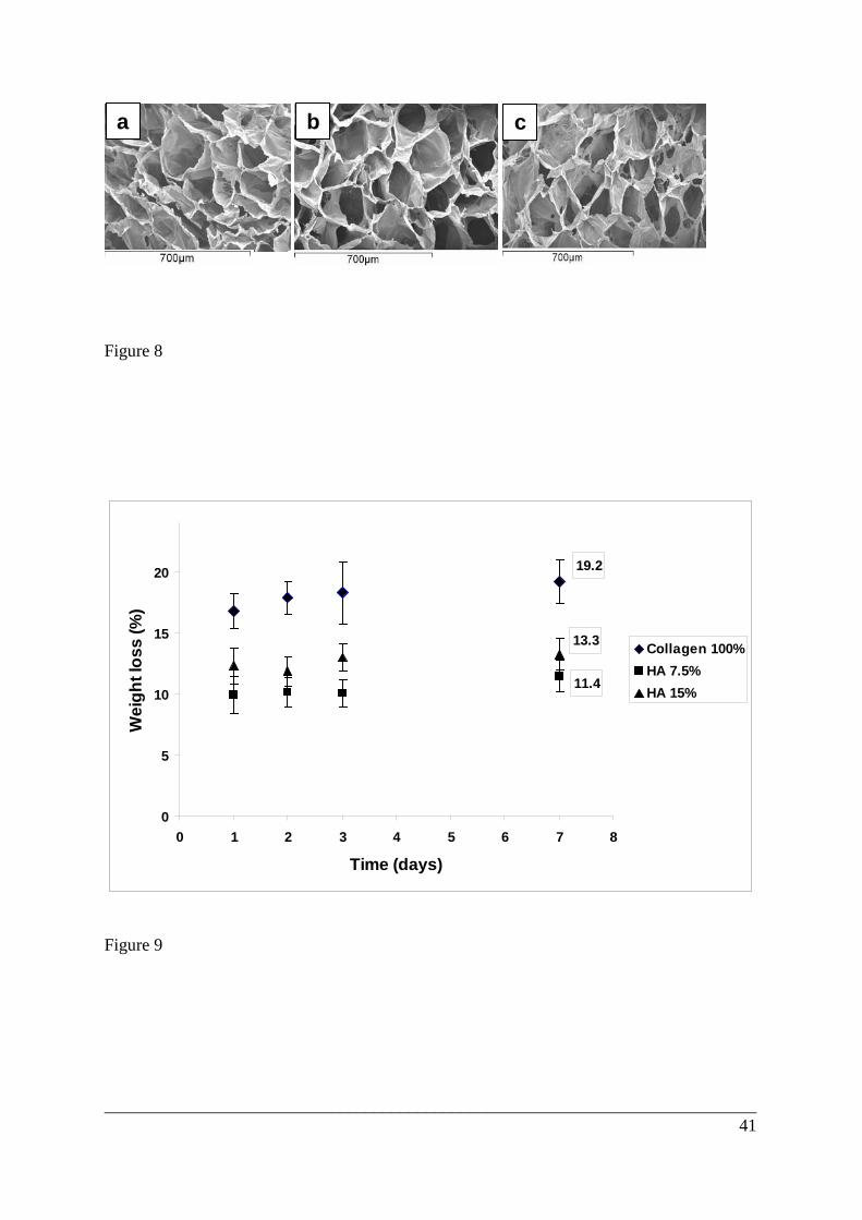
The structural integrity of scaffolds after water-uptake studies was confirmed by
analysing SEM images of samples which were swollen in water at 37°C for 10 days and then
re-frozen and re-lyophilized using the previous freeze-drying cycle. From these images (Fig.
8) it can be seen that there is no visible change in the morphology of the scaffolds, which
indicates that the structural integrity of all scaffold compositions remain unaltered in the
aqueous environment over this period of time. This suggests that even though there is likely
to be some pore closure in water in the scaffolds with lower HA levels, the overall structural
integrity remains good after 10 days.
3.6. Dissolution
The resistance to dissolution of various crosslinked scaffolds is shown in Fig. 9. All
samples appeared to remain intact throughout the dissolution study. The pure collagen
matrices lost more weight during their period in water (19.2% in 7 days), and the majority of

___________________________________________________________________________ 19
mass loss (16.8%) occurred within 24 h , probably due to migration of unbound or weakly
crosslinked collagen molecules from the scaffold structure. The collagen-HA scaffolds
exhibited a slower dissolution rate (11.4% and 13.3% in 7 days for collagen-7.5HA and
collagen-15HA, respectively), which could be a sign of the higher crosslinked degree of these
biopolymer composites. The slightly greater percentage weight loss for scaffold richer in HA
might result from the dissolution and removal of free HA molecules [35, 38, 39], a
phenomenon which should increase with increasing HA content. The increase of the scaffold
resistance to dissolution in the presence of HA could indicate the improvement in the overall
structural integrity of the scaffolds which should delay their hydrolytic dissolution. This
effect is important in the control of mechanical strength of the scaffold during tissue
regeneration. The results obtained are in agreement with those reported for water-uptake
studies (Fig. 6) where the higher swelling percentage of collagen-HA scaffolds as a whole
was explained by the contribution of HA to the intermolecular bonding with collagen, leading
to enhancement of the structural stability of the scaffold matrix in water.
It is appropriate to point out that these results reveal the resistance to dissolution of
the scaffolds under study in an aqueous media lacking in enzymatic activity. However it is
known that hyaluronic acid can be degraded hydrolytically by a family of enzymes called
hyaluronidases (HAse, EC 3.2.1.35) and that in the presence of these enzymes the internal N-
acetyl-hexosamine glucosidic linkages in the hyaluronan structure may be cleaved to liberate
oligosaccharides (residues of glucuronic acid and N-acetylglucosamine). These degradation
products could migrate from the scaffold to the dissolution media, so altering the apparent
rate of their dissolution. As HA based scaffolds developed in this work could be used for a
number of different applications, including those in vitro and in vivo, the assessment of their
dissolution rates in the presence of enzymes would be of interest and of practical use to
determine scaffold stability in specific culture media. On the other hand, in this particular
___________________________________________________________________________ 20
study we believe that the presence of serum in our culture media may act as a blocking agent
to any inherent enzymatic activity. The inhibitory effect of serum on hyaluronidase activity
has been noted in a number of clinical conditions. In addition hyaluronidase has not been
directly implicated in mammary gland development so we restricted the dissolution
experiments solely to assessing scaffold behavior in distilled water.
The hydrolytic degradation of free or weakly bound collagen/HA molecules extracted
from scaffolds caused, in all cases, a very similar drop in pH values (from ∼5.78 to ∼3.6),
which mainly occurred within 24 h of soaking. After this time the pH of all aqueous solutions
remained almost unchanged.
3.7. Mechanical Properties
It has already been demonstrated that scaffold microstructure and stiffness affects the
bioactivity of these systems [57]. The migration speed of cells on two-dimensional (2D)
substrates depends on the stiffness of the substrate. Characterization of the stiffness of the
tissue-engineered scaffold would allow the effect of this parameter on cell activity in a more
realistic 3D environment to be determined.
Different kinds of mechanical testing are used to determine the effect of various
parameters on the stiffness of scaffolds. Many studies carry out tensile testing to determine,
for example, the influence of crosslinking on the mechanical properties of collagen-type
matrices. However, it was shown [43, 49] that the compressive properties such as Young’s
modulus and the compressive strength, are of greater interest when studying the impact of
substrate properties on cellular activity because cells, through their action, tend to bend and
buckle individual struts within the scaffold [58]. So in this work compressive testing in the
wet stage of all scaffolds prepared was carried out to determine the influence of scaffold
composition on the compressive modulus and other important mechanical characteristics of
these matrices.
___________________________________________________________________________ 21
Typical profiles of compressive stress–strain curves obtained for hydrated collagen
and collagen-HA scaffolds are shown in Fig. 10. To assess the scaffold’s behaviour under
successive compressions the same sample specimen were repeatedly testing at least three
times. Strain-stress curves corresponding to this testing (Fig. 10a) revealed no significant
changes in their profiles up to a stain of 0.5 (the second and third testing curves overlay each
other). The profiles are characterized by three distinct regimes: a linear elastic regime, a
collapse plateau and a densification zone (Fig. 10) which are typically observed in
deformation (under compression) of low-density, open-cell foams with an interconnected
network of struts [49, 59]. As the microstructure of the studied scaffolds resembles that of
low-density, open-cell foams, the analogous deformation mechanisms in compression for
both systems are expected. Models describing strain-stress behaviour of these foams [59] can
be used to explain the compressive performance of these scaffolds. According to these
models, each regime of the stress–strain curve provide information about different structural
changes occurring in the 3D network: the linear elastic regime is controlled by struts bending;
a collapse plateau zone reflects struts buckling and the beginning of pore collapse when a
densification regime responds to the completion of pore collapse throughout the material
[59].
Mechanical characteristics which describe stress–strain performance were calculated
from each curve as explained in the Materials section. According to the cellular solid theory,
almost all of these characteristics depend on the relative density (ρ*/ρs) of the foam, so that
for all scaffolds studied this parameter was calculated from the dry density of each freeze-
dried sample (ρ*) and the known dry density of solid collagen (ρs = 1.3 g cm-3) [46] and HA
(ρs = 1.0 g cm-3) [60]. As expected, no significant variation in ρ*/ρs (see Table 2) was found
among the scaffolds. This result could be expected since all scaffolds were produced from the
same suspension concentration (1%-wt) under identical experimental conditions. The effect

___________________________________________________________________________ 22
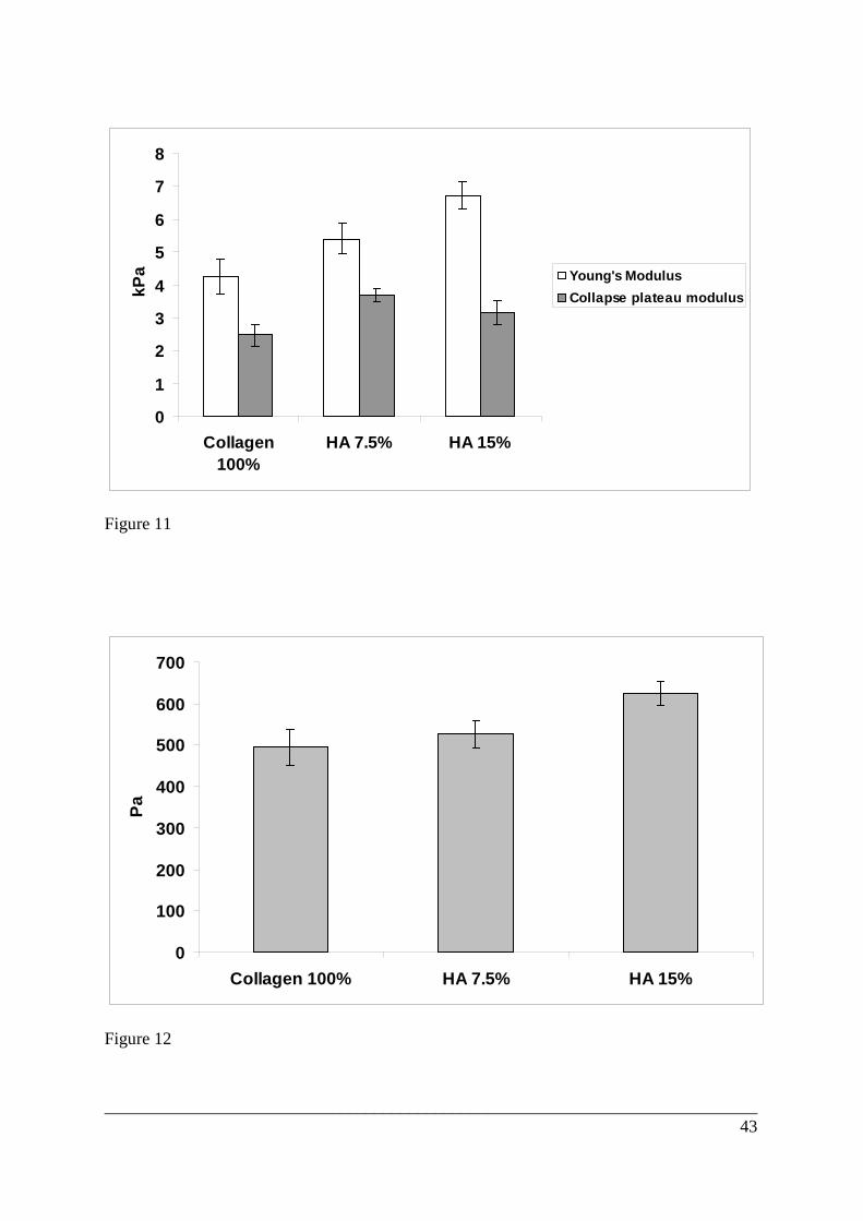
of scaffold composition on all mechanical properties calculated in the wet stage is reported in
Table 2. The mechanical performance of these systems in terms of compressive modulus
(E*), collapse plateau modulus ( and elastic collapse stress (σ*el) are also shown
in Figures 11 and 12. It can be seen that collagen-HA scaffolds exhibited higher values of
these parameters compared to the pure collagen matrix and the increase in HA content
enhanced the values of E* and σ*el (Fig. 11 and 12). In accordance with the cellular solid
theory the Young’s modulus (E*) and elastic plateau stress σ*el (also called the compressive
strength) depend on the foam relative density ( ρ*/ρs), the Young’s modulus of the solid from
which the foam is made (Es), and a constant related to the cell geometry. As ρ*/ρs and cell
geometry are the same for all samples studied, it is logical to assume that the presence of HA
contributes to the enhancement of Es, by increasing the crosslinked density of the scaffold
struts. Therefore, the inclusion of HA in the scaffold composition increases the stiffness of
these systems by the formation of additional crosslinks between collagen and HA molecules,
in this way contributing to the resistance of the struts to bending under compression. These
results are in agreement with those of Harley et al [49] where a significant effect of cross-
linking efficiency on E* and σ*el was observed for collagen-GAG sponges. It was also
reported [45, 47] that the presence of GAG contributes to the formation of extra cross-links
via EDC in collagen-type scaffolds.
The values of collapse plateau modulus were also higher for collagen-HA scaffolds
(Table 2, Fig. 11) but this parameter did not increase steadily with rise in HA content. This
may possibly suggest that the strut buckling (∆σ/∆ε), which is expected to be the primary
mechanism for pore collapse, is not equally affected as strut bending (E*) by stiffness of
scaffold material.
___________________________________________________________________________ 23
On the other hand, elastic collapse stain, ε*e, was found to be constant for all scaffolds
(Table 2). These results are compatible with those obtained for collagen-GAG scaffolds [46]
where no effect of crosslink density on the ε*el – a criterion expected to be characteristic of
the scaffold microstructure and not stiffness- was detected. In addition, these results can be
reasonably explained by the cellular solid theory, which states that the elastic collapse stain is
independent of strut stiffness, Es, and, as a consequence, should not be affected by the
differences in scaffold cross-linking density.
The error in values of mechanical properties could be the result of some heterogeneity
in scaffold microstructures which, as have been shown [61, 62], has a significant effect on
mechanical properties and could contribute to the variation in the measured values.
3.8. Three-dimensional preadipocyte cell culture and adipogenesis
Collagen and collagen-HA scaffolds supported both cell proliferation and adipogenic
differentiation. Cells not exposed to the adipogenic cocktail (control conditions) maintained a
higher proportion of cycling cells than those exposed to the adipogenic cocktail in all scaffold
types, by measure of Ki-67 positive nuclei. In addition cells within collagen-15HA scaffolds
displayed an increased percentage of cycling cells versus those in pure collagen scaffolds
under control conditions, whilst similarly under differentiating conditions cell proliferation in
these scaffolds was elevated over both pure collagen and collagen-7.5HA (p<0.05) (Fig 13
A). An end-stage analysis of adipogenesis was confirmed by oil-red-O staining of lipid
containing vesicles in differentiated 3T3-L1 cells in both pure collagen and HA mixed
scaffolds (Fig 13 B,C. Lipid (l) indicated by arrows). It was noted that lipid was more
confined to the edges of HA mixed scaffolds than pure collagen (data not shown). Gene
expression of adipsin, an enzyme involved in lipid metabolism predominantly located in
mature adipocytes [63] was significantly elevated in HA-mixed collagen scaffolds (p<0.05,
student T-test) subjected to 8 days of adipogenic culture against control culture conditions
___________________________________________________________________________ 24
(Fig 13 D). In a similar manner PPARγ, a principle transcription factor of adipogenesis [64]
was also elevated in HA mixed scaffolds compared to control samples, although unlike
adipsin expression, PPARγ exhibits a transient expression profile throughout an adipogenic
time course [65] meaning that any significant modulation in the expression of this gene may
have been missed. A concominant role for proliferation with adipogenesis has been
described, where re-entry into the cell cycle with several rounds of mitotic division following
confluence are a preliminary feature of the adipocyte terminal differentiation programme
[66]. Therefore, by supporting cell proliferation, the inclusion of HA to collagen scaffolds
may aid adipogenesis both by hastening cell-contacted growth arrest prior to adipogenic
conversion, a noted requirement for adipocyte conversion [65], and support the early stages
of the differentiation programme itself. Although hyaluronic acid based scaffolds have been
shown to be supportive of adipocyte culture in vivo [31] and in vitro [32,33], this is the first
time that varying HA concentration within a 3D substrate has been shown to influence both
the differentiation and proliferation characteristics of 3T3-L1 preadipocytes. Further work
will more comprehensively investigate the performance of these scaffolds in vivo and in vitro,
specifically as 3D substrates for mammary fat-pad tissue engineering, supporting the
development of both stromal and epithelial mammary tissues.
4. Conclusions
Highly porous collagen-HA scaffolds containing different amounts of HA, with a
uniform interconnected pore structure of diameter 150-200 µm, were produced in this study
by a controlled freeze-drying technique. Crosslinking of collagen-HA scaffolds using an
EDS/NHS system proves to be an easy, effective and contaminant-free technique. The
characteristic of the scaffolds obtained is that of a fairly uniform, uni-axial, interconnecting
___________________________________________________________________________ 25
porous network, which should assist cell ingrowths, rapid vascularisation, and transfer of
oxygen, nutrients and waste products [15-17].
XRD and FTIR analyses indicated that no other chemical entities were introduced into
the crosslinked scaffolds, except for the formation of stable amide linkages. As such, the
ability to custom-make collagen-HA scaffolds with appropriate ratios, can be easily
controlled by altering the initial ratios of the two starting biopolymers.
Swelling tests showed that the water uptake of the scaffold as a whole was higher for
collagen-HA samples and increased with HA content. However, the scaffold material alone,
(once the water in the pores was discounted) showed the opposite trend. The pure collagen
sample also lost more weight during the period of dissolution study, and the majority of mass
loss occurred within 24 h. The collagen-HA scaffolds exhibited a slower dissolution rate in
distilled water due to the higher degree of cross-linking in the biopolymer composite. These
phenomena could be due to the enhancement of bonding between collagen and HA during
crosslinking and improvement in the overall structural integrity of the scaffold, thereby
delaying the process of dissolution. This effect is important in the control of mechanical
strength of the scaffold during cell culture and tissue regeneration.
The increase in HA content gives rise to an increase in the stiffness of these systems
which enhanced the values of the mechanical characteristics by the formation of additional
crosslinks between collagen and HA molecules, and thus contributed to the resistance of the
struts to bending under compression.
Collagen HA mixed scaffolds increased the proportion of proliferative 3T3-L1
preadipocytes in addition to enhancing adipogenic conversion with adipogenic media
supplementation.
These results indicate that freeze-dried collagen mixed HA scaffolds may offer robust,
freely permeable three-dimensional matrices that enhance mammary stromal tissue

___________________________________________________________________________ 26
development in vitro. In summary, collagen-HA can be considered as a valuable biomaterial
for constructing tissue-specific scaffolds for adipose tissue engineering applications.
Acknowledgements
The authors acknowledge support from the Biotechnology and Biological Sciences
Research Council, United Kingdom and the NC3Rs initiative.
___________________________________________________________________________ 27
References
1. Sakakura T, Nishizuka Y, Dawe CJ. Mesenchyme-dependent morphogenesis and
epithelium-specific cytodifferentiation in mouse mammary-gland. Science
1976;194(4272):1439-1441.
2. Naylor M, Ormandy C. Mouse strain-specific patterns of mammary epithelial ductal side
branching are elicited by stromal factors. Dev Dyn 2002;225(1):100-105.
3. Beahm E, Walton R, Patrick Jr CW. Progress in adipose tissue construct development.
Clin Plast Surg 2003;30:547-558.
4. Patrick Jr CW. Breast tissue engineering. Ann Review Biomed Eng 2004;6: 109-130.
5. Soofi LJA, Liliensiek SJ, Nealey PF,Murphy CJ. The elastic modulus of MatrigelTM as
determined by atomic force microscopy. J Struct Biol 2009;167:216-219.
6. Gentleman E. NEA, Livesay GA, Dee KC. Collagen composite biomaterials resist
contraction while allowing development of adipocytic soft tissue in vitro. Tissue Eng
2006;12(6):1639-1649.
7. Zhu YK, Umino T, Liu XD, Wang HJ, Romberger DJ, Spurzem JR, Rennard SI.
Contraction of fibroblast-containing collagen gels: initial collagen concentration regulates
the degree of contraction and cell survival. In Vitro Cell Dev-An 2001;37:10-16.
8. Shimonishi M, Sato J, Takahashi N, Komatsu M. Expression of type IV collagen and
laminin at the interface between epithelial cells and fibroblasts from human periodontal
ligament. Eur J Oral Sci 2005;113(1):34.
9. Che Z, Jung T, Choi J, Yoon D, Jeong H, Lee E, et al. Collagen-based co-culture for
invasive study on cancer cells-fibroblasts interaction. Biochemical and biophysical research
communications, 2006. p. 268-275.
___________________________________________________________________________ 28
10. Phillips JB. Tissue engineered approaches to modelling the nervous system. Keynote
lecture at “Tissue Engineering: a new dimension to animal replacement”, NC3Rs&BBSRS,
1-2April, London, UK, 2009.
11. Brown RA, Phillips JB. Cell responses to biomimetic protein scaffolds used in tissue
repair and engineering. Int Rev Cytol 2007;262:75-150.
12. O’Brien FJ, Harley BA, Yannas IV, Gibson LJ. The effect of pore size on cell adhesion
in collagen–gag scaffolds. Biomaterials 2005;26:433–41.
13. Zeltinger J, Sherwood JK, Graham DA, Mueller R, Griffith LG. Effect of pore size and
void fraction on cellular adhesion, proliferation, and matrix deposition. Tissue Eng
2001;7:557–72.
14. Wake MC, Patrick Jr CW, Mikos AG. Pore morphology effects on the fibrovascular
tissue growth in porous polymer substrates. Cell Transplant 1994;3:339–43.
15. Cao Y, Croll TI, Lee JG, Tuch BE, Cooper-White JJ. Scaffolds, stem cells, and tissue
engineering: a potent combination. Aust J Chem 2005;58:691.
16. Hutmacher DW. Scaffold design and fabrication technology for engineering tissues -
state of the art and future perspectives. J Biomr Sci-Polym E 2001;12:107-124.
17. Ma PX. Scaffolds for tissue fabrication. Mater Today 2004;7(5):30-40.
18. Peyton SR, Putnam AJ. Extracellular matrix rigidity governs smooth muscle cell motility
in a biphasic fashion. J Cell Physiol 2005;204:198–209.
19. Grinnell F, Ho CH, Tamariz E, Lee DJ, Skuta G. Dendritic fibroblasts in three-
dimensional collagen matrices. Mol Biol Cell 2003;14:384–95.
20. Yeung T et al. Effects of substrate stiffness on cell morphology, cytoskeletal structure,
and adhesion. Cell Motil Cytoskel 2005;60:24–34.
21. Chapekar MS. Tissue Engineering: Challenges and Opportunities. J Biomed Mater Res
2000;53:617-620.

___________________________________________________________________________ 29
22. Lee CH, Singla A, Lee Y. Biomedical applications of collagen. Int J Pharm 2001;221:1-
22.
23. Nimni ME, Cheung D, Strates B, Kodama M, Sheikh K. Chemically modified collagen: a
natural biomaterial for tissue replacement. J Biomed Mater Res 1987;21:741-771.
24. Hilliou F, Pairault J, Dominice J, Redziniak G. Growth and differentiation of 3T3-F442A
preadipocytes in three-dimensional gels of native collagen. Exp Cell Res 1988;177:372-381.
25. Huss FR, Kratz G. Mammary epithelial cell and adipocyte co-culture in a 3-D matrix: the
first step towards tissue-engineered human breast tissue. Cells Tissues Organs 2001;169:361-
367.
26. Brekke JH, Thacker K. In: Hyaluronan as a Biomaterial (S. Guelcher, J.O. Hollinger,
Eds.) An Introduction to Biomaterials, CRC Press, Boca Raton: Florida, 2005. p. 219-240.
27. West D, Kumar S. The effect of hyaluronate and its oligosaccharides on endothelial cell
proliferation and monolayer integrity. Exp Cell Res 1989;183(1):179-196.
28. Banerjee S, Toole B. Hyaluronan-binding protein in endothelial cell morphogenesis. J
Cell Biol 1992;119(3):643.
29. Gerecht S, Burdick J, Ferreira L, Townsend S, Langer R, Vunjak-Novakovic G.
Hyaluronic acid hydrogel for controlled self-renewal and differentiation of human embryonic
stem cells. Proc Nat Acad Sci 2007;104(27):11298.
30. Hemmrich K, von Heimburg D, Rendchen R, Di Bartolo C, Milella E, Pallua N.
Implantation of preadipocyte-loaded hyaluronic acid-based scaffolds into nude mice to
evaluate potential for soft tissue engineering. Biomaterials 2005; 26:7025-7037.
31. Flynn L, Prestwich GD, Semple JL, Woodhouse KA. Adipose tissue engineeringin
vivowith adipose-derived stem cells on naturally derived scaffolds. J Biomed Mater Res
2009; 89A(4): 929-941.

___________________________________________________________________________ 30
32. Flynn L, Prestwich G, Semple J, Woodhouse K. Adipose tissue engineering with
naturally derived scaffolds and adipose-derived stem cells. Biomaterials 2007;28(26):3834-
3842.
33. Halbleib M, Skurk T, de Luca C, von Heimburg D, Hauner H. Tissue engineering of
white adipose tissue using hyaluronic acid-based scaffolds. I: in vitro differentiation of
human adipocyte precursor cells on scaffolds. Biomaterials 2003; 24:3125-3132.
34. Huang-Lee LLH, Nimmi M. Crosslinked CNBr-activated hyaluronan-collagen matrices:
Effects on fibroblast contraction. Matrix Biol 1994;14:147-157.
35. Park SN, Lee HJ, Lee KH, Suh H. Biological characterization of EDC-crosslinked
collagen-hyaluronic acid matrix in dermal tissue restoration. Biomaterials 2003;24:1631-
1641.
36. Tang S, Spector M. Incorporation of hyaluronic acid into collagen scaffolds for the
control of chondrocyte-mediated contraction and chondrogenesis. Biomed Mater
2007;2:135-141.
37. Kim TG, Chung HJ, Park TG. Macroporous and nanofibrous hyaluronic acid/collagen
hybrid scaffold fabricated by concurrent electrospinning and deposition/leaching of salt
particles. Acta Biomater 2008;doi:10.1016/j.actbio.2008.06.008.
38. Park SN, Park JC, Kim HO, Song MJ, Suh H. Characterization of porous
collagen/hyaluronic acid scaffold modified by 1-ethyl-3-(3-dimethylaminopropyl)
carbodiimide cross-linking. Biomaterials 2002;23:1205-1212.
39. Rehakova M, Bakos D, Vizarova K, Soldan M, Jurickova M. Properties of collagen and
hyaluronic acid composite materials and their modification by chemical crosslinking. J
Biomed Mater Res 1996;30:369-372.
40. Tang S, Vickers SM, Hsu HP, Spector M. Fabrication and characterization of porous
hyaluronic acid-collagen composite scaffolds. J Biomed Mater Res 2007;82A:323-335.

___________________________________________________________________________ 31
41. O’Brien FJ, Harley BA, Yannas IV, Gibson L. Influence of freezing rate on pore
structure in freeze-dried collagen-GAG scaffolds. Biomaterials 2004;25:1077-1086.
42. Ueda H, Hong L, Yamamoto M, Shigeno K, Inoue M, Toba T, Yoshitani M, Nakamura
T, Tabata Y, Shimuzu Y. Use of collagen sponge incorporating transforming growth factor-
beta1 to promote bone repair in skull defects in rabbits. Biomaterials 2002;23:1003-1010.
43. Haugh MG. The development of novel scaffolds for tissue engineering with a range of
structural and mechanical properties. PhD Thesis, Trinity College Dublin, Ireland, 2008.
44. Hollister SJ, Maddox RD, Tabas JM. Optimal design and fabrication of scaffolds to
mimic tissue properties and satisfy biological constraints. Biomaterials 2002;23:4095-4103.
45. Pieper JS, Oosterhol A, Dijkrsta PJ, Veerkamp JH, van Kuppevelt TH. Preparation and
characterization of porous crosslinked collagenous matrices containing bioavailable
chondroitin sulphate. Biomaterials 1999;20:847-858.
46. Khor E. Methods for the treatment of collagenous tissues for bioprostheses Biomaterials
1997;18:95-105.
47. Pieper JS, Hafmans T, Veerkamp JH, van Kuppevelt TH. Development of tailor-made
collagen–glycosaminoglycan matrices: EDC/NHS crosslinking, and ultrastructural aspects.
Biomaterials 2000;21:581-593.
48. Wang X, Seed B. A PCR primer bank for quantitative gene expression analysis. Nucleic
Acids Res 2003;31(24):e154.
49. Harley BA, Leung JH, Silva ECCM, Gibson LJ. Mechanical characterization of
collagen–glycosaminoglycan scaffolds. Acta Biomater 2007;3:463–474.
50. Camacho Nr, West P,Torzilli PA,Mendelson R. FTIR Microscopic Imaging og Collagen
and Proteoglican in Bovin Cartilage. Biopolymers (Biospectroscopy) 2001;62,1-8.
51. Wang XH, Li DP, Wang WJ, Feng QL, Cui FZ, Xu YX, Song XH, van der Wert M.
Biomaterials 2003;24:3213-3220.
___________________________________________________________________________ 32
52. Pietrucha K. Changes in denaturation and rheological properties of collagen-hyaluronic
acid scaffolds as a result of temperature dependences. Int J Biol Macromol 2005;36:299-304.
53. Tsereteli GI, Belopolskaya TV, Grunina NA. Dehydreted native biopolymers – a unique
representative of glassy systems. J Therm Anal Calorim 2008;92:711–716
54. Friess W, Lee G. Basic thermoanalytical studies of insoluble collagen matrices.
Biomaterials 1996;17:2289-2294.
55. Engler A, Bacakova L, Newman C, Hategan A, Griffin M, Discher D. Substrate
compliance versus ligand density in cell on gel responses. Biophys J 2004;J86:617–28.
56. Wang Y, Yang C, Chen X, Zhao N. Development and characterization of novel
biomimetic composite scaffolds based on bioglass-collagen-hyaluronic acid-
phosphatidylserine for tissue engineering applications. Macromol Mater Eng 2006;291:254-
262.
57. Yannas IV. Tissue and organ regeneration in adults. New York: Springer, 2001.
58. Freyman TM, Yannas IV, Pek Y-S, Yokoo R, Gibson LJ. Micromechanics of fibroblast
contraction of a collagen–gag matrix. Exp Cell Res 2001;269:140–53.
59. Gibson LJ, Ashby MF. Cellular solids: structure and properties. 2nd ed. Cambridge, UK:
Cambridge University Press, 1997.
60. Gomez-Alejandre S, Sanchez de la Blanca E, Abradelo de Usera C, Rey-Stolle MF,
Hernandez-Fuentes I. Partial specific volume of hyaluronic acid in different media and
conditions Int J Biol Macromol 2000;27:287–290.
61. Onck PR, Andrews EW, Gibson LJ. Size effects in ductile cellular solids. Part I
Modelling. Int J Mech Sci 2001;43:681–99.
62. Andrews EW, Gioux G, Onck P, Gibson LJ. Size effects in ductile cellular solids. Part II:
Experimental results. Int J Mech Sci 2001;43:701–13.

___________________________________________________________________________ 33
63. Wilkison W, Min H, Claffey K, Satterberg B, Spiegelman B. Control of the adipsin gene
in adipocyte differentiation. Identification of distinct nuclear factors binding to single-and
double-stranded DNA. J Biol Chem 1990;265(1):477.
64. Fajas L, Debril M, Auwerx J. Peroxisome proliferator-activated receptor-gamma: from
adipogenesis to carcinogenesis. J Mol Endocrinol 2001;27(1):1.
65. Avram M, Avram A, James W. Subcutaneous fat in normal and diseased states 3.
Adipogenesis: from stem cell to fat cell. J Am Acad Dermatol 2007;56(3):472-492.
66. Richon V, Lyle R, McGehee R. Regulation and expression of retinoblastoma proteins
p107 and p130 during 3T3-L1 adipocyte differentiation. J Biol Chem 1997;272(15):10117.
67. Ailhaud G, Dani C, Amri E, Djian P, Vannier C, Doglio A, et al. Coupling growth arrest
and adipocyte differentiation. Environ Health Persp 1989;80:17.
___________________________________________________________________________ 34
Captions
Figure 1 µCT images of different cross-sections of freeze-dried scaffolds. (A) pure
collagen, transverse view and (B-C) longitudinal views of B-collagen, C-
collagen-7.5HA and (d) collagen-15HA. A: area 1- larger pore zone; A: area
2- material densification zone.
Figure 2 SEM micrographs of the longitudinal sections of freeze-dried scaffolds with
different content of HA (a)-collagen 100%; (b)-collagen-7.5HA; c)- collagen-
15HA. SEM micrographs of the transverse sections of the same scaffolds: (d)-
collagen 100%; (e)-collagen-7.5HA; f)- collagen-15HA
Figure 3 XRD patterns of pure collagen and various collagen-HA scaffolds
Figure 4 FTIR spectra of pure collagen and various collagen-HA scaffolds
Figure 5 Example of TG and DSC profile of thermal decomposition of scaffolds
Figure 6 The effect of scaffold composition on the overall water uptake at different
soaking time
Figure 7 Swelling percentage corresponding to the scaffold material itself in
dependence of its composition and soaking time
Figure 8 SEM micrographs of the longitudinal sections of freeze-dried scaffolds after
being swollen in water at 37 oC 10 days: (a)-collagen 100%; (b)-collagen-
7.5HA; c) - collagen-15HA.
Figure 9 Weight loss (%) of different scaffold compositions versus time in distilled
water at 37 0 C
Figure 10 Typical profiles of compressive stress–strain curves observed for hydrated
collagen and Coll-HA scaffolds:
___________________________________________________________________________ 35
a) Three repeated testing of the same scaffold specimen in the entire strain
range (ε: 0-0.8): 1- linear elastic zone; 2- collapse plateau; 3- densification
regime.
b) Strain sub- range ε: 0-0.4 to better show the linear elastic region and the
calculation of mechanical properties
Figure 11 Linear elastic (Young’s) modulus (E*) and Collapse plateau modulus (∆σ/∆ε)
for all hydrated scaffolds
Figure 12 Elastic plateau stress σ*el (compressive strength) for all hydrated scaffolds
Figure 13 Cell proliferation and adipogenesis of 3T3-L1 preadipocytes within collagen
and collagen-mixed HA scaffolds. Cell proliferation (A) following 8 days of
adipogenic culture expressed as the number of Ki-67 positive cells versus total
cell numbers per frame. n=8, * indicates significant elevation versus all other
groups by student T-test (p<0.05). Lipid staining by Oil-Red O following 8
days of adipogenic culture within collagen 100% (B) and collagen-7.5HA (C)
scaffolds. S and l indicate regions of scaffold and lipid respectively (bar
indicates 100µm). Mean quantitative PCR for Adipsin (D) and PPARγ (E)
mRNA abundance relative to a pooled sample standard and normalized to
housekeeping gene cyclophillin A. n≥4 for each sample.
___________________________________________________________________________ 36
Figure 1
Figure 2
a c b
d e f
1 2
A B C D

___________________________________________________________________________ 37
5 10 15 20 25 30 35
2Theta (degrees)
Inte
nsity
(a.u
.)characteristic of equatorial
peak of collagen
characteristic of interchain spacingof collagen triple helix
pure collagen
collagen - 7.5HA
collagen - 15HA
Figure 3
1000 1500 2000 2500 3000 3500 4000
Wavenumber (cm-1)
Inte
nsity
(a.u
.)
Amide N-H stretching vibration
Amide I band due toC=O stretching vibration
Amide II band due to motion combining both theamide N-H deformation and C-N stretching vibrationSecondary
amide
Primary amide
pure collagen
collagen - 7.5HA
collagen - 15HA
Figure 4

___________________________________________________________________________ 38
Figure 5

___________________________________________________________________________ 39
Figure 6

___________________________________________________________________________ 40
Figure 7
___________________________________________________________________________ 41
Figure 8
19.2
11.4
13.3
0
5
10
15
20
0 1 2 3 4 5 6 7 8
Time (days)
Wei
ght l
oss
(%)
Collagen 100%HA 7.5%HA 15%
Figure 9
a b c

___________________________________________________________________________ 42
a)
0
1000
2000
3000
4000
5000
6000
7000
8000
9000
10000
0 0.1 0.2 0.3 0.4 0.5 0.6 0.7 0.8 0.9
Strain
Stre
ss, P
a
First TestingSecond TestingThird Testing
1 32
b)
E*
0
200
400
600
800
1000
1200
1400
0 0.1 0.2 0.3 0.4 0.5
Strain
Stre
ss, P
a
ε∗el
∆σ/∆ε
σ∗ el
Figure 10
___________________________________________________________________________ 43
0
1
2
3
4
5
6
7
8
Collagen100%
HA 7.5% HA 15%
kPa
Young's ModulusCollapse plateau modulus
Figure 11
0
100
200
300
400
500
600
700
Collagen 100% HA 7.5% HA 15%
Pa
Figure 12

___________________________________________________________________________ 44
Figure 13
___________________________________________________________________________ 45
Table 1 DSC and TG analysis of Scaffolds.
Td: thermal denaturation temperature; T1-T3: maximum temperatures for thermal transitions;
Δm1: mass loss at denaturation transition; Δm2: mass loss at T1-T3 transitions; Δm3: total
mass loss at 496 0C.
Scaffold Td (0C) Δm1 (%)
T1 T2 T3 Δm2 (%)
Δm3 (%)
Collagen
100%
79.4± 3.4 10.6 177.5 225.4
310.1 67.1 77.7± 2.0
Collagen
-7.5HA
80.2±2.6 11.2 180.4 218.3
306.7
63.2 74.4± 1.5
Collagen
-15HA
86.2±1.7 11.3 182.5 219.7
296.0
62.3 73.6± 2.1
Note: Each parameter is the mean of two readings
Table 2 Mechanical properties and relative density of the tested scaffolds
Scaffolds
Relative Density
ρ*/ρs
Young's modulus E* (kPa)
Collapse plateau modulus
∆σ/∆ε(kPa)
Elastic collapse stress
σ* el (Pa)
Elastic collapse strain
ε* el
Collagen 100%
0.0113±0.0003 4.26±0.53 2.48±0.34 495±43 0.11±0.01
Collagen-
7.5HA 0.0124±0.0006
5.39±0.46 3.69±0.19 526±33 0.09±0.01
Collagen-15HA
0.0121±0.0008 6.73±0.41 3.17±0.36 625±29 0.10±0.01
Note: Each parameter is the average value of five separate measurements