declarations: management of a pulmonary arteriovenous
TRANSCRIPT

Li et al. BMC Surg (2021) 21:101 https://doi.org/10.1186/s12893-021-01103-8
CASE REPORT
Declarations: management of a pulmonary arteriovenous fistulae by uniportal video‐assisted thoracoscopic surgery: a case reportR. Li1, Y. Zhou1, S. Kang2, F. Kong1, L. Guan1, Y. Zhao1 and X. Yin1*
Abstract
Background: A pulmonary arteriovenous fistula (PAVF) is arare condition that is associated with pulmonary arterio-venous malformation(PAVM). Few reports have described managing PAVMs using uniportalvideo-assisted thoraco-scopic surgery (VATS).
Case presentation: A 13-year-old child with PAVF in the left inferior pulmonary artery was treated by uniportal VATS with left lower lobectomy. After surgery, hemoptysis did not recur and there were no postoperative complications. Six months after the operation, postoperative review of computerized tomography showed no recrudescence of PAVF.
Conclusions: PAVF is a rare case that should be diagnosed and treated early. 3D- computerized tomography (CT) reconstruction is useful for diagnosis and preoperative assessment. The case shows that PAVF can be managed with uniportal VATS.
Keywords: Pulmonary arteriovenous fistula, 3D-CT reconstruction, Uniportal VATS, Case report
© The Author(s) 2021. Open Access This article is licensed under a Creative Commons Attribution 4.0 International License, which permits use, sharing, adaptation, distribution and reproduction in any medium or format, as long as you give appropriate credit to the original author(s) and the source, provide a link to the Creative Commons licence, and indicate if changes were made. The images or other third party material in this article are included in the article’s Creative Commons licence, unless indicated otherwise in a credit line to the material. If material is not included in the article’s Creative Commons licence and your intended use is not permitted by statutory regulation or exceeds the permitted use, you will need to obtain permission directly from the copyright holder. To view a copy of this licence, visit http://creat iveco mmons .org/licen ses/by/4.0/. The Creative Commons Public Domain Dedication waiver (http://creat iveco mmons .org/publi cdoma in/zero/1.0/) applies to the data made available in this article, unless otherwise stated in a credit line to the data.
BackgroundA pulmonary arteriovenous fistula (PAVF) is a rare con-dition, first described by Churton in 1897 [1], that is associated with pulmonary arteriovenous malforma-tion (PAVM), which allows abnormal direct communi-cation between the pulmonary arteries and pulmonary veins [2]. The most common cause of PAVM is heredi-tary hemorrhagic telangiectasia, also known as Osler–Weber–Rendu syndrome [3]. Patients without obvious symptoms account for approximately 57 % of PAVF cases in the early stage of the disease. However, PAVF can also exhibit diverse symptoms, including repeated hemop-tysis, nosebleeds, difficulty catching breath, an increase in hemoglobin levels, and hemoptysis, which can lead to sudden fatal rupture of the veins [4, 5]. Patients with
these symptoms are treated with interventional therapy [6] or surgical procedures including lung wedge resection, lobectomy, and pneumonectomy [7, 8]. The following is a case report of a 13-year-old patient with PAVF in the left inferior pulmonary. The patient, who exhibited hemopty-sis and dyspnea, was diagnosed by preoperative 3D-com-puted tomography (3D-CT) reconstruction and treated by uniportal video-assisted thoracoscopic surgery (VATS).
Case presentationA 13-year-old child developed symptoms of mild hem-optysis and dyspnea that persisted for more than two months and did not respond to oral medication. Physical examination showed only a mild cyanosis of lips, with no other obvious findings. The patient had no relevant previ-ous medical history and there was no relevant family his-tory. Chest radiography indicated that a mass was located in the left inferior pulmonary. Non-enhanced chest computerized tomography (CT) and subsequent com-puterized tomographic angiography (CTA) confirmed
Open Access
*Correspondence: [email protected] Department of Thoracic Surgery, The First Affiliated Hospital of Kunming Medical University, Kunming 650032, Yunnan, ChinaFull list of author information is available at the end of the article
Page 2 of 4Li et al. BMC Surg (2021) 21:101
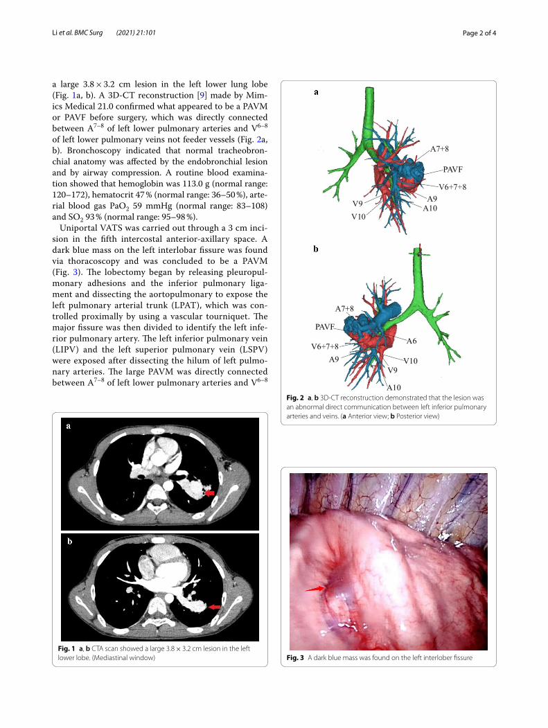
a large 3.8 × 3.2 cm lesion in the left lower lung lobe (Fig. 1a, b). A 3D-CT reconstruction [9] made by Mim-ics Medical 21.0 confirmed what appeared to be a PAVM or PAVF before surgery, which was directly connected between A7–8 of left lower pulmonary arteries and V6–8 of left lower pulmonary veins not feeder vessels (Fig. 2a, b). Bronchoscopy indicated that normal tracheobron-chial anatomy was affected by the endobronchial lesion and by airway compression. A routine blood examina-tion showed that hemoglobin was 113.0 g (normal range: 120–172), hematocrit 47 % (normal range: 36–50 %), arte-rial blood gas PaO2 59 mmHg (normal range: 83–108) and SO2 93 % (normal range: 95–98 %).
Uniportal VATS was carried out through a 3 cm inci-sion in the fifth intercostal anterior-axillary space. A dark blue mass on the left interlobar fissure was found via thoracoscopy and was concluded to be a PAVM (Fig. 3). The lobectomy began by releasing pleuropul-monary adhesions and the inferior pulmonary liga-ment and dissecting the aortopulmonary to expose the left pulmonary arterial trunk (LPAT), which was con-trolled proximally by using a vascular tourniquet. The major fissure was then divided to identify the left infe-rior pulmonary artery. The left inferior pulmonary vein (LIPV) and the left superior pulmonary vein (LSPV) were exposed after dissecting the hilum of left pulmo-nary arteries. The large PAVM was directly connected between A7–8 of left lower pulmonary arteries and V6–8
Fig. 1 a, b CTA scan showed a large 3.8 × 3.2 cm lesion in the left lower lobe. (Mediastinal window)
Fig. 2 a, b 3D-CT reconstruction demonstrated that the lesion was an abnormal direct communication between left inferior pulmonary arteries and veins. (a Anterior view; b Posterior view)
Fig. 3 A dark blue mass was found on the left interlober fissure

Page 3 of 4Li et al. BMC Surg (2021) 21:101
of left lower pulmonary veins, and this was consistent with the results of preoperative 3D-CT reconstruc-tion. The left inferior pulmonary artery and vein were ligated using staplers (Fig. 4a, b). The bronchus of the left lower lobe was then clamped by using staples and removed during the operation. A pathological diagnosis of PAVF was confirmed (Fig. 5).
Hemoptysis and postoperative complications did not occur after surgery. The tube was removed after review-ing the chest X-ray film for pneumothorax and abnor-mal pleural effusion. The patient was discharged from hospital five days after surgical resection. Six months after the operation, postoperative review of computed tomography showed no recrudescence of PAVF (Fig. 6).
Discussion and conclusionsThe condition of PAVF occurs mainly due to defects in pulmonary capillary development. Abnormal commu-nications between pulmonary arteries and veins form a right-to-left shunt causing blood flow without oxygena-tion and an increase in red cells. Complications may be caused by PAVMs due to direct intercommunica-tion between pulmonary arteries and pulmonary veins, including exertional dyspnea, swirl, cyanopathy, poly-cythemia, bacterial infections, and brain abscesses. It can appear as a mass on routine chest radiography and abnormal direct communication between pulmonary arteries and pulmonary veins on CT or 3D-CT.
Appropriate therapies for PAVF are interventional therapy or surgical treatment. As in this case, a patient with serious PAVM and hemoptysis should be treated to prevent possible severe dyspnea due to life-threat-ening hemorrhage and hypoxemia caused by PAVM. In this case, because the diameter of the PAVF was limited to a lobe and was larger than 2 cm, with a.
Fig. 4 Sketch profile of the pulmonary arteriovenous fistula
Fig. 5 A pathological diagnosis of PAVF
Fig. 6 Postoperative review of computed tomography showed no recrudescence of PAVF. (Mediastinal window)

Page 4 of 4Li et al. BMC Surg (2021) 21:101
feeding artery over 3 mm in diameter, it was decided that interventional therapy would not solve the prob-lem and that a surgical procedure should be chosen if the operative risk could be controlled. Because the PAVF of the patient was located deeply, it was hard to remove using transcatheter embolotherapy and lung wedge resection. Therefore, left lower lobectomy was selected.
Three points were analyzed to reduce the thoracoscopic surgical risk during this operation. The 3D-CT recon-struction was used to confirm the structure of the PAVM before the operation. Preoperative simulation based on 3D-CT reconstruction is useful for accurate diagnosis, and in this case, 3D-CT reconstruction revealed a huge malformed hemangioma that was directly connected between the trunk of left lower pulmonary arteries and the left lower pulmonary veins not feeder vessels, which was coincident with the intraoperative situation. The use of 3D-CT reconstruction also showed the anatomical variation of the blood vessels, so life-threatening hem-orrhage could be avoided during surgery. Risk analysis through 3D-CT reconstruction indicated that the left pulmonary trunk should be controlled proximally by using a vascular tourniquet to avoid uncontrolled arte-rial bleeding. This control of the left pulmonary trunk allowed the distally involved pulmonary parenchyma to be safely resected. More importantly, precise patient positioning for thoracoscopic pulmonary lobectomy allowed a suitable surgical plan for the operating surgeon and reduced the risks involved in thoracoscopic surgery. Few cases of PAVF have been published, so no outcome data were available for this surgical procedure. Uniportal VATS was performed for the PAVF in this case, because it was a secure and feasible technique in thoracic surgery and provided a better surgical field. To reduce surgical trauma, there was only one surgical hole. As such, there were fewer complications, less pain, and, consequently, a shorter postoperative hospital stay [10].
In general, PAVF is a rare case which should be diag-nosed and treated early. 3D-CT reconstruction may be useful for diagnosis and preoperative assessment. The case could provide the experience and feasibil-ity for PAVF managed with uniportal video-assisted thoracoscopy.
AbbreviationsPAVF: Pulmonary arteriovenous fistulae; PAVM: Pulmonary arteriovenous malformation; 3D-CT: Three dimensional-computed tomography; VATS: Video-assisted thoracoscopic surgery; CT: Computed tomography; CTA : Computed tomograhy angiograhy; LPAT: Left pulmonary arterial trunk; LIPV: Left inferior pulmonary vein; LSPV: Left superior pulmonary vein.
AcknowledgementsWe would like to thank X Yin and Y Zhao, at The First Affiliated Hospital of Kunming Medical University, for study design and critical review. We thank
International Science Editing (http://www.inter natio nalsc ience editi ng.com) for editing this manuscript.
Authors’ contributionsRL is the first author of the manuscript. SK made the 3D-CT reconstruction. YZ, FK and LG treated the patient after surgery. XY and YZ performed the operation, and substantially contributed to the drafting and revision of the manuscript. XY is the corresponding author. All authors read and approved the final manuscript.
FundingNone.
Availability of data and materialsThe data and materials sets support the conclusions of this article.
Ethics approval and consent to participateNot applicable.
Consent for publicationWritten informed consent for the publication of these case reports and accompanying images was obtained from the guardians of our patients.
Competing interestsAll authors declare that they have no competing interests.
Author details1 Department of Thoracic Surgery, The First Affiliated Hospital of Kunming Medical University, Kunming 650032, Yunnan, China. 2 Department of Imaging, The First Affiliated Hospital of Kunming Medical University, Kunming 650032, Yunnan, China.
Received: 30 September 2020 Revised: 11 February 2021 Accepted: 15 February 2021
References 1. Churton T. Leeds and west-riding medico-chirurgical society: multiple
aneurysms of pulmonary artery. Brit Med J. 1897;1:1223. 2. Shovlin CL. Pulmonary arteriovenous malformations. Am J Respir Crit Care
Med. 2014;190:1217–28. 3. Holzer RJ, Cua CL. Pulmonary arteriovenous malformations and risk of
stroke. Cardiol Clin. 2016;34:241–6. 4. Gh O, Jb S, Aw S, et al. Paradoxical cerebrovascular embolism associated
with pulmonary artefiovenousfistula: contrast transoesophageal echocar-diographic diagnosis. Eur J Echocardiogr. 2001;2(3):207–11.
5. Liao Y, Chen KH, Huang GY, Song W. Pulmonary arteriovenous malforma-tions presenting as refractory heart failure. J Thorac Dis. 2014;6(9):E169-72.
6. Contegiacomo A, Ciello AD, Rella R, et al. Pulmonary arteriovenous malfor-mations: what the interventional radiologist needs to know. La radiologia medica. 2019;124:10.
7. Yufei Wang K, Wang C, Guo Z, Guo. Successful treatment of multiple pulmonary arteriovenous fistulae with thoracoscopy. Thorac Cancer. 2018;9(8):1082–6.
8. Teng P, Li W, Ni Y. Surgical lobectomy of pulmonary arteriovenous malforma-tions in a patient with presentations regarded as sequela of tuberculosis: a case report. J Cardiothorac Surg. 2020. https ://doi.org/10.1186/s1301 9-020-01319 -4.
9. Fan C, Cheng J, Wu S, et al. Pulmonary arteriovenous malformation detected by three-dimensional computed tomographic angiography. Heart Lung Circ. 2017;26(8):e59-61.
10. Gonzalez-Rivas. Diego. Unisurgeon’ uniportal video-assisted thoracoscopic surgery lobectomy. J Vis Surg. 2017;3:163.
Publisher’s noteSpringer Nature remains neutral with regard to jurisdictional claims in pub-lished maps and institutional affiliations.