accompanying pulmonary arteriovenous malformation … [email protected] this is an open access...
TRANSCRIPT
Copyrights © 2017 The Korean Society of Radiology 339
Accompanying Pulmonary Arteriovenous Malformation in Patient with Hydatidiform Mole: A Case Report 포상기태 환자에서 동반된 폐동정맥기형: 증례 보고
So Hyeon Bak, MD1, Hye-Kyung Yoon, MD1*, Young Wook Jeon, MD1, Jung Hwa Ko, MD2, Bong-Ki Lee, MD3
1Department of Radiology, Kangwon National University Hospital, Chuncheon, Korea2Department of Obstetrics and Gynecology, Kangdong Sacred Heart Hospital, Hallym University College of Medicine, Seoul, Korea3Division of Cardiology, Department of Internal Medicine, Kangwon National University School of Medicine, Kangwon National University Hospital, Chuncheon, Korea
Case ReportpISSN 1738-2637 / eISSN 2288-2928J Korean Soc Radiol 2017;77(5):339-343https://doi.org/10.3348/jksr.2017.77.5.339
INTRODUCTION
Gestational trophoblastic disease (GTD) comprises a broad spectrum of disorders that arise from uncontrolled growth of placental trophoblastic tissue, and hydatidiform mole is a pre-malignant lesion of GTD originating from aberrant fertilization (1). GTD refers to a type of tumor which is, characteristically, highly vascularized and can cause heavy or life-threatening bleeding (2, 3). The development of uterine arteriovenous malformations (AVMs) can be found in patients with GTD because of abnormal trophoblastic proliferation and increased angiogenesis caused by high production of human chorionic gonadotropin (hCG) (4).
The primary site of metastasis in GTD is the lung, with an in-cidence of 76–87% (1). However, the formation of AVMs with-in pulmonary metastatic lesions is rare in patients with GTD. We are reporting on a case of AVMs which developed in the areas
of pulmonary metastases before treatment in a patient with hy-datidiform mole. This report was approved by the Institutional Review Board of our institution.
CASE REPORT
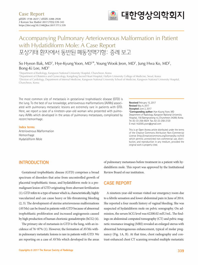
A nineteen-year-old woman visited our emergency room due to a febrile sensation and lower abdominal pain in June of 2014. She reported a four month history of vaginal bleeding. She was suspected of hydatidiform mole on pelvic sonography. On ad-mission, the serum hCG level was 62200.02 mIU/mL. The find-ings on abdominal computed tomography (CT) and pelvic mag-netic resonance imaging (MRI) revealed an enlarged uterus with abnormal heterogeneous enhancement, typical of molar preg-nancy (Fig. 1A, B). At that time, chest radiography and con-trast-enhanced chest CT scanning revealed multiple metastatic
The most common site of metastasis in gestational trophoblastic disease (GTD) is the lung. To the best of our knowledge, arteriovenous malformations (AVMs) associ-ated with pulmonary metastatic lesions are extremely rare in patients with GTD. Here, we report a case of a nineteen-year-old woman who presented with pulmo-nary AVMs which developed in the areas of pulmonary metastases, complicated by recent hemorrhage.
Index termsArteriovenous MalformationHemorrhageHydatidiform Mole
Received February 15, 2017Revised May 4, 2017Accepted June 2, 2017*Corresponding author: Hye-Kyung Yoon, MDDepartment of Radiology, Kangwon National University Hospital, 156 Baengnyeong-ro, Chuncheon 24289, Korea.Tel. 82-33-258-9024 Fax. 82-33-258-2120E-mail: [email protected]
This is an Open Access article distributed under the terms of the Creative Commons Attribution Non-Commercial License (http://creativecommons.org/licenses/by-nc/4.0) which permits unrestricted non-commercial use, distri-bution, and reproduction in any medium, provided the original work is properly cited.

340
Arteriovenous Malformation within Pulmonary Metastasis
jksronline.orgJ Korean Soc Radiol 2017;77(5):339-343
BA
C
DFig. 1. A 19-year-old woman with hydatidiform mole, metastasizing to the lung accompanying with the pulmonary; arteriovenous malformation.A. Contrast-enhanced abdomen CT shows an enlarged uterus with abnormal heterogeneous enhancement of the endometrium (white thick ar-rows). Multiple air bubbles are found in the uterine cavity because dilatation and curettage is performed on the same day.B. Axial T2-weighted (left) and T1-weighted gadolinium-enhanced (right) pelvic MR images demonstrate strong enhancement (white thin ar-rows) of the endometrium with multiple T2-hyperintense cystic foci (a black thick arrow).C. Axial contrast-enhanced chest CT shows multiple nodules consistent with pulmonary metastases. Some nodules communicate with a curvilin-ear structure (white thick arrows), indicating arteriovenous malformations. Ground-glass halo around a metastatic nodule in the inferior lingular segment of the left upper lobe represents hemorrhage (a black arrow).D. Pulmonary angiography and coil embolization. Selective left pulmonary arteriogram shows pulmonary AVM (white thick arrows). Embolization coil (a black arrow) is deployed and postembolization pulmonary arteriogram shows complete occlusion of AVM.AVM = arteriovenous malformation, CT = computed tomography

341
So Hyeon Bak, et al
jksronline.org J Korean Soc Radiol 2017;77(5):339-343
nodules in both lungs, and there were curvi-linear structures that were connected to some metastatic nodules in both lower lobes and the inferior lingular segment of the left upper lobe. Around the metastatic nodule, there were patchy ground-glass opacity lesions in the inferior lingular segment of the left upper lobe, consistent with pulmonary hemorrhage. The findings on chest CT consisted of multiple pulmonary metastases with AVMs, complicated by recent hemorrhage (Fig. 1C). No other metastasis was found in the brain or abdomen on abdominal
CT or brain MRI. During admission, she was in septic shock (body temperature; 38.5°C, blood pressure; 80/60 mm Hg, heart rate; 120 beats per minute). After uterine embolization, dilatation and curettage was performed twice. Pulmonary arte-riography confirmed the presence of two left AVMs and pul-monary AVMs were successfully embolized with a 2 × 5 mm coil one week later, as shown in Fig. 1D. Then, she was given three cycles of Methotrexate weekly and she attained a normal hCG level (Fig. 1E). She was regularly followed up and the find-
70000.00
60000.00
50000.00
40000.00
30000.00
20000.00
10000.00
0.00
Hum
an c
horio
nic
gona
dotr
opin
(mIU
/mL)
2014-10-252014-09-252014-08-25
Methetrexate
Dilatation and currettage
Date
2014-07-252014-06-25
Fig. 1. A 19-year-old woman with hydatidiform mole, metastasizing to the lung accompanying with the pulmonary; arteriovenous malformation.E. hCG levels during the course of treatment. At the time of diagnosis, the serum hCG level was 62,200.02 mIU/mL. The serum hCG level de-creased to 3897.82 mIU/mL after two dilatation and curettage on June 26 and July 2, 2014. From July 28 to August 11, the patient was given three cycles of Methotrexate weekly. The serum hCG level decreased to 9.85 mIU/mL on first follow-up examination after Methotrexate adminis-tration. Thereafter, the serum hCG level had decreased continuously, and it was less than 0.5 mIU/mL in the last examination performed on July 6, 2016.F. Follow-up contrast-enhanced chest computed tomography obtained after embolization and three courses of methotrexate shows that multi-ple metastatic nodules with residual arteriovenous malformations (white thick arrows) in both lungs have markedly decreased in size or disap-peared (arrows).hCG = human chorionic gonadotropin
E
F

342
Arteriovenous Malformation within Pulmonary Metastasis
jksronline.orgJ Korean Soc Radiol 2017;77(5):339-343
ings on follow-up chest CT showed that multiple metastatic nodules with residual AVMs in both lungs had markedly de-creased in size in January 2015 (Fig. 1F).
DISCUSSION
GTD is a rare entity, and it manifests as an hCG-producing hy-pervascular tumor (2). The key regulatory role of hCG in angio-genesis and vascular function is well known (4). AVMs in the uterus are a relatively frequent event, and the relationship be-tween GTD and uterine AVMs is well-documented by abnor-mal trophoblastic proliferation and angiogenesis induced by high hCG production (2, 4, 5).
Pulmonary metastases of GTD are seen most often and are thought to be most common. Angiographic studies have shown that pulmonary metastases of GTD are hyper-vascular tumors, and are supplied by the pulmonary artery (6). Pulmonary AVMs are abnormally dilated vessels which provide an anatomic right-to-left shunt between the pulmonary artery and the pulmonary vein without an intervening pulmonary capillary system (7). Most pulmonary AVMs are congenital in etiology, and are asso-ciated with hereditary hemorrhagic telangiectasia. Acquired pul-monary AVMs can occur in the context of a variety of physical injuries or disease processes such as liver cirrhosis, chest trauma, infection, metastatic carcinoma, mitral stenosis or systemic am-yloidosis (7, 8). Pulmonary metastases of GTD have the poten-tial to form pulmonary AVMs, which can cause life-threatening hemorrhage or hemoptysis (2). A few studies have shown that pulmonary AVMs were secondary to GTD at the metastatic site during or after chemotherapy (2, 6, 9, 10). Our patient showed multiple pulmonary metastases and AVMs within these lesions accompanied by pulmonary hemorrhage even before the onset of chemotherapy.
Uterine AVMs in patients with GTD have the potential risk of heavy vaginal bleeding or intraperitoneal hemorrhage result-ing from disorganized vascular structures and high blood flow (5). Uterine or pulmonary AVMs may be spontaneously resolved with successful chemotherapy, and these patients were treated for persistent or life-threatening bleeding with embolization (3, 5). However, AVMs might persist after successful completion of chemotherapy (2, 9, 10). In our case, embolization was performed to reduce the bleeding risk, because our patient had pulmonary
hemorrhage associated with pulmonary AVMs. In addition to metastatic nodules, residual pulmonary AVMs had disappeared or were markedly decreased with successful chemotherapy.
In conclusion, AVMs associated with pulmonary metastatic lesions are extremely rare. When the early diagnosis of pulmo-nary AVM using CT scanning is established, appropriate emboli-zation and chemotherapy could be considered in an effort to re-duce the risk of bleeding and to improve the outcome.
Acknowledgments
The authors would like to thank the CT, MR, intervention technologists at the Department of Radiology, Kangwon Na-tional University Hospital.
REFERENCES
1. Shanbhogue AK, Lalwani N, Menias CO. Gestational tropho-
blastic disease. Radiol Clin North Am 2013;51:1023-1034
2. McDonald-Burrows Z, Davies R, Goode E, Clarke C, Jackson
J, Seckl M, et al. Haemoptysis from a pulmonary arteriove-
nous malformation in a post molar pregnancy gestational
trophoblast tumour patient managed by radiological em-
bolisation: a case report. J Med Case Rep 2014;8:117
3. McGrath S, Harding V, Lim AK, Burfitt N, Seckl MJ, Savage P.
Embolization of uterine arteriovenous malformations in pa-
tients with gestational trophoblastic tumors: a review of pa-
tients at Charing Cross Hospital, 2000-2009. J Reprod Med
2012;57:319-324
4. Zygmunt M, Herr F, Münstedt K, Lang U, Liang OD. Angio-
genesis and vasculogenesis in pregnancy. Eur J Obstet Gyne-
col Reprod Biol 2003;110 Suppl 1:S10-S18
5. Touhami O, Gregoire J, Noel P, Trinh XB, Plante M. Uterine ar-
teriovenous malformations following gestational tropho-
blastic neoplasia: a systematic review. Eur J Obstet Gynecol
Reprod Biol 2014;181:54-59
6. Green JD, Carden TS Jr, Hammond CB, Johnsrude IS. Angio-
graphic demonstration of arteriovenous shunts in pulmonary
metastatic choriocarcinoma. Radiology 1973;108:67-70
7. Shovlin CL. Pulmonary arteriovenous malformations. Am J
Respir Crit Care Med 2014;190:1217-1228
8. Pick A, Deschamps C, Stanson AW. Pulmonary arteriovenous
fistula: presentation, diagnosis, and treatment. World J Surg

343
So Hyeon Bak, et al
jksronline.org J Korean Soc Radiol 2017;77(5):339-343
포상기태 환자에서 동반된 폐동정맥기형: 증례 보고
박소현1 · 윤혜경1* · 전영욱1 · 고정화2 · 이봉기3
임신융모질환의 가장 흔한 전이 부위는 폐이다. 임신융모질환 환자에서 폐전이와 동반된 폐동정맥기형은 매우 드물다. 저
자들은 포상기태로 진단된 환자에서 폐전이 결절에 동반된 폐동정맥기형의 증례를 경험하였기에 문헌고찰과 함께 보고하
고자 한다.
1강원대학교병원 영상의학과, 2한림대학교 의과대학 강동성심병원 산부인과학교실, 3강원대학교 의과대학 강원대학교병원 심장내과학교실
1999;23:1118-1122
9. Casson AG, McCormack D, Craig I, Inculet R, Levin L. A per-
sistent pulmonary lesion following chemotherapy for met-
astatic choriocarcinoma. Chest 1993;103:269-270
10. Choi SH, Goo JM, Kim HC, Im JG. Pulmonary arteriovenous
fistulas developed after chemotherapy of metastatic chorio-
carcinoma. AJR Am J Roentgenol 2003;181:1544-1546