cytotoxicity of polyaminobenzene …hashsham/courses/ene806/docs/ene806...an experimental setup with...
TRANSCRIPT

CYTOTOXICITY OF POLYAMINOBENZENE SULFONIC
ACID FUNCTIONALIZED SINGLE WALLED CARBON
NANOTUBES ON ESCHERICHIA COLI K12 CELLS IN
SUSPENSION AND IN BIOFILM
Submitted by
Indumathy Jayamani
Tracy Lynne Repp
On
April 30th
, 2008
Submitted to:
Dr. Syed Hashsham
Engineering Research Complex
Department of Civil and Environmental Engineering
Michigan State University
East Lansing, MI 48824

2
TABLE OF CONTENTS
ACKNOWLEDGMENT .................................................................................................. 3
ABSTRACT....................................................................................................................... 4
1. INTRODUCTION......................................................................................................... 5
2. BACKGROUND ....................................................................................................... 6
3. METHODOLOGY ................................................................................................... 7
4. EXPERIMENTAL SETUP...................................................................................... 8
5. EXPERIMENTAL PROCEDURE........................................................................ 11
5. 1 GROWTH OF E. COLI – CELL CULTURE AND INOCULATION .................................. 11
5.2 SAMPLING FOR INITIAL CELL COUNT TO CHECK FOR SUFFICIENT GROWTH ......... 12
5.3 PLATE COUNT METHOD ...................................................................................... 13
5.4 EXPOSURE TO SWCNT PAB’S .......................................................................... 16
6. RESULTS ................................................................................................................ 17
7. FUTURE STUDY.................................................................................................... 19
8. REFERENCES........................................................................................................ 20
APPENDIX A................................................................................................................... 21
APPENDIX B................................................................................................................... 23
APPENDIX C................................................................................................................... 26

3
ACKNOWLEDGMENT
We express our sincere thanks to Dr. Syed Hashsham, for this wonderful
opportunity to work on a topic of our interest. We also thank him for his
continuous guidance and support throughout the period of the experiment.
We thank Lab Manager Joseph Nguyen for his help and guidance in building
the setup. Thanks to graduate students Alla Alpatova, Yu Yang and Robert
D. Stedtfeld for their help during the course of this experiment. Finally we
also would like to thank our friends who supported and helped us in
conceiving the idea for the experiment.
Date : 30th April, 08 Indumathy Jayamani
Tracy Lynn Repp

4
ABSTRACT
Carbon nanotubes (CNT’s) are considered to be a very promising material
and are widely used in various technological applications. But their wide
spread use also increases the risk of increased release of these particles into
the environment. CNT’s are used in various forms for various applications. To
add to, microorganisms in the environment prefer to form biofilms and are
found to be more resistant to chemical disinfection. This study aimed at
studying the cyctotoxity of polyaminobenzene sulfonic acid functionalized
CNT’s (SWCNT PAB’s) on Escherichia coli (E. coli) cells in biofilm and in
suspension. An experimental setup with the CDC bioreactor as a CSTR was
build to grow E. coli in biofilm and in suspension. Favorable conditions for E.
coli growth were maintained throughout the study. A cell plate count test
was conducted on the third day of the study to establish the cell count
method for the exposure run and as well to check for sufficient growth of E.
coli cells in biofilm. The SWCNT PAB has 5mg/mL solubility in water and
can be added to the reactor through the inoculation port. The exposure run
was planned to be conducted on day 5 of the experiment but not carried out
due to time constraint.
5
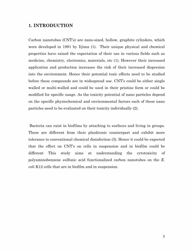
1. INTRODUCTION
Carbon nanotubes (CNT’s) are nano-sized, hollow, graphite cylinders, which
were developed in 1991 by Iijima (1). Their unique physical and chemical
properties have raised the expectation of their use in various fields such as
medicine, chemistry, electronics, materials, etc (1). However their increased
application and production increases the risk of their increased dispersion
into the environment. Hence their potential toxic effects need to be studied
before these compounds are in widespread use. CNT’s could be either single
walled or multi-walled and could be used in their pristine form or could be
modified for specific usage. As the toxicity potential of nano particles depend
on the specific physiochemical and environmental factors each of these nano
particles need to be evaluated on their toxicity individually (2).
Bacteria can exist in biofilms by attaching to surfaces and living in groups.
These are different from their planktonic counterpart and exhibit more
tolerance to conventional chemical disinfection (3). Hence it could be expected
that the effect on CNT’s on cells in suspension and in biofilm could be
different This study aims at understanding the cytotoxicity of
polyaminobenzene sulfonic acid functionalized carbon nanotubes on the E.
coli K12 cells that are in biofilm and in suspension.

6
2. BACKGROUND
CNT’s could be either single walled (SWCNT) or multi walled (MWCNT).
CNT’s in their pristine form are hydrophobic and hence they form into
aggregates when discharged in water. However they might get dispersed in
water given a longer duration of time. CNT’s when functionalized with
another group like amide, carboxylic group etc may become water soluble due
to the hydrophilic nature of the functional group attached. Functionalized
CNT’s are widely used in biology and biomedical application for carrying
drugs inside the human system (4).
Recent studies have indicated the toxicity of SWCNT’s is more than that of
MWCNT’s (5). A study conducted by Elimelech et al. (2007), confirms the
cytotoxicity of pristine SWCNT’s on E. coli cells by the cell wall piercing
mechanism. However oxidized MWCNT’s are more toxic than pristine
MWCNT’s (1). Functionalized CNT’s are found to be less or not toxic due to
their hydrophilic nature. This was confirmed by the study on human T cells
with functionalized CNT’s (4). Functionalized CNT’s are rapidly taken up by
the human B and T lymphocytes but does not affect viability of the cells (4).
However no literature was found relating to their effect on microorganisms.

7
3. METHODOLOGY
To test the cytotoxicity of SWCNT-PAB’s on E. coli K12 cells in suspension
and in biofilm, E. coli was grown in a CDC bioreactor, which is a continuously
stirred bioreactor. This reactor helps in growing the cells both in suspension
and in film. The reactor was maintained under aerobic conditions and well
fed with a continuous supply of fresh media to sustain the growth of E. coli
K12. The effluent of dead cells was constantly removed by pumping. After
approximately 3 days of inoculating the reactor with cells, samples
(triplicates) were collected from both the biofilm and the cells in suspension
and a plate count test was conducted to check for sufficient growth of the
cells. After 5 days, the exposure test was planned, by conducting plate count
on samples (triplicates) collected just before the application of the SWCNT’s
and immediately after the application of the SWCNT’s. Another cell count
test was planned to be conducted after 24 hours of application of the
SWCNT’s. The detailed methodology is described in the following sections.
8
4. EXPERIMENTAL SETUP
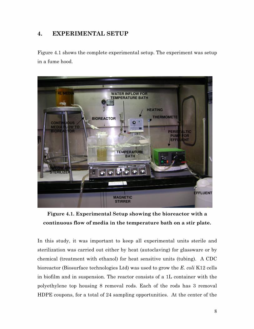
Figure 4.1 shows the complete experimental setup. The experiment was setup
in a fume hood.
Figure 4.1. Experimental Setup showing the bioreactor with a
continuous flow of media in the temperature bath on a stir plate.
In this study, it was important to keep all experimental units sterile and
sterilization was carried out either by heat (autoclaving) for glassware or by
chemical (treatment with ethanol) for heat sensitive units (tubing). A CDC
bioreactor (Biosurface technologies Ltd) was used to grow the E. coli K12 cells
in biofilm and in suspension. The reactor consists of a 1L container with the
polyethylene top housing 8 removal rods. Each of the rods has 3 removal
HDPE coupons, for a total of 24 sampling opportunities. At the center of the
4L MEDIA
BIOREACTOR
CONTINUOUS MEDIA FLOW TO BIOREACTOR
THERMOMETER
HEATING
WATER INFLOW FOR TEMPERATURE BATH
TEMPERATURE BATH
MAGNETIC STIRRER
PERISTALTIC PUMP FOR EFFLUENT
EFFLUENT
INFLOW AIR STERILIZER

9
reactor there is a detachable magnetic stirrer with a baffle wall attached.
This was not used in this experiment as the baffle wall did not rotate
continuously and was replaced by a normal magnetic stir bar. There is an
outlet for the effluent at just above the 400 mL mark of the container.
However the effective volume of the liquid in the reactor was estimated to be
350 mL as the rods displaced approximately 50 mL of liquid. The reactor
consists of three ports on the polyethylene top, one for media flow, one for
inoculation and one for airflow.
A magnetic stirrer was placed at the bottom of the reactor. LB media was
prepared as per the protocol in Appendix 2 and was transferred aseptically to
the reactor up to the 410 mL mark (i.e. just up to the outlet). The reactor was
closed with the polyethylene top along with the rods, and was sealed with
autoclave tape. The effluent outlet and the three inlet ports on the top of the
reactor were then closed with aluminum foil. The reactor was then sealed
with autoclave tape autoclaved for 15 mins at slow speed.
Fresh media needs to be supplied continuously to the reactor to maintain
growth of the E. coli in the reactor during the study period. A four-liter bottle
with an outlet at the bottom was used as the reservoir of fresh media. The
flow from reservoir bottle to the reactor was designed to be by gravity. The
bottle was sealed at the top and at the bottom outlet by an aluminum foil.
The aluminum foil is then taped with autoclave tape and autoclaved for 15
mins at slow speed. The sterilized bottle is then connected with sterile tubing
to the media inlet of the reactor and a pinch unit was used to control the flow
through the tube. The bottle is then filled with 4L of autoclaved media
aseptically. The bottle is then kept at a suitable height from the reactor
height to facilitate flow by gravity. However the media flow was not turned
on for the first 24 hours.
10
The outlet of the reactor was connected to the ORCBS waste container using
chemically sterilized tubing. The effluent outflow was controlled by a
peristaltic pump maintained at a lower flow rate (20 rpm) to ensure a
constant volume of liquid suspension in the reactor. The reactor was then
placed in the temperature bath maintained at 37°C by a heating unit. The air
supply of the fume hood was connected to a conical flask (with a side outlet)
with sterile cotton balls, and the outlet of the conical flask was connected to
the air inlet of the reactor. Tubing’s and connectors for connecting the various
units (air inflow, media inflow, effluent outflow, tubing for the inoculation
port along with the control value) were washed with ethanol before
connecting to sterilize them.
11
5. EXPERIMENTAL PROCEDURE
5. 1 GROWTH OF E. COLI – CELL CULTURE AND INOCULATION
A 10 mL cell culture tube was inoculated aseptically a day before with E. coli
K12. Yu Yang, a graduate student, Department of Plants and Soil Sciences,
Michigan State University, provided the E. coli K12 agar culture. Few
milliliters’ of the culture was then aseptically transferred to the reactor,
through the inoculation port on the reactor. The temperature bath was
placed over a magnetic stir plate, and the magnetic stirrer within the reactor
was set to stir at a slow mixing rate (40-50 rpm approximately) to enable a
continuously stirred tank reactor (CSTR) condition. The airflow to the reactor
also was turned on to make the reactor aerobic. The media flow from the
media reservoir was turned on a day after inoculation, and was set at a rate
such that it would compensate the evaporation losses as well as to maintain
sufficient media for the growth of E. coli cells. It was not set to a particular
value and was later estimated during the experiment.
The system was allowed to run for 3 days continuously with periodic
monitoring. The temperature bath, magnetic stirrer, media flow, effluent
outflow were periodically monitored to ensure that everything was working
fine throughout the three day period. Also the tubing’s and the media
reservoir bottle were visually checked for cross contamination either by
environmental microbes or by E. coli backward growth.
As E. coli might take 2-5 days to grow into at least a thin biofilm (6), a plate
count test was planned on day 3 to check for sufficient growth of biofilms and
to establish the sampling and plate count method. Also the CNT’s exposure
was planned after 5 days of growth.

12
5.2 SAMPLING FOR INITIAL CELL COUNT TO CHECK FOR
SUFFICIENT GROWTH
On the third day from inoculating the reactor, samples of the cells in biofilm
and in suspension were collected to perform a cell count by the pour plate
count method. Adequate amount of 1.5 mL centrifuge tubes were autoclaved
and aseptically filled with 900 µL buffered dilution water. These tubes were
arranged in a holder and the sampling and dilution series was performed
under the fume hood. The fume hood was first sterilized by spraying ethanol
and then the experiment was conducted near a flame to avoid contamination.
The sampling was done in triplicates.
One of the rods from the reactor was removed along with the three coupons
present on it and a sterile plug is placed in place of the rod in the reactor to
prevent contamination. The coupons were removed and placed in separate
sterile containers. The coupons were first washed in phosphate buffer
solution (Appendix 1) to remove any unattached cells from the coupon. Then
they were transferred to a 30 mL cell culture tube with 10 mL of phosphate
buffer and sonicated for 2 mins (7). 100 µL of this solution was used for
further dilution (10-2 dilution).
Around the same time when the rod was removed, three aliquots of 100µL
samples of the suspended cells were also collected in a 1.5 mL vial containing
900µL of dilution water (10-1 dilution).
13
5.3 PLATE COUNT METHOD
The plate count method is a direct cell count method, which helps in counting
the viable cells only. It works on the assumption that each viable cell yields
one colony on the plate after the incubation period. Also by the use of a
specific medium to plate the plate count method can help in counting only the
target species. For conducting plate count method, the sample has to be first
diluted before plating. If the right dilution is not plated, there will be either
too many colonies on the plate (grown into each other making counting
difficult) or little/no cells (not being representative of the sample). Since the
right dilution is not known more than one dilution was made.
14
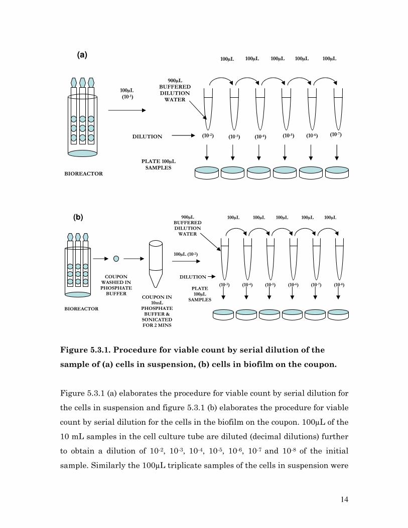
Figure 5.3.1. Procedure for viable count by serial dilution of the
sample of (a) cells in suspension, (b) cells in biofilm on the coupon.
Figure 5.3.1 (a) elaborates the procedure for viable count by serial dilution for
the cells in suspension and figure 5.3.1 (b) elaborates the procedure for viable
count by serial dilution for the cells in the biofilm on the coupon. 100µL of the
10 mL samples in the cell culture tube are diluted (decimal dilutions) further
to obtain a dilution of 10-2, 10-3, 10-4, 10-5, 10-6, 10-7 and 10-8 of the initial
sample. Similarly the 100µL triplicate samples of the cells in suspension were
PLATE 100µL
SAMPLES
900µL BUFFERED
DILUTION
WATER
100µL
(10-1)
(10-2) DILUTION (10-3) (10-4) (10-5) (10-6) (10-7)
100µL 100µL 100µL 100µL 100µL (a)
BIOREACTOR
COUPON WASHED IN
PHOSPHATE BUFFER
COUPON IN 10mL
PHOSPHATE
BUFFER & SONICATED FOR 2 MINS
100µL (10-2)
(10-3)
100µL 100µL 100µL 100µL 100µL
(10-4) (10-5) (10-6) (10-7) (10-8)
900µL BUFFERED DILUTION
WATER
PLATE 100µL
SAMPLES
DILUTION
(b)
BIOREACTOR

15
also diluted to obtain a dilution of 10-1, 10-2, 10-3, 10-4, 10-5, 10-6 and 10-7 of the
initial sample. Sterile pipette tips were used for dilution and the tips were
changed between dilutions. All dilutions were made using the buffered
dilution water (Appendix 2).
The plate count was performed by the spread plate method. 100µL of each
dilution was poured using a pipette to the center (introduces another 10-1
dilution) of a sterile agar plate and spread using a sterile bend glass
spreader. The glass spreader was sterilized by dipping in ethanol followed by
heating in flame. The spreader was first cooled in the agar on the side of the
plate and then was used to spread the cells by rotating the plate. To avoid
contamination while plating, the lid of the plate was opened only slightly to
allow the pipette tip.
The plates were then incubated at 37°C in an incubator. The number of
colony forming units (CFU’s) was counted after 24 hours of plating using a
Quebec colony counter (Appendix 3). The colonies counted (Appendix 1) were
then multiplied by the dilution factor, which is the inverse of the dilution.
16
5.4 EXPOSURE TO SWCNT PAB’S
The impact of the SWCNT’s was to be studied on the fifth day of growth of
the biofilm in the reactor. The SWCNT PAB’s are available in powder form
(Carbon Solutions Inc.,) and have a water solubility of 5mg/mL. The
concentration of the SWCNT’s planned to be exposed was 50µg/mL. Thus 17.5
mg of the SWCNT’s could be mixed with 3.5 mL of water and sonicated for 30
minutes in water (1).
Samples of cells in suspension and cells in biofilm should be collected as
described in section 5.2 and are plated as described in section 5.3. These are
the initial number of CFU’s before exposure. After collecting and plating
samples, the SWCNT solution should be added to the reactor through the
inoculation port. Samples of cells in suspension and cells in biofilm should
again be collected to study the immediate impact of exposure to SWCNT
PAB’s. These are again diluted and plated as described in sections 5.2 and
5.3. Another set of samples should be collected 24 hours after the dosage of
SWCNT’s to study the long term impact of the CNT’s on the E. coli cells.

17
6. RESULTS
Table 6.1 gives the results of the cell count test conducted on day 3 of the
experiment. Though all dilutions were plated, the less diluted samples plated
had too many colonies and the highly diluted samples plated didn’t have any
colonies on them. Hence only those plates that had countable colonies
(between 25 to 250 colonies) are presented in this report (Appendix 1).
Table 6.1. Plate count results for cells in biofilm and in suspension
Trial 1 Trial 2 Trial 3 Mean SD
Biofilm on coupon
Log CFU's/mm2 5.358 5.057 5.233 5.216 0.15
Suspended cells
Log CFU's/mL 8.427 8.455 8.476 8.452 0.025
The media flow rate from the media reservoir to the bioreactor was estimated
to be 0.083 mL/min. The height and the diameter of individual coupons were
measured to be 3.9 mm and 12.5 mm using vernier calipers. The surface area
exposed for biofilm formation was thus calculated to be 122.72 mm2. The data
obtained from the triplicate samples was averaged and the standard
deviation was calculated. The results expressed as mean±standard deviation
are 5.21 ± 0.15 log CFU/mm for the biofilm per coupon and 8.45 ± 0.02 log
CFU/mL in suspension (Figure 6.3).
18
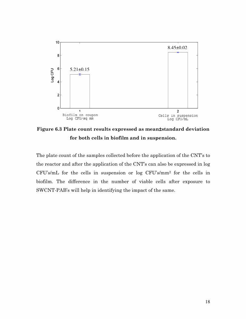
Figure 6.3 Plate count results expressed as mean±±±±standard deviation
for both cells in biofilm and in suspension.
The plate count of the samples collected before the application of the CNT’s to
the reactor and after the application of the CNT’s can also be expressed in log
CFU’s/mL for the cells in suspension or log CFU’s/mm2 for the cells in
biofilm. The difference in the number of viable cells after exposure to
SWCNT-PAB’s will help in identifying the impact of the same.
5.21±0.15
8.45±0.02
19
7. FUTURE STUDY
The experimental run to study the exposure effects of SWCNT-PAB’s was not
completed due to time constraint and could be completed as part of future
study. However since some primary studies have found that functionalized
SWCNT’s are not toxic as that of pristine CNT’s, it could be expected that the
exposure run with SWCNT-PAB’s may not produce a significant decrease in
the number of viable counts (4). Also as the standard deviation of the
triplicate cell counts by the plate count method is only in the order of 0.15 log
CFU’s/mL it is not expected to interfere with the decrease in cell counts due
to SWCNT’s. However if need may arise alternate methods like direct
microbial count, or viewing the cells under a scanning electron microscope
may help in studying the impact further.
Alternatively, this set up could also be used to study the impact of pristine
CNT’s on the E. coli cells in suspension and as biofilm. Though CNT’s are not
very easily soluble in water, the study by Cheng et al. (1) has reported that a
water solubility of 50 mg/L can be achieved by continuously stirring the
CNT’s in water for 30 mins. Thus, a similar procedure can be adopted to test
the cytotoxicity of the CNT solution on the E. coli cells in suspension and in
biofilm.

20
8. REFERENCES
1. Cheng, J., E. Flahaut, S. H. Cheng. 2006. Effect of carbon
nanotubes on developing zebrafish (Danio rerio) embryos.
Environmental toxicology and chemistry. 26:708-716.
2. Helland, A., P. Wick, A. Koehler, K. Schmid, C. Som. 2007.
Reviewing the environmental and human health knowledge base of
carbon nanotubes. Environmental health perspectives. 115(8):1125-
1131.
3. Goeres. D. M., K. Buckingham-Meyer, M. A. Hamilton. 2007.
Comparative evaluation of biofilm disinfectant efficacy tests. Journal
of microbiological methods. 70:236-244.
4. Yang, W., P. Thordarson, J. J. Gooding, S. P. Ringer, F. Braet.
2007. Carbon nanotubes for biological and biomedical applications.
Nanotechnology. 412001:12pp.
5. Elimelech, M., S. Kang, M. Piault, L.D. Pfefferle. 2007. Single-
walled carbon nanotubes exhibit strong antimicrobial activity.
Langmuir. 23:8670-8673.
6. Ghigo, JM., C. Beloin, J. Valle, P. Latour-Lambert, P. Faure, M.
Kzreminski, D. Balestrino, J. A. J. Haagensen, S. Molin, G.
Prensier, B. Arbeille. 2004. Global impact of mature biofilm lifestyle
on Escherichia coli K-12 gene expression. Molecular microbiology.
51(3):659-674.
7. Kim H-J., V.L. Dorn, J. M. VanBriesen. 2004. The efficacy of
ethylenediaminetetraacetic acid (EDTA) against biofilm bacteria.
Biomedical engineering annual fall meeting.
8. Standard Methods for the Examination of Water and Wastewater, 20th
Ed.
21
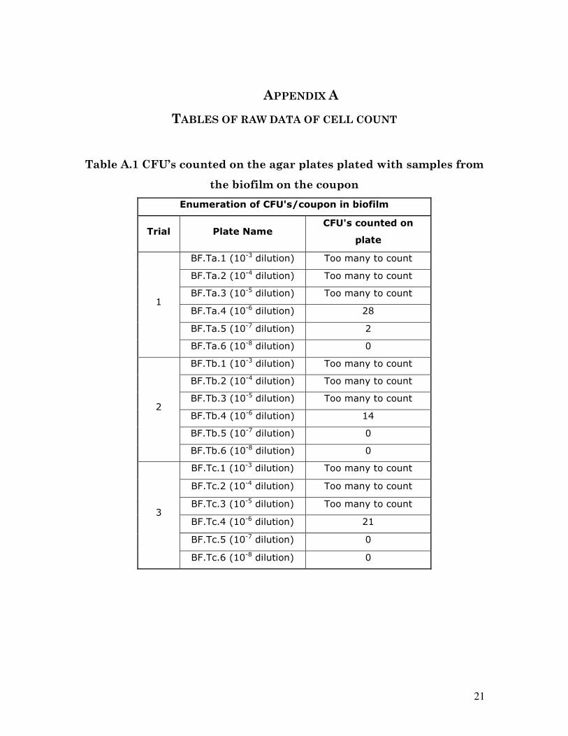
APPENDIX A
TABLES OF RAW DATA OF CELL COUNT
Table A.1 CFU’s counted on the agar plates plated with samples from
the biofilm on the coupon
Enumeration of CFU's/coupon in biofilm
Trial Plate Name CFU's counted on
plate
BF.Ta.1 (10-3 dilution) Too many to count
BF.Ta.2 (10-4 dilution) Too many to count
BF.Ta.3 (10-5 dilution) Too many to count
BF.Ta.4 (10-6 dilution) 28
BF.Ta.5 (10-7 dilution) 2
1
BF.Ta.6 (10-8 dilution) 0
BF.Tb.1 (10-3 dilution) Too many to count
BF.Tb.2 (10-4 dilution) Too many to count
BF.Tb.3 (10-5 dilution) Too many to count
BF.Tb.4 (10-6 dilution) 14
BF.Tb.5 (10-7 dilution) 0
2
BF.Tb.6 (10-8 dilution) 0
BF.Tc.1 (10-3 dilution) Too many to count
BF.Tc.2 (10-4 dilution) Too many to count
BF.Tc.3 (10-5 dilution) Too many to count
BF.Tc.4 (10-6 dilution) 21
BF.Tc.5 (10-7 dilution) 0
3
BF.Tc.6 (10-8 dilution) 0

22
Table A.2 CFU’s counted on the agar plates plated with samples from
the biofilm on the coupon
Enumeration of CFU's/mL in suspension
Trial Plate Name CFU's counted on
plate
S.Ta.1 (10-2 dilution) Too many to count
S.Ta.2 (10-3 dilution) Too many to count
S.Ta.3 (10-4 dilution) Too many to count
S.Ta.4 (10-5 dilution) Too many to count
S.Ta.5 (10-6 dilution) 267
1
S.Ta.6 (10-7 dilution) 26
S.Tb.1 (10-2 dilution) Too many to count
S.Tb.2 (10-3 dilution) Too many to count
S.Tb.3 (10-4 dilution) Too many to count
S.Tb.4 (10-5 dilution) Too many to count
S.Tb.5 (10-6 dilution) 285
2
S.Tb.6 (10-7 dilution) 51
S.Tc.1 (10-2 dilution) Too many to count
S.Tc.2 (10-3 dilution) Too many to count
S.Tc.3 (10-4 dilution) Too many to count
S.Tc.4 (10-5 dilution) Too many to count
S.Tc.5 (10-6 dilution) 299
3
S.Tc.6 (10-7 dilution) 40
23
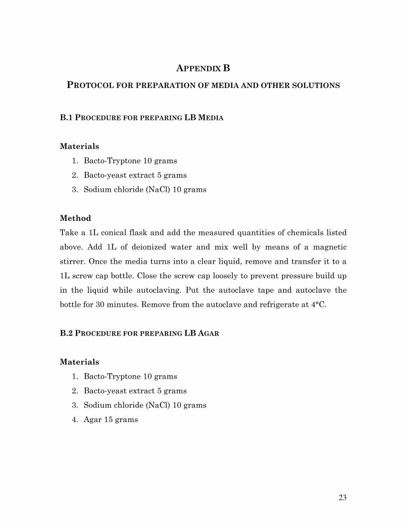
APPENDIX B
PROTOCOL FOR PREPARATION OF MEDIA AND OTHER SOLUTIONS
B.1 PROCEDURE FOR PREPARING LB MEDIA
Materials
1. Bacto-Tryptone 10 grams
2. Bacto-yeast extract 5 grams
3. Sodium chloride (NaCl) 10 grams
Method
Take a 1L conical flask and add the measured quantities of chemicals listed
above. Add 1L of deionized water and mix well by means of a magnetic
stirrer. Once the media turns into a clear liquid, remove and transfer it to a
1L screw cap bottle. Close the screw cap loosely to prevent pressure build up
in the liquid while autoclaving. Put the autoclave tape and autoclave the
bottle for 30 minutes. Remove from the autoclave and refrigerate at 4°C.
B.2 PROCEDURE FOR PREPARING LB AGAR
Materials
1. Bacto-Tryptone 10 grams
2. Bacto-yeast extract 5 grams
3. Sodium chloride (NaCl) 10 grams
4. Agar 15 grams
24
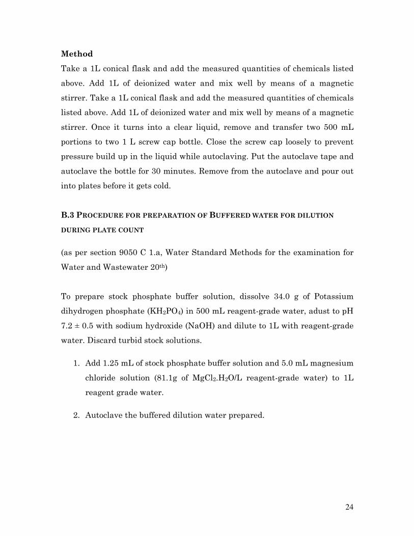
Method
Take a 1L conical flask and add the measured quantities of chemicals listed
above. Add 1L of deionized water and mix well by means of a magnetic
stirrer. Take a 1L conical flask and add the measured quantities of chemicals
listed above. Add 1L of deionized water and mix well by means of a magnetic
stirrer. Once it turns into a clear liquid, remove and transfer two 500 mL
portions to two 1 L screw cap bottle. Close the screw cap loosely to prevent
pressure build up in the liquid while autoclaving. Put the autoclave tape and
autoclave the bottle for 30 minutes. Remove from the autoclave and pour out
into plates before it gets cold.
B.3 PROCEDURE FOR PREPARATION OF BUFFERED WATER FOR DILUTION
DURING PLATE COUNT
(as per section 9050 C 1.a, Water Standard Methods for the examination for
Water and Wastewater 20th)
To prepare stock phosphate buffer solution, dissolve 34.0 g of Potassium
dihydrogen phosphate (KH2PO4) in 500 mL reagent-grade water, adust to pH
7.2 ± 0.5 with sodium hydroxide (NaOH) and dilute to 1L with reagent-grade
water. Discard turbid stock solutions.
1. Add 1.25 mL of stock phosphate buffer solution and 5.0 mL magnesium
chloride solution (81.1g of MgCl2.H2O/L reagent-grade water) to 1L
reagent grade water.
2. Autoclave the buffered dilution water prepared.
25
B.4 PROCEDURE FOR PREPARATION OF PHOSPHATE BUFFER
(as per section 9216 B 3.a, Standard Methods for the examination for Water
and Wastewater 20th)
1. Dissolve 13.6g KH2PO4 in water and dilute to 1L
2. Adjust to pH 7.2 if necessary, filter through 0.2µm membrane filter.
26
APPENDIX C
LIST OF FIGURES
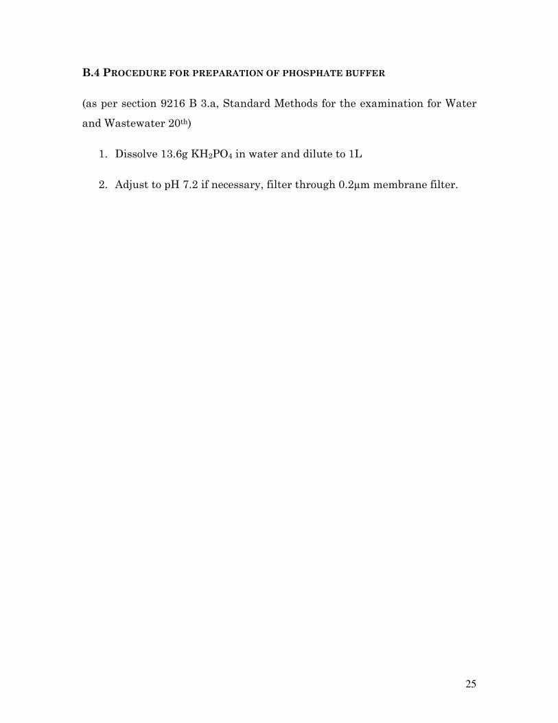
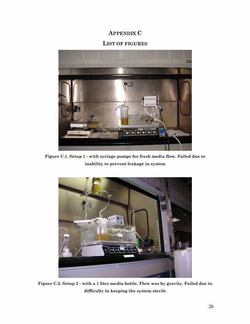
Figure C.1. Setup 1 - with syringe pumps for fresh media flow. Failed due to
inability to prevent leakage in system
Figure C.2. Setup 2 - with a 1 liter media bottle. Flow was by gravity. Failed due to
difficulty in keeping the system sterile
27
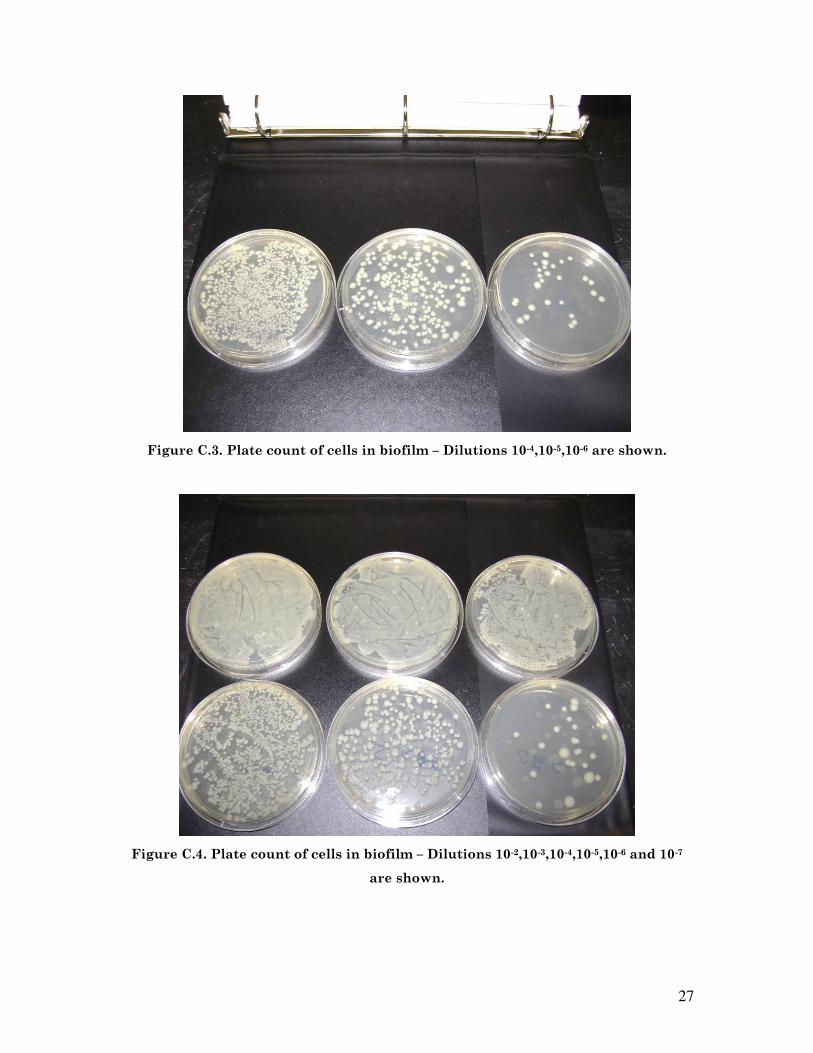
Figure C.3. Plate count of cells in biofilm – Dilutions 10-4,10-5,10-6 are shown.
Figure C.4. Plate count of cells in biofilm – Dilutions 10-2,10-3,10-4,10-5,10-6 and 10-7
are shown.

28
Figure C.5. Quebec colony counter