cystic lung disease - medstar health · •late: predominantly advanced bullous and cystic disease...
TRANSCRIPT

CYSTIC LUNG DISEASE
Stephen R Selinger MD
November 17, 2017
Case Presentation: 24 year old woman
with Pleuritic Pain
• 24 year old woman
• Right sided pleuritic pain of sudden onset
• 3 months of mild exertional dyspnea
• No cough, sputum nor wheezing
• Mild fatigue
• No infectious symptoms
• ROS negative
Case Presentation: 24 year old woman
with Pleuritic Pain
• Normal Vital Signs
• Decreased breath sounds bilaterally
• Chest hyperinflated
• Right sided chest tube with significant air leak
• No other abnormal finding
Case Presentation: 24 year old woman
with Pleuritic Pain
• WBC 5000/mm3
• Hgb 13 gm/dl
• Electrolytes normal
• Creatinine 0.7 mg/dl
• PaO2 70, PaCO2 37 on RA
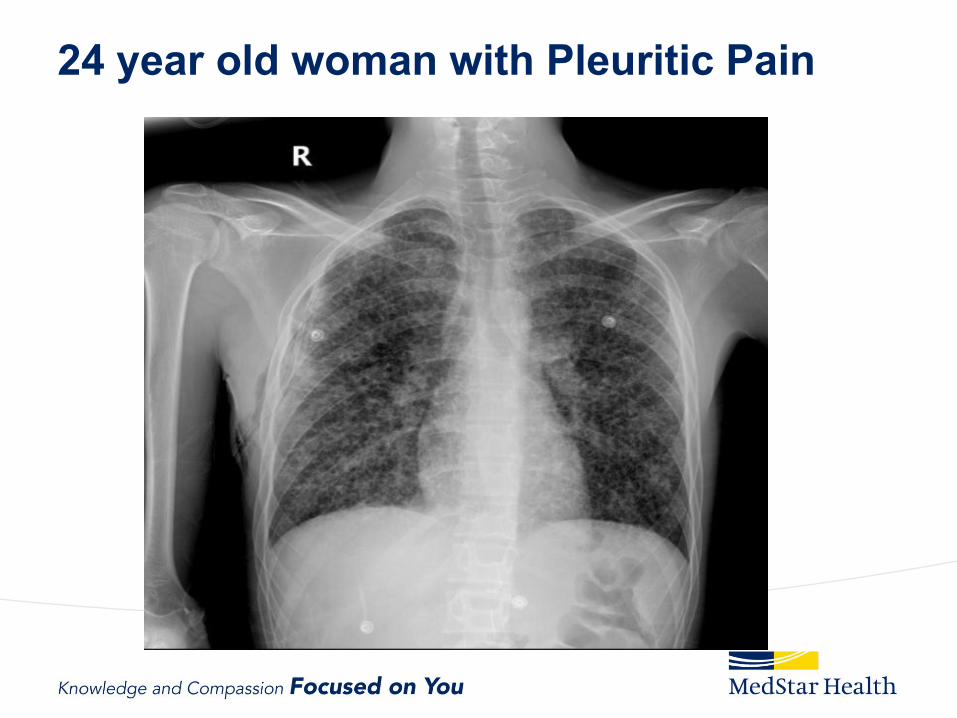
24 year old woman with Pleuritic Pain
Hospital Day 3
• 4-5 large cups of water were at the bedside
• A diagnostic Test was performed
Diagnosis:
Pulmonary Langerhans cell
Histiocytosis
Cystic Lung Diseases
November 21, 2017
8
Fleischner Society Definition
• Cyst: Thin walled (<2mm) spherical parenchymal lucency, interfaced with normal lung
• Cavity: Gas filled space within pulmonary consolidation, mass or nodule; typically thick walled (>2mm) and more irregularly shaped than cysts
• Bulla: Spherical focal lucency, > 1 cm, bounded by a thin wall (<1mm). Usually emphesematous change in adjacent lung
• Bleb: Cystic Airspace bounded by a thin wall adjacent to the visceral pleura, typically <1 cm in size
• Pneumatocoele: Approximately round, thin walled, air-filled space. Generally caused by infection, trauma, aspiration of hydrocarbon, usually transient
Cystic Lung Diseases
• Broad differential diagnosis
• HRCT narrows the differential, often providing a
definitive diagnosis
– Neoplastic
– Congenital,
– Genetic,
– Developmental,
– Lymphoproliferative,
– Inflammatory or Infectious
– Smoking Related

Mechanisms of Cyst Formation
• Check Valve Obstruction
• Destruction of the bronchiolar wall
• Ischemia
• Remodeling by MMP and other matrix degrading
enzymes
Cystic Lung Diseases
• Langerhans Cell Histiocytosis
• Lymphangioleiomyomatosis
• Birt-Hogg Dube
• Others
Langerhans Cell Histiocytosis
November 21, 2017
13

Langerhans Cell Histiocytosis
• 3-4% of Lung Biopsies
• Age 20-40
• 90% are smokers
• Can be multisystem especially in the pediatric
population
Langerhans Cell Histiocytosis: A
Cigarette Smoke Promoted Dendritic
Neoplasm?
• Langerhans cells are dendritic cells that regulate
mucosal immunity
• S100 staining
• Cd1a surface antigen
• Birbeck rod shaped inclusions

Langerhans Cell Histiocytosis: A
Cigarette Smoke Promoted Dendritic
Neoplasm?
• Cigarette smoking activates Langerhans Cells
via GMCSF and TGF B
• Clonal pattern with extrapulmonary LCH
• Clonal or polyclonal in PLCH
• BRAF, ARAF and MAP2K1 mutations in LCH
and PLCH (50%)
• C/W a myeloid neoplasm
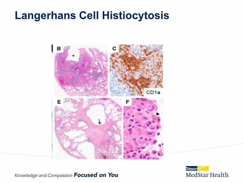
Langerhans Cell Histiocytosis
• Langerhans cells and other immune cells
produce bronchiolcentric nodules that precede
airway remodeling and cyst formation
• Eosinophilic infiltration with other inflammatory
cells
• Metallomatrix proteins lead to cyst formation
• Late: predominantly advanced bullous and
cystic disease
Langerhans Cell Histiocytosis
Pulmonary Langerhans Cell Histiocytosis:
Presentation
• 1/3 asymptomatic at diagnosis
• 2/3 cough, dyspnea, fatigue
• 1/5 constitutional symptoms such as weight loss or
fever
• 10-20% present with pneumothorax
• 10-15% extrapulmonary including skin, bone,
hypothalamus, lymph nodes
• 90% are smokers
Pulmonary Langerhans Cell Histiocytosis:
PFT
• 1/5 normal at diagnosis
• 2/3 reduced DlCO
• Often obstructed
• Lung volumes reduced, normal or increased
Pulmonary Langerhans Cell
Histiocytosis: Imaging
• Thin walled Cysts
• Nodules several mm to 2 cm
• Cysts with Bizarre shape
• Apical and midlung predominant
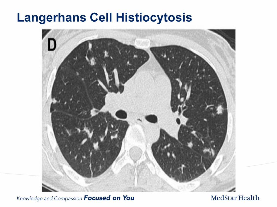
Langerhans Cell Histiocytosis
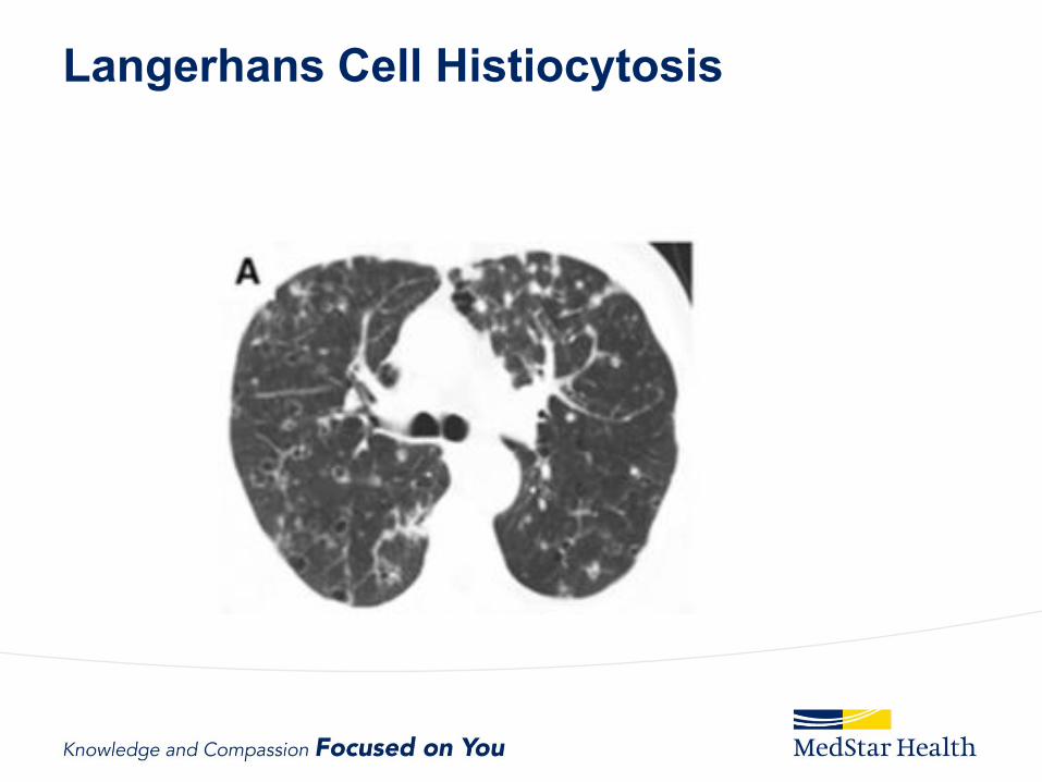
Langerhans Cell Histiocytosis

Langerhans Cell Histiocytosis
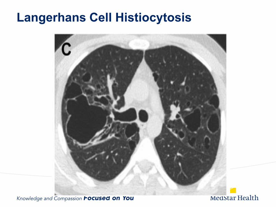
Langerhans Cell Histiocytosis
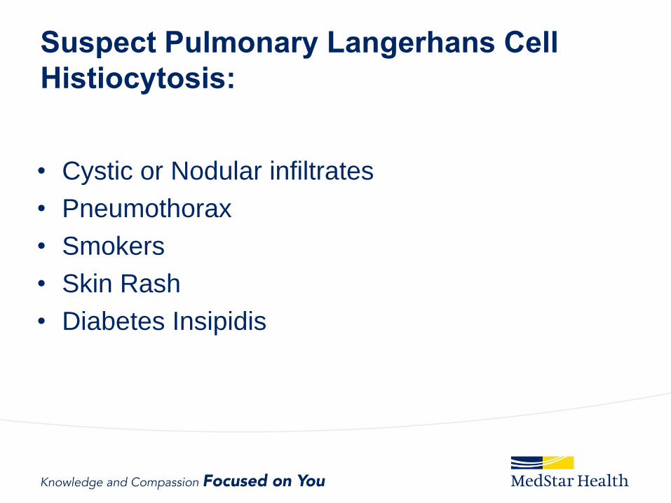
Suspect Pulmonary Langerhans Cell
Histiocytosis:
• Cystic or Nodular infiltrates
• Pneumothorax
• Smokers
• Skin Rash
• Diabetes Insipidis
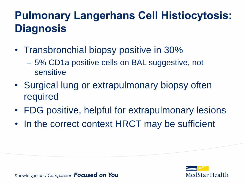
Pulmonary Langerhans Cell Histiocytosis:
Diagnosis
• Transbronchial biopsy positive in 30%
– 5% CD1a positive cells on BAL suggestive, not
sensitive
• Surgical lung or extrapulmonary biopsy often
required
• FDG positive, helpful for extrapulmonary lesions
• In the correct context HRCT may be sufficient
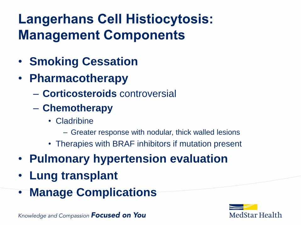
Langerhans Cell Histiocytosis:
Management Components
• Smoking Cessation
• Pharmacotherapy
– Corticosteroids controversial
– Chemotherapy
• Cladribine
– Greater response with nodular, thick walled lesions
• Therapies with BRAF inhibitors if mutation present
• Pulmonary hypertension evaluation
• Lung transplant
• Manage Complications
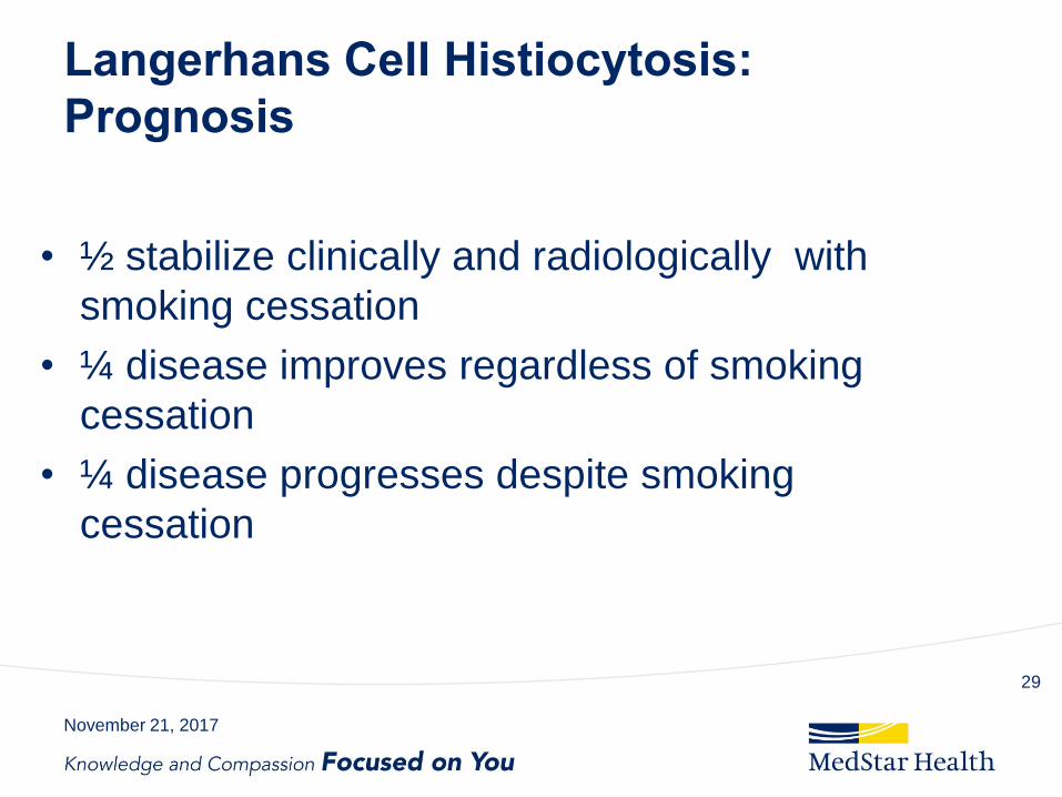
Langerhans Cell Histiocytosis:
Prognosis
• ½ stabilize clinically and radiologically with
smoking cessation
• ¼ disease improves regardless of smoking
cessation
• ¼ disease progresses despite smoking
cessation
November 21, 2017
29
Langerhans Cell Histiocytosis:
Prognosis
• Prognosis better with early diagnosis
• Impact of immune suppression on mortality not
clear
• Decline in PFT associated with worse prognosis
• ? Higher risk of neoplasms
November 21, 2017
30
Lymphangioleiomyomatosis
November 21, 2017
31

Lymphangleiomyomatosis:
A Disorder of TSC Genes
• Infiltration of lung by smooth muscle (LAM) cells
arising from unknown source
• LAM cells circulate and metastasize
• Conducting lymphatics extensively infiltrated by
LAM cells
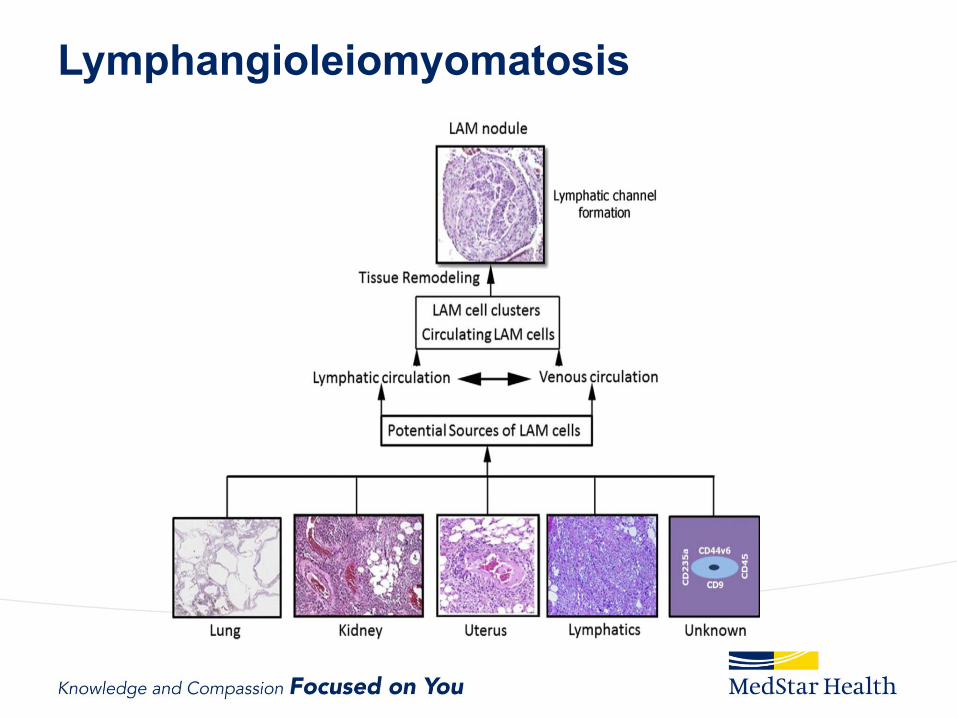
Lymphangioleiomyomatosis
Lymphangleiomyomatosis:
A Disorder of TSC Genes
• Growth activating mutations in tuberous
sclerosis genes
• TSC-LAM: Tuberous sclerosis complex
– 30% of women with tuberous sclerosis, 10-15% of
men with TSC
• S-LAM: Sporadic without tuberous sclerosis
– Entirely in women
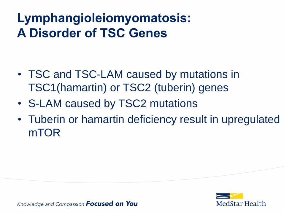
Lymphangioleiomyomatosis:
A Disorder of TSC Genes
• TSC and TSC-LAM caused by mutations in
TSC1(hamartin) or TSC2 (tuberin) genes
• S-LAM caused by TSC2 mutations
• Tuberin or hamartin deficiency result in upregulated
mTOR
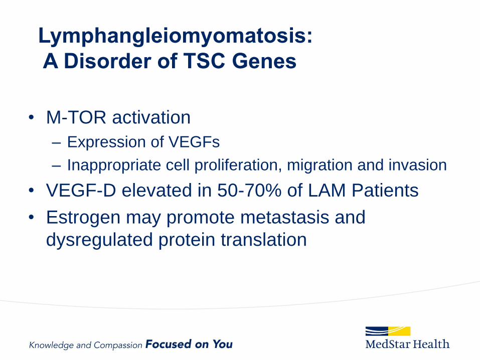
Lymphangleiomyomatosis:
A Disorder of TSC Genes
• M-TOR activation
– Expression of VEGFs
– Inappropriate cell proliferation, migration and invasion
• VEGF-D elevated in 50-70% of LAM Patients
• Estrogen may promote metastasis and
dysregulated protein translation
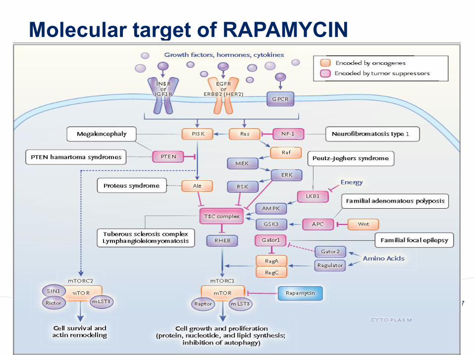
Molecular target of RAPAMYCIN
November 21, 2017
37

Pathology: Smooth Muscle Infiltration of
Parenchyma, Airways, Lymphatics with
Areas of Thin Walled Cystic Change
Lymphangioleiomyomatosis:
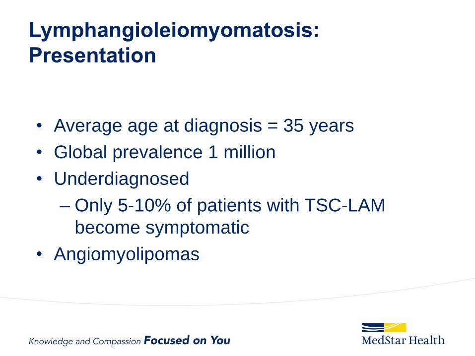
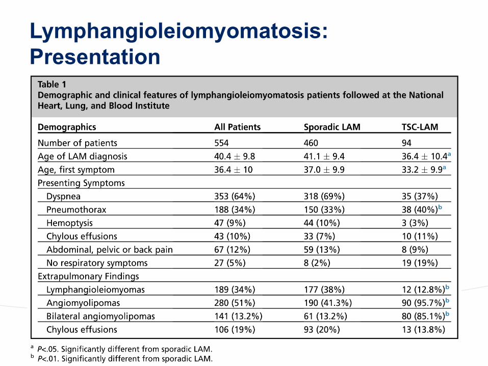
Presentation
• Average age at diagnosis = 35 years
• Global prevalence 1 million
• Underdiagnosed
– Only 5-10% of patients with TSC-LAM
become symptomatic
• Angiomyolipomas
Lymphangioleiomyomatosis:
Presentation
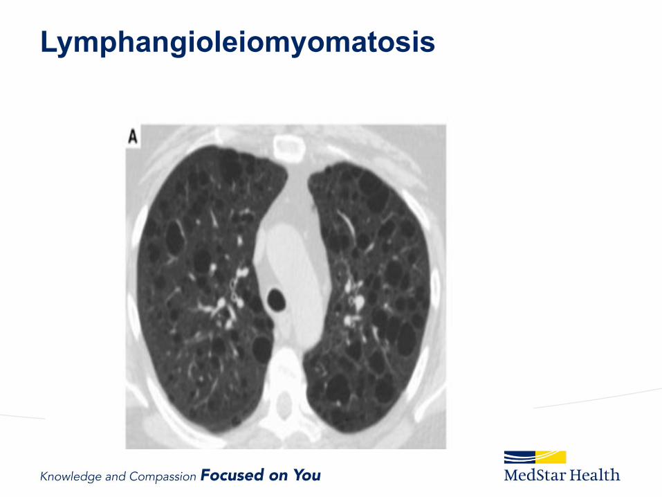
Lymphangioleiomyomatosis
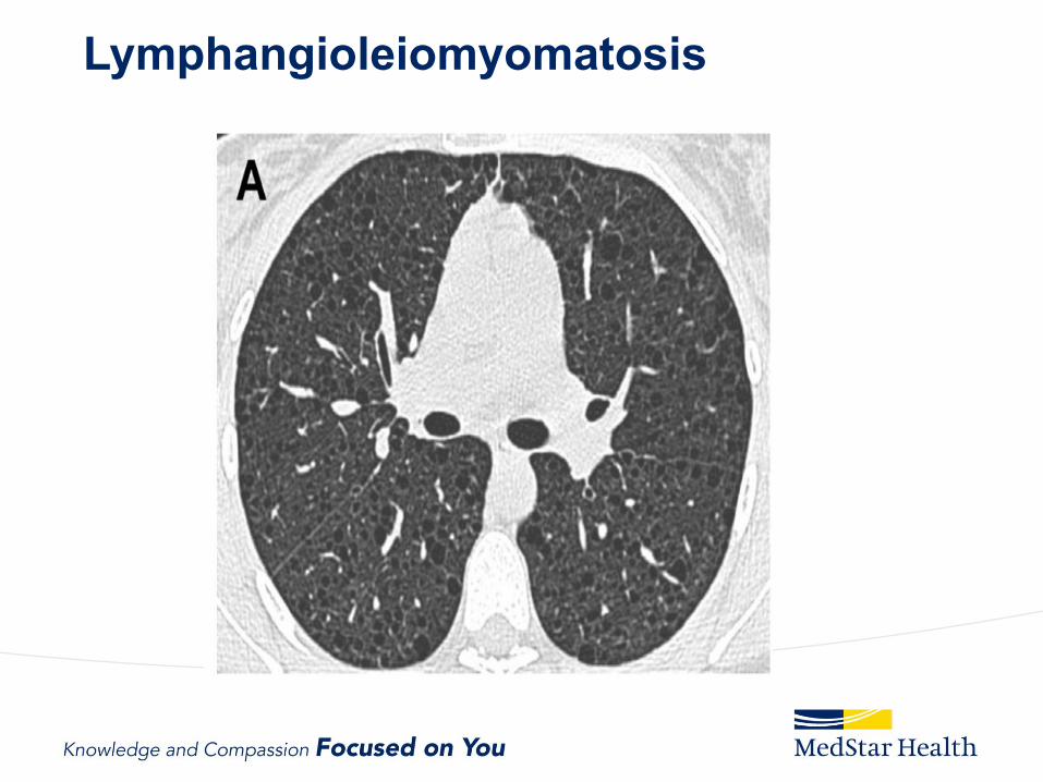
Lymphangioleiomyomatosis
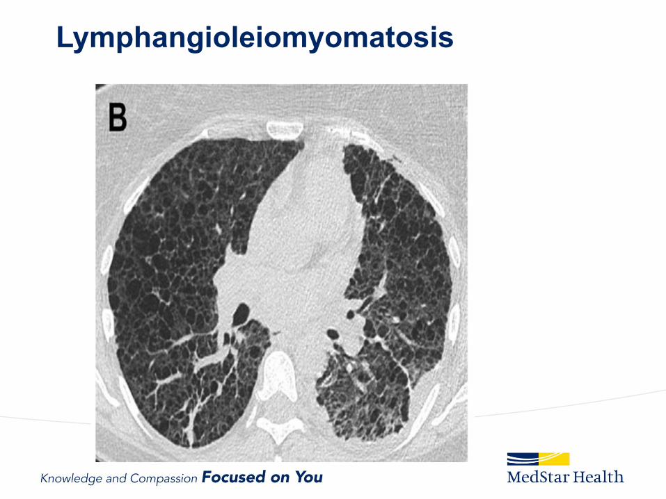
Lymphangioleiomyomatosis
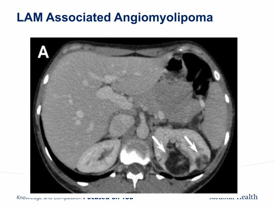
LAM Associated Angiomyolipoma
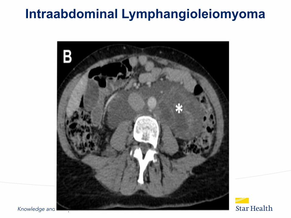
Intraabdominal Lymphangioleiomyoma
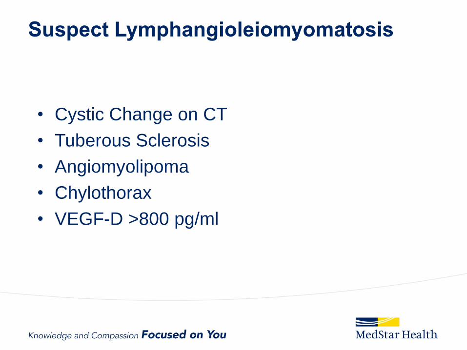
Suspect Lymphangioleiomyomatosis
• Cystic Change on CT
• Tuberous Sclerosis
• Angiomyolipoma
• Chylothorax
• VEGF-D >800 pg/ml
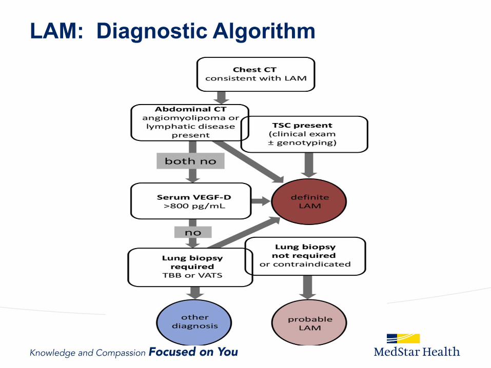
LAM: Diagnostic Algorithm
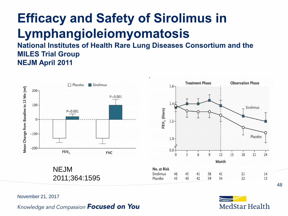
Efficacy and Safety of Sirolimus in
Lymphangioleiomyomatosis National Institutes of Health Rare Lung Diseases Consortium and the
MILES Trial Group
NEJM April 2011
November 21, 2017
48
NEJM
2011;364:1595
Lymphangioleiomyomatosis:
Management
• Sirolimus if FEV1<70%
• Accelerated decline in pregnancy
• Accelerated decline with estrogen use
• Lung transplant
NEJM
2011;364:1595
Lymphangioleiomyomatosis:
Management
• Screen for angiomyolipomas
– >4cm likely to bleed
– Treat with MTOR inhibitors and or embolization
• 70 % recurrence of PTX
– Pleurodese first episode
NEJM
2011;364:1595

Birt-Hogg-Dube
November 21, 2017
51
Birt-Hogg-Dube:
Folliculin Gene Mutation
• Autosomal Dominant
• Genetic testing available
• Mutations in folliculin (FLCN) gene
(a tumor supressor protein)
• Dysregulation in M-TOR signaling
• Hair Follicle tumors
• Renal Neoplasms
Birt-Hogg-Dube;
Pulmonary Cysts
• Basal subpleural thin walled Cysts
• Typically seen in 4th -5th decade
• By age 50 there is an 80% penetrance
• 24% of those with cysts develop pneumothorax
• Pneumothorax 32 times that of general
population
• Prevalence in young patients presenting with
PTX 5-10%
• Pneumothorax recurrence 75%
Birt-Hogg-Dube:
Renal Neoplasms
• Renal Cancer in 25%
– Mean age 50.4
– Chromophobe adenomas and oncocytomas
– Bilateral and multifocal in >50%
• Screen for renal neoplasms age 20
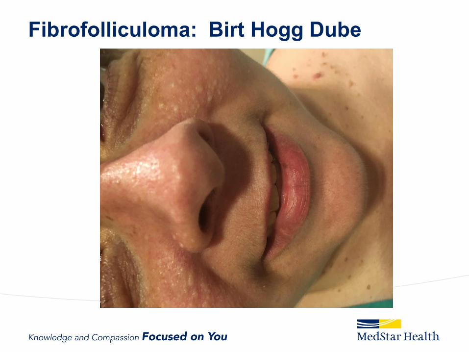
Fibrofolliculoma: Birt Hogg Dube
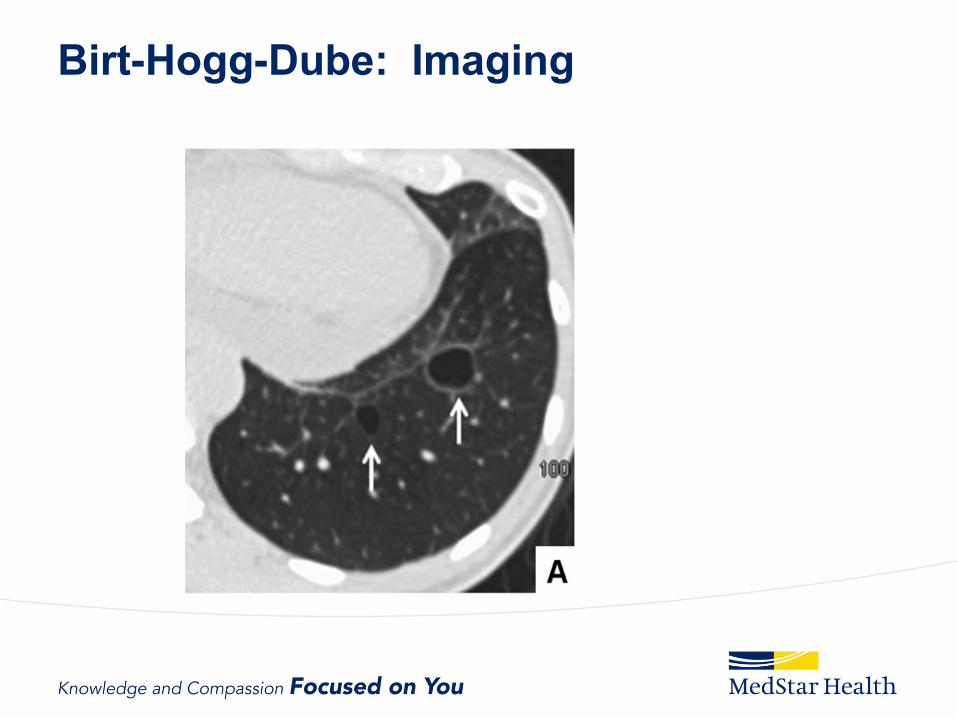
Birt-Hogg-Dube: Imaging
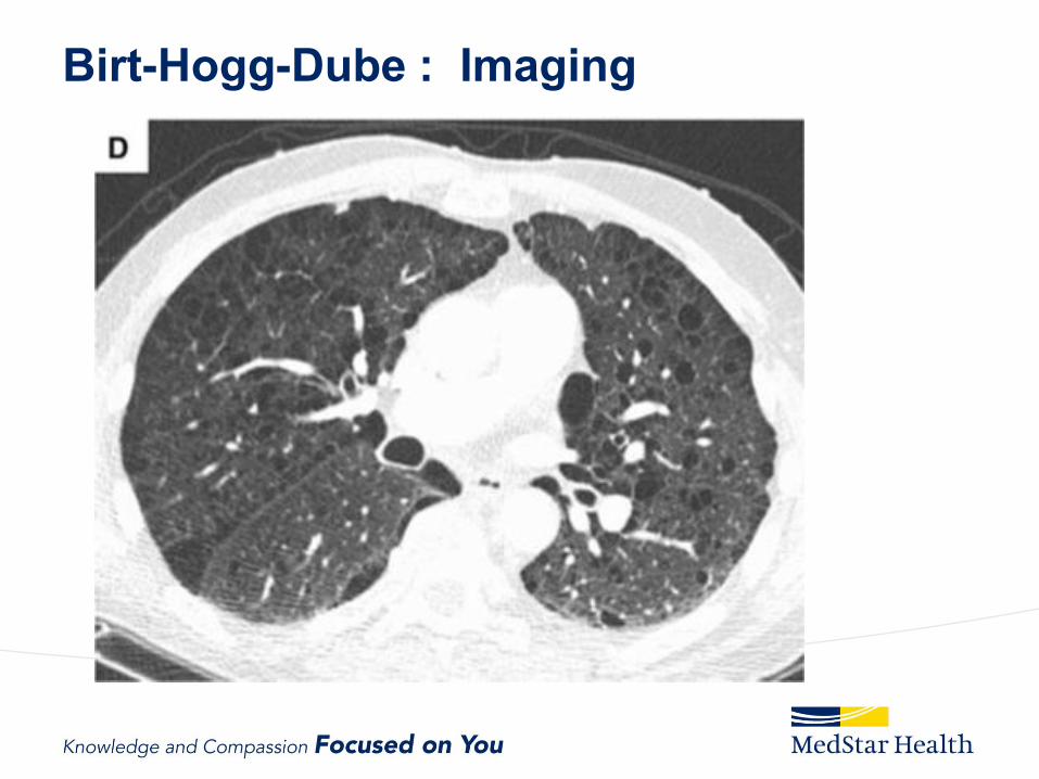
Birt-Hogg-Dube : Imaging

Other ILDs with Cystic Change
• IPF
• Chronic
Hypersensitivity
pneumonitis
• Sarcoidosis
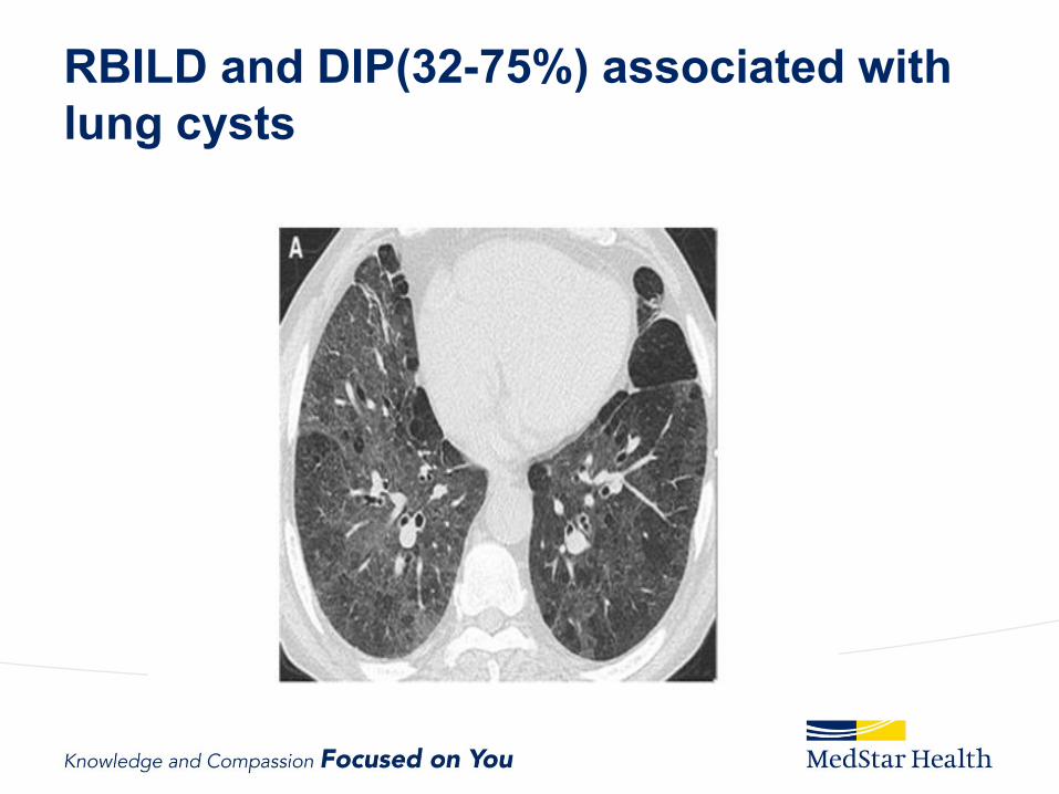
RBILD and DIP(32-75%) associated with
lung cysts
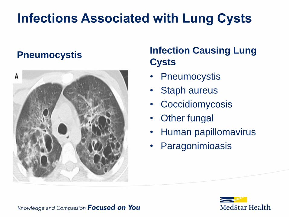
Infections Associated with Lung Cysts
Pneumocystis Infection Causing Lung
Cysts
• Pneumocystis
• Staph aureus
• Coccidiomycosis
• Other fungal
• Human papillomavirus
• Paragonimioasis
CYSTIC LUNG DISEASE AND
LYMPHOPROLIFERATIVE DISORDERS
• Follicular Bronchiolitis or LIP
– Autoimmune
• Sjogrens syndrome
• SLE
• RA
– Immunodeficiency
• HIV
• Common variable immunodeficiency
• Lymphoid infiltrates with B and T cells
– Germinal Centers in FB and LIP associated with SS
LIP/FB
• Ground Glass
• Centrilobular nodules
• Cysts in 68%
• May be associated with MALT lymphoma
• Malignant transformation to lymphoma is rare
• Median survival LIP 5-10 years
Sjogrens Syndrome
• HRCT abnormal in 60-90%
• Cysts in 10-50%
– Random
– Internal structure
– Can be associated with amyloidosis
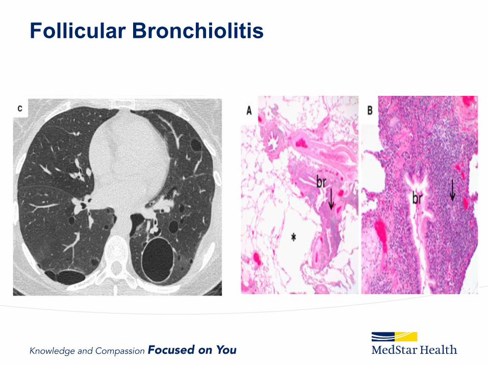
Follicular Bronchiolitis
Other Disease In the
Lymphoproliferative Spectrum
• Amyloidosis
– Cavitary nodules
– Diffuse cystic lung disease
– Associated with MALT lymphoma
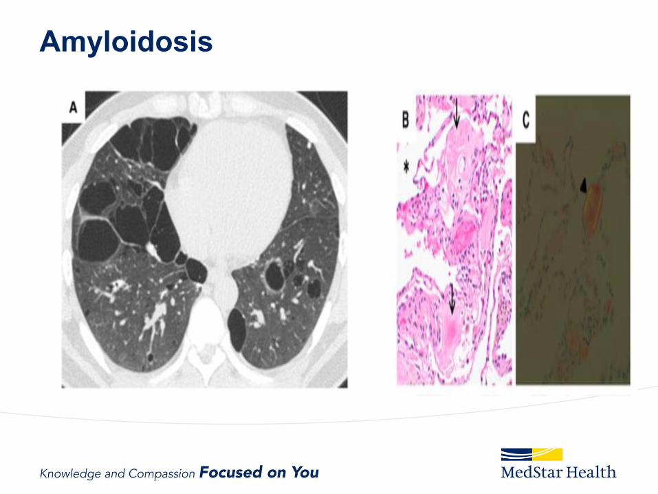
Amyloidosis

Other Diseases In the Lymphoproliferative
Spectrum
• Light Chain Deposition Disease
– Lymphoproliferative diseases with renal
involvement
– Diffuse cystic lung disease
• Multiple small diffuse
• Large cystic spaces with nodules mimic PLCH
• Has been associated with B cell clone in lung
suggestive of lymphoproliferative disorder
• Often progressive leading to respiratory failure
Cystic Lung Diseases
• Wide spectrum of disease
• Considerable progress in the last decade
• Many with neoplastic characteristics
• Prognosis and course highly variable
• Diagnosis often made based on imaging
characteristics and ancillary findings
November 21, 2017
68

Diagnostic Approach to DCLD

Questions
November 21, 2017
70