cucurbita argyrosperma seed extracts attenuate
TRANSCRIPT

Article 1
Cucurbita argyrosperma seed extracts attenuate 2
angiogenesis in a corneal chemical burn model 3
María Fernanda Estrella-Mendoza 1, Francisco Jiménez-Gómez2, Adolfo López-Ornelas2, Rosa 4 Martha Pérez-Gutiérrez 1, and Javier Flores-Estrada 2,* 5
1 Laboratorio de Investigacíon de productos naturales, Escuela de Ingeniería Química e Industrias 6 Extractivas, Instituto Politécnico Nacional. Unidad Profesional Adolfo López Mateos, Av. Instituto 7 Politécnico Nacional S/N, 07708 Ciudad de México, México; [email protected]; 8 [email protected] 9
2 División de Investigación, Hospital Juárez de México. Av. Instituto Politécnico Nacional 5160, Magdalena 10 de las Salinas, Gustavo A. Madero, 07760, Ciudad de México, México; [email protected] 11
* Correspondence: [email protected]; Tel.: +52-55-5747-7560 (ext.7476) 12 Received: date; Accepted: date; Published: date 13
Abstract: Cornea severe inflammation produces opacity or even perforation, scarring, and 14 angiogenesis, resulting in blindness. The cornea can be used to study the effect of new anti-15 angiogenic chemopreventive agents. We researched the anti-angiogenic effect of two extracts, 16 Methanol (Met) and Hexane (Hex), from the seed of Cucurbita argyrosperma, in the inflamed corneas. 17 The corneas of Wistar rats were alkali-injured and treated intragastrically for seven successive days. 18 Clinical manifestation as opacity score, corneal neovascularization (CNV) area, re-epithelialization 19 percentage, and histological evaluation were performed. Inflammatory (COX-2, NF-κB, and IL-1β), 20 and angiogenic (VEGF-A, VEGFR1, VEGFR2) markers were assessed by immunohistochemistry. 21 Cox-2, Il-1β, and Vegf-a mRNA levels were also determined. After treatments, we observed slim 22 corneal thickness with lower opacity scores and low cell infiltration compared to untreated rats. 23 Treatment also accelerated wound healing and decreased CNV area. The staining of inflammatory 24 and angiogenic factors was significantly decreased. These effects are related to a down-expression 25 of Cox-2, Il-1β, and Vegf. These results suggest that intake of C. argyrosperma seed can be used to 26 attenuate the angiogenesis secondary to inflammation in corneal chemical damage. 27
Keywords: C. argyrosperma; corneal chemical burn; angiogenesis; corneal neovascularization (CNV); 28 Vascular Endothelial Growth Factor (VEGF); Interleukin-1β (IL-1β); Cyclooxigenase-2 (COX-2); 29 Nuclear Factor-kappaB (NF-κB). 30
31
1. Introduction 32 Angiogenesis, the formation of new blood vessels from the pre-existing ones, inflammation, and 33
oxidative stress are important factors that predispose and promote the progression of degenerative 34 diseases such as tumors and diabetes, and corneal diseases are not an exception. Corneal 35 neovascularization (CNV), caused by viral infections, autoimmune diseases, and chemical burns 36 could progress into defective healing with persistent perforation and ulceration, resulting in 37 blindness or failure in the penetrating keratoplasty when not treated in a timely manner [1]. Under 38 this condition, NF-κB signaling pathway in inflammatory, epithelial, and endothelial cells, is a key 39 step for the transcriptional overexpression of pro-inflammatory and proangiogenic factors, including 40 interleukin-1β (IL-1β), cyclooxygenase 2 (COX-2), and vascular endothelial growth factor A (VEGF-41 A). COX-2 enzyme increases the synthesis of prostaglandins to modulate cell proliferation, cell death, 42 and tumor invasion in many types of cancer. IL-1β, IL-6 or TNF-α, regulate COX-2, therefore, they 43 are overexpressed during inflammation [2-4]. 44
45
Preprints (www.preprints.org) | NOT PEER-REVIEWED | Posted: 17 April 2019 doi:10.20944/preprints201904.0193.v1
© 2019 by the author(s). Distributed under a Creative Commons CC BY license.
Peer-reviewed version available at Nutrients 2019, 11, 1184; doi:10.3390/nu11051184
2 of 12
In the alkali-burn corneal injury, VEGF-A is an important factor of angiogenesis when is 46 expressed in macrophages and epithelial cells. VEGF-A binds to two main tyrosine kinase receptors, 47 VEGFR1 and VEGFR2, on the vascular endothelial cell to promote migration, proliferation, and 48 formation of capillaries, along with monocyte/macrophage migration in the microenvironment 49 injured [1,5,6]. Several therapeutic strategies to reduce CNV are studied based on these observations, 50 topical or subconjunctival treatments, mainly corticosteroids and non-steroidal anti-inflammatory 51 agents [7], have limited use and potential side effects as an impediment of wound healing [8,9]. Anti-52 VEGF therapy in chemically burned ocular tissues results in a substantial reduction of angiogenesis 53 both animal studies and clinical trials [10,11]. However, to establish its safety and efficacy, controlled 54 and randomized trials to justify their continued use are required. Besides, systemic drug treatment is 55 not recommended because of adverse effects. Thus, it is important the search for new drugs for the 56 systemic treatment of these disorders. 57
58 Current data in CNV models shown that natural extracts from plants or the bioactive 59
compounds in its extracts have angiogenic suppressing activity [12-14]. The genus Cucurbita 60 (pumpkin) belongs to one of the 300 genera of the Cucurbitaceae family and it is one of the most 61 popular vegetables eaten in the world. Recently, pumpkin was recognized as a functional food and 62 Cucurbita pepo, C. maxima, C. moschata, C. andreana, and C. ficifolia are the most cultivated species [15]. 63 Nutritionally, pumpkin seed contains a high amount of polyunsaturated fatty acids as well as 64 proteins, vitamins, several minerals, and other phytochemicals. The anti-diabetic, antioxidant, anti-65 carcinogenic, anti-inflammatory properties of this seed are studied due to its high content natural 66 bioactive compounds, such as carotenoids, tocopherols, and sterols [16-19]. 67
68 Cucurbita argyrosperma is an economically important species cultivated in Mesoamerica. 69
Isozyme, morphological, and ecological analysis suggest that it was probably domesticated from the 70 Mexican wild squash C. sororia [20]. Seed is usually consumed as a snack or as an ingredient in 71 traditional stews, although the scientific findings of its beneficial effects on human health have not 72 been sufficiently proven and the anti-neovascular effects of the secondary metabolites remain 73 unknown. However, conceivably its phytochemical composition could be like the related species, 74 showing anti-inflammatory effects as suggested. Besides, proangiogenic factors such as COX-2, IL-75 1β, and, VEGF, including to VEGFR1 and -R2, induced by the inflammatory agents has not studied 76 these plants. 77
78 The aim of this study was to contribute with new evidence of the effect of seed extracts from C. 79
argyrosperma in the inflammatory and angiogenic process attenuation. Here show that hexanic and 80 methanolic extracts from C. argyrosperma seed significantly attenuates the expression of 81 proangiogenic factors during the inflammation using CNV model. Also, we observed by clinical 82 manifestation that both extracts significantly diminish corneal neovascularization area. Remarkably, 83 corneal re-epithelialization was higher in the hexane extract treatment than methanol extract. 84
85
2. Materials and Methods 86 87 2.1 Extract Preparation 88
The pumpkins of C. argyrosperma were harvested in an agricultural field of the Michoacán 89 Province, México and identified by a botanist in the herbarium of The National Polytechnic Institute 90 (IPN). Voucher specimen number 4532 was deposited in the herbarium of the National School of 91 Biological Sciences of IPN. One kilogram of seed was extracted with 3L of hexane (50% v/v) and left 92 to macerate for 8 days at room temperature. Crude extract was filtered for one hour in 8 μM-medium 93 flow filter paper (Whatman®), concentrated using a rotary vacuum evaporator and taken to dryness 94 at 60°C in a vacuum rotator until the complete removal of the solvent, obtaining a viscous residue 95
Preprints (www.preprints.org) | NOT PEER-REVIEWED | Posted: 17 April 2019 doi:10.20944/preprints201904.0193.v1
Peer-reviewed version available at Nutrients 2019, 11, 1184; doi:10.3390/nu11051184
3 of 12
(8.38 g/L). The same procedure was applied to the residue, using methanol for a sequential separation 96 of the seed components. Each extract was stored in the dark at 4°C until use. 97 98 2.2 Animal model 99
Twenty-eight male Wistar rats weighing 200-250 g were used. Water and standard food were 100 available ad libitum. The care and management of experimental animals were performed according 101 to the guidelines of the National Institutes of Health Guide for the Care and Use of Laboratory 102 Animals, the standards described by ARVO (Association for Research in Vision and Ophthalmology), 103 and Official Mexican Standard NOM-062-ZOO-1999. 104
105 2.3 Experimental design 106
Each extract was dissolved in 0.5 ml (water) and Tween-80 (20%), which was also used as the 107 vehicle (Veh). Rats were randomly divided into four groups (n=7 each): Non-chemically burned 108 healthy corneas (Non-CB) treated only with the vehicle; chemically burned (CB) corneas treated with 109 the vehicle (CB-Veh); hexanic extract treatment (CB-Hex); and methanolic extract treatment (CB-110 Met). Groups were intragastrically injected with 400 mg/kg of Hex/Met extracts or 0.5 ml of the 111 vehicle in a single dose, at the same time daily (10:00 am). 48h later, animals were intraperitoneally 112 anesthetized with pentobarbital sodium (0.5 mg/kg), inhaled sevoflurane, and one drop of 113 ophthalmic tetracaine, to perform the chemical cauterization of the cornea. Central corneas from the 114 right eyes were burned by applying a 3-mm-diameter filter paper saturated with 1M NaOH solution 115 for 30 seconds and immediately washing with 10 ml of saline solution. To avoid infection, a drop of 116 ophthalmic ciprofloxacin was applied every 24 hours until the end of the study. Animals were 117 euthanized with an overdose of pentobarbital. 118
119 2.4 Clinical manifestation 120
Corneal opacity, epithelial defects, and the CNV area were clinically evaluated eight days after 121 CB. Corneal opacity was scored using a scaling system from 0 to 4: 0=no opacity, completely clear 122 cornea; 1=slightly hazy, iris and lens visible; 2=moderately opaque, iris and lens still detectable; 123 3=severely opaque, iris and lens hardly visible; and 4=completely opaque, with no visibility of the iris 124 and lens [21]. 125
126 The measurement of the CNV area (mm2) was performed in vivo using a ruler under a 127
microscope and photographed. The software program Image-Pro Plus version 6.0 software (by Media 128 Cybernetics, Inc., Rockville, MD, USA) was used. Inferonasal quadrant was selected to calculate the 129 neovascularized area, according to previous reports [22]. 130
131 To evaluate corneal wound re-epithelialization, we used corneal fluorescein staining. Briefly, the 132
lateral conjunctival sac was stained using fluorescein sodium ophthalmic strips. Corneas were 133 examined using a slit lamp biomicroscope with cobalt blue light. Injured epithelial tissues retain the 134 fluorescein staining, meanwhile, the lack of stain indicates a re-epithelialization. The re-135 epithelialization percentage was evaluated from corneal central burning, considering that a total area 136 of approx. 7 mm2 is 100 percent (3 mm disc 1M NaOH-embedded). 137
138 2.5 Histological evaluation 139
Enucleated eyes (n=4 by group) were immediately fixed in neutral formalin. Cut tissue slides (3-140 5 mm) were made, anteroposterior, and included the optic nerve. Slides were dehydrated in graded 141 alcohols and embedded in paraffin. Histological sections of 2μm were processed and stained with 142 hematoxylin-eosin (H&E). We measured the corneal thickness and cell infiltration in the peripheral 143 region (500 μm beyond the limb area) using light microscopy (axioscope 2 plus, Carl Zeiss). The 144 percentage of the infiltration was calculated in a masked fashion based on the density in the corneal 145 stroma of CB-Veh group. 146
147
Preprints (www.preprints.org) | NOT PEER-REVIEWED | Posted: 17 April 2019 doi:10.20944/preprints201904.0193.v1
Peer-reviewed version available at Nutrients 2019, 11, 1184; doi:10.3390/nu11051184
4 of 12
Other 2μm-sections were dewaxed and rehydrated up to antigen recovery solution 148 (ImmunoDNA Retriever 20X with Citrate; BioSB). Slides were then loaded into a Shandon Sequenza 149 chamber (Thermo Fisher Scientific, Inc). We used the procedure described for the polymer-based 150 immunodetection system (PolyVue® mouse/rabbit DAB detection system, Diagnostic BioSystems, 151 Pleasanton, CA, USA). We applied 100 μl of IL-1β (Cat. No. sc-7884), NF-κB p65 (sc-8008), COX-2 (sc-152 1746), VEGF-A (sc-7269), VEGFR1 (sc-31173). All antibodies were purchased from Santa Cruz 153 Biotechnology, Inc. (Santa Cruz, CA, USA). VEGFR2 antibody (MAB3571) was purchased from R&D 154 systems. All dilutions were at 1:200 and incubated overnight at 4°C. Later, the enhancers Polyvue 155 Plus and HRP were added and incubated with DAB plus/chromogen substrate and counterstained 156 hematoxylin. An Axio Imager.A2 microscope with an integrated camera (axiocam ICc5; Carl Zeiss 157 Microscopy GmbH, Germany) was used for histological observation and image capture. Micrographs 158 of the peripheral region of the cornea (3 fields per side and 500 mm above the limb, at 200X of 159 magnitude) were taken to measure the mean staining intensity of these markers. Images were 160 analyzed with the Image-Pro Plus software version 6.0. 161 162 2.6 Quantitative-reverse-transcription polymerase chain reaction (qRT-PCR) 163
Total RNA was isolated from corneal tissue using TRIzol™ Reagent (Invitrogen, Boston, MA, 164 USA) (n=3 by group). One microgram of DNase I-treated RNA (Roche Applied Science, Germany) 165 was reverse transcribed with SuperScript® II Reverse Transcriptase system. Quantification of mRNA 166 was carried out using qPCR with SYBR green and the following primers: Cox-2 (5'-167 CTGAGGGGTTACCACTTCCA-3´; and 5'-CTTGAACACGGACTTGCTCA-3); Il-1β (5'-168 AGGCTTCCTTGTGCAAGTGT-3' and 5'-TGAGTGACACTGCCTTCCTG-3'); Vegf-a (5'- 169 GCCCATGAAGTGGTGAAGTT-3´ and 5'-ACTCCAGGGCTTCATCATTG-3'); and Gapdh (5'-170 CTCATGACCACAGTCCATGC-3' and 5'-TTCAGCTCTGGGATGACCTT-3'). The cycling protocol was 171 as follows: denaturation (95°C for 10 min), 45 cycles of amplification (95°C for 15 s, 59°C for 15 s, and 172 72 ° C for 20 s), and a final extension at 72°C. A melting curve analysis was also performed to ascertain 173 the specificity of the amplified product. The expression for each gene was normalized to Gapdh. 174 Expression was quantified as fold-change using the ∆∆Ct method. 175
176 2.7 Statistical analysis 177
We used GraphPad Prism software (La Jolla, CA, USA) (version 5.0). Values are mean with 178 standard deviation (mean ± SD). In all cases, we used unifactorial analysis of variance followed by 179 Tukey’s post hoc analysis. 180
181
3. Results 182 3.1 Amelioration of corneal wound repair 183
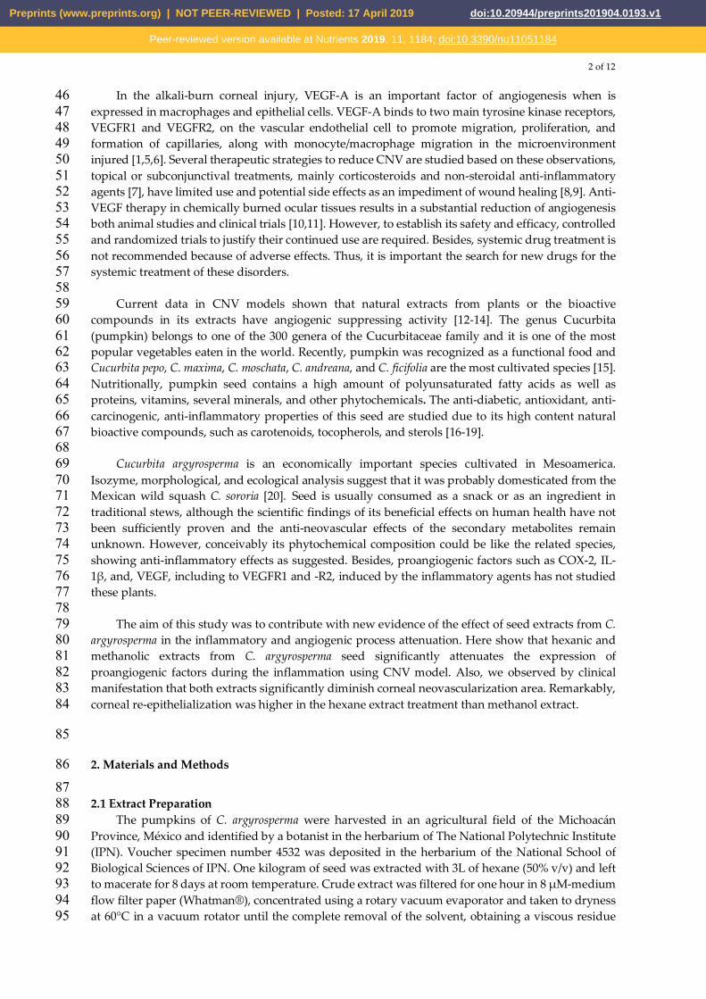
We evaluated corneal wound healing mediated by the extracts in the alkali-burn corneal model. 184 Figure 1a shows that treated groups had a significant reduction in corneal opacity score and CNV 185 area compared to CB-Veh (p<0.05 and p<0.001, respectively). However, CNV in CB-Met was lower 186 that CB-Hex treatment (p<0.05). Furthermore, CB-Met does not show significant differences in the 187 percentage of re-epithelialization compared to CB-Veh, inverse to CB-Hex treatment (Figure 1b). 188
H&E stained slides (Figure 1c), non-CB group had an average corneal thickness, 336.7±39.5μm, 189 with integral epithelial layers and a dense stroma, consisting of keratocytes. There are neither 190 inflammatory cells nor blood vessels. Conversely, corneal integrity in CB-Veh group was severely 191 impaired, with the loss of the epithelial cell layers, an extreme denaturation of stromal collagen fibers, 192 and an average cell infiltration of 81±11.9%. Corneal thickness was 592.3±112.1μm. Whereas, CB-Hex 193 tissues display a 40±8.1% infiltration, with a corneal thickness of 427.2±113.7μm (p<0.001 compared 194 to CB-Veh). In CB-Met group, there is a significant decrease in cell infiltration (33.2±5.9%) and 195 thickness of 295.2±62.67μm (p<0.001 compared to CB-Veh), but corneal thickness in CB-Met no shown 196 difference with Non-CB. 197
Preprints (www.preprints.org) | NOT PEER-REVIEWED | Posted: 17 April 2019 doi:10.20944/preprints201904.0193.v1
Peer-reviewed version available at Nutrients 2019, 11, 1184; doi:10.3390/nu11051184
5 of 12
198 199
Figure 1. Methanol (Met) and hexane (Hex) extracts of C. argyrosperma seed in alkali-injured corneas 200 (CB) compared to the untreated group (CB-Veh). (a) average of opacity score (Opa) and corneal 201 neovascularization area (CNV) in mm2. (b) re-epithelialization percentage (RE) and (c) corneal 202 thickness in microns (THK) and infiltration cell percentage (INF). Average value±SD; *p<0.05; **p< 203 0.01, and αp<0.001 compared to CB-Veh. @p<0.05 compared to CB-Met. Gray dotted lines show studied 204 area and black lines are geometrical axis. Arrows indicate the lumen of stromal blood vessels. (Scale 205 bar = 100 μm). 206
2 Anti-inflammatory effect 207
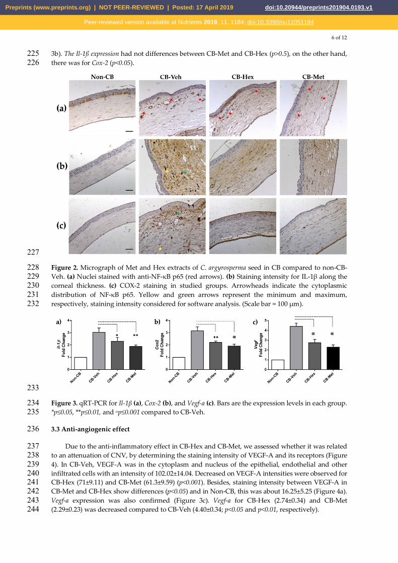
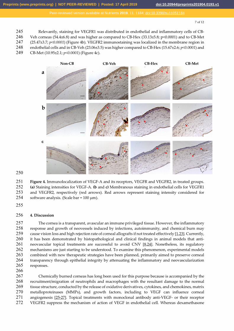
Several inflammatory cytokines implicated in alkali-induced corneal injury are regulated by the 208 nuclear internalization of active NF-κB. Hence, we glance for the staining location of NF-κB in the 209 corneal lesions. In Non-CB, NF-κB was restricted in the cytoplasm of epithelial cells in the basal layer 210 (Figure 2a). In CB-Veh and both extracts-treated groups, NF-κB was distributed in the nuclear 211 compartment of endothelial and inflammatory cells. However, staining density decreased in CB-Hex 212 (41.13±9.6) and CB-Met (32.73±8.1) compared to CB-Veh (73.31±10.4; p<0.0001 both). Additionally, we 213 performed measurements of the staining intensity for IL-1β (Figure 2b) and COX-2 (Figure 2c). In 214 Non-CB there was a low staining intensity for IL-1β (9.28±2.6) compared to CB-Veh (75.95±12.16; 215 p<0.0001), showing a distribution along the corneal stroma, as well as endothelial cells. Meanwhile, 216 the intensities in CB-Hex (42.16±9.14) and CB-Met (38.21±7.9) were lower than CB-Veh group 217 (p<0.0001). Staining intensity for IL-1β between CB-Met and CB-Hex have no differences (p>0.5). 218 Likewise, the intensity for COX-2 was significantly different when comparing CB-Veh (102.6±13.08) 219 to CB-Hex (68.79±10.73) and CB-Met (37.15±7.18) (p<0.0001). Non-CB has a detectable expression of 220 7.49±3.48. Staining intensity for IL-1β and COX-2 in the cornea was also confirmed at the level of 221 mRNA (Figure 3), Il-1β expression for CB-Met (2.31±0.30) and CB-Hex (1.89±0.11) was decreased 222 compared to CB-Veh (3.03±0.35; p<0.05 and p<0.01, respectively) (Figure 3a). Cox-2 in CB-Hex 223 (2.22±0.10) and CB-Met (1.91±0.15) was also diminished compared to Veh (3.15±0.31; p<0.01) (Figure 224
Preprints (www.preprints.org) | NOT PEER-REVIEWED | Posted: 17 April 2019 doi:10.20944/preprints201904.0193.v1
Peer-reviewed version available at Nutrients 2019, 11, 1184; doi:10.3390/nu11051184
6 of 12
3b). The Il-1ß expression had not differences between CB-Met and CB-Hex (p>0.5), on the other hand, 225 there was for Cox-2 (p<0.05). 226
227
Figure 2. Micrograph of Met and Hex extracts of C. argyrosperma seed in CB compared to non-CB-228 Veh. (a) Nuclei stained with anti-NF-κB p65 (red arrows). (b) Staining intensity for IL-1β along the 229 corneal thickness. (c) COX-2 staining in studied groups. Arrowheads indicate the cytoplasmic 230 distribution of NF-κB p65. Yellow and green arrows represent the minimum and maximum, 231 respectively, staining intensity considered for software analysis. (Scale bar = 100 μm). 232
233
Figure 3. qRT-PCR for Il-1β (a), Cox-2 (b), and Vegf-a (c). Bars are the expression levels in each group. 234 *p≤0.05, **p≤0.01, and αp≤0.001 compared to CB-Veh. 235
3.3 Anti-angiogenic effect 236
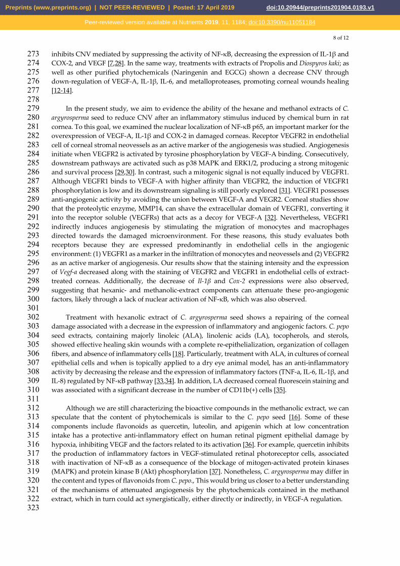
Due to the anti-inflammatory effect in CB-Hex and CB-Met, we assessed whether it was related 237 to an attenuation of CNV, by determining the staining intensity of VEGF-A and its receptors (Figure 238 4). In CB-Veh, VEGF-A was in the cytoplasm and nucleus of the epithelial, endothelial and other 239 infiltrated cells with an intensity of 102.02±14.04. Decreased on VEGF-A intensities were observed for 240 CB-Hex (71±9.11) and CB-Met (61.3±9.59) (p<0.001). Besides, staining intensity between VEGF-A in 241 CB-Met and CB-Hex show differences (p<0.05) and in Non-CB, this was about 16.25±5.25 (Figure 4a). 242 Vegf-a expression was also confirmed (Figure 3c). Vegf-a for CB-Hex (2.74±0.34) and CB-Met 243 (2.29±0.23) was decreased compared to CB-Veh (4.40±0.34; p<0.05 and p<0.01, respectively). 244
Preprints (www.preprints.org) | NOT PEER-REVIEWED | Posted: 17 April 2019 doi:10.20944/preprints201904.0193.v1
Peer-reviewed version available at Nutrients 2019, 11, 1184; doi:10.3390/nu11051184
7 of 12
Relevantly, staining for VEGFR1 was distributed in endothelial and inflammatory cells of CB-245 Veh corneas (54.4±6.8) and was higher as compared to CB-Hex (33.13±5.8; p<0.0001) and to CB-Met 246 (25.47±3.7; p<0.0001) (Figure 4b). VEGFR2 immunostaining was localized in the membrane region in 247 endothelial cells and in CB-Veh (23.06±3.5) was higher compared to CB-Hex (15.67±2.6; p<0.0001) and 248 CB-Met (10.95±2.1; p<0.0001) (Figure 4c). 249
250
Figure 4. Immunolocalization of VEGF-A and its receptors, VEGFR and VEGFR2, in treated groups. 251 (a) Staining intensities for VEGF-A. (b and c) Membranous staining in endothelial cells for VEGFR1 252 and VEGFR2, respectively (red arrows). Red arrows represent staining intensity considered for 253 software analysis. (Scale bar = 100 μm). 254
255
4. Discussion 256
The cornea is a transparent, avascular an immune privileged tissue. However, the inflammatory 257 response and growth of neovessels induced by infections, autoimmunity, and chemical burn may 258 cause vision loss and high rejection rate of corneal allografts if not treated effectively [1,23]. Currently, 259 it has been demonstrated by histopathological and clinical findings in animal models that anti-260 neovascular topical treatments are successful to avoid CNV [8,24]. Nonetheless, its regulatory 261 mechanisms are just starting to be understood. To examine this phenomenon, experimental models 262 combined with new therapeutic strategies have been planned, primarily aimed to preserve corneal 263 transparency through epithelial integrity by attenuating the inflammatory and neovascularization 264 responses. 265
266 Chemically burned corneas has long been used for this purpose because is accompanied by the 267
recruitment/migration of neutrophils and macrophages with the resultant damage to the normal 268 tissue structure, conducted by the release of oxidative derivatives, cytokines, and chemokines, matrix 269 metalloproteinases (MMPs), and growth factors, including to VEGF can influence corneal 270 angiogenesis [25-27]. Topical treatments with monoclonal antibody anti-VEGF- or their receptor 271 VEGFR2 suppress the mechanism of action of VEGF in endothelial cell. Whereas dexamethasone 272
Preprints (www.preprints.org) | NOT PEER-REVIEWED | Posted: 17 April 2019 doi:10.20944/preprints201904.0193.v1
Peer-reviewed version available at Nutrients 2019, 11, 1184; doi:10.3390/nu11051184
8 of 12
inhibits CNV mediated by suppressing the activity of NF-κB, decreasing the expression of IL-1β and 273 COX-2, and VEGF [7,28]. In the same way, treatments with extracts of Propolis and Diospyros kaki; as 274 well as other purified phytochemicals (Naringenin and EGCG) shown a decrease CNV through 275 down-regulation of VEGF-A, IL-1β, IL-6, and metalloproteases, promoting corneal wounds healing 276 [12-14]. 277
278 In the present study, we aim to evidence the ability of the hexane and methanol extracts of C. 279
argyrosperma seed to reduce CNV after an inflammatory stimulus induced by chemical burn in rat 280 cornea. To this goal, we examined the nuclear localization of NF-κB p65, an important marker for the 281 overexpression of VEGF-A, IL-1β and COX-2 in damaged corneas. Receptor VEGFR2 in endothelial 282 cell of corneal stromal neovessels as an active marker of the angiogenesis was studied. Angiogenesis 283 initiate when VEGFR2 is activated by tyrosine phosphorylation by VEGF-A binding. Consecutively, 284 downstream pathways are activated such as p38 MAPK and ERK1/2, producing a strong mitogenic 285 and survival process [29,30]. In contrast, such a mitogenic signal is not equally induced by VEGFR1. 286 Although VEGFR1 binds to VEGF-A with higher affinity than VEGFR2, the induction of VEGFR1 287 phosphorylation is low and its downstream signaling is still poorly explored [31]. VEGFR1 possesses 288 anti-angiogenic activity by avoiding the union between VEGF-A and VEGR2. Corneal studies show 289 that the proteolytic enzyme, MMP14, can shave the extracellular domain of VEGFR1, converting it 290 into the receptor soluble (VEGFRs) that acts as a decoy for VEGF-A [32]. Nevertheless, VEGFR1 291 indirectly induces angiogenesis by stimulating the migration of monocytes and macrophages 292 directed towards the damaged microenvironment. For these reasons, this study evaluates both 293 receptors because they are expressed predominantly in endothelial cells in the angiogenic 294 environment: (1) VEGFR1 as a marker in the infiltration of monocytes and neovessels and (2) VEGFR2 295 as an active marker of angiogenesis. Our results show that the staining intensity and the expression 296 of Vegf-a decreased along with the staining of VEGFR2 and VEGFR1 in endothelial cells of extract-297 treated corneas. Additionally, the decrease of Il-1β and Cox-2 expressions were also observed, 298 suggesting that hexanic- and methanolic-extract components can attenuate these pro-angiogenic 299 factors, likely through a lack of nuclear activation of NF-κB, which was also observed. 300
301 Treatment with hexanolic extract of C. argyrosperma seed shows a repairing of the corneal 302
damage associated with a decrease in the expression of inflammatory and angiogenic factors. C. pepo 303 seed extracts, containing majorly linoleic (ALA), linolenic acids (LA), tocopherols, and sterols, 304 showed effective healing skin wounds with a complete re-epithelialization, organization of collagen 305 fibers, and absence of inflammatory cells [18]. Particularly, treatment with ALA, in cultures of corneal 306 epithelial cells and when is topically applied to a dry eye animal model, has an anti-inflammatory 307 activity by decreasing the release and the expression of inflammatory factors (TNF-a, IL-6, IL-1β, and 308 IL-8) regulated by NF-κB pathway [33,34]. In addition, LA decreased corneal fluorescein staining and 309 was associated with a significant decrease in the number of CD11b(+) cells [35]. 310
311 Although we are still characterizing the bioactive compounds in the methanolic extract, we can 312
speculate that the content of phytochemicals is similar to the C. pepo seed [16]. Some of these 313 components include flavonoids as quercetin, luteolin, and apigenin which at low concentration 314 intake has a protective anti-inflammatory effect on human retinal pigment epithelial damage by 315 hypoxia, inhibiting VEGF and the factors related to its activation [36]. For example, quercetin inhibits 316 the production of inflammatory factors in VEGF-stimulated retinal photoreceptor cells, associated 317 with inactivation of NF-κB as a consequence of the blockage of mitogen-activated protein kinases 318 (MAPK) and protein kinase B (Akt) phosphorylation [37]. Nonetheless, C. argyrosperma may differ in 319 the content and types of flavonoids from C. pepo., This would bring us closer to a better understanding 320 of the mechanisms of attenuated angiogenesis by the phytochemicals contained in the methanol 321 extract, which in turn could act synergistically, either directly or indirectly, in VEGF-A regulation. 322
323
Preprints (www.preprints.org) | NOT PEER-REVIEWED | Posted: 17 April 2019 doi:10.20944/preprints201904.0193.v1
Peer-reviewed version available at Nutrients 2019, 11, 1184; doi:10.3390/nu11051184
9 of 12
Corneal inflammation eventually causes vision loss due to CNV. Corneal alkali-injury not only 324 raised to NF-κB, IL-1β, and COX-2 expression, but also significantly increase VEGF and their 325 receptors VEGFR1 and VEGFR2 in endothelial cells. This work demonstrates for the first time, that 326 methanolic or hexanic extracts of C. argyrosperma seed (400 mg/kg/7d) improves the healing of corneal 327 wound injured by a chemical agent and may contribute to the anti-inflammatory properties of the 328 phytochemicals in its composition. In addition, a significant reduction of the CNV was related to the 329 attenuation of proangiogenic factors. Significantly, our results indicate that C. argyrosperma Hex-330 extract is better than Met-extract to reduce the corneal re-epithelialization time, improving the 331 healing process and thus preventing the entrance of microorganisms and inflammatory mediators 332 into the deeper layers, probably through the inhibition of the NF-κB pathway during at least seven 333 days after corneal alkali-burn. 334
335 Consequently, ingestion of the seed can be an option to prevent the corneal angiogenesis. As 336
well, it might benefit wound healing or inhibit neovascularization in other degenerative pathologies. 337 Further pharmacological and phytochemical studies are required to identify its constituents and 338 accurately assess this activity. 339
340 Author Contributions: Conceptualization, R.M.P-G; Formal analysis, J.F-E; Investigation, M.F.E-M, 341 F. J-G, R.M.P-G and J.F-E; Methodology, M.F.E-M, F. J-G and R.M.P-G; Project administration, J.F-E; 342 Resources, F. J-G and R.M.P-G; Supervision, J.F-E; Visualization, A.L-O and J.F-E; Writing – original 343 draft, A.L-O and J.F-E; Writing – review & editing, A.L-O and J.F-E. 344
345 Funding: This research received no external funding 346 Acknowledgments: We thank Julia D. Toscano-Garibay for her numerous comments on the 347 manuscript. This research supported by the Juárez of México Hospital and School of Chemical 348 Engineering and Extractive Industries from the National Polytechnic Institute. 349 Conflicts of Interest: The authors declare no conflict of interest. 350
351
Preprints (www.preprints.org) | NOT PEER-REVIEWED | Posted: 17 April 2019 doi:10.20944/preprints201904.0193.v1
Peer-reviewed version available at Nutrients 2019, 11, 1184; doi:10.3390/nu11051184

10 of 12
References 352 1. Azar, D.T. Corneal angiogenic privilege: angiogenic and antiangiogenic factors in corneal 353
avascularity, vasculogenesis, and wound healing (an American Ophthalmological Society thesis). 354 Trans Am Ophthalmol Soc 2006, 104, 264-302. 355
2. Kuwano, T.; Nakao, S.; Yamamoto, H.; Tsuneyoshi, M.; Yamamoto, T.; Kuwano, M.; Ono, 356 M. Cyclooxygenase 2 is a key enzyme for inflammatory cytokine-induced angiogenesis. FASEB 357 journal : official publication of the Federation of American Societies for Experimental Biology 2004, 18, 300-358 310, doi:10.1096/fj.03-0473com. 359
3. Nakao, S.; Kuwano, T.; Tsutsumi-Miyahara, C.; Ueda, S.; Kimura, Y.N.; Hamano, S.; 360 Sonoda, K.H.; Saijo, Y.; Nukiwa, T.; Strieter, R.M., et al. Infiltration of COX-2-expressing macrophages 361 is a prerequisite for IL-1 beta-induced neovascularization and tumor growth. The Journal of clinical 362 investigation 2005, 115, 2979-2991, doi:10.1172/jci23298. 363
4. Sobolewski, C.; Cerella, C.; Dicato, M.; Ghibelli, L.; Diederich, M. The role of 364 cyclooxygenase-2 in cell proliferation and cell death in human malignancies. Int J Cell Biol 2010, 2010, 365 215158, doi:10.1155/2010/215158. 366
5. Cursiefen, C.; Chen, L.; Borges, L.P.; Jackson, D.; Cao, J.; Radziejewski, C.; D'Amore, P.A.; 367 Dana, M.R.; Wiegand, S.J.; Streilein, J.W. VEGF-A stimulates lymphangiogenesis and 368 hemangiogenesis in inflammatory neovascularization via macrophage recruitment. The Journal of 369 clinical investigation 2004, 113, 1040-1050, doi:10.1172/jci20465. 370
6. Sivak, J.M.; Ostriker, A.C.; Woolfenden, A.; Demirs, J.; Cepeda, R.; Long, D.; Anderson, K.; 371 Jaffee, B. Pharmacologic uncoupling of angiogenesis and inflammation during initiation of 372 pathological corneal neovascularization. J Biol Chem 2011, 286, 44965-44975, 373 doi:10.1074/jbc.M111.294967. 374
7. Nakao, S.; Hata, Y.; Miura, M.; Noda, K.; Kimura, Y.N.; Kawahara, S.; Kita, T.; Hisatomi, T.; 375 Nakazawa, T.; Jin, Y., et al. Dexamethasone inhibits interleukin-1beta-induced corneal 376 neovascularization: role of nuclear factor-kappaB-activated stromal cells in inflammatory 377 angiogenesis. The American journal of pathology 2007, 171, 1058-1065, doi:10.2353/ajpath.2007.070172. 378
8. Gupta, D.; Illingworth, C. Treatments for corneal neovascularization: a review. Cornea 2011, 379 30, 927-938, doi:10.1097/ICO.0b013e318201405a. 380
9. Sheppard, J.D.; Comstock, T.L.; Cavet, M.E. Impact of the Topical Ophthalmic 381 Corticosteroid Loteprednol Etabonate on Intraocular Pressure. Advances in therapy 2016, 33, 532-552, 382 doi:10.1007/s12325-016-0315-8. 383
10. Al-Debasi, T.; Al-Bekairy, A.; Al-Katheri, A.; Al Harbi, S.; Mansour, M. Topical versus 384 subconjunctival anti-vascular endothelial growth factor therapy (Bevacizumab, Ranibizumab and 385 Aflibercept) for treatment of corneal neovascularization. Saudi journal of ophthalmology : official journal 386 of the Saudi Ophthalmological Society 2017, 31, 99-105, doi:10.1016/j.sjopt.2017.02.008. 387
11. Zhou, C.; Robert, M.C.; Kapoulea, V.; Lei, F.; Stagner, A.M.; Jakobiec, F.A.; Dohlman, C.H.; 388 Paschalis, E.I. Sustained Subconjunctival Delivery of Infliximab Protects the Cornea and Retina 389 Following Alkali Burn to the Eye. Invest Ophthalmol Vis Sci 2017, 58, 96-105, doi:10.1167/iovs.16-20339. 390
12. Keshavarz, M.; Mostafaie, A.; Mansouri, K.; Shakiba, Y.; Motlagh, H.R. Inhibition of corneal 391 neovascularization with propolis extract. Archives of medical research 2009, 40, 59-61, 392 doi:10.1016/j.arcmed.2008.10.004. 393
13. Oguido, A.; Hohmann, M.S.N.; Pinho-Ribeiro, F.A.; Crespigio, J.; Domiciano, T.P.; Verri, 394 W.A., Jr.; Casella, A.M.B. Naringenin Eye Drops Inhibit Corneal Neovascularization by Anti-395 Inflammatory and Antioxidant Mechanisms. Invest Ophthalmol Vis Sci 2017, 58, 5764-5776, 396 doi:10.1167/iovs.16-19702. 397
14. Yang, S.J.; Jo, H.; Kim, K.A.; Ahn, H.R.; Kang, S.W.; Jung, S.H. Diospyros kaki Extract 398 Inhibits Alkali Burn-Induced Corneal Neovascularization. Journal of medicinal food 2016, 19, 106-109, 399 doi:10.1089/jmf.2014.3404. 400
15. Kocyan, A.; Zhang, L.B.; Schaefer, H.; Renner, S.S. A multi-locus chloroplast phylogeny for 401 the Cucurbitaceae and its implications for character evolution and classification. Molecular 402 phylogenetics and evolution 2007, 44, 553-577, doi:10.1016/j.ympev.2006.12.022. 403
Preprints (www.preprints.org) | NOT PEER-REVIEWED | Posted: 17 April 2019 doi:10.20944/preprints201904.0193.v1
Peer-reviewed version available at Nutrients 2019, 11, 1184; doi:10.3390/nu11051184
11 of 12
16. Seun F. Akomolafe; Ganiyu Oboh; Sunday I. Oyeleye; Olorunfemi R. Molehin; Opeyemi B. 404 OgunsuyiPeiretti, P.G. Phenolic Composition and Inhibitory Ability of Methanolic Extract from 405 Pumpkin (Cucurbita pepo L) Seeds on Fe-induced Thiobarbituric acid reactive species in Albino Rat’s 406 Testicular Tissue In-Vitro. Journal of Applied Pharmaceutical Science 2016, 6, 115-120, 407 doi:10.7324/JAPS.2016.60917 408
17. Yadav, M.; Jain, S.; Tomar, R.; Prasad, G.B.; Yadav, H. Medicinal and biological potential of 409 pumpkin: an updated review. Nutrition research reviews 2010, 23, 184-190, 410 doi:10.1017/s0954422410000107. 411
18. Bardaa, S.; Ben Halima, N.; Aloui, F.; Ben Mansour, R.; Jabeur, H.; Bouaziz, M.; Sahnoun, Z. 412 Oil from pumpkin (Cucurbita pepo L.) seeds: evaluation of its functional properties on wound 413 healing in rats. Lipids in health and disease 2016, 15, 73, doi:10.1186/s12944-016-0237-0. 414
19. Medjakovic, S.; Hobiger, S.; Ardjomand-Woelkart, K.; Bucar, F.; Jungbauer, A. Pumpkin 415 seed extract: Cell growth inhibition of hyperplastic and cancer cells, independent of steroid hormone 416 receptors. Fitoterapia 2016, 110, 150-156, doi:10.1016/j.fitote.2016.03.010. 417
20. Sanchez-de la Vega, G.; Castellanos-Morales, G.; Gamez, N.; Hernandez-Rosales, H.S.; 418 Vazquez-Lobo, A.; Aguirre-Planter, E.; Jaramillo-Correa, J.P.; Montes-Hernandez, S.; Lira-Saade, R.; 419 Eguiarte, L.E. Genetic Resources in the "Calabaza Pipiana" Squash (Cucurbita argyrosperma) in 420 Mexico: Genetic Diversity, Genetic Differentiation and Distribution Models. Frontiers in plant science 421 2018, 9, 400, doi:10.3389/fpls.2018.00400. 422
21. Yoeruek, E.; Ziemssen, F.; Henke-Fahle, S.; Tatar, O.; Tura, A.; Grisanti, S.; Bartz-Schmidt, 423 K.U.; Szurman, P. Safety, penetration and efficacy of topically applied bevacizumab: evaluation of 424 eyedrops in corneal neovascularization after chemical burn. Acta ophthalmologica 2008, 86, 322-328, 425 doi:10.1111/j.1600-0420.2007.01049.x. 426
22. Rogers, M.S.; Birsner, A.E.; D'Amato, R.J. The mouse cornea micropocket angiogenesis 427 assay. Nature protocols 2007, 2, 2545-2550, doi:10.1038/nprot.2007.368. 428
23. Dana, M.R.; Streilein, J.W. Loss and restoration of immune privilege in eyes with corneal 429 neovascularization. Invest Ophthalmol Vis Sci 1996, 37, 2485-2494. 430
24. Roshandel, D.; Eslani, M.; Baradaran-Rafii, A.; Cheung, A.Y.; Kurji, K.; Jabbehdari, S.; Maiz, 431 A.; Jalali, S.; Djalilian, A.R.; Holland, E.J. Current and emerging therapies for corneal 432 neovascularization. The ocular surface 2018, 16, 398-414, doi:10.1016/j.jtos.2018.06.004. 433
25. Choi, H.; Phillips, C.; Oh, J.Y.; Stock, E.M.; Kim, D.K.; Won, J.K.; Fulcher, S. Comprehensive 434 Modeling of Corneal Alkali Injury in the Rat Eye. Current eye research 2017, 42, 1348-1357, 435 doi:10.1080/02713683.2017.1317817. 436
26. Shakiba, Y.; Mansouri, K.; Arshadi, D.; Rezaei, N. Corneal neovascularization: molecular 437 events and therapeutic options. Recent Pat Inflamm Allergy Drug Discov 2009, 3, 221-231. 438
27. Edelman, J.L.; Castro, M.R.; Wen, Y. Correlation of VEGF expression by leukocytes with the 439 growth and regression of blood vessels in the rat cornea. Invest Ophthalmol Vis Sci 1999, 40, 1112-1123. 440
28. Nakao, S.; Zandi, S.; Lara-Castillo, N.; Taher, M.; Ishibashi, T.; Hafezi-Moghadam, A. Larger 441 therapeutic window for steroid versus VEGF-A inhibitor in inflammatory angiogenesis: surprisingly 442 similar impact on leukocyte infiltration. Invest Ophthalmol Vis Sci 2012, 53, 3296-3302, 443 doi:10.1167/iovs.11-8114. 444
29. Kim, Y.M.; Hwang, S.; Kim, Y.M.; Pyun, B.J.; Kim, T.Y.; Lee, S.T.; Gho, Y.S.; Kwon, Y.G. 445 Endostatin blocks vascular endothelial growth factor-mediated signaling via direct interaction with 446 KDR/Flk-1. J Biol Chem 2002, 277, 27872-27879, doi:10.1074/jbc.M202771200. 447
30. Gee, E.; Milkiewicz, M.; Haas, T.L. p38 MAPK activity is stimulated by vascular endothelial 448 growth factor receptor 2 activation and is essential for shear stress-induced angiogenesis. J Cell Physiol 449 2010, 222, 120-126, doi:10.1002/jcp.21924. 450
31. Koch, S.; Claesson-Welsh, L. Signal transduction by vascular endothelial growth factor 451 receptors. Cold Spring Harbor perspectives in medicine 2012, 2, a006502, 452 doi:10.1101/cshperspect.a006502. 453
Preprints (www.preprints.org) | NOT PEER-REVIEWED | Posted: 17 April 2019 doi:10.20944/preprints201904.0193.v1
Peer-reviewed version available at Nutrients 2019, 11, 1184; doi:10.3390/nu11051184
12 of 12
32. Han, K.Y.; Chang, J.H.; Lee, H.; Azar, D.T. Proangiogenic Interactions of Vascular 454 Endothelial MMP14 With VEGF Receptor 1 in VEGFA-Mediated Corneal Angiogenesis. Invest 455 Ophthalmol Vis Sci 2016, 57, 3313-3322, doi:10.1167/iovs.16-19420. 456
33. Erdinest, N.; Shmueli, O.; Grossman, Y.; Ovadia, H.; Solomon, A. Anti-inflammatory effects 457 of alpha linolenic acid on human corneal epithelial cells. Invest Ophthalmol Vis Sci 2012, 53, 4396-4406, 458 doi:10.1167/iovs.12-9724. 459
34. Erdinest, N.; Shohat, N.; Moallem, E.; Yahalom, C.; Mechoulam, H.; Anteby, I.; Ovadia, H.; 460 Solomon, A. Nitric oxide secretion in human conjunctival fibroblasts is inhibited by alpha linolenic 461 acid. Journal of inflammation (London, England) 2015, 12, 59, doi:10.1186/s12950-015-0104-1. 462
35. Rashid, S.; Jin, Y.; Ecoiffier, T.; Barabino, S.; Schaumberg, D.A.; Dana, M.R. Topical omega-463 3 and omega-6 fatty acids for treatment of dry eye. Archives of ophthalmology (Chicago, Ill. : 1960) 2008, 464 126, 219-225, doi:10.1001/archophthalmol.2007.61. 465
36. Chen, R.; Hollborn, M.; Grosche, A.; Reichenbach, A.; Wiedemann, P.; Bringmann, A.; 466 Kohen, L. Effects of the vegetable polyphenols epigallocatechin-3-gallate, luteolin, apigenin, 467 myricetin, quercetin, and cyanidin in primary cultures of human retinal pigment epithelial cells. 468 Molecular vision 2014, 20, 242-258. 469
37. Lee, M.; Yun, S.; Lee, H.; Yang, J. Quercetin Mitigates Inflammatory Responses Induced by 470 Vascular Endothelial Growth Factor in Mouse Retinal Photoreceptor Cells through Suppression of 471 Nuclear Factor Kappa B. International journal of molecular sciences 2017, 18, doi:10.3390/ijms18112497. 472
473
474
Preprints (www.preprints.org) | NOT PEER-REVIEWED | Posted: 17 April 2019 doi:10.20944/preprints201904.0193.v1
Peer-reviewed version available at Nutrients 2019, 11, 1184; doi:10.3390/nu11051184