crispr mediated somatic cell genome engineering …gene-targeted knockout technologies are...
TRANSCRIPT
Developmental Biology 407 (2015) 68–74
Contents lists available at ScienceDirect
Developmental Biology
http://d0012-16
n CorrE-m1 Th
journal homepage: www.elsevier.com/locate/developmentalbiology
CRISPR mediated somatic cell genome engineering in the chicken
Nadège Véron, Zhengdong Qu, Phoebe A.S. Kipen, Claire E. Hirst 1, Christophe Marcelle n,1
EMBL Australia, Australian Regenerative Medicine Institute, Monash University, Building 75, Clayton, VIC 3800, Australia
a r t i c l e i n f o
Article history:Received 7 May 2015Received in revised form7 August 2015Accepted 11 August 2015Available online 13 August 2015
Keywords:CRISPRChickenCas9Gene-targeted knockoutElectroporationPAX7
x.doi.org/10.1016/j.ydbio.2015.08.00706/& 2015 Elsevier Inc. All rights reserved.
esponding author.ail address: [email protected] authors contributed equally to this work
a b s t r a c t
Gene-targeted knockout technologies are invaluable tools for understanding the functions of genes in-vivo. CRISPR/Cas9 system of RNA-guided genome editing is revolutionizing genetics research in a widespectrum of organisms. Here, we combined CRISPR with in vivo electroporation in the chicken embryo toefficiently target the transcription factor PAX7 in tissues of the developing embryo. This approachgenerated mosaic genetic mutations within a wild-type cellular background. This series of proof-of-principle experiments indicate that in vivo CRISPR-mediated cell genome engineering is an effectivemethod to achieve gene loss-of-function in the tissues of the chicken embryo and it completes thegrowing genetic toolbox to study the molecular mechanisms regulating development in this importantanimal model.
& 2015 Elsevier Inc. All rights reserved.
1. Introduction
Recent advances in the targeted modification of complex eu-karyotic genomes have unlocked a new era of genome engineer-ing. From the pioneering work using zinc-finger nucleases (ZFNs(Porteus and Carroll, 2005)) and transcription activator-like ef-fector nucleases (TALEN (Miller et al., 2011)), to the recent devel-opment of the highly accessible clustered, regularly interspacedpalindromic repeats (CRISPR)/Cas9 methodologies (Barrangou,2014; Jinek et al., 2012; Pennisi, 2013; Wu et al., 2014), we nowpossess an unprecedented ability to analyze developmental pro-cesses using sophisticated designer genetic tools (Peng et al.,2014).
While CRISPR-mediated gene editing is widely used to generateloss-of-function in embryos and Primordial Germ Cells (PGCs)(Doudna and Charpentier, 2014; Hsu et al., 2014), this technologyhas also been used in mice to perform in vivo genome editing ofsomatic cells (Sánchez-Rivera et al., 2014; Wang et al., 2014; Xueet al., 2014; Yin et al., 2014). This approach creates genetic muta-tions in a subset of cells within a wild-type background, a tech-nology that was used extensively in the Drosophila field to studycomplex biological processes (Blair, 2003). The electroporationtechnique, extensively used in the chicken embryo (Itasaki et al.,1999; Nakamura and Funahashi, 2013; Scaal et al., 2004; Serralboet al., 2013; Voiculescu et al., 2008; Yokota et al., 2011), also results
(C. Marcelle)..
in the mosaic expression of constructs, which, combined to loss-of-function approaches, could provide similar advantages as in fly.However, gene inactivation in the chicken has been limited toknockdown by RNA interference- and morpholino-based meth-odologies (Das et al., 2006; Gros et al., 2009; Hou et al., 2011;Norris and Streit, 2014; Rios et al., 2011; Serralbo and Marcelle,2014; Voiculescu et al., 2008) that each have their own limitations,including variability in the level of knockdown, off target effects,and transient inhibition of transcripts.
Here we show that CRISPR-mediated gene targeting is anamenable method of in vivo somatic cell genome editing in thechicken. This technology will allow refining the spatial and tem-poral roles of genes during embryonic development. Additionallythis technology opens the door to further advances in the geneticmanipulations of avian species. Indeed, recent reports of geneti-cally modified chickens using targeted gene knockout using theTALEN technology in isolated chicken Primordial Germ Cells(PGCs) (Park et al., 2014) suggests that the same strategy combinedwith the easier CRISPR technique may soon be used at a large scaleto generate specific genome-edited avian lines.
2. Results and discussion
2.1. Design of an inducible CRISPR-mediated gene-targeting system
We examined whether the CRISPR mediated gene-targetingmethodologies could be combined with the in ovo electroporationtechnique for loss-of-function experiments in the chicken embryo.As a proof-of-principle, we targeted the transcription factor PAX7,
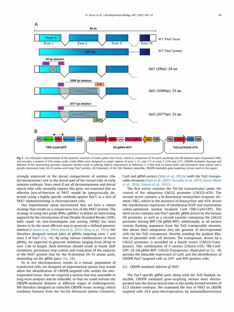
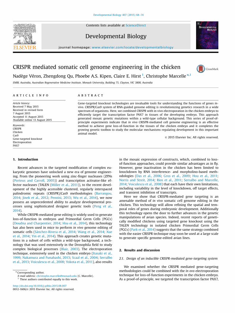
Fig. 1. (A) Schematic representation of the genomic structure of Gallus gallus Pax7 locus, which is comprised of 10 exons spanning over 86 kilobase pairs of genomic DNA,and encodes a protein of 524 amino acids. Guide RNAs were designed to target regions of exon 1 (1.1 and 1.7) or exon 2 (2.16 and 2.17). CRISPR-mediated cleavage anddeletion of the intervening genomic sequence should result in splicing defects represented as deletions 1–3 that lead to frame-shifts and premature stop codons and agreatly shortened (only 24–33 amino-acid long) Pax7 protein. (B) Schematic of the Tol2 flanked, inducible, CRISPR mediated gene-targeting vectors used in this paper.
N. Véron et al. / Developmental Biology 407 (2015) 68–74 69
strongly expressed in the dorsal compartment of somites (thedermomyotome) and in the dorsal part of the neural tube in earlyamniote embryos. Since most if not all dermomyotome and dorsalneural tube cells normally express this gene, we reasoned that aneffective loss-of-function of PAX7 would be unequivocally de-tected (using a highly specific antibody against Pax7) as a lack ofPAX7 immunostaining in electroporated cells.
Our experimental setup necessitated that we have a robuststrategy that results in a conspicuous loss of the PAX7 protein. Thestrategy of using two guide RNAs (gRNAs) to delete an interveningsegment by the introduction of two Double Stranded Breaks (DSB),with repair via non-homologous end joining (NHEJ) has beenshown to be the most efficient way to generate a defined genomicdeletion (Canver et al., 2014; Ran et al., 2013; Zhou et al., 2014). Wetherefore designed several pairs of gRNAs targeting exon 1 andexon 2 of Pax7 (Fig. 1A). By using various combinations of thesegRNAs, we expected to generate deletions ranging from 28 bp toover 2 kb in length. Such deletions should result in frame shiftmutations, premature stop codons and truncation of the majorityof the PAX7 protein (but for the N-terminal 24–33 amino acids,depending on the gRNA pairs; Fig. 1A).
As in ovo electroporation results in a mosaic population oftransfected cells, we designed an experimental system that wouldallow the identification of CRISPR-targeted cells within the elec-troporated tissue. Also we required a system that was amenable tolong-term analyses and be inducible, so that we could activate theCRISPR-mediated deletion at different stages of embryogenesis.We therefore designed an inducible CRISPR vector strategy, whichcombines features from the Tet-On Advanced system (Clontech),
Cas9 and gRNA vectors (Mali et al., 2013a) with the Tol2 transpo-sable elements (Sato et al., 2007; Serralbo et al., 2013; Sieiro-Mostiet al., 2014; Yokota et al., 2011).
The first vector contains the Tet-On transactivator under thecontrol of the ubiquitous CAGGS promoter (CAGGS-rtTA). Thesecond vector contains a bi-directional tetracycline-response ele-ment (TRE), which in the presence of doxycycline and rtTA, drivesthe simultaneous expression of membranal EGFP and mammaliancodon-optimized, nuclear localized Cas9 (TRE-Cas9-GFP). Thethird vector contains one Pax7-specific gRNA driven by the humanU6 promoter, as well as a second cassette containing the CAGGSpromoter driving RFP (U6-gRNA-RFP). Additionally, as all vectorscontain flanking sequences from the Tol2 transposable element,this allows their integration into the genome of electroporatedcells via the Tol2 transposase, thereby avoiding the gradual dilu-tion of plasmids with cell division. The transposase, driven by aCAGGS promoter, is provided on a fourth vector (CAGGS-Trans-posase). This combination of 5 vectors (CAGGS-rtTA; TRE-Cas9-GFP; 2X U6-gRNA-RFP; CAGGS-Transposase; illustrated in Fig. 1B)permits the inducible expression of Cas9, and the identification ofCRISPR Pax7-targeted cells as GFP- and RFP-positive cells.
2.2. CRISPR mediated deletion of PAX7
The Pax7-specific gRNA pairs along with the Tol2 flanked, in-ducible, CRISPR mediated gene-targeting vectors were electro-porated into the dorsal neural tube or the newly formed somites ofE2.5 chicken embryos. We examined the loss of PAX7 in CRISPRtargeted cells 24 h post-electroporation, by immunofluorescence

N. Véron et al. / Developmental Biology 407 (2015) 68–7470
using the PAX7 monoclonal antibody. In addition, we performedPCR-based mutation analysis to characterize the correspondingdeletion. At E3.5, PAX7 is highly expressed in the dorsal-mostportion of the neuroepithelium (Jostes et al., 1990; Marcelle et al.,1995). Electroporation of the Pax7-gRNA pair (1.1 and 1.7) into theneural tube along with the CRISPR mediated gene-targeting vec-tors resulted in a significant decrease or a loss in PAX7 staining inCas9/eGFP and gRNA/RFP positive cells, when compared to controlcells within the neural tube (Fig 2A–C). Quantitative analysis of thePAX7 staining showed a robust decrease of PAX7 expression, sinceonly 20.95% of electroporated cells displayed normal levels ofPAX7 staining when compared to control electroporated cells,suggesting that PAX7 knockdown has occurred in approximately80% of cells (Fig 2G). As controls, we verified that this proceduredid not affect the expression of the closely related transcriptionfactor PAX3, or of the acetylated form of Tubulin. In addition, DAPIstaining confirmed that it did not lead to any visible increase inapoptosis (Supplementary Fig. 1).
PAX7 gene knockdown was also performed in the DML of so-mites. Initially homogenously expressed in the dermomyotome,Pax7 expression becomes progressively weaker in differentiatingsomites in the DML as cells within this structure initiate theirmyogenic program (Galli et al., 2008; Rios et al., 2011). Conse-quently, we observed that approximately 65% of cells within theDML of control electroporated somites expressed high levels ofPax7. In comparison, only 14.5% of Cas9/eGFP and gRNA/RFP la-beled cells within somites electroporated with the Pax7-gRNA pair(1.1 and 1.7) expressed normal levels of Pax7 (Fig. 2D–F, H), againdemonstrating a significant decrease in Pax7 staining.
Electroporation of other combinations of Pax7-gRNA pairs (1.7with either 2.16 or 2.17) into the dorsal dermomyotome also re-sulted in a drastic reduction of Pax7 staining (Fig 3A–I). Quanti-tative measurement of the Pax7 staining demonstrated that less
Neural Tube DML
Pax7
Cas
9/eG
FPca
axgR
NA
/RFP
Cas
9/eG
FPca
axPa
x7 g
RN
A/R
FP
Fig. 2. The neural tube or newly formed somites in E2.5 chicken embryos were electrowith doxycycline added at the time of the electroporation. (A–C) Transverse sections oelectroporation demonstrating the loss of Pax7 immunostaining specifically in the GFP anof Pax7 in the (G) neural tube or (H) DML of cells electroporated with Pax7 targeted gRNAbars: 25 μm.
than 25% of the Cas9/eGFP and gRNA/RFP labeled cells have nor-mal levels Pax7 staining (Fig 3J) when compared to control gRNAelectroporated somites where over 95% of cells have high levels ofPax7 staining. The reduction of PAX7 expression we observed withwith all gRNA pairs suggested that the protocol and approach wefollowed are very efficient.
However, it was important to verify that the reduction or loss ofPax7 expression was not artefactual but actually due to genomeediting in electroporated cells. To verify this, genomic DNA wasextracted from electroporated tissues and the Pax7 gene wasanalyzed by PCR. We first analyzed genomic DNA extracted fromcells electroporated with gRNAs from exon 1 (1.1 and 1.7, i.e.leading to a 28 bp deletion). We did not observe the two (wildtype and mutated) DNA fragments after PCR amplification of thatregion, and T7 Endonuclease I assay was also unsuccessful toidentify the mutant genomic DNA. We believe this is due not onlyto the small deletion, but more importantly to the relatively smallpercentage of Cas9/GFP expressing cells within the dissected andanalyzed tissues. Rather than resorting to cumbersome cell-sort-ing of electroporated cells to enrich for mutated cells (Wang et al.,2014), we reasoned that a PCR reaction of a mixed population ofnormal and mutant DNA would favor the detection of the latter asdeletions became larger. We therefore tested whether deletions ofabout 2 kb would be easier to detect using the same strategy. In-deed, the analysis of tissues collected from embryos electro-porated with Pax7 gRNA pairs targeting exon 1 and exon 2 (1.7with either 2.16 or 2.17, which resulted in a greater than 2 kbgenomic deletion), using a nested PCR approach, amplified DNAfragments, readily visible as a smear of about the expected size(800 bp and smaller) on a DNA gel (not shown). These were clonedand sequenced. Analysis of sequences from individual clonesgenerated from these gRNA pairs confirmed the deletion, however,we observed important variations in the size of the indels in these
0
20
40
60
80
100
% P
ax7
hi
0
20
40
60
80
100
% P
ax7
hi
NT - gRNA
NT + Pax7 gRNA
DML - gRNA
DML + Pax7 gRNA
***
***
porated with the Tol2 flanked, inducible, CRISPR mediated gene-targeting vectors,f the neural tube or (D–F) confocal slice through the DML of a somite, 24 h post-d RFP labeled cells. Graphs illustrating the percentage of cells expressing high levelss versus control (i.e. no gRNA) electroporations. *** denotes P value of r0.001. Scale

1.7 & 2.171.7 & 2.16Control
contro
l
1.7 &
2.16
1.7 &
2.17
0
20
40
60
80
100
%Pa
x7H
i
*** ***
Pax7
Cas
9/eG
FPca
axgR
NA
/RFP
Cas
9/eG
FPca
axPa
x7 g
RN
A/R
FP
Fig. 3. The dorsal dermomyotome of newly formed somites in E2.5 chicken embryos were electroporated with the Tol2 flanked, inducible, CRISPR mediated gene-targetingvectors, with doxycycline added at the time of the electroporation, then analyzed 24 h post-electroporation. Confocal stacks showing the dorsal dermomyotome of somiteselectroporated with (A–C) control gRNA versus gRNAs targeting various portions of exons 1 and 2 of Pax7 (D–F) 1.7 with 2.16 or (G–I) 1.7 with 2.17. Immunostaining for Pax7within the GFP and RFP labeled cells showing the decrease or lack of Pax7 staining within the Pax7 gRNA targeted cells. (J) Graphs illustrating the percentage of ‘Pax7 hi’ cellswithin the dermomyotome of control gRNAs versus Pax7 targeted gRNA electroporated somites. *** denotes P value of r0.001. Scale bars: 25 μm.
N. Véron et al. / Developmental Biology 407 (2015) 68–74 71
clones (Fig. 4). Only 1 of 18 clones from Pax7 gRNA pair 1.7 and2.16, and 1 of 14 clones from 1.7 and 2.17 contained the exactpredicted deletion, while the majority of cloned sequences con-tained much larger deletions, with up to an additional 341 bp ofgenomic sequence deleted in one instance, although on average130 bp of sequence were deleted. This is surprising, as previousreports of CRISPR mediated gene targeting in Xenopus, Zebrafishand Drosophila embryos suggest that the NHEJ repair mechanismis very precise (Gratz et al., 2014; Guo et al., 2014; Hruscha et al.,2013; Irion et al., 2014; Shen et al., 2014). However, our resultswould suggest that the process of NHEJ can be error prone, maybein the specific context of electroporation. Importantly, regardlessof the range in size of the Pax7 genomic deletions, sequenceanalysis of the CRISPR targeted clones confirmed that all thesedeletions resulted in aberrant mRNA splicing, changes to the openreading frame and the presence of premature stop codons thatwould result in truncated Pax7 protein, thereby explaining the lossof Pax7 staining, detected by immunofluorescence in these cells.
3. Conclusion
This series of proof-of-principle experiments indicate thatin vivo CRISPR-mediated cell genome engineering is a highly ef-fective method to achieve genetic mutations in a subset of cells ofthe chicken embryo. Future improvements to the technology de-scribed here will aim to reduce the number of co-electroporatedvectors: we show here that the co-electroporation of five vectors isefficient. However, this may become a limiting factor if an alter-native strategy using modified Cas9 “nickase” is utilized. While themain advantage of the Cas9 nickase is to greatly reduce the off-target activity of the Cas9 endonuclease, it utilizes paired gRNAs tocreate targeted double strand breaks in the genome (Mali et al.,2013a; Ran et al., 2013). Large deletions described here would thenrequire four gRNAs (i.e. four U6-gRNA-RFP vectors). MultiplexgRNA cloning kits that gather two or even four gRNAs at once in asingle vector (Systembio) would very effectively reduce thisnumber. Despite, this advancement in gene engineering now

Fig. 4. Strategy for CRISPR mediated deletion of exon 2 of Pax7, illustrating position of gRNAs targeting (A) exon 1 (1.7) and exon 2 (2.16) or (C) exon 1 (1.7) and exon 2 (2.17)of Pax7. Gray box illustrates the expected deletion of (A) 2086 bp or (C) 2077 bp. Mutant alleles of Pax7 were identified using a nested PCR approach initially using primers107 and 106, then primers 74 and 106 (black arrows). (B and D) Sequences of unique Pax7 mutant alleles compared to wildtype sequence. Underlined sequence indicatesgRNA target sequence with PAM in bold. Gray box illustrates the expected deletion. Red letters indicate inserted sequences. Dots indicate deleted bases. The total size of theindel is shown in parentheses.
N. Véron et al. / Developmental Biology 407 (2015) 68–7472
permits large scale loss-of-function analysis of candidate genes inthe chicken and the entry of this important animal model in thegenetic era.
4. Materials and methods
4.1. CRISPR mediated gene-targeting vectors
The Tol2-pTRE-BI-eGFPcaax vector was derived from the bi-directional doxycycline inducible pTRE-BI-eGFP vector (Clontech)by the replacement of eGFP with a membrane-localized eGFP(containing the CAAX prenylation sequence from H-Ras) alongwith the addition of flanking Tol2 integration sequences, intowhich nuclear localized, human codon optimized Cas9 (Addgene#41815, (Mali et al., 2013b)) was cloned to generate the TRE-Cas9-GFP. We previously showed that this inducible plasmid system is
Table 1Primers used to clone Pax7 targets in hU6 gRNA plasmid.
Pax7 1.1 F TTTCTTGGCTTTATATATCTTGTGG
Pax7 1.1 R GACTAGCCTTATTTTAACTTGCTAT
Pax7 1.7 F TTTCTTGGCTTTATATATCTTGTGG
Pax7 1.7 R GACTAGCCTTATTTTAACTTGCTAT
Pax7 2.16 F TTTCTTGGCTTTATATATCTTGTGG
Pax7 2.16 R GACTAGCCTTATTTTAACTTGCTAT
Pax7 2.17 F TTTCTTGGCTTTATATATCTTGTGG
Pax7 2.17 R GACTAGCCTTATTTTAACTTGCTAT
silent without doxycyclin (Rios et al., 2011). Tol2-CAGGS-rtTA hasbeen described previously (Serralbo et al., 2013). The hU6-gRNAempty vector was obtained from Addgene (#41824, (Mali et al.,2013b)). The hU6-gRNA scaffold cassette was cloned into Tol2-CAGGS-RFP (Serralbo and Marcelle, 2014) to generate the U6-gRNA-RFP. Pax7 target gRNAs were designed and selected as de-scribed in Mali et al. (2013b), with further verification of specificityand off target effects using the CRISPR design tool (CRISPR.mit.edu,(Hsu et al., 2014)). The verified gRNA target sequences (Table 1)were cloned into the hU6 promoter gRNA scaffold primers in thehU6 gRNA vector as described (Mali et al., 2013b). An empty gRNAvector was used for control electroporations.
4.2. In ovo electroporation and imaging
Fertilized chicken eggs were incubated at 38 °C in a humidifiedincubator. Embryos were staged according to days of incubation
AAAGGACGAAACACCggtaccgcggatgatgcgcc
TTCTAGCTCTAAAACggcgcatcatccgcggtacc
AAAGGACGAAACACCgggcagaactacccgcgcac
TTCTAGCTCTAAAACgtgcgcgggtagttctgccc
AAAGGACGAAACACCgcccagggtgagtgcaatgt
TTCTAGCTCTAAAACacattgcactcaccctgggc
AAAGGACGAAACACCgccccacattgcactcaccc
TTCTAGCTCTAAAACgggtgagtgcaatgtggggc

Table 2Primers used to detect Pax7 deleted genomic region.
74 Pax7 gDNA F1 ctccgccgccccccgctatgg
106 Pax7 gDNA R2 ccgtccttcagcagcctgtcccgg
107 Pax7 gDNA F2 ggctgggagacctccgaaagc
N. Véron et al. / Developmental Biology 407 (2015) 68–74 73
and number of somites. The neural tube and somites were elec-troporated as previously described (Gros et al., 2004; Rios et al.,2011). The final concentration for each plasmid in the electro-poration mix was 1 mg/ml. Doxycycline (300 ml of 15 mg/ml) wasadded at the time of electroporation. Embryos were analyzedunder UV examination 24 h after electroporation and correctlyelectroporated embryos (i.e. high expression of the fluorescentreporters and no visible malformation due to electroporation)were dissected and fixed for 1 h in 4% formaldehyde.
Embryos were either analyzed by whole mount im-munostaining or embedded in 15% sucrose/7.5% gelatin/PBS solu-tion and sectioned into 20 mm slices using a cryostat (as describedin Serralbo and Marcelle (2014)). Immunohistochemistry on sec-tions or whole mount embryos was performed with the followingantibodies: chicken polyclonal antibody against GFP (Abcam#ab13970), rabbit polyclonal anti RFP (Abcam #ab62341), mousemonoclonal Acetylated Tubulin (Sigma #T7451), mouse mono-clonal anti Pax7 (Hybridoma Bank), mouse monoclonal anti Pax3(Hybridoma Bank) detected with species-specific secondary anti-bodies coupled to AlexaFluor-488,-555, or -647 (LifeTechnologies).Whole-mount embryos and sections were imaged using a LeicaSP5 confocal microscope running LAS AF software (LeicaMicroSystems).
Image stacks were analyzed by using either (i) the Imarissoftware package (Bitplane, version 7.5.2) or (ii) ImageJ software.Using the “spot” module of the Imaris, the region of interest wasmanually specified for each somite and an initial quality count forgRNA-containing cells was performed. Selected cells were thenfiltered based on central intensity of Pax7 staining, and the in-tensity peak value recorded as the percentage of positivecells. Manual cell counting was performed using the cell counterplugin (Kurt De Vos, University of Sheffield) within ImageJ(Schindelin et al., 2012) Statistical analyses were performed usingthe GraphPad Prism software. Student's t-test was applied to po-pulations to determine the P values indicated in the figures. Ineach graph, columns correspond to the mean and standard de-viation. *** denotes Po0.001.
4.3. PCR analysis
Twenty-four hours after electroporation, tissue expressing bothGFP and RFP was dissected and pooled from three to five embryosand genomic DNA extracted. The CRISPR targeted region wasamplified by a nested PCR approach with primers detailed in Ta-ble 2, that preferentially amplified the CRISPR deleted region bylimiting the extension time of the PCR to 1 min. In the first roundof PCR amplification of the wildtype band (with forward and re-verse primers 107 and 106) would result in an �3 kb band. Asecond round of PCR was then performed (with forward and re-verse primers 74 and 106) which preferentially amplified the�750 bp CRISPR deleted band over the �2.9 kb wildtype band.The PCR products were gel purified and cloned into the pGEMt-Easy vector (Promega) by TA cloning. Ten single colonies wererandomly picked for DNA sequencing analysis to detect the in-sertion or deletion of bases. Sequences were aligned and analyzedusing SnapGene (GSL Biotech).
Author contributions
CH and CM planned the experiments and wrote the manu-script, CH, NV and ZQ performed and analyzed the experiments.PK quantified the experiments and provided the correspondinggraphs.
Acknowledgments
The authors thank Daniel Sieiro-Mosti for assistance withimaging and provision of vectors. The authors also acknowledgethe help from Monash Micro Imaging, Monash University, Victoria,Australia. This work was funded by grants (APP1034736 andAPP1065954) from the National Health and Medical ResearchCouncil (NHMRC, Australia) and (DP130103680) from the Aus-tralian Research Council (ARC) to CM. CM is a Senior ResearchFellow of the NHMRC.
Appendix A. Supplementary material
Supplementary data associated with this article can be found inthe online version at http://dx.doi.org/10.1016/j.ydbio.2015.08.007.
References
Barrangou, R., 2014. Cas9 targeting and the CRISPR revolution. Science 344,707–708. http://dx.doi.org/10.1126/science.1252964.
Blair, S.S., 2003. Genetic mosaic techniques for studying Drosophila development.Development 130, 5065–5072. http://dx.doi.org/10.1242/dev.00774.
Canver, M.C., Bauer, D.E., Dass, A., Yien, Y.Y., Chung, J., Masuda, T., Maeda, T., Paw, B.H., Orkin, S.H., 2014. Characterization of genomic deletion efficiency mediatedby clustered regularly interspaced palindromic repeats (CRISPR)/Cas9 nucleasesystem in mammalian cells. J. Biol. Chem. 289, 21312–21324.
Das, R.M., Van Hateren, N.J., Howell, G.R., Farrell, E.R., Bangs, F.K., Porteous, V.C.,Manning, E.M., McGrew, M.J., Ohyama, K., Sacco, M.A., Halley, P.A., Sang, H.M.,Storey, K.G., Placzek, M., Tickle, C., Nair, V.K., Wilson, S.A., 2006. A robust systemfor RNA interference in the chicken using a modified microRNA operon. Dev.Biol. 294, 554–563. http://dx.doi.org/10.1016/j.ydbio.2006.02.020.
Doudna, J.A., Charpentier, E., 2014. The new frontier of genome engineering withCRISPR–Cas9. Science, 346.
Galli, L.M., Knight, S.R., Barnes, T.L., Doak, A.K., Kadzik, R.S., Burrus, L.W., 2008.Identification and characterization of subpopulations of Pax3 and Pax7 ex-pressing cells in developing chick somites and limb buds. Dev. Dyn. 237,1862–1874. http://dx.doi.org/10.1002/dvdy.21585.
Gratz, S.J., Ukken, F.P., Rubinstein, C.D., Thiede, G., Donohue, L.K., Cummings, A.M.,O’Connor-Giles, K.M., 2014. Highly specific and efficient CRISPR/Cas9-catalyzedhomology-directed repair in Drosophila. Genetics 196, 961–971.
Gros, J., Scaal, M., Marcelle, C., 2004. A two-step mechanism for myotome formationin chick. Dev. Cell 6, 875–882. http://dx.doi.org/10.1016/j.devcel.2004.05.006.
Gros, J., Serralbo, O., Marcelle, C., 2009. WNT11 acts as a directional cue to organizethe elongation of early muscle fibres. Nature 457, 589–593. http://dx.doi.org/10.1038/nature07564.
Guo, X., Zhang, T., Hu, Z., Zhang, Y., Shi, Z., Wang, Q., Cui, Y., Wang, F., Zhao, H., Chen,Y., 2014. Efficient RNA/Cas9-mediated genome editing in Xenopus tropicalis.Development 141, 707–714. http://dx.doi.org/10.1242/dev.099853.
Hou, X., Omi, M., Harada, H., Ishii, S., Takahashi, Y., Nakamura, H., 2011. Conditionalknockdown of target gene expression by tetracycline regulated transcription ofdouble strand RNA. Dev., Growth Differ. 53, 69–75. http://dx.doi.org/10.1111/j.1440-169X.2010.01229.x.
Hruscha, A., Krawitz, P., Rechenberg, A., Heinrich, V., Hecht, J., Haass, C., Schmid, B.,2013. Efficient CRISPR/Cas9 genome editing with low off-target effects in zeb-rafish. Development 140, 4982–4987. http://dx.doi.org/10.1242/dev.099085.
Hsu, P.D., Lander, E.S., Zhang, F., 2014. Development and applications of CRISPR–Cas9 for genome engineering. Cell 157, 1262–1278. http://dx.doi.org/10.1016/j.cell.2014.05.010.
Irion, U., Krauss, J., Nüsslein-Volhard, C., 2014. Precise and efficient genome editingin zebrafish using the CRISPR/Cas9 system. Development 141, 4827–4830.
Itasaki, N., Bel-Vialar, S., Krumlauf, R., 1999. ‘Shocking’ developments in chickembryology: electroporation and in ovo gene expression. Nat. Cell Biol. 1,E203–E207. http://dx.doi.org/10.1038/70231.
Jinek, M., Chylinski, K., Fonfara, I., Hauer, M., Doudna, J.A., Charpentier, E., 2012. Aprogrammable dual-RNA–guided DNA endonuclease in adaptive bacterial im-munity. Science 337, 816–821. http://dx.doi.org/10.1126/science.1225829.
Jostes, B., Walther, C., Gruss, P., 1990. The murine paired box gene, Pax7, is

N. Véron et al. / Developmental Biology 407 (2015) 68–7474
expressed specifically during the development of the nervous and muscularsystem. Chick Dev. Biol. 33, 27–37. http://dx.doi.org/10.1016/0925-4773(90)90132-6.
Mali, P., Aach, J., Stranges, P.B., Esvelt, K.M., Moosburner, M., Kosuri, S., Yang, L.,Church, G.M., 2013a. CAS9 transcriptional activators for target specificityscreening and paired nickases for cooperative genome engineering. Nat. Bio-technol. 31, 833–838. http://dx.doi.org/10.1038/nbt.2675.
Mali, P., Yang, L., Esvelt, K.M., Aach, J., Guell, M., DiCarlo, J.E., Norville, J.E., Church, G.M., 2013b. RNA-guided human genome engineering via Cas9. Science 339,823–826. http://dx.doi.org/10.1126/science.1232033.
Marcelle, C., Wolf, J., Bronner-Fraser, M., 1995. The in vivo expression of the FGFreceptor FREK mRNA in avian myoblasts suggests a role in muscle growth anddifferentiation. Dev. Biol. 172, 100–114. http://dx.doi.org/10.1006/dbio.1995.0008.
Miller, J.C., Tan, S., Qiao, G., Barlow, K.A., Wang, J., Xia, D.F., Meng, X., Paschon, D.E.,Leung, E., Hinkley, S.J., Dulay, G.P., Hua, K.L., Ankoudinova, I., Cost, G.J., Urnov, F.D., Zhang, H.S., Holmes, M.C., Zhang, L., Gregory, P.D., Rebar, E.J., 2011. A TALEnuclease architecture for efficient genome editing. Nat. Biotechnol. 29,143–148. http://dx.doi.org/10.1038/nbt.1755.
Nakamura, H., Funahashi, J., 2013. Electroporation: past, present and future. Dev.Growth Differ. 55, 15–19. http://dx.doi.org/10.1111/dgd.12012.
Norris, A., Streit, A., 2014. Morpholinos: studying gene function in the chick. Dev.Biol. 66, 454–465. http://dx.doi.org/10.1016/j.ymeth.2013.10.009.
Park, T.S., Lee, H.J., Kim, K.H., Kim, J.-S., Han, J.Y., 2014. Targeted gene knockout inchickens mediated by TALENs. Proc. Natl. Acad. Sci. 111, 12716–12721.
Peng, Y., Clark, K.J., Campbell, J.M., Panetta, M.R., Guo, Y., Ekker, S.C., 2014. Makingdesigner mutants in model organisms. Development 141, 4042–4054.
Pennisi, E., 2013. The CRISPR craze. Science 341, 833–836. http://dx.doi.org/10.1126/science.341.6148.833.
Porteus, M.H., Carroll, D., 2005. Gene targeting using zinc finger nucleases. Nat.Biotechnol. 23, 967–973. http://dx.doi.org/10.1038/nbt1125.
Ran, F.A., Hsu, P.D., Lin, C.-Y., Gootenberg, J.S., Konermann, S., Trevino, A.E., Scott, D.A., Inoue, A., Matoba, S., Zhang, Y., Zhang, F., 2013. Double nicking by RNA-guided CRISPR Cas9 for enhanced genome editing specificity. Cell 154,1380–1389. http://dx.doi.org/10.1016/j.cell.2013.08.021.
Rios, A.C., Serralbo, O., Salgado, D., Marcelle, C., 2011. Neural crest regulates myo-genesis through the transient activation of NOTCH. Nature 473, 532–535. http://dx.doi.org/10.1038/nature09970.
Sato, Y., Kasai, T., Nakagawa, S., Tanabe, K., Watanabe, T., Kawakami, K., Takahashi,Y., 2007. Stable integration and conditional expression of electroporatedtransgenes in chicken embryos. Dev. Biol. 305, 616–624. http://dx.doi.org/10.1016/j.ydbio.2007.01.043.
Sánchez-Rivera, F.J., Papagiannakopoulos, T., Romero, R., Tammela, T., Bauer, M.R.,Bhutkar, A., Joshi, N.S., Subbaraj, L., Bronson, R.T., Xue, W., Jacks, T., 2014. Rapid
modelling of cooperating genetic events in cancer through somatic genomeediting. Nature 516, 428–431. http://dx.doi.org/10.1038/nature13906.
Scaal, M., Gros, J., Lesbros, C., Marcelle, C., 2004. In ovo electroporation of aviansomites. Dev. Dyn. 229, 643–650. http://dx.doi.org/10.1002/dvdy.10433.
Schindelin, J., Arganda-Carreras, I., Frise, E., Kaynig, V., Longair, M., Pietzsch, T., Pre-ibisch, S., Rueden, C., Saalfeld, S., Schmid, B., Tinevez, J.-Y., White, D.J., Harten-stein, V., Eliceiri, K., Tomancak, P., Cardona, A., 2012. Fiji: an open-source platformfor biological-image analysis. Nat Meth 9, 676–682. http://dx.doi.org/10.1038/nmeth.2019.
Serralbo, O., Marcelle, C., 2014. Migrating cells mediate long-range WNT signaling.Development 141, 2057–2063. http://dx.doi.org/10.1242/dev.107656.
Serralbo, O., Picard, C.A., Marcelle, C., 2013. Long term, inducible gene loss-of-function in the chicken embryo. Genesis . http://dx.doi.org/10.1002/dvg.22388,n/a-n/a.
Shen, B., Zhang, W., Zhang, J., Zhou, J., Wang, J., Chen, L., Wang, L., Hodgkins, A., Iyer,V., Huang, X., Skarnes, W.C., 2014. Efficient genome modification by CRISPR–Cas9 nickase with minimal off-target effects. Nat. Methods 11, 399–402. http://dx.doi.org/10.1038/nmeth.2857.
Sieiro-Mosti, D., La, Celle, De, M., Pelé, M., Marcelle, C., 2014. A dynamic analysis ofmuscle fusion in the chick embryo. Development 141, 3605–3611.
Voiculescu, O., Papanayotou, C., Stern, C.D., 2008. Spatially and temporally con-trolled electroporation of early chick embryos. Nat. Protocols 3, 419–426. http://dx.doi.org/10.1038/nprot.2008.10.
Wang, S., Sengel, C., Emerson, M.M., Cepko, C.L., 2014. A gene regulatory networkcontrols the binary fate decision of rod and bipolar cells in the vertebrate re-tina. Dev. Cell 30, 513–527.
Wu, X., Scott, D.A., Kriz, A.J., Chiu, A.C., Hsu, P.D., Dadon, D.B., Cheng, A.W., Trevino,A.E., Konermann, S., Chen, S., Jaenisch, R., Zhang, F., Sharp, P.A., 2014. Genome-wide binding of the CRISPR endonuclease Cas9 in mammalian cells. Nat. Bio-technol. 32, 670–676. http://dx.doi.org/10.1038/nbt.2889.
Xue, W., Chen, S., Yin, H., Tammela, T., Papagiannakopoulos, T., Joshi, N.S., Cai, W.,Yang, G., Bronson, R., Crowley, D.G., Zhang, F., Anderson, D.G., Sharp, P.A., Jacks,T., 2014. CRISPR-mediated direct mutation of cancer genes in the mouse liver.Nature 514, 380–384. http://dx.doi.org/10.1038/nature13589.
Yin, H., Xue, W., Chen, S., Bogorad, R.L., Benedetti, E., Grompe, M., Koteliansky, V.,Sharp, P.A., Jacks, T., Anderson, D.G., 2014. Genome editing with Cas9 in adultmice corrects a disease mutation and phenotype. Nat. Biotechnol. . http://dx.doi.org/10.1038/nbt.2884
Yokota, Y., Saito, D., Tadokoro, R., Takahashi, Y., 2011. Genomically integratedtransgenes are stably and conditionally expressed in neural crest cell-specificlineages. Dev. Biol. 353, 382–395.
Zhou, J., Wang, J., Shen, B., Chen, L., Su, Y., Yang, J., Zhang, W., Tian, X., Huang, X.,2014. Dual sgRNAs facilitate CRISPR/Cas9-mediated mouse genome targeting.FEBS J. 281, 1717–1725. http://dx.doi.org/10.1111/febs.12735.