crispr-cas9 screening for target indentification · crispr-cas9 screening for target identification...
TRANSCRIPT
CRISPR-Cas9 Screening for Target IdentificationSteffen Lawo, Tim Scales, Alessandro Riccombeni, Benedict CS Cross and Jonathan D MooreHorizon Discovery, Cambridge UK
IntroductionThe identification and validation of novel drug targets is a challenging process for drug discovery programmes. Forward genetic screening with the RNA-guided nuclease Cas9 provides a new and powerful tool to interrogate gene function on a genome-wide level. In contrast to RNAi technology, which can be prone to off-target effects and variable knock-down efficiencies, the combination of bacterial CRISPR (clustered regularly interspaced short palindromic repeat)-associated Cas9 nuclease and pooled short guide RNA (sgRNA) libraries aims to generate complete knock-outs with high on-target specificity.
We have adapted a pooled-based screening protocol, where lentivirus transduction delivers both the Cas9 endonuclease and the sgRNA to the cells. We have used both custom libraries and the GeCKOv2 whole-genome library, which is comprised of sgRNAs designed to target Cas9 to initial consecutive exons of the open reading frame (ORF). The nuclease then introduces double strand breaks that induce non-homologous end joining (NHEJ), resulting in frame shift mutations and premature stop codons in the ORF. The prospect of a complete loss of gene function enables a biologically robust interrogation of phenotypes to identify novel hits in positive and negative selection screens (Figure 1).
Figure 1. Workflow of a typical CRISPR-Cas9 screen.
Results and DiscussionAs a proof of concept, we have repeated a genome-wide positive selection screen to identify resistance factors against Vemurafenib (PLX-4032) – a BRAF kinase inhibitor – in A375 melanoma cells, which have a BRAF V600E gain-of-function mutation (Shalem et al., 2013; Flaherty et al., 2010; Davies et al., 2002). We used the GeCKOv2 knock-out library containing 6 guide RNAs against 19,050 genes (Sanjana et al., 2014). Cells became cytostatic as a consequence of PLX-4032 treatment, which allowed a subset of cells with resistance-conferring gene editing events to accumulate. Cell pellets were taken at the end of the screen and data was resolved by next-generation sequencing. Briefly, the abundance of each guide RNA in each condition is used to assess drop-out or enrichment of cells with corresponding CRISPR-Cas9 dependent perturbations.
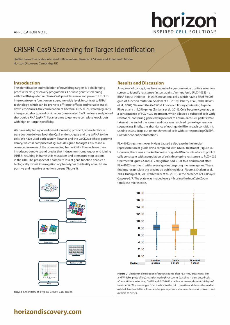
PLX-4032 treatment over 14 days caused a decrease in the median representation of guide RNAs compared with DMSO treatment (Figure 2). However, there was a marked increase of guide RNA counts of a sub pool of cells consistent with a population of cells developing resistance to PLX-4032 treatment (Figures 2 and 3). 228 sgRNAs had >100-fold enrichment after PLX-4032 treatment, with several guides targeting the same genes. These findings recapitulate the previously published data (Figure 3, Shalem et al., 2013; Huang et al., 2012; Whittaker et al., 2013). In the presence of CellPlayer Caspase-3/7. The plate was imaged every 4 h using the IncuCyte Zoom timelapse microscope.
Figure 2. Change in distribution of sgRNA counts after PLX-4032 treatment. Box and Whisker plots of log2-transformed sgRNA counts (baseline – transduced cells after antibiotic selection; DMSO and PLX-4032 – cells at screen end-point (14 days of treatment)). The box ranges from the first to the third quartile and shows the median as black line. In addition, lower and upper adjacent values are shown as whiskers, and outliers as circles.
horizondiscovery.com
APPLICATION NOTE

Figure 3. Enrichment of sgRNAs after PLX-4032 treatment. Scatterplot of log2-transformed sgRNA counts in screen conditions after 14 days of DMSO (x-axis) versus PLX-4032 treatment (y-axis). Among the guides that were enriched in the PLX-4032 treated condition (circled) was a set of guide RNAs targeting the same genes multiple times (highlighted in colour).
We use a dedicated CRISPR-Cas9 screen analysis platform, adapted from the MAGeCK workflow (Li et al., 2014), which enables individual sgRNA analysis and gene level hit ranking. For the PLX-4032 screen, the highest ranking genes (MED12, NF1, CUL3, NF2, TADA2B and TADA1) were those whose loss is known to confer resistance to Vemurafenib (Figure 4). Loss of additional members of the STAGA histone acetyl transferase complex (TAFL5/PAF65β) and the Mediator complex (MED23) were also found to confer resistance in our screen.
Figure 4. Ranking of screen hits by the MAGeCK hit calling algorithm. On the y-axis, genes are ranked by robust ranking aggregation values for their enrichment after Vemurafenib treatment. In addition, the mean log2-fold change of sgRNAs targeting the same gene are plotted on the x-axis.
When sgRNAs for each of these hits are evaluated individually, it is apparent that although the majority of sgRNAs were enriched after PLX treatment, not all sgRNAs performed equally well (Figure 5). This emphasizes that for successful screening, the library composition and complexity (number of targets and number of guides per target) is of paramount importance.
Figure 5. Efficiency of individual sgRNAs against 8 hit genes. For each guide RNA, the log2-fold change in sgRNA frequency between PLX-4032 and DMSO treatment is plotted.
SummaryIn summary, we have used a well-established biological paradigm to examine the power of whole-genome CRISPR-Cas9 screening. We were able to successfully repeat previously published data, unambiguously identifying all validated hits whose depletion can confer resistance to Vemurafenib, as wells as additional genes from the same pathways.
MethodsThe screen was carried out at 300-fold coverage of the GeCKOv2 library, i.e. 300 cells per sgRNA. A375 cells were infected with the lentivirus library at a low MOI to ensure single integration events, followed by an antibiotic selection treatment to eliminate non-transduced cells prior to the start of the screen. Once selection was complete, cells were grown out to allow the Cas9 enzyme to function and for the resulting phenotypic effects to manifest. At this point, a baseline pellet was harvested to allow time-resolved comparison of the transduced pool. Cells were then cultured in media containing DMSO control or 2 µM PLX-4032 for 14 days or 17 cell doublings. During this time, a minimum of 36x106 cells were split every 3-4 days and reseeded into medium containing DMSO/PLX-4032. At the screen end-point, cells were harvested, genomic DNA extracted and guide RNA frequency in each condition resolved by next-generation sequencing.
References• Shalem et al., Science. 2014 Jan 3;343(6166):84-7• Flaherty et al., N Engl J Med. 2010 Aug 26;363(9):809-19• Davies et al., Nature. 2002 Jun 27;417(6892):949-54• Sanjana et al., Science. 2014 Jan 3;343(6166):84-7• Huang et al., Cell. 2012 Nov 21;151(5):937-50• Whittaker et al., Cancer Discov. 2013 Mar;3(3):350-62• Li et al., Genome Biol. 2014;15(12):554
© 2
018v
-02
If you have any questions
t +44 (0) 1223 976 000 (UK) or +1 800 235 9880 (USA); +1 303 604 9499 (USA)f + 44 (0)1223 655 581w horizondiscovery.com/contact-us or dharmacon.horizondiscovery.com/service-and-support Horizon Discovery, 8100 Cambridge Research Park, Waterbeach, Cambridge, CB25 9TL, United Kingdom
©2018 Horizon Discovery Group Company—All rights reserved. First published March 2018. UK Registered Head Office: Building 8100, Cambridge Research Park, Cambridge, CB25 9TL, United Kingdom.
mKate2 is a trademark of Evrogen. 1x Protease Inhibitor Mix is a trademark of GE Healthcare. FastDigest™ NheI enzyme NuPAGE™ 4X LDS sample buffer and NuPAGE Sample Reducing Agent (10X), Phusion™, Invitrogen™, RNase A, Novex™ 4-20% Tris Glycine Mini Protein Gel, SuperSignal™ West Dura Substrate are trademarks of Thermo Fisher Scientific, Inc. FLAG is a trademark of Sigma-Aldrich Co. LLC. ©2018 Horizon Discovery Group Company.