cp4 5 assessment of ecological risks at the dumping...
TRANSCRIPT

Reportontheassessmentofecologicalriskatthedumpingsites
"Application of ecosystem principles for the location and management of offshore dumping sites in
SE Baltic Region (ECODUMP)”
Monika Michałek, Andrzej Osowiecki, Magdalena Błeńska, Katarzyna Smolarz
Department of Aquatic Ecology
Maritime Institute in Gdańsk
Gdańsk
December 2014

1. Introduction
This report constitutes a part of the project "Application of ecosystem principles for the location and
management of offshore dumping sites in SE Baltic Region (ECODUMP).
Marine dumping site is an area of sea bottom specifically designated for dredged material disposal.
The material comes from dredging activities carried out continually in order to maintain the
standards of waterways, particularly their depth and width, to provide convenient ships accessibility
to harbours and shipyards through safe waterways. For economic reasons, most of the dredged
material is disposed in off‐shore dumping sites (Simonini et al. 2005).
The offshore dredged sediment disposal poses a considerable threat to most of the benthic species
due to their sessile way of life. Studies on dumping activities impact on soft‐bottom benthos have
revealed that its scale ranged from large and long‐term impacts to few or non‐detectable effects
(Simonini et al. 2005). In case the impact was detected, it was primarily manifested by decrease in
the taxonomic diversity of the benthic communities caused either by burial or by changing sediment
properties.
The strength of the dumping impact depends on several factors: chemical and physical
characteristics, volume of the sediment, water depth, sedimentary and hydrological regime of
dumping site, time of the year, similarity of sediment in dredged and disposal areas, contamination
of dredged material, structure and composition of benthic assemblages in the dumping site and
nearby areas (Essink 1999).
According to the Polish environmental regulations only non‐contaminated sediments can be dumped
in the marine areas (Regulation of the Minister of Environment of 16 April 2002, on ...the type and
concentration of substances that indicate that the dredged material is contaminated... Journal of
Laws No. 55, item 498). However, dumping of non‐contaminated dredged sediments, may also have
adverse effect on the functioning of the aquatic ecosystem.
The aim of the study was to assess the ecological risk the dumping site poses to selected biotic
elements of the marine environment. Most of actively living animals can avoid adverse conditions,
especially if they occur in a restricted area. On the other hand bottom macroinvertebrate
communities consist mostly of sessile organisms, thus are continuously affected by possible dredged
material originated factors.
The case study was performed in the Gdynia dumping site. The qualitative and quantitative structure
of the macrozoobenthos as well as the ecological status of the dumping area (acc. to Polish
regulations) were investigated. Additionally, the impact of Persistent Organic Pollutants (POPs) on
benthic malacofauna was analysed on the basis of selected cytogenetic and histological biomarkers
(Arkhipchuk et al. 2005). The results of macrozoobenthos investigations carried out in 2011 and
2012 were compared with historical data, particularly those obtained within the State Environmental
Monitoring programme (Warzocha et al. 2011, Radziejewska et al. 2012 a & b) and the research
project carried out in the area in 2008 (Bełdowski et al. 2008).

2. Case study – Gdynia dumping site Gdynia dumping site is situated in the outer part of the Puck Bay. The average depth of this basin is
20.5 m, maximum 54 m. Sediments are primarily sandy and muddy. The Puck Bay is considerably
eutrophic due to runoff from three sewage treatment facilities and seven rivers outflow. Eutrophic
character of the basin is reflected in the macrozoobenthos structure, which is dominated by
suspension and deposit feeders; bivalves and polychaetes constituting respectively 89% and 7% of
the total biomass (Kruk‐Dowgiałło & Szaniawska 2008).
The assessment of the Gdynia dumping site is based on macrozoobenthos, which is defined as a
group of benthic invertebrates that retain on a 1 mm mesh size sieve (HELCOM 1988). The group
includes both organisms living on the surface of the benthic sediments (epifauna) and below the
surface (infauna). Macrozoobenthos consists of a number of taxonomically diversified invertebrate
organisms occupying almost all aquatic ecosystems. These are mostly sessile species with long life‐
cycles (at least one year). Macrozoobenthos can be regarded as a good indicator of biological water
quality (Diaz & Rosenberg 1995; Gray et al. 2002, Rosenberg et al. 2002, Karlson et al. 2002).
Deterioration of ecological status, e.g. as a result of the progressing eutrophication, causes alteration
in benthic populations, biomass and composition of species (Cederwall & Elmgren 1990, Rumohr et
al. 1996).
2.1. Biological sampling
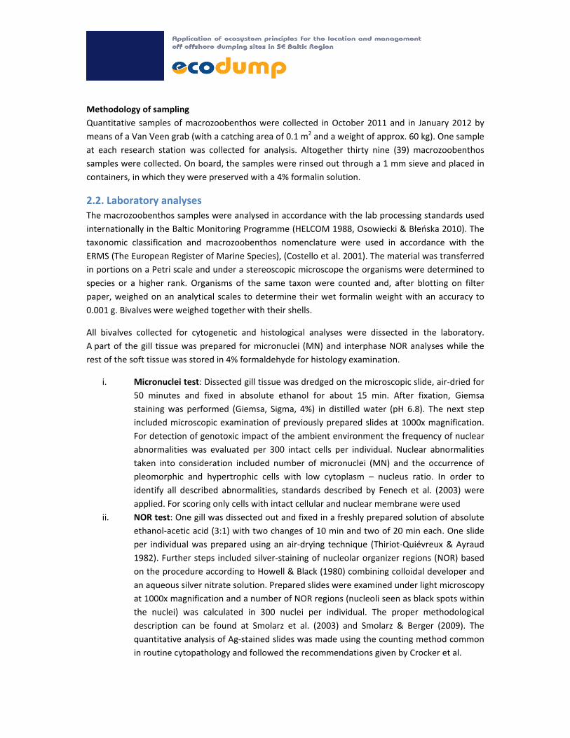
Location of sampling stations
The representative number of sampling stations were determined in the aftermath of the sonar and
bathymetric maps analyses (Fig. 1, Fig. 2). The same stations samples were collected for
physicochemical measurements and for macrozoobenthos. Bivalves for cytogenetic and histological
analyses were collected at four stations.
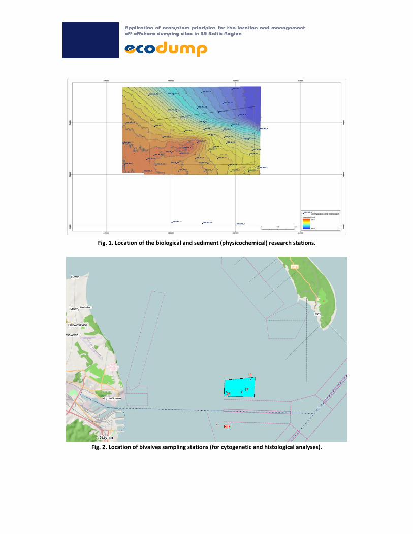
Fig. 1. Location of the biological and sediment (physicochemical) research stations.
Fig. 2. Location of bivalves sampling stations (for cytogenetic and histological analyses).
Methodology of sampling
Quantitative samples of macrozoobenthos were collected in October 2011 and in January 2012 by
means of a Van Veen grab (with a catching area of 0.1 m2 and a weight of approx. 60 kg). One sample
at each research station was collected for analysis. Altogether thirty nine (39) macrozoobenthos
samples were collected. On board, the samples were rinsed out through a 1 mm sieve and placed in
containers, in which they were preserved with a 4% formalin solution.
2.2. Laboratory analyses
The macrozoobenthos samples were analysed in accordance with the lab processing standards used
internationally in the Baltic Monitoring Programme (HELCOM 1988, Osowiecki & Błeńska 2010). The
taxonomic classification and macrozoobenthos nomenclature were used in accordance with the
ERMS (The European Register of Marine Species), (Costello et al. 2001). The material was transferred
in portions on a Petri scale and under a stereoscopic microscope the organisms were determined to
species or a higher rank. Organisms of the same taxon were counted and, after blotting on filter
paper, weighed on an analytical scales to determine their wet formalin weight with an accuracy to
0.001 g. Bivalves were weighed together with their shells.
All bivalves collected for cytogenetic and histological analyses were dissected in the laboratory.
A part of the gill tissue was prepared for micronuclei (MN) and interphase NOR analyses while the
rest of the soft tissue was stored in 4% formaldehyde for histology examination.
i. Micronuclei test: Dissected gill tissue was dredged on the microscopic slide, air‐dried for
50 minutes and fixed in absolute ethanol for about 15 min. After fixation, Giemsa
staining was performed (Giemsa, Sigma, 4%) in distilled water (pH 6.8). The next step
included microscopic examination of previously prepared slides at 1000x magnification.
For detection of genotoxic impact of the ambient environment the frequency of nuclear
abnormalities was evaluated per 300 intact cells per individual. Nuclear abnormalities
taken into consideration included number of micronuclei (MN) and the occurrence of
pleomorphic and hypertrophic cells with low cytoplasm – nucleus ratio. In order to
identify all described abnormalities, standards described by Fenech et al. (2003) were
applied. For scoring only cells with intact cellular and nuclear membrane were used
ii. NOR test: One gill was dissected out and fixed in a freshly prepared solution of absolute
ethanol‐acetic acid (3:1) with two changes of 10 min and two of 20 min each. One slide
per individual was prepared using an air‐drying technique (Thiriot‐Quiévreux & Ayraud
1982). Further steps included silver‐staining of nucleolar organizer regions (NOR) based
on the procedure according to Howell & Black (1980) combining colloidal developer and
an aqueous silver nitrate solution. Prepared slides were examined under light microscopy
at 1000x magnification and a number of NOR regions (nucleoli seen as black spots within
the nuclei) was calculated in 300 nuclei per individual. The proper methodological
description can be found at Smolarz et al. (2003) and Smolarz & Berger (2009). The
quantitative analysis of Ag‐stained slides was made using the counting method common
in routine cytopathology and followed the recommendations given by Crocker et al.

iii. (1989). In general each NOR cluster was considered as one when a dot aggregation could
not be resolved in individual NORs by focusing.
iv. Preparation of histological blocks from the soft tissues of sampled bivalves. Preparation
of histological slides included fixation in the 4% buffered formaldehyde, dehydration of
the soft tissues using different ethanol concentrations (from 70% to absolute ethanol),
immersion in xylene, embedding in paraffin and final preparation of histological blocks.
All steps were done using an automated procedure with a tissue processor. Prepared
blocks were cut at 1‐2 µm slides using semi‐automatic microtome (Leica). The final step
included staining with Hematoxylin and Eosin (H&E). Next, microscope examination was
performed.
v. Sexing and gonad development was assessed based on histological examination of
previously prepared slides. The presence of female or male gonads classified the
individuals as male or female. Assessing of gonad development was based on a five stage
scale after Wenne (1985).
2.3. Quantitative analyses
The following biocenotic indices were used in quantitative analyses:
Dominance Index D
S
SD a100
where: Sa – total number/biomass of individuals belonging to taxon a in all samples
S – total number/biomass of individuals in all samples
Multimetric Index B
This index assesses the ecological quality of the environment based on macrozoobenthos studies. It
combines the results of the quantitative measurements of the species abundance and diversity with
the qualitative information about the ecological tolerance of the particular taxa to a single index
value.
)1log()(
1
1
1
n
iin
ii
n
iiii
DD
SensDwB
where:
Di – number of taxa belonging to the particular domination classes (D1, D2, D3)
wi – weight of domination classes (respectively: 3 for D1, 2 for D2 and 1 for D3)
Sensi – tolerance/sensitivity of a given taxon to stress caused by anthropogenic impact (3 – sensitive
taxa, 2 – semi‐tolerant taxa, 1 – tolerant taxa)
n – number of taxon groups within the domination, sensitivity classes w, by domination classes

Domination classes, which were assumed according to Trojan (1980), include:
dominants (D1) – species comprising more than 10% of the total abundance/biomass at the
station;
influents (D2) – species comprising from 5 to 10% of the total abundance/biomass at the
station;
accessory species (D3) – species comprising less than 5% of the total abundance/biomass at
the station.
A weight was assigned to each domination class reflecting their role in the environment, because not
all organisms influence the character and functioning of the biocoenosis to the same extent (Odum
1982). Dominants were assigned weight 3, since they are the primary drivers structuring living
conditions of other organisms; hence they have the greatest bearing on the biocoenosis. Influents
were assigned weight 2, and accessory species weight 1.
The range of tolerance to stress caused by excessive amounts of organic matter in sediments as
result of the progressing eutrophication was determined for each taxa. By using a 3‐grade scale of
tolerance (sensitivity), the taxa identified in the Polish area of the Baltic Sea were divided into the
following groups:
sensitive taxa that have a narrow range of tolerance, the indicator species of unpolluted
seafloor (Sensi=3);
semi‐tolerant taxa that provide no direct indication of the quality of the ecosystem (Sensi=2);
resistant taxa with a wide range of tolerance to significant amounts of organic matter in
sediments, the indicator species of polluted seafloor (Sensi=1).
Taxa division into three groups of ecological tolerance was determined by expert judgment following
consultations with specialists who study macrozoobenthos of the Baltic Sea (Osowiecki et al. 2012),
(Table1).

Table 1. Numerical values of the ecological tolerance of taxa (Sensi) recorded in the study area
No Taxa1 Sensi
1. Halicryptus spinulosus 2
2. Nemertina2 2
3. Bylgides sarsi 1
4. Hediste diversicolor 1
5. Pygospio elegans 3
6. Marenzelleria neglecta 1
7. Oligochaeta2 1
8. Idotea chelipes 3
9. Saduria entomon 2
10. Jaera 2 3
11. Bathyporeia pilosa 3
12. Monoporeia affinis 3
13. Pontoporeia femorata 3
14. Gammarus2 2
15. Corophium volutator 1
16. Diastylis rathkei 2
17. Hydrobia2 2
18. Mytilus edulis trossulus 2
19. Cerastoderma glaucum 3
20. Macoma balthica 1
21. Mya arenaria 1 1 terminology in compliance with the ERMS – European Register of Marine Species (Costello et al. 2001)
2 ecological tolerance refers to any species within a specified systematic group
For each station, the state of macrozoobenthos assemblages was determined using index B.
Classification of the ecological status of the dumping area was performed acc. to Regulation on the
method of classification of the status of surface water bodies of 9 November 2011 (Journal of Laws
No. 257, item 1545).
Statistics
For the qualitative characteristics the test ratio, whereas for quantitative traits non‐parametric
Mann‐Whitney U test and non‐parametric Kruskal‐Wallis ANOVA were performed (Towned 2002).
The statistical calculations were performed in Statistica 8 (Statsoft).
The similarity analyses were carried out using Primer V5 software, for the analysis of the relationship
between the parameters of the sediments and zoobenthos structure Microsoft Office 2007
application was used.
3. Results
3.1. Assessment of persistent organic pollutants’ (POPs) impact on malacofauna
To assess the persistent organic pollutants’ (POPs) impact on malacofauna in the Gdynia dumping
area selected non‐specific biomarkers (micronuclei test, histopathological changes and NOR regions)
were used.
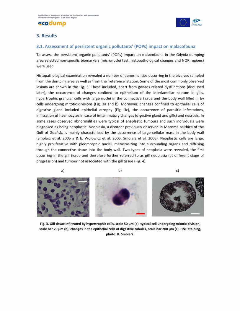
Histopathological examination revealed a number of abnormalities occurring in the bivalves sampled
from the dumping area as well as from the ‘reference’ station. Some of the most commonly observed
lesions are shown in the Fig. 3. These included, apart from gonads related dysfunctions (discussed
later), the occurrence of changes confined to epithelium of the interlamellar septum in gills,
hypertrophic granular cells with large nuclei in the connective tissue and the body wall filled in by
cells undergoing mitotic divisions (Fig. 3a and b). Moreover, changes confined to epithelial cells of
digestive gland included epithelial atrophy (Fig. 3c), the occurrence of parasitic infestations,
infiltration of haemocytes in case of inflammatory changes (digestive gland and gills) and necrosis. In
some cases observed abnormalities were typical of anaplastic tumours and such individuals were
diagnosed as being neoplastic. Neoplasia, a disorder previously observed in Macoma balthica of the
Gulf of Gdańsk, is mainly characterized by the occurrence of large cellular mass in the body wall
(Smolarz et al. 2005 a & b, Wołowicz et al. 2005, Smolarz et al. 2006). Neoplastic cells are large,
highly proliferative with pleomorphic nuclei, metastasizing into surrounding organs and diffusing
through the connective tissue into the body wall. Two types of neoplasia were revealed, the first
occurring in the gill tissue and therefore further referred to as gill neoplasia (at different stage of
progression) and tumour not associated with the gill tissue (Fig. 4).
a)
b)
c)
Fig. 3. Gill tissue infiltrated by hypertrophic cells, scale 50 µm (a); typical cell undergoing mitotic division,
scale bar 20 µm (b); changes in the epithelial cells of digestive tubules, scale bar 200 µm (c). H&E staining,
photo: K. Smolarz.
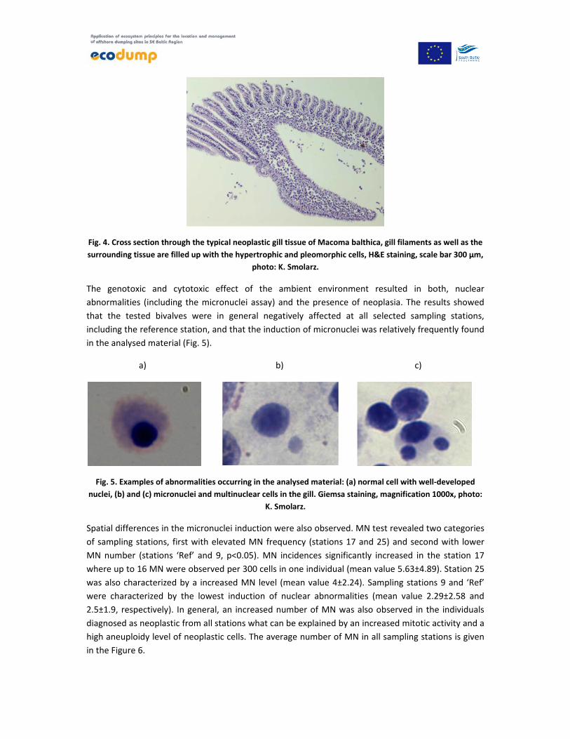
Fig. 4. Cross section through the typical neoplastic gill tissue of Macoma balthica, gill filaments as well as the
surrounding tissue are filled up with the hypertrophic and pleomorphic cells, H&E staining, scale bar 300 µm,
photo: K. Smolarz.
The genotoxic and cytotoxic effect of the ambient environment resulted in both, nuclear
abnormalities (including the micronuclei assay) and the presence of neoplasia. The results showed
that the tested bivalves were in general negatively affected at all selected sampling stations,
including the reference station, and that the induction of micronuclei was relatively frequently found
in the analysed material (Fig. 5).
a)
b)
c)
Fig. 5. Examples of abnormalities occurring in the analysed material: (a) normal cell with well‐developed
nuclei, (b) and (c) micronuclei and multinuclear cells in the gill. Giemsa staining, magnification 1000x, photo:
K. Smolarz.
Spatial differences in the micronuclei induction were also observed. MN test revealed two categories
of sampling stations, first with elevated MN frequency (stations 17 and 25) and second with lower
MN number (stations ‘Ref’ and 9, p<0.05). MN incidences significantly increased in the station 17
where up to 16 MN were observed per 300 cells in one individual (mean value 5.63±4.89). Station 25
was also characterized by a increased MN level (mean value 4±2.24). Sampling stations 9 and ‘Ref’
were characterized by the lowest induction of nuclear abnormalities (mean value 2.29±2.58 and
2.5±1.9, respectively). In general, an increased number of MN was also observed in the individuals
diagnosed as neoplastic from all stations what can be explained by an increased mitotic activity and a
high aneuploidy level of neoplastic cells. The average number of MN in all sampling stations is given
in the Figure 6.

Fig. 6. Average number of MN in the gill tissue from Macoma balthica.
Mitotic divisions were also observed in the studied material. The occurrence of mitotic figures was in
general very low for most studied material but increased significantly in the animals diagnosed as
neoplastic. Cytogenetic diagnosis was based on the occurrence of hypertrophic cells with low nuclei‐
cytoplasm ratio (Fig. 7 a) and the presence of abnormal cell division resulting in high aneuploidy level
(Fig. 7 b). This diagnosis was also confirmed by histology examination (see above). Out of all studied
sampling locations, stations 17 and 25 were the most affected by both types of neoplasia and
substantially differed in that respect from the stations 9 and ‘Ref’ (p<0,05), (Fig. 8). However, it shall
be noted that the gill neoplasia was found in the bivalves sampled from all sampling stations.
a)
b)
Fig. 7. Cytopathologies found in Macoma balthica, (a) hypertrophic and pleomorphic cells typical for
neoplasia (arrow) (note significantly smaller haemocytes nearby), (b) mitotic divisions in the gill tissue
(arrow). Giemsa staining, scale bar 50 µm, photo: K. Smolarz

Fig. 8. Frequency of neoplasia occurring in Macoma balthica from the studied area.
Taking into account the interphase NORs analysis, no clear pattern in response to the ambient
environment was observed. In most cases 1 or 2 nucleoli per nucleus were present but occasionally 4
to 6 nucleoli per interphase cells were found (Fig. 9 b). The highest mean number of nucleoli was
observed at the station 17, but in general, all stations located in the dumping area were
characterized by increased number of NORs (Fig. 10). The mean number of nucleoli in the material
from the ‘Ref’ station was 2.1 ± 0.63 and was generally lower than in the dumping area, however the
difference was statistically insignificant (p>0.05).
a)
b)
Fig. 9. NORs occurring in the gill tissue of Macoma balthica, (a) single, double and tripnucleolated nuclei, (b)
tetranucleolated nuclei. Silver staining, magnification 1000x, photo: K. Smolarz.

Fig. 10. Mean NOR number in the gill tissue from the Baltic clam M. balthica. SD for ‘Ref’ station was 0.63, for
the remaining stations was between 0.54 (station 9) and 0.72 (station 17 and 25).
Extensive histopathological observations enabled to study the sex ratio (SR) and the gonad
development (GI) in the studied bivalves. Some lesions observed in both, male and female gonad
included gonad inflammation, primary and secondary germ cell necrosis and ova cell necrosis.
Additionally, the skewed sex ratio in favour of males have been observed in all sampling stations
located at the dumping area (p<0.05), (Fig. 11 a).
There were no significant differences in the GI parameter among studied clams, implying the same
level of acclimatization/adaptation of each group to the specific environmental conditions (Fig. 11 b).
GI for all clams was above 2.5 showing ongoing oogenesis and spermatogenesis processes.
Moreover, in the studied material the occurrence of partial or total gonad regression was observed.
A phenomenon were characterised by contracted follicles, recolonisation of the interfollicular space
by the connective tissue, reduction of gonad follicles, degeneration of ovocytes and were mostly
observed in bivalves from the station 25 and ‘Ref’ (3 and 2 individuals displayed this characteristics,
respectively). Occasionally the occurrence of hermaphroditic (intersex) gonad was found. The lesion
was found in individuals from the stations 9 and 25 (Fig. 11 d). Histological section through
hermaphroditic gonad and partial regression is shown in the Figure 12 a and b.

a)
b)
c)
d)
Fig. 11. Sex ratio (a), gonad index (b), the prevalence of regression in gonads (c) and the occurrence of
hermaphroditism (c) in the gonads from the Baltic clam Macoma balthica.
a)
b)
Fig. 12. Cross section through the gonads of Macoma balthica. Intersex individual with oocytes (OVA) and
spermatocytes (SPE) (a), area with partial regression of female gonads (arrow) (b). H&E staining,
magnification 200x, photo: K. Smolarz.
0
0,5
1
1,5
9 17 25 Ref
Sex ratio [f/m]
Sampling station
0
1
2
3
4
9 17 25 Ref
IG
Sampling station
0
10
20
30
40
50
9 17 25 Ref
Regression in
gonad
s [%
]
0
10
20
30
40
50
9 17 25 Ref
Prevalence of bisexual
organ
isms [%
]
OVA
SPE

Standard histopathological endpoints used as biomarkers in the present study base on changes or
alteration of cells, tissues and organs. Chosen lesions were designed to assess the chronic biological
effect of the dumping area’s sediment to benthic malacofauna and were found in bivalves from all
studied stations. That, and available literature data suggest that the negative environmental
conditions occur not only in the dumping area but also outside of it (Smolarz et al. 2005a & b)
Moreover, all the presented lesions have been found worldwide in individuals inhabiting polluted
areas (Kok‐Leng Tay et al. 2003, Smolarz et al. 2005 a & b, Lehtonen et al. 2006). Most of them are
also used as biomarkers of effect and exposure. Potential contaminants associated histopathological
lesions and cytogenetic abnormalities occurring in the bivalves from the dumping area were:
• Neoplasms
• Induction of MN and other cellular abnormalities
• Specific or unique degeneration such as atrophy, nuclear pleomorphism not linked to
neoplasia, gonad regression, occurrence of bisexual organisms
• Nonspecific necrotic lesions without visible causing agent.
Therefore based on the obtained results, and in particular on increased frequency of nuclear
abnormalities such as MN, two groups of stations were selected. The first group consisted of stations
‘Ref’ and 9 while the second consisted of stations 17 and 25.The bivalves inhabiting stations 17 and
25 were characterized by increased frequency of nuclear abnormalities and therefore are potentially
exposed to worse environmental conditions than the other two tested locations.
Therefore it was concluded that the collected individuals are chronically exposed to an
environmental stress most likely caused by a mixture of contaminants that are potentially
characterized by genotoxic properties. It also appears that bivalves inhabiting stations 17 and 25 are
exposed to worse environmental conditions as the other two tested locations.
Some of the endpoints found in the present study have already been observed in that area before.
That includes the occurrence of neoplasia and skewed sex ratio in favour of males (Smolarz et al.
2005 a & b, Wołowicz et al. 2005). Despite the fact that the origin of neoplasia occurring in the Baltic
clam Macoma balthica is not clear yet much evidence suggest that the aetiology of the disease is also
linked to bad environmental conditions (Elston et al. 1992, Bauman 1998). For example, the
bioavailability of pollutants such as POPs is believed to be the most important environmental factor
contributing to tumour formation in the marine biota. Furthermore, TBT (known endocrine
disruptor) and possible synergies between these and other environmental factors can be the most
important causal factors responsible for disproportions in sex ratios as well as the occurrence of
intersex and imposex in marine molluscs. Despite many regional regulations and the fact that the
European Parliament has taken a decision to ban TBT from 2008 on (decision no. 782/2003, April
2003) there is still cause for concern over its effects to sensitive and key species of the Baltic Sea.
With natural sedimentation processes contaminants such as TBT are accumulated in the bottom
sediments, often in harbour areas, including small boat harbours, dumping areas and
neighbourhoods of major marine routes, so especially burrowing species may be at the potential risk.
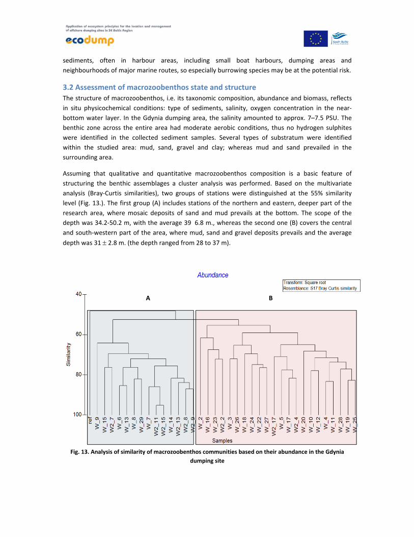
3.2 Assessment of macrozoobenthos state and structure
The structure of macrozoobenthos, i.e. its taxonomic composition, abundance and biomass, reflects
in situ physicochemical conditions: type of sediments, salinity, oxygen concentration in the near‐
bottom water layer. In the Gdynia dumping area, the salinity amounted to approx. 7–7.5 PSU. The
benthic zone across the entire area had moderate aerobic conditions, thus no hydrogen sulphites
were identified in the collected sediment samples. Several types of substratum were identified
within the studied area: mud, sand, gravel and clay; whereas mud and sand prevailed in the
surrounding area.
Assuming that qualitative and quantitative macrozoobenthos composition is a basic feature of
structuring the benthic assemblages a cluster analysis was performed. Based on the multivariate
analysis (Bray‐Curtis similarities), two groups of stations were distinguished at the 55% similarity
level (Fig. 13.). The first group (A) includes stations of the northern and eastern, deeper part of the
research area, where mosaic deposits of sand and mud prevails at the bottom. The scope of the
depth was 34.2‐50.2 m, with the average 39�6.8 m., whereas the second one (B) covers the central
and south‐western part of the area, where mud, sand and gravel deposits prevails and the average
depth was 31 2.8 m. (the depth ranged from 28 to 37 m).
Fig. 13. Analysis of similarity of macrozoobenthos communities based on their abundance in the Gdynia
dumping site
A B

Qualitative composition
A total of 23 species and higher taxonomic units of benthic macrofauna (not identified to the species
level) were found in the studied area (Table 2). In the shallower area diversity was slightly higher.
Table 2. Benthic macroinvertebrate taxa found in A and B area (Gdynia dumping site)
No. Taxa A B
1 Electra crustulenta + +
2 Halicryptus spinulosus + +
3 Nemertina +
4 Bylgides sarsi + +
5 Hediste diversicolor + +
6 Pygospio elegans + +
7 Marenzelleria neglecta + +
8 Oligochaeta + +
9 Balanus improvisus + +
10 Idotea chelipes + +
11 Saduria entomon + +
12 Jaera sp. +
13 Bathyporeia pilosa +
14 Monoporeia affinis + +
15 Pontoporeia femorata + +
16 Gammarus sp. + +
17 Corophium volutator + +
18 Diastylis rathkei + +
19 Hydrobia sp. + +
20 Mytilus edulis trossulus + +
21 Cerastoderma glaucum +
22 Macoma balthica + +
23 Mya arenaria + +
Altogether 19 23
Quantitative composition
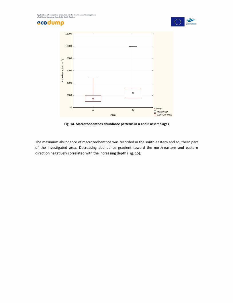
The abundance of benthic fauna in the A region ranged from 810 individuals per 1 m2 at the station
W2_8 up to 2429 per 1 m2 at the station ‘Ref’. The average abundance within A area amounted to
1457 ± 465.2 individuals per 1 m2 (Fig. 14).
In the B area abundance was higher and ranged from 1505 at the station W_20 to 5078 individuals
per 1 m2 at the station W2_2 with an average 2363 ± 791,8 individuals per 1 m2 (Fig. 14).
Mean Mean+SD 1,96*Min-Max
A B
Area
0
2000
4000
6000
8000
10000
12000
Abu
ndan
ce [
ind.
m-2
]
Fig. 14. Macrozoobenthos abundance patterns in A and B assemblages
The maximum abundance of macrozoobenthos was recorded in the south‐eastern and southern part
of the investigated area. Decreasing abundance gradient toward the north‐eastern and eastern
direction negatively correlated with the increasing depth (Fig. 15).

Fig. 15. Macrozoobenthos abundance distribution in the Gdynia dumping site area. A – deeper stations area
and B – shallower stations area (acc. to Bray Curtis similarity analysis based on the abundance).
Dominance patterns associated with the abundance of macrozoobenthos phyla differed in the
particular communities (Fig. 16). Generally bivalves, polychaetes and crustaceans dominated in
abundance. The share of the bivalves was greater in the deeper part of the dumping site, whereas
the polychaetes in the shallower one.
A
B

Fig. 16. Percentage share of macrozoobenthos phyla in abundance in the A and B assemblages within the
research area
A bivalve Macoma balthica and a polychaete Marenzelleria neglecta dominated in both region. Both
species are regarded as tolerant to anthropogenic pressure. Macoma balthica, when covered by a
layer of newly dumped sediment, is able to survive by moving to the sediment or extending its
siphon out of the sediment. Also mobile burrowing organisms e.g. Hediste diversicolor, Marenzelleria
neglecta survived rather successfully in the sediment dumping (Olenin 1992). Another tolerant
crustacean Corophium volutator had a considerable share in the B area (Fig. 17).
A B
Fig. 17. Abundance dominance structure within A and B region
The biomass of macrozoobenthos in the A area varied from 58,802 g per 1 m2 at ‘Ref’ station to
362.402 g per 1 m2 at W_15 with average 176.4 ± 85,32 g per 1 m2. In the B region the biomass was
0
10
20
30
40
50
60
70
80
90
100
A B
Bivalvia
Gastropoda
Crustacea
Oligochaeta
Polychaeta
Nemertina
Priapulida
Macomabalthica
Marenzellerianeglecta
Pontoporeiafemorata
Varia
Macomabalthica
Marenzellerianeglecta
Corophiumvolutator
Pygospioelegans
Varia

slightly higher; ranging from 43,085 g per 1 m2 at the W_23 station to 444,180 g per 1 m2 at the
W_22 station, with the average 199.0 ± 105,69 g per 1 m2 (Fig. 18).
Mean Mean+SD 1,96*Min-Max
A B
Area
0
100
200
300
400
500
600
700
800
900
1000
Bio
mas
s [g
m-2
]
Fig. 18. Macrozoobenthos biomass patterns in selected assemblages (A and B areas)
The biomass distribution was not related to the depth gradient. The highest biomass values were
recorded in the northern and southern parts, whereas the lowest values were observed in the central
part of the dumping site (Fig. 19).

Fig. 19. Macrozoobenthos biomass distribution in the Gdynia dumping site area.
Bivalves considerably dominated in biomass in both regions. The share of the group of other phyla
did not exceed 10 % of the total macrozoobenthos biomass (Fig. 20).
Fig. 20. Percentage share of macrozoobenthos phyla in the particular assemblages (A and B) within the
researching area (biomass).
0
10
20
30
40
50
60
70
80
90
100
A B
Bivalvia
Gastropoda
Crustacea
Oligochaeta
Polychaeta
Nemertina
Priapulida

Analysis of the biomass dominance structure shows that in both regions bivalves species Macoma
balthica and Mytilus trossulus were dominant. In deeper area (A) a crustacean Saduria entomon had
a considerable share in biomass structure (Fig. 21). Distribution of this relict species is restricted to
deeper and colder waters in the Baltic Sea.
A B
Fig. 21. Biomass dominance structure within A and B region
Mean abundance, biomass and macrozoobenthos composition recorded within Gdynia dumping site
were typical of the outer Puck Bay within similar depth range (Kruk‐Dowgiałło & Szaniawska 2008).
The minor differences between A and B region were probably not attributed to dumping activities
but resulted from natural spatial variability of macrozoobenthos assemblages. Negative coefficient of
correlation reflects relationship between the abundance and the depth (R2=‐0,5).
The nearest HELCOM COMBINE Monitoring Programme station ‘P104’ is situated in the vicinity of the
Hel Peninsula at the depth of 56 m, approx. 5 km off the northernmost W_2_11 station of the ‘A’
region. The results of macrozoobenthos monitoring conducted during the period of 2007‐2009
(Warzocha et al. 2011; Radziejewska et al. 2012 a & b) are presented in the table 4. Macoma balthica
dominated in terms of abundance as well as biomass. Significant share of Pontoporeia femorata,
Marenzelleria neglecta and Saduria entomon was noted in abundance dominance structure. Similar
dominance pattern was found in the deeper region (‘A’) during the studies conducted within
‘Ecodump project’ .
Slightly less diversified macrozoobenthic community was noted in 2008 at the 7 stations located in
the dumping site area (Bełdowski et al. 2008), (Table 3). The authors recorded considerable
dominance of Macoma balthica, however the share of Oligochaeta was also noticeable. In shallower
part of the research area, located westward off the dumping area, 14 taxa of macrozoobenthos were
found. Analyses showed that spatial abundance and biomass distribution as well as diversity of
macrozoobenthos depend, first of all, on the depth and bottom sediment granulation, rather than on
the dumping activities (Bełdowski et al. 2008).
Macomabalthica
Mytilus edulistrossulus
Saduriaentomon
Varia
Macomabalthica
Mytilus edulistrossulus
Varia

Table 3. Taxonomic composition of the macrozoobenthos in the area of the Gdynia dumping site in 2007‐
2009 (1Warzocha et al. 2011, 1 Radziejewska et al. 2012 a & b, 2 Bełdowski et al. 2008)
No. Taxa Deeper part Shallower part
P104 (2007‐2009)
1 Bełdowski et al. (2008)
2 Bełdowski et al. (2008)
2
2 Halicryptus spinulosus + +
4 Bylgides sarsi +
5 Hediste diversicolor + +
6 Pygospio elegans + + +
7 Marenzelleria neglecta + +
8 Oligochaeta + + +
11 Saduria entomon + +
14 Monoporeia affinis +
15 Pontoporeia femorata + + +
17 Corophium volutator + + +
18 Diastylis rathkei + + +
19 Hydrobia sp. + +
20 Potamopyrgus jenkinsi +
21 Mytilus edulis trossulus +
23 Macoma balthica + + +
24 Mya arenaria + +
N of taxa 13 7 14
Abundance dominance structure
Macoma balthica, Pontoporeia femorata, Bylgides sarsi, Marenzelleria neglecta
Macoma balthica,Oligochaeta
Macoma balthica,Oligochaeta, Halicryptus spinulosus
Biomass dominance structure Macoma balthica, Saduria entomon
No data No data
3.3 Ecological quality assessment based on macrozoobenthos
The values of the biotic index B within the Gdynia dumping site area varied from 1.87 to 3.66. The
average value of the B index (2.64 ± 0.39) classifies the entire area ecological status as ‘poor’ in the
meaning of the Water Framework Directive, acc. to the Regulation of Ministry of the Environment of
9th November 2011 On classification of ecological status of surface water bodies... ( Journal of Laws
No. 257, item 1545). The environmental conditions at a deeper part (A) were slightly better (B mean
= 2.73 ± 0.41) than in the shallower one (B mean = 2.58 ± 0.38). Some narrow restricted part located
along the eastern border of the area remained in ‘good’ ecological status (Fig. 22).
Similar values of the B index was found in 2011 at the stations investigated within National
Monitoring Programme in the Outer Puck Bay (PLTW III WB 3). Ecological status based on
macrozoobenthos in this transitional water body was assessed as poor (B = 2,59) at the station T12,

whereas at the station OM1 as moderate (B = 3,14) (data from the Regional Inspectorate of
Environmental Protection in Gdańsk).
Fig. 22. Ecological quality status within the Gdynia dumping site area (based on B index macrozoobenthos
assessment)
The lowest value of the B index was found in the southern part of the area, at the station W_20
located at the shallower bottom where species tolerant to unfavourable environmental conditions –
Macoma balthica and Marenzelleria neglecta dominated. The highest value was determined at the
station W2_8, within the deeper area, where a sensitive crustacean Pontoporeia femorata had a
considerable share in total abundance. Additionally the abundance structure was remarkably even.
Generally, values of the B index increased in the eastern direction.
A
B
Conclusions No significant alterations of macrozoobenthic community and structure, that could be
directly connected to dumping activities were found.
The ecological status of the area, acc. to Water Framework Directive classification scheme
was assessed as ‘poor’.
Bivalves collected from the dumping area as well as the ‘Ref’ station appear to be chronically
exposed to an unfavourable environmental conditions what is manifested by e.g. increased
number of histological and nuclear abnormalities.
Taking into account that the analysed endpoints (e.g. neoplasia and skewed sex ratio) were
also observed in other areas of the Gulf of Gdańsk, it can be concluded that the
environmental conditions in the dumping area does not seem to be significantly worse than
in the other deeper parts of the Gulf of Gdańsk.
References Arkhipchuk V. V., Garanko N. N. 2005. Using the nucleolar biomarker and the micronucleus test on In
vivo fish fin cells. Ecotox. Environ. Saf. 62: 45–52.
Baumann P. C. 1998. Epizootics of cancer in fish associated with genotoxins in sediment and water.
Mut. Res. 411: 227–233.
Bełdowski J., Pazdro K., Pempkowiak J., Kotwicki L., Urbański J. 2008. Badanie walorów
przyrodniczych siedliska dennego w rejonie „klapowiska” i toru podejściowego do Portu Gdynia oraz
wpływu projektowanych inwestycji na obszary Natura 2000. Instytut Oceanologii PAN: 12‐42.
Cederwall H., Elmgren R. 1990. Biological effectsof eutrophication in the Baltic Sea, particularly the
coastal zone, Ambio, 19: 109‐112.
Costello M. K., Emblow C. S., White R. 2001. European register of marine species. A check list of the
marine species in Europe and a bibliography of guides to their identification. Patrimoines Naturels
50: 1‐463.
Crocker J., Boldy D. A., Egan M. J. 1989. How should we count AgNORs? Proposal for a standardized
approach. J. Pathol. 158: 185–188.
Diaz R. J., Rosenberg R. 1995. Marine benthic hypoxia: a review of its ecological effects and the
behaviour responses of benthic macrofauna. Oceanogr. Mar. Biol. Ann. Rev. 33: 245‐303.
Elston R. A., Moore J. D., Brooks K. 1992. Disseminated neoplasia of bivalve molluscs. Rev. Aquat.
Org. 6: 405–466.
Essink K. 1999. Ecological effects of dumping of dredged sediments; options for management. Journal
of Coastal Conservation 5: 69‐80.
Fenech M., Chang W. P., Kirsch‐Volders M., Holland N., Bonassi S., Zeiger E. 2003. HUMN project:
detailed description of the scoring criteria for the cytokinesis‐block micronucleus assay using isolated
human lymphocyte cultures. Mut. Res. 534: 5–75.

Gray J. S., Shiu‐Sun Wu R., Ying Or Y. 2002. Effects of hypoxia and organic enrichment on the coastal
marine environment. Mar. Ecol. Prog. Ser. 238: 249‐272.
HELCOM 1988. Guidelines for the Baltic Monitoring Programme for the Third Stage. Helsinki
Commission, Baltic Sea Envir. Proc. 12 D.
Howell W. M., Black D. A. 1980. Controlled silver‐staining of nucleolus organizer regions with a
protective colloidal developer: a 1‐step method. Experientia 36: 1014–1015.
Karlson K., Rosenberg R. Bonsdorff E. 2002. Temporal and spatial large‐scale effects of eutrophication
and oxygen efficiency on benthic fauna in Scandinavian and Baltic waters – a review. Oceanogr. Mar.
Biol. Ann. Rev. 40: 427‐489.
Kruk‐Dowgiałło L., Szaniawska A. 2008. Gulf of Gdańsk and Puck Bay [in] Ecology of Baltic Coastal
Waters. Ecological Studies, Vol. 197, 2, II.B: 139‐165.
Lehtonen K. K., Schiedek D., Kohler A., Lang T., Vouninen P. J., Forlin L., Barsiene J., Pempkowiak J.,
Gercken J. 2006. The BEEP project in the Baltic Sea: Overview of results and outline for a regional
biological effects monitoring strategy. Mar. Pol. Bull. 53: 523–537.
Odum E. 1982. Podstawy ekologii. Wydanie III. Państwowe Wydawnictwo Rolnicze i Leśne,
Warszawa.661 pp.
Olenin, S. 1992. Changes in a south‐eastern Baltic soft‐bottom community induced by dredged spoil
dumping. Proc. 12th Baltic Marine Biologists Symposium. E. Bjørrnestad, L. Hagerman, K. Jensen (ed‐
s). Olsen&Olsen: International Symposium Series, Fredensborg: 119‐123.
Osowiecki A., Błeńska M. 2010. Makrobezkręgowce bentosowe [w:] Przewodniki metodyczne do
badań terenowych i analiz laboratoryjnych fitoplanktonu, innej flory wodnej i makrobezkręgowców
bentosowych w wodach przejściowych i przybrzeżnych. Biblioteka Monitoringu Środowiska. Inspekcja
Ochrony Środowiska, Wyd. Nauk. Inst. Technologii Eksploatacji – PIB, Radom. ISBN 978‐83‐61227‐36‐
6.: 65‐84.
Osowiecki A., Łysiak‐Pastuszak E., Kruk‐Dowgiałło L., Błeńska M., Brzeska P., Kraśniewski W.,
Lewandowski Ł., Krzymiński W. 2012. Development of tools for ecological quality assessment in the
polish marine areas according to the Water Framework Directive. Part IV – preliminary assessment.
Oceanological and Hydrobiological Studies Vol.41(3): 1‐10.
Radziejewska T., Wawrzyniak‐Wydrowska B., Piątkowska Z. 2012 a. Makrobezkręgowce bentosowe
[Benthic macroinvertebrates] [in] Charakterystyka wybranych elementów środowiska. Bałtyk
Południowy w 2008 r. IMGW PIB: 117‐122.

Radziejewska T., Wawrzyniak‐Wydrowska B., Osowiecki A. 2012 b. Makrobezkręgowce bentosowe
[Benthic macroinvertebrates] [in] Charakterystyka wybranych elementów środowiska. Bałtyk
Południowy w 2009 r. IMGW PIB:125‐132.
Regulation of Ministry of the Environment of 9th November 2011 on the method of classification of
the status of surface water bodies (Journal of Laws No. 257, item 1545).
Regulation of the Minister of Environment of 16 April 2002, on the type and concentration of
substances that indicate that the dredged material is contaminated (Journal of Laws No. 55, item
498).
Rosenberg R., Agrenius S., Hellman B., Nilsson H. C., Norling K. 2002. Recovery of benthic habitats
and fauna in a Swedish fjord following improved oxygen conditions. Mar. Ecol. Prog. Ser. 234: 43‐53.
Rumohr H., Bonsdorf E., Pearson T. H. 1996. Zoobenthic successions in Baltic sedimentary habitats.
Arch. Fish. Mar. Res. 44 (3): 179‐214.
Simonini R., Ansaloni I., Cavallini F., Graziosi F., Iotti M., G. Massamba N_Siala,Mauri M, Montanari
G., Preti M., Prevedelli D. 2005. Effects of long‐term dumping of harbor‐dredged material on
macrozoobenthos at four disposal sites along the Emilia‐Romagna coast (Northern Adriatic Sea,
Italy). Marine Pollution Bulletin 50 (2005): 1595–1605.
Smolarz K., Berger A. 2009. Long‐term toxicity of hexabromocyclododecane (HBCDD) to the benthic
clam Macoma balthica (L.) from the Baltic Sea. Aqat. Tox. 95: 239–245.
Smolarz K., Renault T., Soletchnik P., Wołowicz M. 2005 a. Survey for neoplasia in M. balthica from
the Gulf of Gdansk using flow cytometry. Dis. Aquat. Org. 66: 41‐46.
Smolarz K., Thiriot‐Quiévreux C., Wołowicz M. 2005 b. Recent trends in prevalences of neoplasia in
the Baltic clam Macoma balthica (L.) from the Gulf of Gdańsk (Baltic Sea). Oceanologia 47:61–74.
Smolarz K., Renault T., Wołowicz M. 2006. Ultrastructural study of neoplastic cells in Macoma
balthica (Bivalvia) from the Gulf of Gdansk (Poland). J. Invert. Path. 92: 79‐84.
Smolarz K., Wołowicz M., Thiriot‐ Quiévreux C. 2003. Argyrophilic nucleolar organizer regions
(AgNORs) in interphases and metaphases of normal and neoplastic gill cells of Macoma balthica
(Bivalvia: Tellinidae) from the Gulf of Gdańsk, Baltic Sea. Dis. Aquat. Org. 56: 269‐274.
Tay K., Teh S., Doe K., Lee K., Jackman P. 2003. Histopathologic and histochemical biomarker
responses of Baltic clam, Macoma balthica, to contaminated Sydney Harbour sediment, Nova Scotia,
Canada. Environ Health Perspect. 111: 273–280.
Thiriot‐Quievreux C., Ayraund N. 1982. Les caryotypes de quelques espèces de bivalves et
gastéropodes marins. Mar. Biol. 70: 165–172.

Towned J. 2002. Practical statistics for environmental and biological scientists. England. 276 pp.
Trojan P. 1980. Ekologia ogólna. Wydanie IV. PWN, Warszawa. 419 pp.
Warzocha J., Gromisz S., Wróblewska H., Łysiak‐Pastuszak E., Piątkowska Z. 2011. Makrobezkręgowce
bentosowe [Marine invertebrates] [in] Charakterystyka wybranych elementów środowiska. Bałtyk
Południowy w 2007 r. IMGW PIB: 96‐98.
Wenne R. 1985. Microgeographic differentiation of the reproductive cycle of Macoma balthica (l.) in
the Gulf of Gdańsk (South Baltic), and the relationship between this cycle and energy reserve
changes. Pol. Arch. Hydrobiol. 32: 47–63.
Wołowicz M., Smolarz K., Sokołowski A. 2005. Neoplasia in estuarine bivalves: effect of feeding
behaviour and pollution in the Gulf of Gdansk (Baltic Sea, Poland). NATO Science Series IV: Earth and
Environmental Sciences, NATO Special Edition, Dame D. F. and Olenin S. (eds), Springer: 165–182.