connections of the skull - is.muni.cz · humerus + radius + ulna –art. cubiti radius + ulna...
TRANSCRIPT
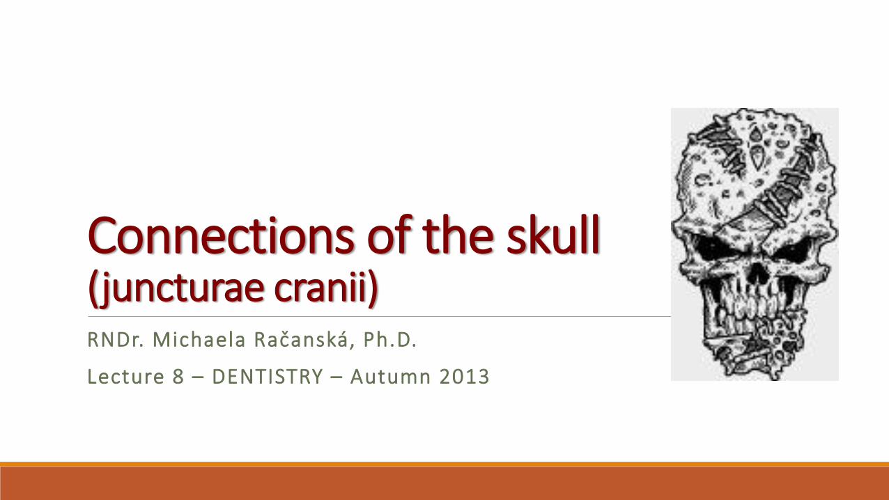
RNDr. Michaela Račanská, Ph.D.
Lecture 8 – DENTISTRY – Autumn 2013
Connections of the skull(juncturae cranii)
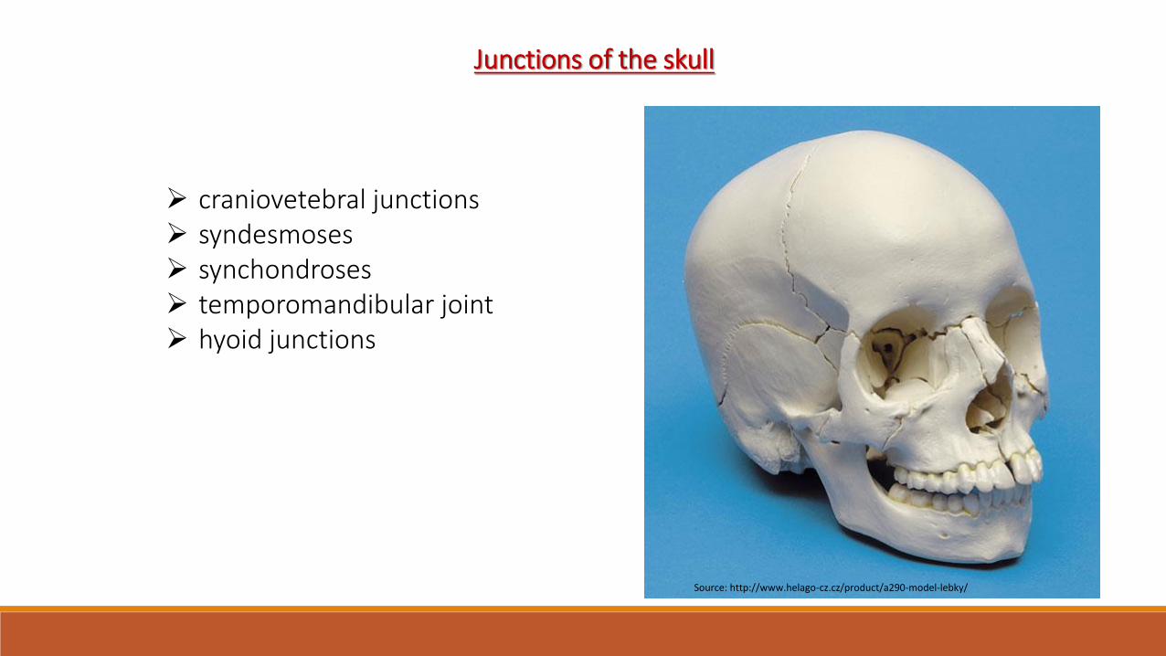
Junctions of the skull
craniovetebral junctions syndesmoses synchondroses temporomandibular joint hyoid junctions
Source: http://www.helago-cz.cz/product/a290-model-lebky/

Craniovertebral junctiones Connection of the skull with the C1 and C2
1. Articulatio atlantooccipitalisPaired joint
AS: condyli occipitales andfoveae articulares superioresof atlasAS: Is attached to the margins ofthe articular surfaces

Special apparatus:membrana atlantooccipitalis anterior and posterior(between arches of atlas and occipital bone) membrana tectoria(cranial continuation of lig. longitudinale posterius, it reaches to clivus)Type of joint: elipsoidal with possibility of flexion and extension of thehead and there are also possible smaller movements sideways

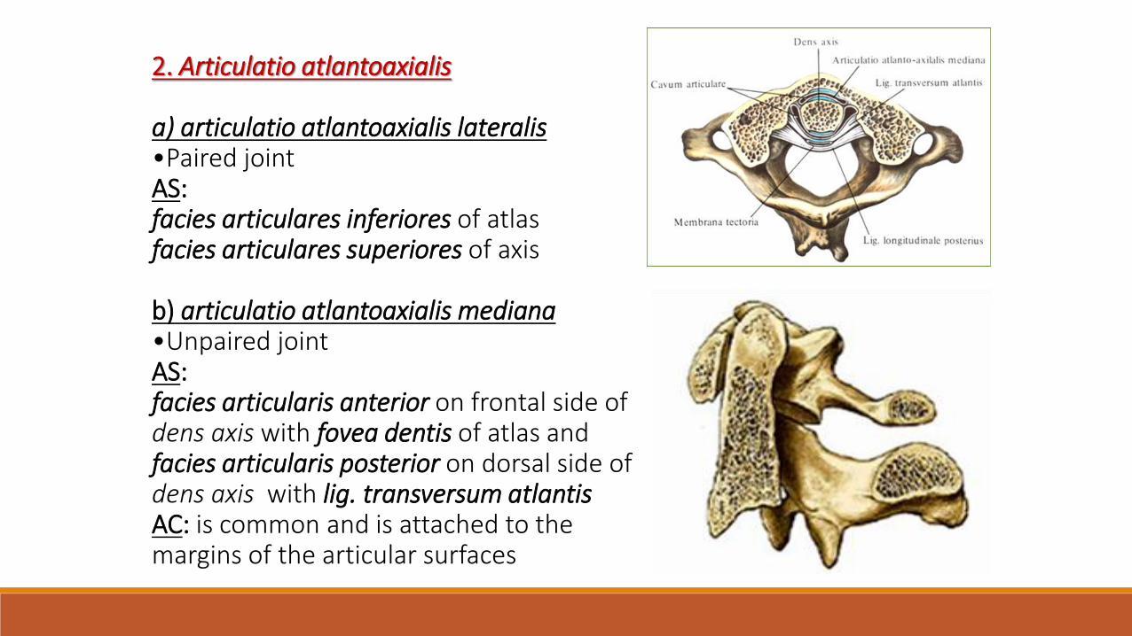
2. Articulatio atlantoaxialis
a) articulatio atlantoaxialis lateralis•Paired jointAS:facies articulares inferiores of atlasfacies articulares superiores of axis
b) articulatio atlantoaxialis mediana•Unpaired jointAS: facies articularis anterior on frontal side ofdens axis with fovea dentis of atlas andfacies articularis posterior on dorsal side ofdens axis with lig. transversum atlantisAC: is common and is attached to themargins of the articular surfaces

Special apparatus: lig. apicis dentis, ligg. alaria,lig. cruciforme atlantis, formed bylig. transversum atlantis and fasciculilongitudinales (vertical fibrous bands goingfrom axis to occipital bone)Type of joint: both joints form onemechanical unit, atlas is rotating along dens axis in range of 60°

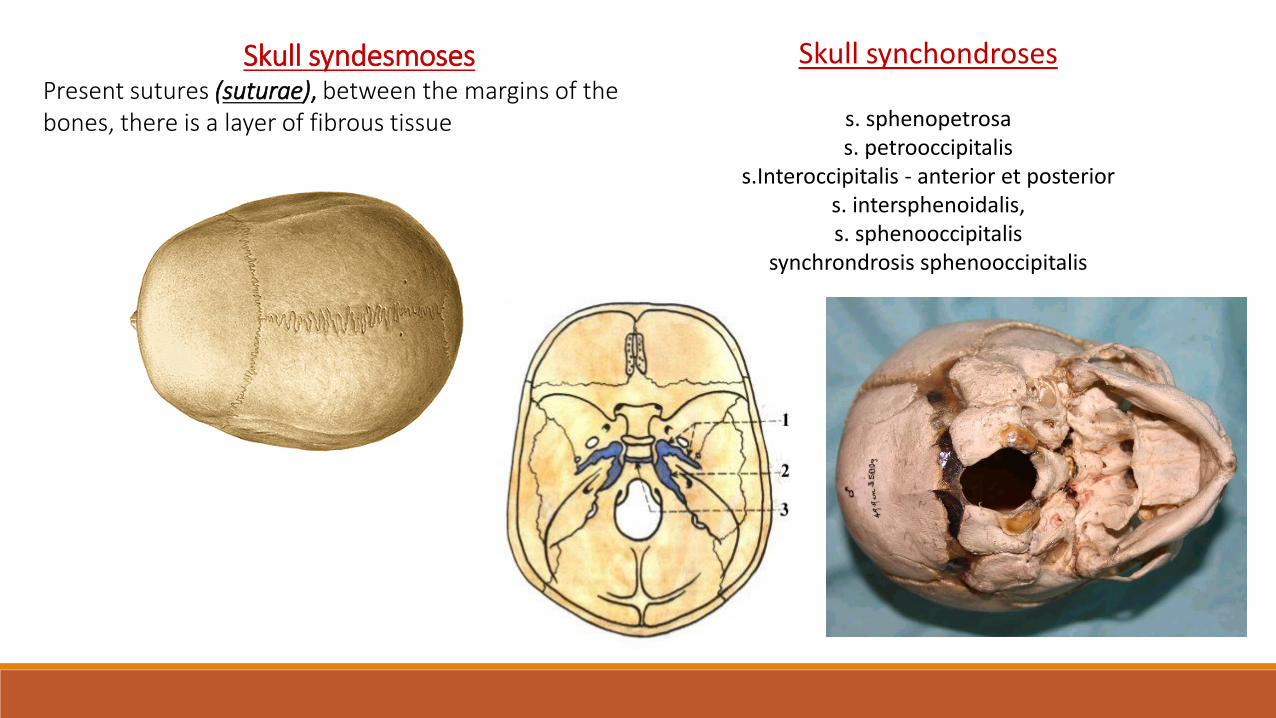
Skull syndesmosesPresent sutures (suturae), between the margins of thebones, there is a layer of fibrous tissue
Skull synchondroses
s. sphenopetrosas. petrooccipitalis
s.Interoccipitalis - anterior et posteriors. intersphenoidalis, s. sphenooccipitalis
synchrondrosis sphenooccipitalis

Temporomandibular joint (articulatio temporomandibularis)
AS: caput mandibulae connects with fossa mandibularis and tuberculum articulare of temporal bone
AC: is attached to the margins of the articular surfaces, its medial part is very strong, it rows together with discusarticularis
Type of joint: gynglimus (hinge)Elevation – closing of the mouthDepresion – opening of the mouthProtraction – shifting od the chin forwardsRetraction – shifting od the chin backwards
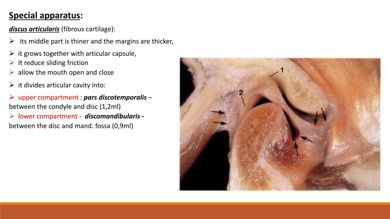
Special apparatus:
discus articularis (fibrous cartilage):
its middle part is thiner and the margins are thicker,
it grows together with articular capsule,
It reduce sliding friction
allow the mouth open and close
it divides articular cavity into:
upper compartment : pars discotemporalis –
between the condyle and disc (1,2ml)
lower compartment - discomandibularis -
between the disc and mand. fossa (0,9ml)

Ligaments - extraarticular
on lateral side: lig. laterale
around the joint: lig. sphenomandibulare (runs from the styloid process→ the posterior edge of the angle of the mandible)
lig. stylomandibulare (runs from the styloid process → the posterioredge of the angle of the mandible)
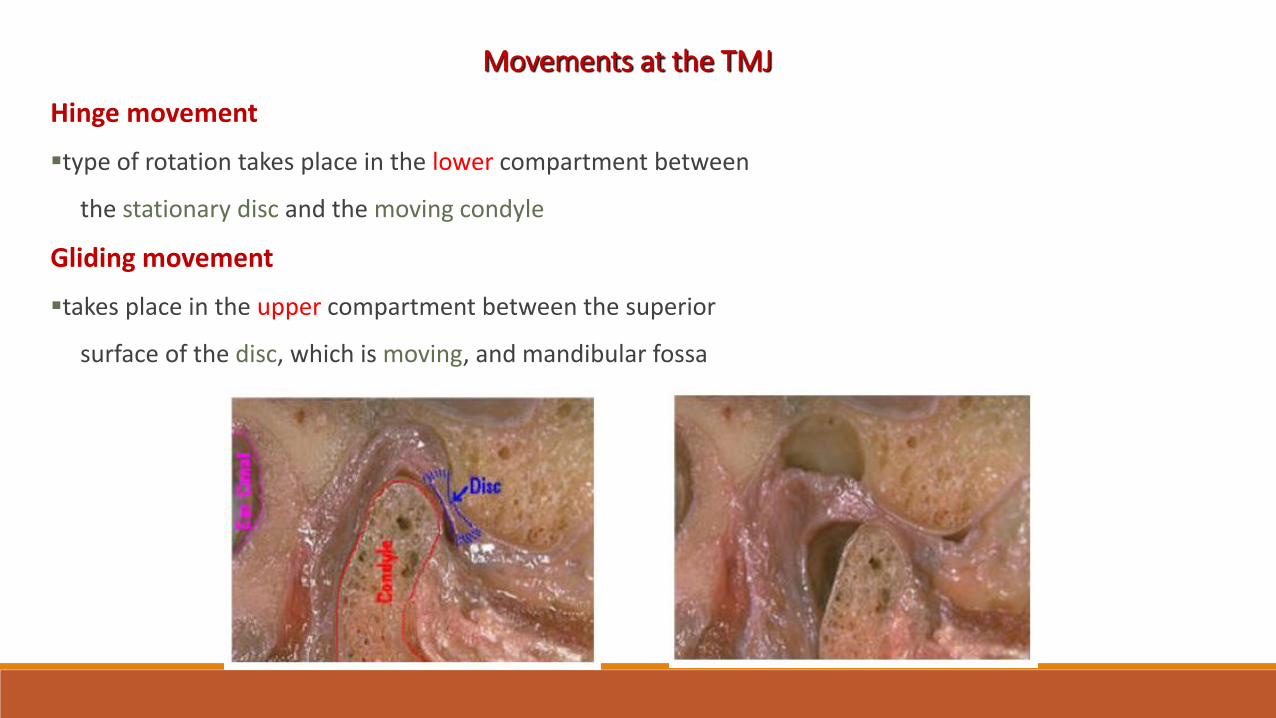
Movements at the TMJ
Hinge movement
type of rotation takes place in the lower compartment between
the stationary disc and the moving condyle
Gliding movement
takes place in the upper compartment between the superior
surface of the disc, which is moving, and mandibular fossa

Depression - the opening
with simple rotation at the joint can be achieved 15 - 20mm
intericisor distance
during translation, the disc and condyle move under the
articular eminence
Elevation – the closing
translation - the condyles move backward and upward along
the articular eminence
rotation upward to attain final position

Protrusion
slide the mandible forward
maximal protrusion results in the lower incisors being a few mm
anterior to the maxillary incisors
Retrusion
move the mandible posteriorly
condyles move backward and upward and reoccupy the
mandibular fossa
Laterotrusion
the condyle move to the right or to the left side
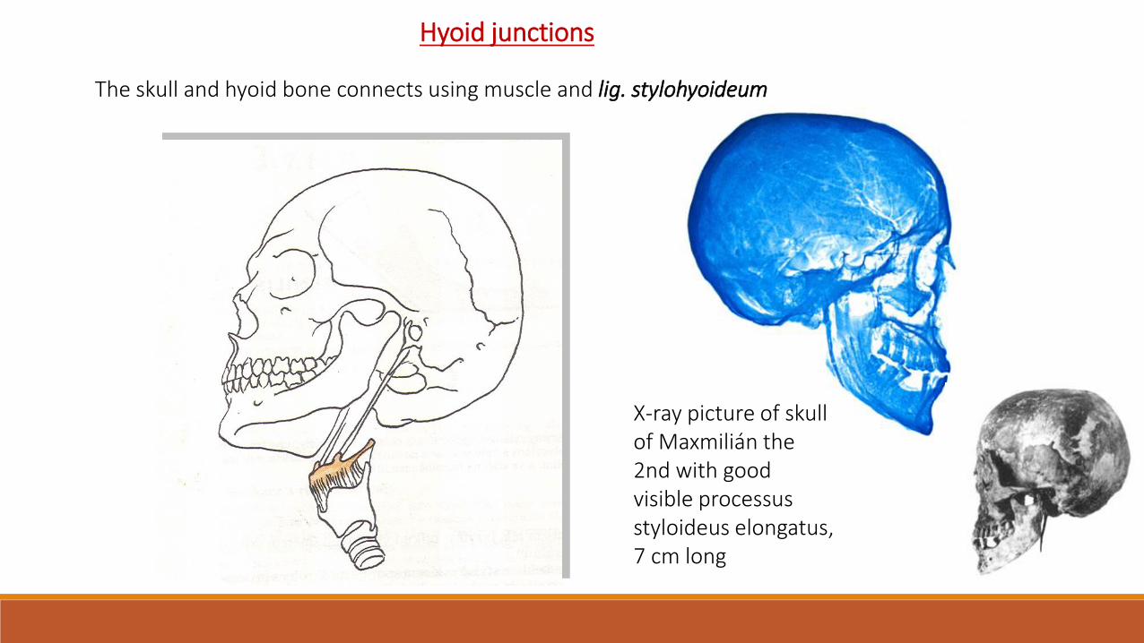
Hyoid junctions
The skull and hyoid bone connects using muscle and lig. stylohyoideum
X-ray picture of skullof Maxmilián the2nd with goodvisible processusstyloideus elongatus, 7 cm long

Connections of the upper limb(juncturae ossium membri superioris)
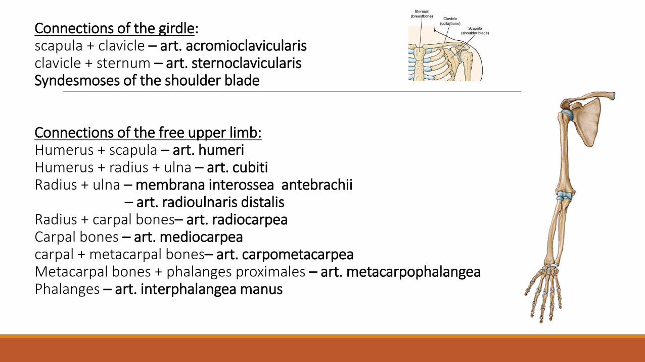
Connections of the girdle:scapula + clavicle – art. acromioclavicularisclavicle + sternum – art. sternoclavicularisSyndesmoses of the shoulder blade
Connections of the free upper limb:Humerus + scapula – art. humeriHumerus + radius + ulna – art. cubitiRadius + ulna – membrana interossea antebrachii
– art. radioulnaris distalisRadius + carpal bones– art. radiocarpeaCarpal bones – art. mediocarpeacarpal + metacarpal bones– art. carpometacarpeaMetacarpal bones + phalanges proximales – art. metacarpophalangeaPhalanges – art. interphalangea manus

I. Articulatio sternoclavicularis
Type: compound joint- discus articularisball and socket (movements in connection to the scapulamovements)A. head: facies articularis sternalis claviculaeA. fossa: incisura clavicularis manubrii sterniAC: tough, shortLigaments:
lig. sternoclaviculare anteriuslig. sternoclaviculare posteriuslig. interclavicularelig. costoclaviculare
Movements: small, to all direction
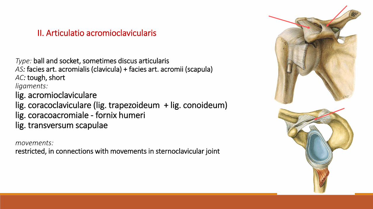
Type: ball and socket, sometimes discus articularisAS: facies art. acromialis (clavicula) + facies art. acromii (scapula)AC: tough, shortligaments:lig. acromioclavicularelig. coracoclaviculare (lig. trapezoideum + lig. conoideum) lig. coracoacromiale - fornix humerilig. transversum scapulae
movements:restricted, in connections with movements in sternoclavicular joint
II. Articulatio acromioclavicularis


Syndesmoses of the shoulder blade:
- lig. transversum scapulae
- lig. coracoacromiale - fornix humeri
Movements of the scapula:
- Retraktion
- Protraktion
- Elevation
- Depresion
- Rotation
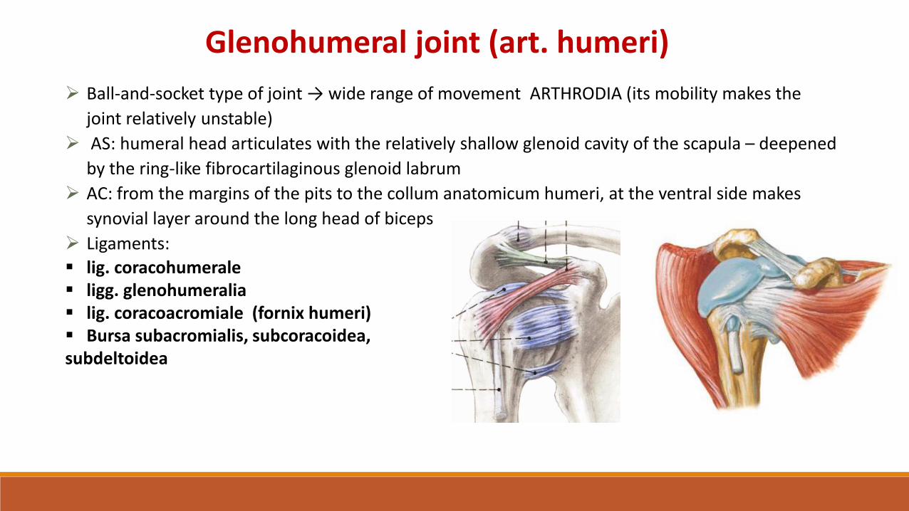
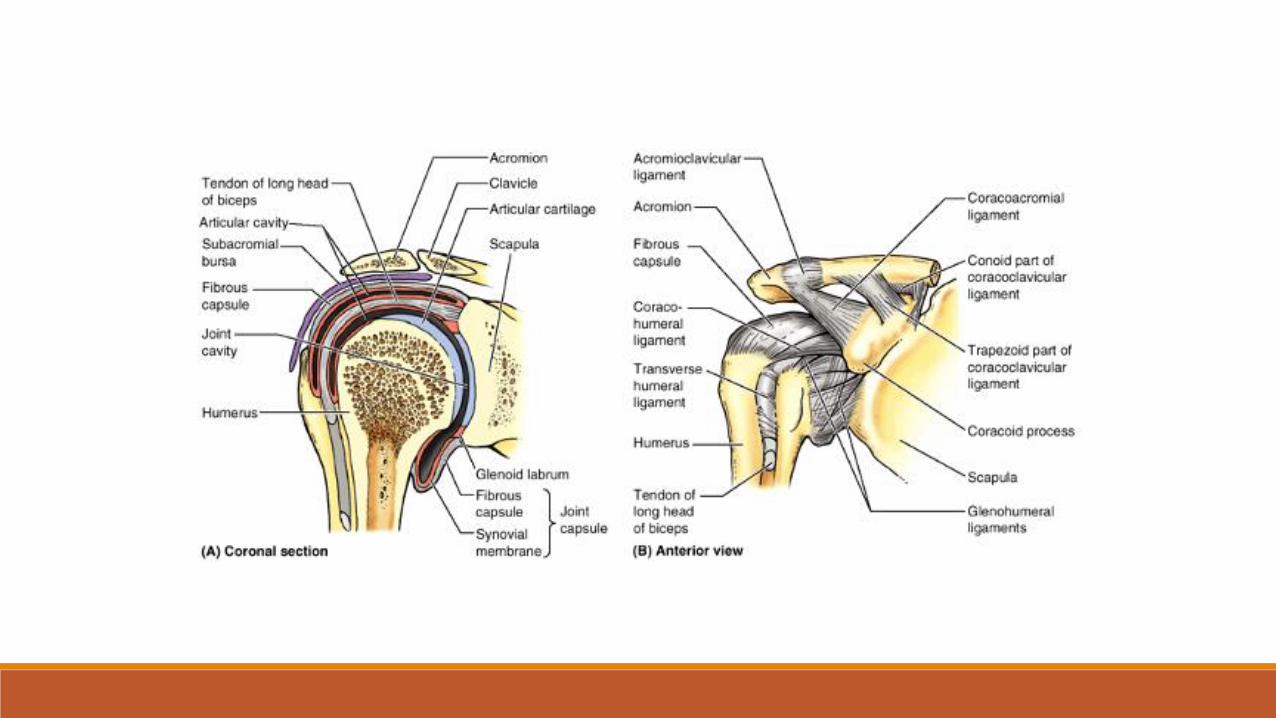
Glenohumeral joint (art. humeri)
Ball-and-socket type of joint → wide range of movement ARTHRODIA (its mobility makes the
joint relatively unstable)
AS: humeral head articulates with the relatively shallow glenoid cavity of the scapula – deepened
by the ring-like fibrocartilaginous glenoid labrum
AC: from the margins of the pits to the collum anatomicum humeri, at the ventral side makes
synovial layer around the long head of biceps
Ligaments:
lig. coracohumerale ligg. glenohumeralia lig. coracoacromiale (fornix humeri) Bursa subacromialis, subcoracoidea, subdeltoidea


MOVEMENTS:Ventral and dorsal flexionabduktion(from the horizontal plane togetherwith movements of the scapula)adduktionrotation - supination, pronationMiddle position:Slow flexion and small abduktion


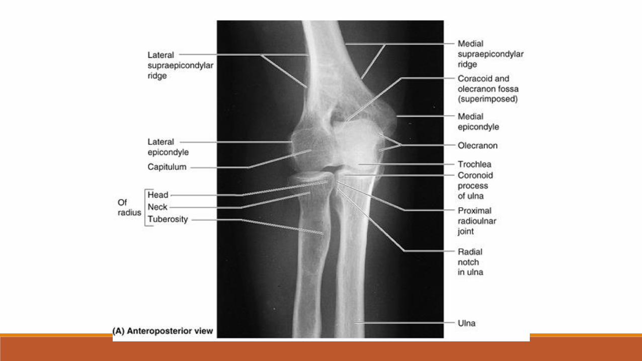
Articulations of the forearmELBOW JOINT (ART. CUBITI)Type: compound joint
Articulatio humeroulnarisType: hingeA. head: trochlea humeriA. fossa: incisura trochlearis ulnae
Articulatio humeroradialisType: ball and socketA. head: capitulum humeriA. fossa: fovea articularis radii
Articulatio radioulnaris proximalisType: pivotA. head: circumferentia articularis radii A. fossa: incisura radialis ulnae

AC: common for all three parts, attach to the margins of AS, at radius to the collum - recessus sacciformisLigamnets: lig. collaterale radialelig. collaterale ulnare
lig. obliquumlig. anulare radii
Movements: flexion, extensionArt. radioulnaris proximalis togetherWith art. radioulnaris distalis – pronation and supinationMiddle position: in slight flexion and pronation
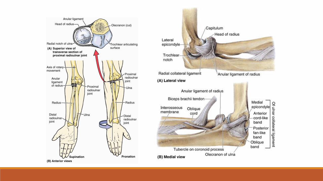


SYNDESMOSES RADIOULNARIS
Interosseous membrane (chorda obliqua)

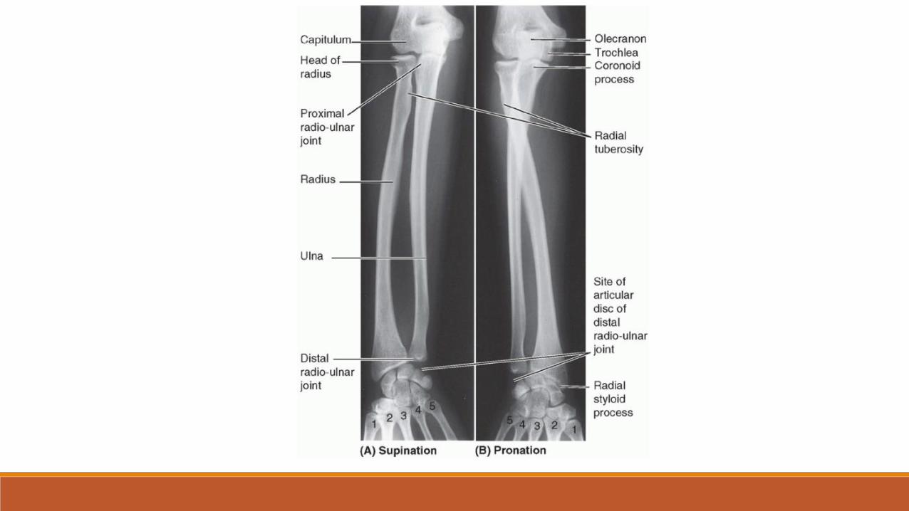
Distal Radioulnar Joint (pivot)
head of the ulna articulates with the ulnar
notch on the medial side of the distal end of
the radius
AC: free, anables rotaion of the distal part of
the radius around the head of the ulna
articular disc binds the ends of the ulna and
radius together
movements - supination and pronation

ARTICULATIO RADIOCARPALIS Radius and carpal bones
ARTICULATIO MEDIOCARPALISbetween proximal and distal row of carpal bones
ARTICULATIONES INTERCARPALESconestions between carpal bones
ARTICULATIONES CARPOMETACARPALESdistal row of carpal bones with metacarpals
ARTICULATIONES INTERMETACARPALESbetween bases of metacarpal bones
ARTICULATIONES METACARPOPHALANGEALESheads of the metacarpals with the proximal row ofphalanges
ARTICULATIONES INTERPHALANGEALESBetween phalangesretinaculum musculorum flexorum(lig. carpi transversum) between eminentia carpi radialis et ulnaris -> canalis carpi
Articulationes manus

Articulatio radiocarpalis
Type: compound, ellipsoidA. head: os scaphoideum, os lunatum, os triquetrumA. fossa: facies articularis carpalis radii, discus articularisAC: firm and shortLigaments: common with art. mediocarpalisMovements: functional unit with medicarpal, intercarpal, carpometacarpal jointsPalmar and dorsal flexionradial and ulnar duktioncircumduktion

Articulatio mediocarpalis
Type: elipsoid, compound, in the shape of horizontaly placed "S"A. head, ulnar side: os hamatum, os capitatumA. head, Radial side: os scaphoideum
A. fossa, ulnar side: os scaphoideum, os lunatum, os triquetrumA. fossa, radila side: os trapezium, os trapezoideumAC: firm and short
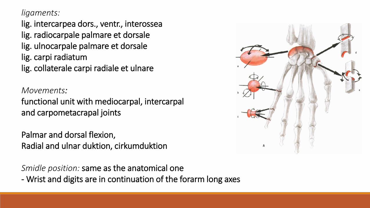
ligaments:lig. intercarpea dors., ventr., interossealig. radiocarpale palmare et dorsale lig. ulnocarpale palmare et dorsale lig. carpi radiatumlig. collaterale carpi radiale et ulnare
Movements:functional unit with mediocarpal, intercarpaland carpometacrapal joints
Palmar and dorsal flexion, Radial and ulnar duktion, cirkumduktion
Smidle position: same as the anatomical one- Wrist and digits are in continuation of the forarm long axes

Articulatio ossis pisiformis:
os pisiforme and os triquetrum - amphiarthrosislig. pisohamatumlig. pisometacarpeum(continuation of the tendon of m. flexor carpi ulnaris)
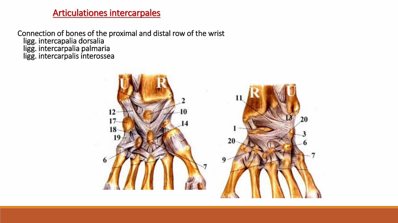
Articulationes intercarpales
Connection of bones of the proximal and distal row of the wrist• ligg. intercapalia dorsalia• ligg. intercarpalia palmaria• ligg. intercarpalis interossea

Articulationes carpometacarpales II.-V.
Type: compoundAS: base of the MC II - os trapezium, os trapezoideum, os capitatumbase of the MC III - os capitatumbase of the MC IV and V - os hamatumMC bases in betweenAC: short, toughligaments:ligg. carpometacarpalia dorsalialigg. carpometacarpalia palmarialigg. carpometacarpalia interossealigg. metacarpea palm., dors., interosseaMovements: amphiarthrosis

Articulatio carpometacarpalis pollicis
Type: saddleAH: basis ossis metacarpale IAF: os trapeziumAC: freeMovements: abduktion, adduktion
oposition, reposition
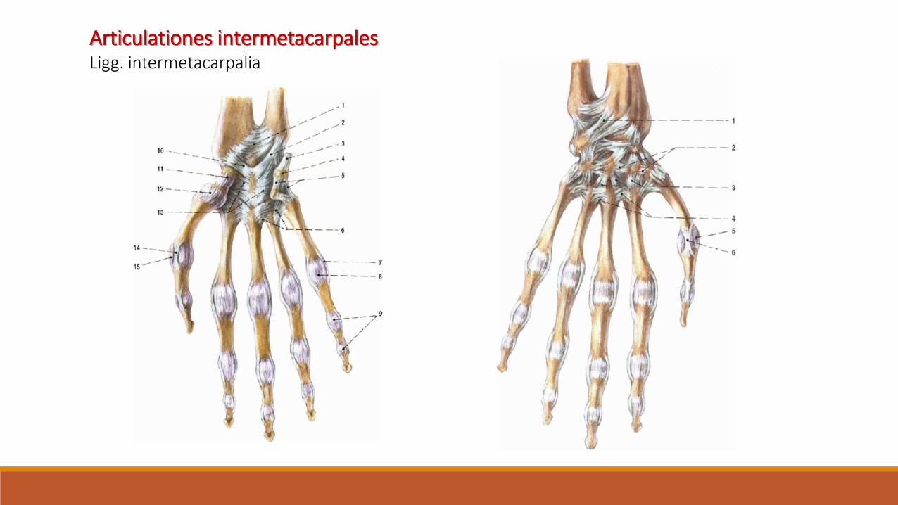
Articulationes intermetacarpalesLigg. intermetacarpalia

Art. metacarpophalangeales
Type: ball and socketAH: caput ossis metacarpalisAF: basis phalangisAC: freeLigaments: ligg. collateralialigg. palmaria - fibrocartilago palmarislig. metacarpale transversum profundumMovements: flexion a etensionabduktion and adduktion – in not flexed finger
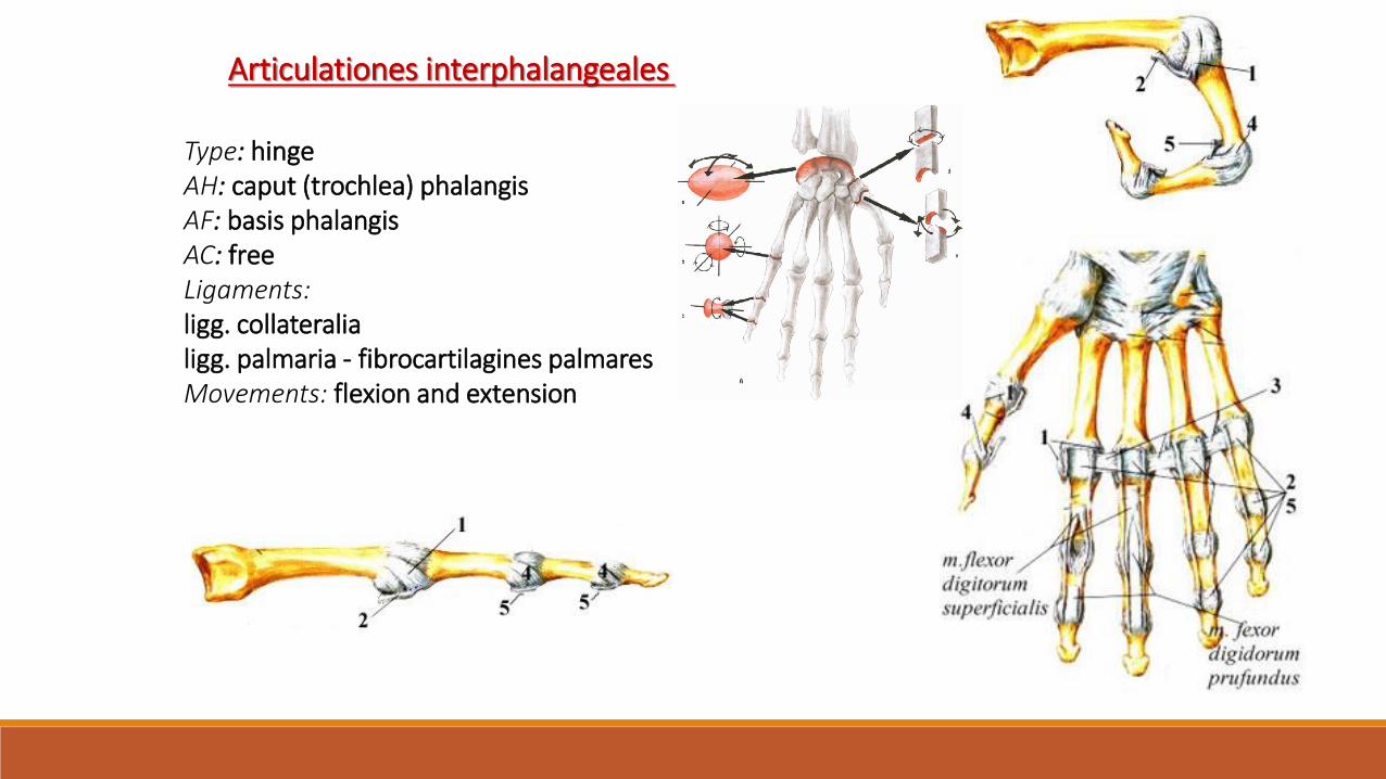
Type: hingeAH: caput (trochlea) phalangisAF: basis phalangisAC: freeLigaments:ligg. collateralialigg. palmaria - fibrocartilagines palmaresMovements: flexion and extension
Articulationes interphalangeales


Thank you for your attention!!
The pictures for the presentation were taken from:
Atlas der Anatomie des Menschen/Sobotta. Putz,R., und Pabst,R. 20. Auflage.
München:Urban & Schwarzenberg, 1993
Netter: Interactive Atlas of Human Anatomy.
Naňka, Elišková: Přehled anatomie. Galén, Praha 2009.
Čihák: Anatomie I, II, III.
Drake et al: Gray´s Anatomy for Students. 2010
Archiv of the lecturer, archiv of the Department of Anatomy, MU, Brno