congenital anomalies in north western indian population...
TRANSCRIPT

Congenital anomalies in North Western Indian population –
a fetal autopsy study
ORIGINAL ARTICLE Eur. J. Anat. 17 (3): 166-175 (2013)
Kanchan Kapoor1, Kashish Singh
1, Anshu Sharma
1, Balbir Singh
1,
Anju Huria2 and Suman Kochhar
3
1Department of Anatomy, GMCH, Chandigarh, India, 2Department of Obst. and Gynae, GMCH,
Chandigarh, India and 3Department of Radiodiagnosis, GMCH, Chandigarh, India
SUMMARY
Better knowledge of unexpected fetal loss is the promise for better parental counseling and for prevention of recurrences. Fetal autopsy can pro-vide a clue to ascertain cause of death in these cases. Variations in the incidence can be attrib-uted to multiple factors. The present study was carried out to help us to develop a database con-cerning number of autopsies, incidence and types of congenital malformations (CMF) in the North-Western Indian population. The period of study was from January 2010 to November 2011.
Autopsy was carried out on 150 fetuses follow-ing guidelines provided by a fetal autopsy proto-col. Prior to autopsy, prenatal investigations such as ultrasound and radiographs were procured; a brief maternal and family history was noted. Out of a total of 150 autopsies, 87(58%) were induced abortions and 63(42%) spontaneous abortions. In total, the incidence of CMF was 104(69%) of fetal autopsies. The types of CMF were classified as central nervous system defects (CNS) in 49 (33%), gastrointestinal tract (GIT) disorders in 48 (32%), musuculoskeletal (MS) disorders in 31 (21%), genito-urinary (GU) in 25 (17%), and ge-netic disorders in 12 (8%). Multiple anomalies were present in 40 (27%) fetuses. Anencephaly (meroencephaly) turned out to be the most preva-lent anomaly (29%). A few cases showed the occurrence of some uncommon syndromes. Ma-
jor CMFs manifested very early in intra-uterine life, and could lead to termination of pregnancy (spontaneous or induced) in the 2
nd trimester of
gestation. Hence the presence of any CMF at the time of birth cannot provide the total percentage of CMF occurring in a given population. The above findings are discussed in the light of the available literature.
Key words: Fetal – Autopsy – Congenital – Ma-jor anomalies – Malformations – Fetus – Abortion
INTRODUCTION
Congenital Malformation is a physical, meta-bolic or anatomic defect which is apparent before birth, at birth, or detected during the first year of life. CMF can present itself in a single organ, sys-tem, or may involve multiple organs of the body.
Early detection of major anomalies can be indica-tive of induced abortion to reduce the high mor-bidity of neonates due to congenital malforma-tions (Siebert and Kapur, 2001).
The evaluation of CMF can be done by ultra-sound, maternal serum analysis, etc., prenatally and by autopsy after the fetal death. Fetal autop-sies, if done, can provide a clue to ascertain cause of death in these cases. Etiological diagno-sis in unexplained fetal deaths is possible with detailed evaluation of fetus. Fetal autopsy is con-firmative in 28.6-89%, diagnostic in 10-38%; it provided additional information in 3.9-24% cases; it changed the predicted probability in 18% cases
166
Submitted: 17 October, 2012. Accepted: 18 April, 2013.
Corresponding author: Kanchan Kapoor. Department of
Anatomy, Government Medical College and Hospital (GMCH),
Sarai Building, Sector 32, Chandigarh, India.
Tel: +91-9646121528 . E-mail: [email protected]

Congenital anomalies in North Western Indian
167
(Mohan et al., 2004; Boyd et al., 2004; Sankar and Phadke, 2006; Sankar, 2011). In addition, the data pertaining to demography, socio-economic status, and maternal health is helpful to pinpoint the factors behind the occurrence of fetal loss.
Various studies (Choudhury et al., 1989; Swain et al., 1994; Malla, 2007; Saini and Kumar, 2009; Shamim et al., 2010) have described the inci-dence of congenital malformations in neonates (still and live born). The studies (Pahi et al., 1998) on the incidence of CMFs in fetuses are less. However there is a marked difference in the per-centage of different CMFs in newborn vs. fetuses. Fetuses afflicted with major malformations result in spontaneous or induced abortions. Therefore a large number of malformations do not get counted in studies done at birth.
This study is an attempt to find the incidences of congenital anomalies out of total fetal autopsies in a given time. Further the classification of anomalies may help to identify the root cause of a specific disorder. It will also help us to develop a database containing number of fetal autopsies, incidence and types of CMF, and other related information in North-Western Indian population.
MATERIALS AND METHODS
The present study included a profile of 150 fetal autopsies being routinely done in this institute from January 2010 till Nov, 2011. Prior to the au-topsy, the prenatal investigations like ultrasound, radiographs carried out by the respective depart-ments were procured. Consent for the autopsy was taken from the parents/ relatives on a Per-forma prepared in accordance with guidelines provided by the ethics committee which conforms to the provisions of the Declaration of Helsinki in 1995. A brief maternal and family history regard-ing any history of disease, intake of drugs, socio-economic status, etc., was noted down. The infor-mation regarding the prenatal genetic screening, if done, was noted and correlated with the pres-ence of malformation.
Each fetus was sent for radiographic examina-tion to note any skeletal abnormality. The various morphometric parameters including body weight, CRL (crown rump length), and CHL (crown heel length), head, thorax and abdominal circumfer-
ences, etc., were measured. The placenta and umbilical cord were examined for any gross ab-normality. The external appearance of the fetus was recorded.
The autopsies were performed as per guide-lines provided by fetal autopsy protocol (Siebert and Kapur, 2001; WHO, 2007; Agarwal et al., 2008). A longitudinal incision followed by a hori-zontal incision was given to retract the skin flap. The thoracic and abdominal cavities were opened and any deviation from the normal anatomy was photographed and noted. In relevant cases, tis-sues from various organs were sent for histologi-cal examination.
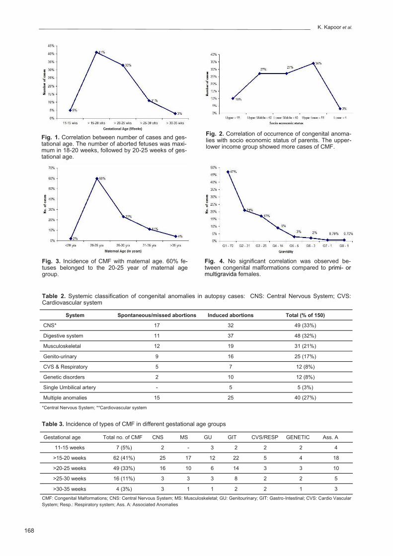
All the available information was collated to as-certain the cause of death. In this retrospective study, data from 150 fetal autopsies were com-puted to note the incidence of spontaneous abor-tions, intrauterine deaths, incidence and type of congenital malformations, and presence of syn-dromes. Graphs were plotted to correlate the number of cases with gestational age, socioeco-nomic status, maternal age, and gravidity of mother (Figs. 1-4).
RESULTS
Out of total 150 fetal autopsies, 87 (58%) speci-mens were from induced abortions, and 63 (42%) from spontaneous abortions and intrauterine deaths. In total, the incidence of congenital mal-formations was 104 (69%, Table1). Out of 150 fetuses, 74 were males and 76 female fetuses; out of which the incidence of CMF in males and females was 75% and 83% respectively.
The congenital malformations were grouped according to the system affected, using World Health Organization classification (2007) of con-genital malformation (Table 2). In 32.6% fetuses, defects in the central nervous system (CNS) were most prevalent. Gastrointestinal tract (GIT) anomalies were found in another 32%, followed by musculoskeletal (MS) anomalies in 21%, and genitourinary (GU) system in 17% specimens each; genetic disorders were the main cause in 8% of fetal deaths. Multiple anomalies manifested in 27% fetuses, giving rise to various syndromes.
When the gestational age was taken into ac-count, the presentation and subsequent fetal loss
Table 1. Profile of 150 fetal autopsy cases. CMF, congenital malformations
Total Normal Macerated CMF*
Spontaneous/missed abortions 63(42%) 19 (30%) 8 (13%) 36 (57%)
Induced abortions 87(58%) 11 (13%) 8 (9%) 68 (78%)
Total 150 30 (20%) 16 (11%) 104 (69%)
*congenital malformations
K. Kapoor et al.
168
Fig. 1. Correlation between number of cases and ges-tational age. The number of aborted fetuses was maxi-mum in 18-20 weeks, followed by 20-25 weeks of ges-tational age.
Fig. 2. Correlation of occurrence of congenital anoma-lies with socio economic status of parents. The upper-lower income group showed more cases of CMF.
Fig. 3. Incidence of CMF with maternal age. 60% fe-tuses belonged to the 20-25 year of maternal age group.
Fig. 4. No significant correlation was observed be-tween congenital malformations compared to primi- or multigravida females.
Table 3. Incidence of types of CMF in different gestational age groups
Gestational age Total no. of CMF CNS MS GU GIT CVS/RESP GENETIC Ass. A
11-15 weeks 7 (5%) 2 - 3 2 2 2 4
>15-20 weeks 62 (41%) 25 17 12 22 5 4 18
>20-25 weeks 49 (33%) 16 10 6 14 3 3 10
>25-30 weeks 16 (11%) 3 3 3 8 2 2 5
>30-35 weeks 4 (3%) 3 1 1 2 2 1 3
Table 2. Systemic classification of congenital anomalies in autopsy cases: CNS: Central Nervous System; CVS: Cardiovascular system
System Spontaneous/missed abortions Induced abortions Total (% of 150)
CNS* 17 32 49 (33%)
Digestive system 11 37 48 (32%)
Musculoskeletal 12 19 31 (21%)
Genito-urinary 9 16 25 (17%)
CVS & Respiratory 5 7 12 (8%)
Genetic disorders 2 10 12 (8%)
Single Umbilical artery - 5 5 (3%)
Multiple anomalies 15 25 40 (27%)
*Central Nervous System; **Cardiovascular system
CMF: Congenital Malformations; CNS: Central Nervous System; MS: Musculoskeletal; GU: Genitourinary; GIT: Gastro-Intestinal; CVS: Cardio Vascular
System; Resp.: Respiratory system; Ass. A: Associated Anomalies

Congenital anomalies in North Western Indian
169
Fig. 5. A 24+ week female fetus with meningocele. Fig. 6. A 14 week female fetus with complete rachishi-sis.
Fig. 7. Bilateral polycystic kidneys in a 28 week male fetus.
Fig. 8. Enlarged cystic urinary bladder with dilated uret-ers.
Table 4. Classification of defects in the Central Nerv-ous system: VC, vertebral column
Anencephaly 14 (29%)
Rachischisis (Spina bifida) 11 (22%)
Meningocoele 7 (14%)
Meningomyelocoele 10 (20%)
Encephalocoele 9 (18%)
Hydrocephalous 10 (20%)
Abnormal flexion of VC* 9 (18%)
Total 49 (33%)
Table 5. Classification of defects in the Genitourinary system
Agenesis 8 (32%)
Congenital polycystic kidneys 6 (24%)
Horse-shoe kidneys 3 (12%)
Undifferentiated gonads 2 (8%)
Hypospadias 1 (4%)
Oligohydramnios 4 (16%)
Dilated pelvis/ureter 2 (8%)
Absent/Cystic urinary bladder 1 (4%)
Total 25 (17%) *vertebral column

K. Kapoor et al.
170
(either by spontaneous or induced abortions) oc-curred maximum (41%) in >15-20 wks, followed by 33% in >20-25 wks. The fetal loss was 11% in >25-30 wks, and only 5% in 11-15 wks (Table 3). Out of this total loss, CNS defects were the main cause in all the age groups followed by almost equal proportion of musculoskeletal, genito-urinary and GIT disorders (Table 3; Fig. 1).
Anencephaly (Mero-encephaly) turned out to be the most prevalent anomaly in CNS (28.5%); it was often associated with Meningocele (Fig. 5) or Meningomyelocele. Incidence of Rachischisis (spina bifida, Fig. 6) was 22.4% in cases with CNS anomalies. Hydrocephalus was observed in 20.4% cases and in 18% fetuses there was an abnormal flexion of vertebral column (kyphosis, scoliosis, etc.) (Table 4).
Amongst the defects in genito-urinary system, a congenital polycystic kidney (Fig. 7) was found in 24% cases; Agenesis of kidneys/ suprarenal/ go-
nad was observed in 32% cases. The kidney was horse-shoe shaped in 3 specimens; there was dilated pelvis/ureter in 2 specimens, and in other 3 cases there was abnormal opening of ureter. The urinary bladder had a large cyst in one fetus (Fig. 8). Two specimens presented with undiffer-entiated gonads (Table 5).
Exomphalos (omphalocele) was found in 12 fetuses (25%) in the GIT defects (Fig. 9). Ab-sence of a digestive organ was present in 7 specimens, whereas there was non/mal- rotation of gut in 7 cases. Other anomalies of GIT in-cluded hepato/splenomegaly, etc. (Table 6).
Musculoskeletal anomalies included diaphrag-matic hernia in 16%, joint defects in 29%, club foot in 16%, and cleft lip/palate (Fig. 10) in an-other 16% fetuses. Facial features were dysmor-phic in 4 fetuses (Table 7).
Out of total 87 induced abortions, 12 (8%) specimens were triple-test positive when a ge-
Fig. 9. A case of omphalocele with abdominal visceral herniation enclosed in a transparent amniotic sac.
Fig. 10. A 28-week female fetus presenting complete cleft lip associated with complete cleft palate.
Table 6. Classification of defects in digestive system
Digestive System Total
Omphalocoele 12 (25%)
Non/malformation of gut 7 (14.6%)
Absence (liver/spleen/colon) 7 (14.6%)
Hepato/Splenomegaly 4 (8.3%)
Ascites 16 (33%)
Total 48 (32%)
Diaphragmatic hernia 5 (16%)
Club Foot 5(16%)
Joint defects 9 (29%)
Cleft lip / palate 5 (16%)
Kyphoscoliosis 6 (19%)
Dysmorphic face 4 (13%)
Total 31 (21%)
Table 7. Classification of defects in the musculoskele-tal system

Congenital anomalies in North Western Indian
171
netic analysis was done prenatally. The morpho-logical features of 3 cases resembled those of Down’s syndrome including the presence of sim-ian palmar crease, etc., although these features could not be conclusive of presence of Down’s syndrome. Three fetuses had features similar to Arnold Chiari syndrome, including the small pos-terior cranial fossa, rocker bottom feet, etc. Apart from these, we could find one case each of split notochord syndrome (SNS) (Fig. 11), hand foot genital syndrome (HFGS), Edward’s syndrome, aplasia cutis congenita, and cloacal dysgenesis sequence (Fig. 12). Our limitation with these cases was the lack of postnatal genetic analysis, which could have corroborated the present au-topsy findings.
An attempt was made to correlate socioeco-nomic status and the occurrence of CMFs (Fig. 2). The socioeconomic status was calculated ac-cording to Kuppuswamy scale (Kumar et al., 2007), which is based on three variables – educa-tion, occupation and income. On this basis, popu-lation was divided into upper, upper middle, mid-dle, upper lower, and lower classes. The present study included 54% cases belonging to middle class; 37% from lower class and only 10% from upper class (Fig. 2).
Sexual preponderance was not significant sta-tistically, as 83% female fetuses had CMF as compared to 75% male fetuses. The incidence of CMF was correlated with maternal age (Fig. 3) and status of gravida (Fig. 4). It was observed that 60% of the fetuses belonged to the mothers who were between 20-25 years of age. Also, al-most 50% of the mothers were primigravida, fol-lowed by 2
nd gravida in 21%. Multi-gravida moth-
ers (G4-G8) had only 15% of the congenitally malformed fetuses.
At the time of taking consent, any history of in-take of drugs was enquired. Most of the parents/parent replied in negative. It was also admitted that the mother was not taking any kind of nutri-tional supplements usually given during preg-nancy.
DISCUSSION
CMFs are emerging as important component in the perinatal mortality and morbidity with consid-erable repercussion on the families’ affected. Early diagnosis of life threatening CMF can pave the way for surgical correction or palliation of these infants.
Studies are available (Mohan et al., 2004; Boyd et al., 2004; Sankar and Phadke, 2006) where the presence of CMFs, prenatally diagnosed, are confirmed by fetal autopsy. Autopsies are per-formed to identify a cause of death, to produce epidemiologic data to further the understanding of the patient’s course and disease. Detailed au-topsy, which includes radiological and histological examination, can be a useful tool in the diagno-ses. It can also verify the ultrasound findings in the prenatal period, and can corroborate their results. In a teaching hospital, this kind of feed-back is essential to maintain good standards.
The incidence of congenital anomalies in the total fetal autopsies was found to be 69% in the present observations. In another study (Puri et al., 2009) done in North India, the incidence of fetal anomalies was 63%, which is similar to the present study. Mohan et al. (2004) described the incidence as 38.7% out of 62 perinatal autopsies
Fig. 11. A rare case of split notochord syndrome in a 24 week female fetus where intestinal loops were seen herniating from the posterior abdominal wall.
Fig. 12. Imperforate anus with associated gut anoma-lies in a 16 week male fetus.

K. Kapoor et al.
172
from the same region, which is lesser than the two abovementioned studies.
Most of the available literature pertains to con-genital anomalies found at the time of birth (still or live) (Malla, 2007; Swain et al., 1994; Shamim et al., 2010; Choudhury et al., 1989). Table 6 shows the systemic incidence of various CMFs in these studies. Many authors (Grover, 2000; Mohan et al., 2004; Malla, 2007; Saini and Kumar, 2009; Sankar, 2011) had stated the de-fects in central nervous system as the highest anomaly; the findings are similar to those of the present study. Some studies (Chinara and Singh, 1982; Datta and Chaturvedi, 2000; Boyd et al., 2004; Malla, 2007; Karbasi et al., 2009) men-tioned the musculoskeletal as the most affected system, whereas a few others (Shamim et al., 2010; Dutta et al., 2010) reported GIT anomalies to be the most frequent.
In the present study, the fetal autopsies showed an incidence of NTDs higher than other anoma-lies. This observation is in agreement with the other two fetal studies from the same region (Mohan et al., 2004; Sankar and Phadke, 2006). The apparent cause could be that most of the fetuses afflicted with neural tube disorders were aborted very early in the gestational period. How-ever, more incidences of NTD’s could be due to lower socio economic status and lack of social awareness. In US, Williams et al. (2005) found the prevalence of spina bifida and anencephaly highest among Hispanic births, followed by the
non-Hispanic whites, and the lowest among non-Hispanic black births. To reduce these NTD’S among these racial groups, folic acid fortification programme was launched in US.
Favorito et al. (2004) noted the incidence of UG (urogenital) anomalies in human male fetuses in the age group of 10-36 wks of IU life. They found the incidence of total UG anomalies as 4.2%, out of which renal agenesis was observed in 1.2%. They stated that UG anomalies in human male fetuses are rare. Similar views were expressed by Grover (2000), as they found the incidence of UG anomalies in only 3.8% cases. Some other au-thors (Malla, 2007; Sharma et al., 2009; Saini and Kumar, 2009; Shamim et al., 2010; Dutta et al., 2010)
noted the incidence as 25.8%, 24.8%,
24.5%, 17.3%, and 26.2%, respectively.
Sanghvi et al. (1998) diagnosed congenital re-nal malformations in 0.2% of fetuses by antenatal ultrasound, out of which oligohydramnios was noted in 31%. The other anomalies found were dilated urinary system, polycystic kidneys, and renal agenesis. The incidence of UG anomalies in the present study was 16.6%. Renal agenesis was present in 32%, polycystic kidneys in 24%. Oligohydramnios was associated in 16% renal anomalies. The genital defects were rare; a few cases with defined syndromes had hypospadias or undifferentiated gonads. In comparison to the above mentioned authors, the absence of kidney/kidneys in the present series is on a higher side. This could be explained on the basis that many of
Table 8. Comparison of incidence of CMF with the available literature (%)
CNS: Central Nervous System; GIT: Gastrointestinal System; GU: Genitourinary; MS: Musculoskeletal; CVS: Cardiovascular System
S.no Authors Autopsy CNS GIT GU MS Genetic CVS Multiple
anomalies
Sex
preponderance
1. Sankar et al. (2006)
Fetal 74.2 - 17.2 - - 4.9 8 -
2. Malla (2007) At birth 40 17.3 17.3 18.6 1.3 - - More in females
3. Swain et al. At birth 39.5 10.4 10.4 14.5 4.2 8.3 18.8 No sex diff.
4. Shamim et al. (2010)
- 14 44 24.5 1.7 - 7 - -
5. Grover (2000) At birth 40 - 3.8 23.8 - - - -
6. Chinara & Singh (1982)
At birth 13.5 13.5 10.8 29.7 13.5 2.7 10.8 Slight male
preponderance.
7. Karabasi et al. (2009)
At birth 16.9 11.0 13.23 29.4 4.09 8.8 - No sex diff.
8. Dutta et al. (2010)
Live births
5.6 26 25.8 - - - - More in males
9. Al-Jama (2001)
Live births
49 6 14 11.2 - - - -
10. Kim et al. At birth 8.4 11 11 10 - - - -
11. Present Study Fetal 33 32 17 21 8 8 27 No sex diff.

Congenital anomalies in North Western Indian
173
these studies were done by antenatal ultrasound examination, where the absence of an organ could not be verified as compared to the confirmed autopsy findings.
GIT anomalies constituted 32% in the present ob-servations, and mostly these were represented by exomphalos (omphalocoele), gastroshisis, hepato/splenomegaly, and imperforate anus. Some of the defects were associated with paraumbilical defect of the anterior abdominal wall; some gut anomalies were associated with neural tube defects, and a few cases were part of a syndrome. 54% of GIT defects also had ascites. The incidence of GIT anomalies as reported by various authors (Al-Jama, 2001; Malla, 2007; Saini and Kumar, 2009; Kim et al., 2000) was 6%, 10.4%, 17%, and 1.17/1000. The present data report a higher incidence of GIT anomalies. The agenesis of an organ (liver, spleen, and pancreas) was not very common, as corroborated by other studies as well.
Musculoskeletal deformities included the diaphrag-matic hernia, club foot, cleft lip/palate and dysmor-phic facial features. Diaphragmatic hernias usually occur on left side (16% in the present study), and the heart and lungs are compressed towards the right due to upward displacement of abdominal or-gans. The incidence was reported as 1 in 3000-5000 births (Gilbert and Debich, 2004). Various au-thors (Kim et al., 2000; Al-Jama, 2001; Malla 2007; Saini and Kumar, 2009) have reported the incidence of musculoskeletal anomalies as 10%, 11.2% 15%, and 19%, which is in accordance with present ob-servations.
Racial variations, geographical location and socio-economic status all play an important role in deter-mining the incidence of congenital anomalies in a given population, e.g. most of the studies from Mid-dle East countries refer consanguinity as one of the leading cause of CMF. Various researchers (Kulkarni and Kurian, 1990; Tayebi et al., 2010) have established the fact that consanguinity has a deleterious effect on fetal growth, and it increases the risk of CMF and fetal Loss. Some authors have linked low socio-economic status with consanguine-ous marriages, and therefore increase in the per-centage of CMF is the result of both these factors. The present study did not find even a single case of consanguineous marriage in north-west India. Therefore, the impact of consanguinity on the occur-rence of CMF could not be debated.
According to GAPPS (Global alliance to prevent prematurity) 85% preterm births occur in Africa and Asia (Erickson, 1976). This fact can explain the rea-son of higher incidences of anomalies in the present study as compared to the studies done in other con-tinents.
Fetal factors
In the present study the most vulnerable period of gestation was >15-20 wks followed by >20-25 wks. Although the involvement of many systems might have occurred much earlier during the first trimester, USG examination had confirmed the malformations in the second trimester which was followed by In-duced abortions. None of the available literature had considered the prevalence of CMF according to ges-tational age. This observation has been the basis of our hypothesis that calculations of incidence of CMF at the time of birth do not reflect the accurate data.
Few studies (Chinara and Singh, 1982; Malla, 2007) had observed higher incidence of anomalies in male fetuses as compared to females. Raghura-maiah et al (2010)
stated that NTDs are thrice more
common in females than in males. Contrary to that, no gender preponderance could be established by many authors (Swain et al., 1994; Karbasi et al., 2009; Taksande et al., 2010). Similarly, in the pre-sent study the number of male and female fe-tuses was almost similar, and sexual preponder-ance was statistically not significant.
Maternal factors
The maternal factors considered in the present study were the age of mother and the number of pregnancy. The incidences of CMF were higher in primigravida; concurrently the age of the mother was also in the age group of 20 to 25 years. A few authors (Taksande et al., 2010) have main-tained the higher incidence of CMF in gravida 4 and maternal age also higher than 35 years. Since the present study did not include many cases above 35 yrs. of maternal age, the data is mostly inconclusive. However 47% cases were from primigravida as compared to 9% cases from gravida 4. This observation is consistent with ear-lier studies. However, various studies (Chinara and Singh, 1982; Datta and Chaturvedi, 2000) were of the opinion that frequency of malformed babies was not influenced either by maternal age or parity of the mother.
According to Chinara and Singh (1982), there was a slight increase in the incidence of mal-formed babies in higher income groups. However, their division into different socio-economic groups seems to be arbitrary. Socioeconomic inequalities increased the risk of cardiac defects, GIT disor-ders and multiple anomalies – i.e., anomalies of non-chromosomal origin (Vrijheid et al., 2000). However NTD and oral clefts are not affected by socioeconomic deprivation (Sanghvi et al., 1998). The present study found the incidence to be 54% in middle class, and 37% in lower socio economic class. In India the socio-economic status of the families can cause severe nutritional deficiencies leading to gross congenital anomalies. That is why CNS malformations, particularly the anen-cephaly (mero-encephaly) and rachischisis, are
K. Kapoor et al.
174
the most prevalent anomalies in the present study.
Conclusion
Fetal autopsies performed in cases of induced/spontaneous abortions can provide a clue, and can therefore serve as a purposeful tool in coun-seling the family for future planning.
The incidence of CMF was 69% out of 150 cases of fetal autopsy observed in North-West Indian population in a period of almost two years. The most prevalent CMF was NTD’s (33%) in the form of anencephaly (mero-encephaly) and ra-chischisis. They were followed by anomalies in the digestive system in 32%, the genito-urinary in 25%, the musculoskeletal in 21%, and genetic in 8%. Twenty seven percent of fetuses had multi-ple CMF leading to various syndromes. There was no significant sexual preponderance for the occurrence of CMF. Almost 50% of CMF oc-curred in primigravida and mothers of less than 25 years of age.
It will be erroneous to calculate the incidence of CMF in stillborn /live born fetuses at the time of delivery, as most of the fetuses with major CMF get aborted in the 17-28
th weeks of IU life.
It is well known that surveillance and monitoring of congenital conditions is important for identify-ing patterns of CMF. The emphasis of the pre-sent study would be to sensitize the antenatal health care workers to provide an effective ante-natal screening even in the tertiary or rural healthcare hospitals, and to recognize social fac-tors for the prevention of birth defects.
REFERENCES
Agarwal SS, Kumar L, Chavli K (2008) Autopsy on bodies of fetuses / infants. In: Legal Medicine Man-ual. Jaypee. New Delhi. P.192–202.
Al-Jama F (2001) Congenital malformations in new-borns in a teaching hospital in eastern Saudi Arabia. J Obstet Gynaecol, 21: 595-598.
Boyd PA, Tondi F, Hicks NR, Chamberlain PF (2004) Autopsy after termination of pregnancy for fetal anomaly: retrospective cohort study. BMJ, 328: 137-140.
Chinara PK, Singh S (1982) East West differentials in congenital malformations in India. Indian J Pediatr, 49: 325-329.
Choudhury AR, Mukherjee M, Sharma A, Talukder G, Ghosh PK (1989) Study of 1,26,266 consecutive births for major congenital defects. Indian J Pediatr, 56: 493-499.
Datta V, Chaturvedi P (2000) Congenital malforma-
tions in rural Mahrashtra. Indian Pediatr, 37: 998-1002.
Dutta HK, Bhattacharyya NC, Sarma JN, Kusre G (2010) Congenital malformations in Assam. JIAPS, 15: 53-55.
Erickson JD (1976) Racial variations in the incidence of congenital malformations. Ann Hum Genet, 39: 315-320.
Favorito LA, Cardinot TM, Morais ARM, Sampaio FJ (2004) Urogenital anomalies in human male fetuses. Early Hum Dev, 79: 41-47.
Gilbert BE, Debich SD (2004) Embryo and Fetal Pa-thology. Cambridge University Press, New York, pp 388-400.
Grover N (2000) Congenital malformations in Shimla. Indian J Pediatr, 67: 249-251.
Karbasi SA, Golestan M, Fallah R, Mirnasseri F, Barkhordari K, Bafghee MS (2009) Prevalence of congenital malformations. Acta Medica Iranica, 47: 149-153.
Kim HJ, Kim YK, Koh DK, Kim JH, Choi BW et al. (2000) A prospective epidemiological study on birth defects: A community based pilot study. J Korean Pediatr Soc, 43: 738-745.
Kulkarni ML, Kurian M (1990) Consanguinity and its effect on fetal growth and development: A South In-dian study. J Med Genet, 27: 348-352.
Kumar N, Shekhar C, Kumar P, Kundu AS (2007) Kup-puswamy socioeconomic status scale – updating for 2007. Indian J Pediatr, 74: 1131-1132.
Malla BK (2007) One year review study of congenital anatomical malformation at birth in Maternity Hospital (Prasutigriha), Thapathali, Kathmandu. Kathmandu Univ Med J, 5: 557-560.
MMWR Morb Mortal Wkly Rep (2009) Racial / ethnic differences in the birth prevalence of spina bifida – United States, 1995-2005. 57(53), 1409-1413.
Mohan H, Bhardwaj S, Bal A (2004) Congenital vis-ceral malformations - role of perinatal autopsy in di-agnosis. Fetal Diagn Ther, 19: 131-133.
Pahi J, Phadke SR, Halder A, Gupta A, Pandey R, Agarwal SS (1998) Does autopsy of antenatally diag-nosed malformed foetuses aid genetic counselling? Natl Med J India, 11: 169-170.
Puri RD, Thakur S, Verma IC (2009) Utility of fetal evaluation in stillbirth – application for counseling and decreasing morbidity. 4th International Conference on Birth Defects and Disabilities in the Developing World, New Delhi.
Raghuramaiah G, Sinivasa R, Devi S, Reddy R, Reddy V (2010) Neural tube defects - an incidence ratio between male and female dead foetuses. JASI, 59: 58.
Saini SS, Kumar P (2009) Eight years’ analysis of trend of congenital malformations in a referral hospital in Northern India. 4th International Conference on Birth Defects and Disabilities in the Developing World, New Delhi.

Congenital anomalies in North Western Indian
175
Sanghvi KP, Merchant RH, Gondhalekar A, Lulla CP, Mehta AA, Mehta KP (1998) Antenatal diagnosis of congenital renal malformations using ultrasound. J Tropical Pediatr, 44: 235-240.
Sankar VH, Phadke SR (2006) Clinical utility of fetal autopsy and comparison with prenatal ultrasound findings. J Perinatol, 26: 224-229.
Sankar VH (2011) Fetal autopsy in clinical practice. Genetics Clinics, 4: 5-8.
Shamim S, Chohan N, Qmar S (2010) Pattern of con-genital malformations and their neonatal outcome. J Surg Pakistan (Internat), 15: 34-37.
Sharma A, Chawla D, Jain S, Rani S, Sehgal A, Goel P, Huria A (2009) Congenital malformations amongst live births in a teaching hospital - Nine year experi-ence. 4th International Conference on Birth Defects and Disabilities in the Developing World, New Delhi.
Siebert JR, Kapur RP (2001) Diagnosing congenital malformations in the fetus. Society for Pediatric Pa-
thology Workshops, Seattle, Washington, 3-34.
Swain S, Agrawal A, Bhatia BD (1994) Congenital mal-formations at birth. Indian Pediatr, 31: 1187-1191.
Taksande A, Vilhekar K, Chaturvedi P, Jain M (2010) Congenital malformations at birth in central India: A rural medical college hospital based data. Indian J Human Genet, 16: 159-163.
Tayebi N, Yazdani K, Naghshin N (2010) The preva-lence of congenital malformations and its correlation with consanguineous marriages. OMJ, 25: 37-40.
Vrijheid M, Dolk H, Stone D, Abramsky L, Alberman E, Scott JES (2000) Socio economic inequalities in risk of congenital anomaly. Arch Dis Child, 82: 349-352.
Williams LJ, Rasmussen SA, Flores A, Kirby RS, Ed-monds LD (2005) Decline in the prevalence of spina bifida and anencephaly by race / ethnicity: 1995-2002. Pediatrics, 116: 580-586.
World Health Organization (WHO) (2007) International classification of diseases, Tenth division. Tabular List and Alphabetic Index. Geneva.