complimentary contributor copy · human anatomy and physiology. posture. types, exercises and...
TRANSCRIPT

Complimentary Contributor Copy

Complimentary Contributor Copy

HUMAN ANATOMY AND PHYSIOLOGY
POSTURE
TYPES, EXERCISES AND HEALTH EFFECTS
No part of this digital document may be reproduced, stored in a retrieval system or transmitted in any form orby any means. The publisher has taken reasonable care in the preparation of this digital document, but makes noexpressed or implied warranty of any kind and assumes no responsibility for any errors or omissions. Noliability is assumed for incidental or consequential damages in connection with or arising out of informationcontained herein. This digital document is sold with the clear understanding that the publisher is not engaged inrendering legal, medical or any other professional services.
Complimentary Contributor Copy

HUMAN ANATOMY AND PHYSIOLOGY
Additional books in this series can be found on Nova‘s website
under the Series tab.
Additional e-books in this series can be found on Nova‘s website
under the e-book tab.
Complimentary Contributor Copy

HUMAN ANATOMY AND PHYSIOLOGY
POSTURE
TYPES, EXERCISES AND HEALTH EFFECTS
SARAH A. CURRAN
EDITOR
New York
Complimentary Contributor Copy
Copyright © 2014 by Nova Science Publishers, Inc.
All rights reserved. No part of this book may be reproduced, stored in a retrieval system or
transmitted in any form or by any means: electronic, electrostatic, magnetic, tape, mechanical
photocopying, recording or otherwise without the written permission of the Publisher.
For permission to use material from this book please contact us:
Telephone 631-231-7269; Fax 631-231-8175
Web Site: http://www.novapublishers.com
NOTICE TO THE READER
The Publisher has taken reasonable care in the preparation of this book, but makes no expressed or
implied warranty of any kind and assumes no responsibility for any errors or omissions. No
liability is assumed for incidental or consequential damages in connection with or arising out of
information contained in this book. The Publisher shall not be liable for any special,
consequential, or exemplary damages resulting, in whole or in part, from the readers‘ use of, or
reliance upon, this material. Any parts of this book based on government reports are so indicated
and copyright is claimed for those parts to the extent applicable to compilations of such works.
Independent verification should be sought for any data, advice or recommendations contained in
this book. In addition, no responsibility is assumed by the publisher for any injury and/or damage
to persons or property arising from any methods, products, instructions, ideas or otherwise
contained in this publication.
This publication is designed to provide accurate and authoritative information with regard to the
subject matter covered herein. It is sold with the clear understanding that the Publisher is not
engaged in rendering legal or any other professional services. If legal or any other expert
assistance is required, the services of a competent person should be sought. FROM A
DECLARATION OF PARTICIPANTS JOINTLY ADOPTED BY A COMMITTEE OF THE
AMERICAN BAR ASSOCIATION AND A COMMITTEE OF PUBLISHERS.
Additional color graphics may be available in the e-book version of this book.
Library of Congress Cataloging-in-Publication Data
Library of Congress Control Number: 2014931436
Published by Nova Science Publishers, Inc. † New York
ISBN: 978-1-63117-254-0 (eBook)
Complimentary Contributor Copy

Contents
Preface vii
Chapter 1 Old Problems and New Perspectives for Postural Analysis 1 José Luís Pimentel do Rosário, PT, PhD
Chapter 2 The Clinical Usefulness of Head Posture Assessment for Patients
with Neck Pain 15 Anabela G. Silva, PhD, MSc, BSc, David Punt, PhD
and Mark I. Johnson, PhD, BSc, PGCertHE
Chapter 3 Head and Neck Posture and Upper Spine Morphology in Relation
to the Craniofacial Profile and Orofacial Function 43 Liselotte Sonnesen, DDS, PhD
Chapter 4 Emotion: The Missing Link in Posture 55 José Luís Pimentel do Rosário, PT, PhD
Chapter 5 The Influence of Fear, Happiness and Concern on Posture 71 José Luís Pimentel do Rosário, PT, PhD
Chapter 6 Influence of Hamstring Extensibility on Spinal and Pelvic Postures
in Highly Trained Athletes 81 Pedro A. López-Miñarro, PhD, José M. Muyor, PhD, MSc,
Fernando Alacid, PhD, MSc and Raquel Vaquero, MSc
Chapter 7 Spinal Posture in Cycling 95 José M. Muyor, PhD, Pedro A. López-Miñarro, PhD,
Fernando Alacid, PhD, MSc and Raquel Vaquero-Cristóbal, MSc
Chapter 8 Effects of Physical and Sporting Activities on Postural Stability
in Children 105 Sonia Sahli, PhD, Rym Baccouch, MSc and Haithem Rebai, PhD
Chapter 9 The Role of Unstable Shoe Constructions for the Improvement
of Postural Control 125 Andreia S. P. Sousa, PhD, and João Manuel R. S. Tavares, PhD
Complimentary Contributor Copy

Contents vi
Chapter 10 Efficacy of Modified Yoga Positions and Postural Chains Therapy
for Spinal Pain Treatment 137 José Luís Pimentel do Rosário, PT, PhD
Chapter 11 Yoga Postures and Colon Cleanse 159 Vijaypal Arya, MD
Chapter 12 The Behavior Characteristics and Postural Angles in Teenagers
Who Wear High-Heeled Shoes and How Pilates Can Be Used
for Postural Control 171 Patricia Angélica de Oliveira Pezzan, MSc
and Daniel Marcondes de Freitas Lopes, PT
Index 193
Complimentary Contributor Copy

Preface
―Movement is a medicine for creating change in a person‘s physical,
emotional, and mental states‖.
Carol Welch
One of our most precious possessions for each of us is our body. It performs a series of
automatic, complex and important mechanisms such as breathing, digestion, and ambulating
from one point to the next with little conscious effort. Yet, as a consequence of new
experiences or when circumstances change, such as when an individual becomes ill or injured
the body comes under volitional control. Whilst unique to humans, bipedal stance creates an
inherently unstable platform which can challenge balance and the neuromuscular control
system. Integrated within this is posture, which is described as the position of the human body
and its orientation in space. A more specific definition is provided by Britnell et al. (1) who
suggest that posture is related to musculoskeletal alignment and balance that acts to protect
the human body from injury, progressive deformity and create efficiency. Nevertheless,
movement and posture of the human body changes with the routines of daily life and can be
influenced by a variety of factors that include fatigue, general health, state of mind,
participation in physical activity, and musculoskeletal imbalance and malignment.
In the last 25 years, the quest for knowledge and understanding on the role of posture has
been constant and continues to evolve. In particular, in recent years there has been a growing
recognition of psychosomatic conditions on the influence posture. This however adds further
to the complexity of understanding the variability of physical impairments and compensation
mechanisms each individual may present with. In essence, the clinician must be reflective and
perceptive, and acknowledge that there is no ‗one fix‘ treatment strategy of managing postural
related conditions and pathologies.
This book presents a collection of work on the various aspects of posture, including its
evolutionary trend of knowledge and its rehabilitation. Each invited international author has
contributed significantly in their field and are passionate for continued learning and
understanding that integrates theory into clinical practice. The book provides a timely
reminder to recognise the importance of a healthy and active lifestyle that is synonymous with
modern day living, yet in turn acknowledge the consequences of activity (potential for injury)
and value of exercise on improving posture. The book also provides an opportunity to
appraise the role of yoga and Pilates within rehabilitation programs. This inclusion (along
with other and traditional forms of rehabilitation protocols) echoes the increasing trend of
Complimentary Contributor Copy

Sarah A. Curran viii
individuals participating in yoga and Pilates across the world. To reiterate this point, in 2012,
over 20 million Americans practiced yoga, which represents an increase of 29% from 2008.
In contrast, Pilates, which was introduced by Joseph Pilates in the late 1960s and was often
mistaken for the training of pilots has received an incremental stream of followers. However,
it was not until the arrival of the noughties till it gained a rapid growth in popularity.
The book presents twelve chapters and opens with a chapter by Rosário which provides a
contemporary review based on various optical systems for postural assessment. In the first of
his four chapter contributions, Rosário explores the role of three dimensional and two
dimensional analysis and note the issues related to cost, time, space availability, as well as
marker placement and other sources of errors. He also discusses the role of smart phones and
contribution of three dimensional sensors – made popular by the introduction of Microsoft®
Kinect in association with the X-box 360 games console. Rosário also discusses the
introduction of a new and more accurate type of sensor referred to as ‗time of flight‘ for the
new generation of Kinect which may appeal to postural assessment within a clinical setting.
The clinical usefulness of head posture assessment for patients presenting with neck pain is
presented by Silva et al. in chapter two. Using an accumulation of recent research and their
own findings, the authors set out to determine operational definitions for ideal, normal and
abnormal head posture. The methods used to assess head posture are then explored and the
relationships between head posture and neck pain are comprehensively reviewed. In
particular, the authors note the value of forward head posture assessment for patients with
neck pain, but not for head extension/flexion, side-flexion and rotation. This observation is
based on the lack of evidence used to explore these postural components and the authors
acknowledge the need for further research to enhance the understanding, assessment and
management regimens of head posture and neck pain. The third chapter is written by
Sonnesen who explores the relationship between head and neck posture and upper spine
morphology to the craniofacial profile and orofacial function. The chapter which provides an
appreciation of the standard procedure used to record head and neck posture and upper spine
morphology provides an evidenced-based approach of previous literature. The author points
out that head and neck posture as well as upper spine morphology can influence the
developmental of the craniofacial profile and orofacial function for temporomandibular
disorders as well as obstruction to the upper air ways. These factors are of clinical value for a
range of health professionals which include the dentist, orthodontist, general physician and
physiotherapist.
In an appreciation of the vast array of biomechanical, environmental, neurophysiology,
pathological, physiological and psychological conditions that can influence posture, Rosário
presents two chapters, the first of which is based on acknowledging the role of emotion and
its link with posture. He states the need to approach the management of postural related
conditions from a multi-dimensional purpose, which should include the role of emotions. In
particular, Rosário points out the link between pain and its influence on emotion and posture.
He also shows how the role of body image and the effects of emotional tension including
depression, anger and sadness can have on posture. Rosário‘s third chapter, and chapter five
of the book, builds on from the previous chapter and reports on the findings of a study which
investigated the relationship between body posture on subjective fear, concern and happiness
in adult women. Using a digital camera, each volunteer was photographed standing from a
lateral (right angle) and anterior view. A series of angles were obtained to determine postural
assessment (alignment) from each view. Additional data was obtained from a visual analogue
Complimentary Contributor Copy

Preface ix
scale to document the degree of subjective depression. Rosário shows how current fear, usual
concern, and current and usual happiness is related to various postural alterations. Whilst it is
acknowledged that further research is required, Rosario notes that the findings of this data
may be useful clinically in the identification of a patient‘s emotional status from a non-verbal
perspective.
The next three chapters are based on a range of sporting activities, the first of which is
presented by Minarro et al. who explore the influence of hamstring extensibility on spinal and
pelvic posture in highly trained athletes. From a sample of 260 paddlers, three groups were
created based on the passive straight leg angle raise (group 1 <73˚; group 2 >73 and <84˚;
group 3 >84˚). The Spinal Mouse was used to record sagittal plane spinal curvatures and
pelvic inclination during various test conditions that included standing, maximal trunk flexion
and knee extended (sit-and-reach test) and flexed. A greater thoracic and posterior pelvic tilt
during maximum trunk flexion was noted with the knees extended and flexed. These
observations have the potential to increase the load on the spine and as a consequence support
the need to integrate a systematic stretching program for the hamstrings. Chapter seven is a
comprehensive review written by Muyor et al. and is devoted to the popular sport of cycling
and how it can influence spinal posture and predispose to injury and pain. The authors discuss
the relationship between cycling and low back pain, and spinal posture whilst on the bicycle.
They also note the comparisons of spine morphology of the cyclist during standing and in
various trunk flexed positions. Based on the literature reviewed, the authors also discuss the
influence of age on angular alignment of cyclists. The authors highlight the role of core
stability and strengthening in cycling for improving posture whilst cycling and preventing
low back pain in cyclists. The effects of physical and sporting activities on postural activity in
children are explored by Sahli et al. in chapter eight. Here the authors highlight the anxieties
related to the potential detrimental effect on postural stability and control, and its relationship
to physical and sporting activities, and injury. The authors provide a comprehensive review
and set the scene by exploring the effects of physical and sporting training effects on postural
control in adults. This is supported by a discussion on postural development control in
children that is followed by exploring the effects of physical and sporting training on postural
stability in children. From the literature explored, they note that female children who took
part in gymnastics and circus related activities improved postural stability. Whilst the authors
note further research is required, acknowledgment of the relationship between physical and
sporting activities on postural stability for clinicians may be useful for incorporating
rehabilitation programmes.
Chapter nine considers the role of unstable shoes for the improvement postural control.
Presented by Sousa and Tavares, this informative review considers how the neuromuscular
system alters whilst standing in unstable footwear. What is particularly important in this
chapter is the appreciation of the short and long term postural control reorganisation effects to
the wearing of unstable shoes, and how they have the potential to be integrated into a
rehabilitation protocol to improve postural performance. The last three chapters are related to
use of yoga and Pilates for the management of various postures and control of postural
stability. For his final contribution in the tenth chapter, Rosário presents the efficacy of
postural chains therapy that employs modified yoga positions for spinal treatment. The
chapter presents a study of 200 individuals with spinal related complaints. Two groups (n =
100/group) were formed, with one receiving two 15 minute treatments based on the muscular
postural chain therapy (modified yoga) and the other group (control) receiving two 15
Complimentary Contributor Copy

Sarah A. Curran x
minutes relaxation (supine position). It was noted that the group who received the postural
chain therapy (modified yoga) produced an immediate reduction in musculoskeletal spinal
pain, which was potentially as a result of postural improvement. In a slightly alternative
approach, Arya shows how the role of yoga can be used for colon cleanse. This penultimate
chapter (eleven) documents how the drinking of saline water and a series of postural changes
(5 yoga poses), along with relaxation, deep breathing and meditation can assist with bowel
cleansing via parasympathetic activation. This more natural approach negates the need for
large amounts of normal saline/electrolyte solutions over a prolonged period of time (4 – 6
hours) and has the potential to appeal to those individuals requiring colon cleanse.
Postural angles in adolescents who wear high heels and the integration of a Pilates as a
means of postural control is the focus of the last chapter (chapter twelve) by Pezzan and
Lopes. The authors initially explore the role of posture and the influence of growth and
development. The influence of high heels on posture in general is also discussed. This is
followed by the presentation of findings of a study based on 100 female adolescents. Two
groups were created, one based on non-users of high heeled shoes and the other users of high
heeled shoes. The first part of the study focused on the use habits of high heeled shoes and
noted that the majority of adolescents wore high heeled shoes at some point on a weekly
basis. The second part involved analysis of lumbar spine posture, pelvis and lower limbs and
a series of alterations were observed with the use of high heeled shoes. The authors note the
importance of Pilates and how it could assist in improving postural alignment.
This book is by no means a complete guide to posture and its related factors; however it
does capture the evolutionary trend of knowledge on this fascinating subject. Perhaps more
importantly, it offers an insight into how methods of rehabilitation are being integrated within
clinical practice. Whilst the constant drive towards evidenced-based-practice remains at the
core of clinical practice, the need to demonstrate clinical effectiveness remains a priority. It
has been a privilege to have worked with a range of international authors with various
backgrounds, and each are to be commended and thanked for their valuable contribution. As a
final note: the reader should be open-minded and appreciate alternative approaches and
management strategies to postural related conditions. So…sit up, check your posture and
happy reading!
Dr. Sarah A. Curran
Senior Lecturer
Wales Centre for Podiatric Studies, Cardiff School of Health Sciences
Cardiff Metropolitan University, Western Avenue, Cardiff, CF5 2YB United Kingdom
Tel: +44 (0) 29 2041 7221
December, 2013
References
Britnell SJ, Cole JV, Isherwood L, Sran MM, Britnell N, Burgi, S, et al. Postural health in
women: the role of physiotherapy. J Obstet Gynaecol Can. 2005;27;493-510.
Complimentary Contributor Copy

In: Posture: Types, Exercises and Health Effects ISBN: 978-1-63117-252-6
Editor: Sarah A. Curran © 2014 Nova Science Publishers, Inc.
Chapter 1
Old Problems and New Perspectives
for Postural Analysis
José Luís Pimentel do Rosário,* PT, PhD
Federal University of São Paulo, São Paulo, Brazil
Abstract
The study of posture is not an easy task, mainly because postural assessment is still
scientifically inaccurate. Photographs of bipedalism in the frontal and sagittal planes are
one of the most widely used methods for this assessment. Motion capture allows 3D
assessment with increased accuracy, but is considered expensive yet with the introduction
of new technologies it is becoming more cost effective. This chapter presents a review of
the literature which aims to discuss the advantages and disadvantages of the various types
of optical systems for postural assessment. Medline and Lilacs databases were searched
for the period from 2002 to 2012, using the following key terms: "posture" and
"postural". Articles needed to have a description of an optical postural assessment. From
the literature explored, it was noted that the advantages of photos are relate to their cost
effectiveness and portability. It is even possible to find smart phones apps for postural
analysis. Nevertheless, the chances of errors increase with this method. For example,
issues in pinpointing body landmarks and finding the correct position of the camera
(small rotation can distort the picture) are reported as the two major flaws. They can
however be minimized with a better landmark choice and fixed camera positioning. In
comparison, motion capture systems have several advantages over pictures. It can assess
the patient from different angles simultaneously, assess not just the posture, but also gait
and the dynamical part of the posture, which better satisfies the non-stillness postural
concept. However, besides the same landmarks problems found in pictures, motion
capture systems can be far more expensive and take much more space, and it can be a
challenge to transport the system from one venue to the next. A markerless system using
a good algorithm can be an option to be tested for accuracy. Whilst 3D sensors such as
Microsoft Kinect at present can be considered as not the optimal solution, they may be in
a near future. They are inexpensive, portable and do not require markers to determine
* Contact email: [email protected]
Complimentary Contributor Copy
José Luís Pimentel do Rosário 2
anatomical landmarks, and consequently may overcome the limitations associated with
laboratory-based movement analysis systems. The limitation however is the lack of
precision, but new technologies such as the time of flight cameras are improving in
accuracy. Researchers and clinicians should carefully choose their assessment equipment
based on accuracy, pre-assessment preparation time, space available for equipment
installation and cost.
Keywords: Assessment, photogrammetry, posture, 2D, 3D, time of flight, markerless system,
Kinect
Introduction
The study of human posture is relatively new compared to other areas of medical science.
Certain deviations in posture can adversely affect muscular efficiency, joint positions and
ligaments workload. It can be unsightly as well as predisposing individuals to
musculoskeletal pathologic conditions and pain. [1-3] However, it is not an easy subject to
study, mainly because postural assessments are still scientifically inaccurate. Two methods
are widely used for this assessment: the study of the projection of the center of gravity with
the aid of a force platform; [4] and photography of the standing posture, using both frontal
and sagittal planes. [3] Other methods, such as magnetic resonance imaging are expensive,
while others such as radiographs and computed tomography involve radiation problems. [5-7]
The problem with the first approach is purely semantic. Some studies refer to postural
analysis as measured by the force platform, [8] but this is inaccurate. The force platform
measures the oscillation of the body and the association between the projection of the center
of gravity and the support base, [9] thus providing a balance, not a posture, measurement.
There is some scientific evidence that exists to establish connections between posture and
equilibrium problems with orthopedic and rheumatologic diseases. These include knee
osteoarthritis, ankle instability, neck tension and back pain. [4] Therefore, posture seems to be
strongly related to balance [10,11] and its treatment can be similar, but posture is not the same
thing as balance. It is very difficult to imagine a person with good posture and poor balance,
but it is possible to imagine bad posture with good balance if the misaligned body segments
are compensated so that the resulting projection of the center of gravity is between the feet.
The problem with the second approach is that the adhesive markers are not accurate.
These are used in the demarcation of specific bone landmarks adopted as the reference point
for calculating distances and angles on the photos. Depending on the chosen anatomical
region, it is easy to misplace the exact location. Large measurements, such as the distance
between the shoulders for example, may not suffer so much with this error. However, smaller
distances or angular measurements can be questioned since the displacement of the
anatomical point may completely alter the outcome. [3] Myers [12] asserts that posture,
meaning standing or sitting still, does not exist because humans are never placed in stillness.
In other words, there are always moving, shifting, balancing and adapting. This represents a
further problem for picture-based postural analysis.
The aim of this chapter is to explore the literature in search for new scientific methods to
assess posture using cameras and to discuss among both new and old which are the opitmal
methods for scientific and clinical objectives.
Complimentary Contributor Copy

Old Problems and New Perspectives for Postural Analysis 3
Method
Search Methods
The Medline and Lilacs databases were consulted for relevant articles from 2003 to 2013
with the key words "posture" and "postural". Articles needed to be in English, Portuguese,
French, Italian or Spanish. They also needed to have a description of an optical postural
assessment.
Criteria for Inclusion and Exclusion
All articles that assessed posture with cameras in some way were considered. Reviews of
postural assessment and articles that discussed posture in some manner that could assist with
the discussion were also included. The quality of the article was not considered important if
assessment was not its aim. Empirical research, letters to the editor and conference
proceedings were excluded.
Study Selection
For all research articles identified during the search, the titles, keywords and abstracts of
were read in order to confirm if they satisfied the inclusion criteria. Full text copies were
obtained for analysis and data extraction for all articles that met the inclusion criteria.
Preference was given to recent reviews on postural assessment and researches of new or
unusual forms of evaluation. Older articles that showed the same information contained in the
newer ones were also excluded.
Results
A total of 253 articles were found which assessed posture with cameras in some manner.
It is clear that there are two types of technology currently being used: common cameras (2D
photos) and motion capture (2D or 3D motion recording). This last modality was divided into
motion capture with cameras and 3D sensors. From the 253 articles 49 were selected based on
the inclusion and exclusion criteria.
Discussion
1. Pictures
Fortin et al. [13] stated that the measurement of body angle or distance by photography is
the most promising technique to globally assess posture both in the sagittal (two sides) and
frontal planes (anterior and posterior views) because photograph acquisition is cheap, fast,
Complimentary Contributor Copy

José Luís Pimentel do Rosário 4
and easy. A number of studies have reported a reasonable correlation between radiographic
measurements and the placement of markers. [14-16] A number of authors attempted to
employ methods that would reduce the possibility of error in marking the bony landmarks and
the correct placement of joint centers and axes. [17-21] Sacco et al. [22] studied the reliability
of photographic assessment in relation to goniometry of the lower limbs. Twenty-six
asymptomatic volunteers, with no differences greater than 1cm between the lower limbs were
measured for the following data: ankle, knee flexion/ extension; rear foot; and the Q angle
with a manual goniometer and digital photogrammetry. All of the results were similar, except
for the Q angle. (Table 1) Based on these observations, it can be inferred that the software
does not make much difference in the assessment, since all of the trace angles and distances
were similar. The photograph is therefore quite similar to goniometry in terms of the
assessment result. Smith et al. [23] compared angles of curvature of the spine in photographic
and radiographic assessments, both in the standing position and lateral view of 766 teenagers.
Since the main focus of this article was the association with pain, the authors did not directly
correlate the two types of assessment. However, the classification of the alignment of the
thorax, lumbar spine and pelvis was consistent between the two assessments, suggesting the
use of photographs to avoid exposing the patients to radiation.
Table 1. Mean, standard deviation and p value for the goniometric,
Corel Draw and SAPo assessments according to the work of Sacco et al. [22]
Goniometry Corel Draw SAPo† p
Ankle flexion/ extension 112.3 + 4.0 112.4 + 3.6 112.4 + 3.4 0.9991
Rearfoot angle 7.1 + 3.7 8.1 + 4.5 8.1 + 4.4 0.2159
Q angle 15.0 + 5.6 13.1 + 7.8 13.1 + 7.8 0.0068*
Knee flexion/ extension 184.0 + 4.7 181.7 + 4.1 181.6 + 4.3 0.4027
* Significant statistical difference. †Postural assessment software.
Iunes et al. [24] studied twenty-one volunteers, who were visually assessed by three
experienced physiotherapists and then photographed with markers attached to the skin at
various anatomical sites. The photographs, in turn, were analyzed by three other examiners.
There was statistical concordance (Pearson correlation) between the examiners who used
photogrammetry for all of the segments assessed. Inter-examiner correlations were low
(r = .13 to .59) for 15 anatomical landmarks, moderate (r = .61 to .74) for 10, and high (r =
.81 to .82) for four. Although it is interesting that this study reported few associations
between the visual and photographic assessment, the study contained a methodological error.
It concluded that the gold standard would be postural assessment by photograph. The problem
with this assessment was the placement of the markers, which was unique. There was not a
separate placement of markers for each physiotherapist, which would have assessed the real
inter-correlation of the assessments.
Smith et al. [23] compared the alignment of the knee using photographs and radiographs
and concluded that photos are a viable tool for this purpose. Engsberg et al. [25] compared
skin surface markers and radiographic images and obtained some interesting findings. Skin
surface markers representing bony landmarks were used to obtain distances and angles based
on photographs. Placing metallic markers on C7 and S2 on 28 subjects before a biplanar
radiograph, the authors measured the alignment of the marker and bone. Based on their
Complimentary Contributor Copy

Old Problems and New Perspectives for Postural Analysis 5
findings, they suggested caution in interpreting results from only the surface marker. Fortin et
al. [13] also discussed landmarks, reiterating that the ability to identify them can be a factor
that affects the reliability of postural measures. Other factors that can compromise
measurement are: the subject‘s physiological factors, such as a balance or sway problem
during stance; the number of investigators; the landmarks chosen; and the way that postural
body angles are calculated. [13-26] A review study has shown the most common landmarks
used in science. [26] Table 2 displays these landmarks and Figure 1 shows examples of
landmarks disposition.
Figure 1. Examples of pictures of the frontal plane with ventral (A) and dorsal (B) incidence and
sagittal plane (C). Landmarks were used in order to calculate postural deviations with the Corel Drawtm
software.
In table 1, it is notable that the spinous process of the seventh cervical vertebra is the
most common anatomical landmark used. This point is relatively easy to find and can be used
for many measurements of the spine, head and shoulders. Other spinous processes are
commonly marked and used together to measure lordosis, kyphosis and scoliosis. However,
care must be taken when counting the vertebrae and small Styrofoam balls, glued with double
sided tape, should be used for the photos in lateral views, as reported in the work of Canales
et al. [31] and Ferreira et al. [28]
The malleoli, fibular head, and greater trochanter of the femur are also widely used,
probably because they are small bony prominences and are easy to access. Less common, but
with the same localization logic are tibial tuberosity, chin protuberance, manubrium of the
sternum and posterior calcaneal tuberosity.
The anterior superior and posterior superior iliac spines deserve special attention. These
points are widely used, but are more difficult to find due to increased abdominal fat.
Therefore, the scientific use of these points must be associated with a control parameter of
this tissue, such as the body mass index or abdominal cirtometry in order to not to
compromise the examination.
C. B. A.
Complimentary Contributor Copy

José Luís Pimentel do Rosário 6
Table 2. List of anatomical landmarks used for postural assessment
and the respective authors that utilized them
Anatomical landmarks Authors
First metatarsophalangeal joint Cobb et al. [27]
Midpoint between the second and
third metatarsals
Ferreira et al. [28]
Navicular tuberosity Cobb et al. [27]
Lateral malleolus Miranda et al., [29] Saito et al., [30] Ferreira et al., [28]
Canales et al. [31]
Medial malleolus Miranda et al., [29] Cobb et al., [27], Ferreira et al. [28]
Posterior tuberosity of the
calcaneus
Ferreira et al. [28]
Achilles tendon Miranda et al., [29] Ferreira et al. [28]
The midpoint of the calcaneus Miranda et al. [29]
Fibular head Miranda et al., [29] Saito et al., [30] Canales et al. [31]
Tibial tuberosity Ferreira et al. [28]
Joint line of the knee Ferreira et al. [28]
Middle of the patella Ferreira et al. [28]
Medial femoral condyle Miranda et al. [29]
Greater trochanter of the femur Miranda et al., [29] Saito et al., [30] Ferreira et al., [28]
Canales et al. [31]
Anterior superior iliac spines
Miranda et al., [29] Saito et al., [30] Ferreira et al., [28]
Canales et al., [31] Rosário et al. [3]
Posterior-superior iliac spines
Miranda et al., [29] Saito et al., [30] Ferreira et al., [28]
Canales et al., [31] Rosário et al. [3]
Acromion
Miranda et al., [29] Thigpen et al., [33] Ferreira et al.,
[28] Canales et al., [31] Rosário et al. [3]
Coracoid process Saito et al. [30]
Spinous processes:
C7 Miranda et al., [29] Motta et al., [32] Saito et al., [30]
Thigpen et al., [33] Ferreira et al., [28] Canales et al.,
[31] Engsberg et al., [25] Cuccia et al. [34]
T1 Miranda et al., [29] Claus et al. [35]
T3 Miranda et al., [29] Ferreira et al. [28]
T5 Claus et al. [35]
T6 Miranda et al. [29]
T7 Miranda et al. [29]
T10 Claus et al. [35]
T12 Miranda et al. [29]
L3 Miranda et al., [29] Claus et al. [35]
L5 Miranda et al. [29]
S2 Claus et al., [36] Engsberg et al. [25]
Inferior angle of the scapula
Miranda et al., [29] Saito et al., [30] Ferreira et al., [28]
Rosário et al. [3]
Manubrium of the sternum Motta et al., [32] Rosário et al., [3]
Chin protuberance Motta et al. [32]
Tragus Thigpen et al., [33] Ferreira et al., [28] Cuccia et al.,
[34] Rosário et al. [3]
Complimentary Contributor Copy

Old Problems and New Perspectives for Postural Analysis 7
The inferior angle of the scapula is also an interesting point, this can be easy to find, and
is therefore less likely to lead to a methodological error. On the other hand, the acromion is a
relatively large spot and requires a more specific point. The middle of the patella can easily
generate errors, unless a tape measure is used to find the exact center. However, the patella
can be dislocated in some people and lead to an assessment error despite its central point
being perfectly located. The midpoint of the calcaneus is also a challenging location, since it
is a large and irregular bone. The posterior calcaneal tuberosity however, seems to be a good
substitute. The same problem exists with the femoral condyles, since it is very easy to
produce errors because their size causes confusion in terms of their exact location.
The Achilles tendon is often used to evaluate the position of the rear foot. However,
despite its clinical value, it is difficult to find a precise point for photographic measures
through the tendon alone. If the point is poorly chosen, which can easily happen due to the
length of the tendon, this may result in alterations of the measurement angles. The midpoint
between the second and third metatarsal is also a vague point. The joint line of the knee may
also not be an appropriate choice, because it is not a point but a region.
One exception to the points that are not a bony landmark is the tragus, which is a small
and well-defined structure and as such is an easy location to find. In order to increase
reliability, it is possible to use the intertragic notch at this region, which is even smaller and
better defined.
The recommended landmarks suggested based on that review study were [26]: malleoli;
posterior calcaneal tuberosity; fibular head; tibial tuberosity; greater trochanter of the femur;
angle anterior and/or posterior to the lateral edge of the acromion; spinous processes (in
particular C7); inferior angle of the scapula; manubrium of the sternum; chin protuberance;
and the intertragic notch. Iliac spines, both posterior superior and anterior superior, should
only be used in lean subjects.
From a positive perspective, pictures are appealing due their low cost and portability. It is
even possible to find smart phones apps for postural analysis. On the other hand however, the
chances of errors are significantly increased. Problems in pinpointing the landmarks and
finding the correct position of the camera (small rotation can distort the picture) are the two
major problems. They can be minimized with a more appropriate landmark choice [26] and
fixed camera positioning, even though, there is a bias to consider in the method.
2. Motion Capture
3D posture analysis systems can assess posture in a quantitative fashion and may be more
appropriate for understanding postural measures. However, these systems are not accessible
to most clinicians treating persons with musculoskeletal or neurologic disorders. [13] Usually,
3D analysis reconstructs the body image based on a number of cameras (between 3 and 6) and
reflective markers. [36]
Pazos et al. [37] employed the 3D trunk reconstruction for follow-up of scoliotic
deformity. They noted that for over 20 years, many researchers have sought alternatives to
radiographs for scoliosis assessment. However, correlation studies aimed at establishing a
relationship between surface asymmetry and the spinal deformity underneath have been
disappointing. This however may be as a consequence to the available technologies at that
Complimentary Contributor Copy

José Luís Pimentel do Rosário 8
time. In the same study, the researchers discussed some reliability and accuracy issues in
capturing 3D images, namely, postural sway and breathing patterns.
When 3D images are acquired with cameras and skin markers are used to evidence bony
landmarks, the same problems encountered with pictures emerge. Gorton III et al. [38] also
aimed to study scoliosis tested with a 3D scanner. Considering that surface topography is a
noninvasive alternative to radiography for quantifying the body shape and does not use
landmarks, this may be a promising solution. However, to the author‘s knowledge, the use if
this method still requires validation and further study.
To summarize, 3D images have several advantages over pictures. It can assess the patient
from different angles simultaneously, assess not just the posture, but also gait, and assess the
dynamical part of the posture. This in turn satisfies the non-stillness postural concept. [12]
However, besides the same landmarks problems observed in pictures, motion capture systems
can be far more expensive and take much more space, being a more challenging system to
transport. In contrast, a markerless system using a good algorithm can be an option to be
tested for accuracy.
3. 3D Sensors
Camera-based systems suffer for passive vision problems such as brightness variation,
shadows, perspective ambiguity (monocular), optical occlusions (stereo), correspondence
search (stereo), intrinsic camera calibration and high computational costs. [39] Besides,
existing laboratory-based 3D motion capture systems are of limited use for clinical
environments. Both active (e.g., NDI) and passive (e.g., Vicon Motion Systems) video based
systems are not easy to use in real-world applications due to complexity, bulk and space
requirements. [40]
3D sensors have been increasingly investigated as solutions being able to overcome these
drawbacks. [41] Actually, 3D sensors processes in-hardware processed dense depth maps at
high frame rates and at every point in the scene, which reduces the overall computational
workload.
3D sensors became popular after Microsoft® used their depth and image sensor named
Kinect, usually found together with X-box 360 gaming console. It can detect movements and
identify faces, allowing players to use only their own bodies as controls while playing games.
Unlike previous systems of gesture or movement-based controls, this device does not require
the player to wear any kind of accessory to track the player‘s movements. The Kinect follows
users‘ movements by tracking and identifying their joints in three-dimensional space, which
are obtained from the sensor data. However, the application of this equipment has been used
to other purposes and connected to common computers. It uses infra-red light and a video
camera creating a 3D map of the area in front of its cameras, [42] and uses an algorithm to
automatically determine body landmarks, with a 3D camera based motion analysis system.
[43] An RGB (a camera that delivers the three basic color components - red, green, and blue -
on three different wires) gets a 2-dimensional color video of the scene for facial identification
and for displaying images on the screen during play.
The depth component of the Kinect is made up of two sensors that are the basis for
gesture recognition and skeleton tracking when working together: an infrared projector and a
monochrome CMOS sensor. [44] The infrared light projector shines a grid of infrared light on
Complimentary Contributor Copy

Old Problems and New Perspectives for Postural Analysis 9
the field of view. When the sensor receives back the rays from reflections of the light off of
objects in the scene a depth map is created, which specifies the distance of the surfaces of
objects from the viewpoint of the Kinect.
Dutta [45] compared the Kinect with a Vicon 3D motion capture system and reported that
the former showed less precision than the 3D system. However, the author concluded that
with a small amount of further development, Kinect may become a portable 3D motion
capture system. Clarck et al. [46] stated that the Kinect can be compared to a 3D motion
analysis system when assessing anatomical landmark position and angular displacement
during common postural control tests. According to these authors, Microsoft® Kinect has both
benefits and drawbacks when compared to a marker-based 3D camera system for 3D
anatomical landmark assessment. The major benefits are the cost, portability and that it is
markerless. Using markers presents several potential problems, [47] such as soft tissue artifact
and setup time required to accurately position the markers over the respective anatomical
landmarks, among others. On the other hand, data collection time can be greatly decreased
when using a markerless system, with only the setup time being the calibration protocol and
having the individual change into reasonably tight clothing to ensure data integrity. An
additional benefit is that the anatomical landmark data is automatically determined in close to
real-time by the machine-learning algorithm, and therefore the results can be provided to the
patient almost immediately.
An important drawback of the Kinect may be the presence of proportional biases for
some outcome measures. For example, Della Croce et al. [48] found that the data for the
sternum derived from the Kinect tended to be higher than that derived from the 3D camera
system. Thus, greater movement amplitude of the sternum would potentially increase the
differences between the methods. Of course, this is based on the assumption that the
systematic biases were due primarily to the Kinect and not the full-body plug-in-gait marker
model the authors utilized. Although, it is well known that models using skin based markers
are prone to systematic errors. Despite its lower precision, Kinect has the same advantages
and disadvantages as a markerless 3D motion system with the additional. [45] The major
challenge of motion recognition with Kinect is the noisiness and incompleteness of the
tracked postures, suffering from similar problems with traditional optical motion capturer,
such as occlusions and mixing up of tracked body parts. [49] Another limitation of the Kinect
identified by Clark et al. [46] is the inability to assess internal/external joint rotations in the
peripheral limbs because it does not possess the ability to accurately determine a non-joint
center related orthogonal axis to the primary longitudinal axis.
The new generation of Kinect released in 2013 is using a new type of sensor named ‗time
of flight‘ (ToF), which functions in a similar way but it is more accurate. A ToF camera has a
variety of advantages over alternative 3D scanning technology: It can measure 3D depth maps
at video rate and thus lends itself for integration into a fast object scanner. It senses depth by
emitting a pulse or modulated light signal (infrared) and measures its travel time, and
therefore it does not interfere with the scene in the visual spectrum. Its core components are a
CMOS chip (Complementary Metal-Oxide-Semiconductor) and an infrared light source
which bears the potential for low cost production in big volumes.
ToF sensors are a technology that offers rich sensory information about a large part of the
scene and, at the same time, enables a convenient, non-invasive system setup. These sensors
provide dense depth measurements at every point in the scene at high frame rates. The range
Complimentary Contributor Copy

José Luís Pimentel do Rosário 10
data provided allows easy segmentation of the human body and can also disambiguate poses
that would otherwise have similar appearance and therefore confuse most monocular systems.
To summarize, 3D sensors such as Microsoft® Kinect are not yet the best solution, but
they may be in a near future. They are inexpensive, portable and do not require markers to
determine anatomical landmarks, and consequently may overcome the limitations associated
with laboratory-based movement analysis systems. The drawback is that they still have a lack
of precision. However, new technologies such as the ToF cameras are improving in accuracy.
Conclusion
Posture evaluation is a challenging task. All systems, namely: common cameras, motion
capture systems and 3D sensors, using markers or not, have positive points and drawbacks.
Researchers and clinicians should carefully choose their assessment equipment based on
accuracy, pre-assessment preparation time, space available for equipment installation and
cost. It is anticipated that future technologies tend to be more cost effective and more
accurate.
References
[1] Liebenson C. Postural correction. J Bodyw Move Ther. 2008;12:318-9.
[2] Wallden M. The neutral spine principle. J Bodyw Move Ther. 2009;13:350-61.
[3] Rosário JLP, Nakashima IY, Rizopoulos K, Kostopoulos D, Marques AP. Improving
posture: Comparing Segmental Stretch and Muscular Chains Therapy. Clin
Chiropractic. 2012;15: 121-8.
[4] Missaoui B, Portero P, Bendaya S, Hanktie O, Thoumie P. Posture and equilibrium in
orthopedic and rheumatologic diseases. Neurophysiol Clin. 2008;38:447-57.
[5] Suzuki H, Endo K, Mizuochi J, Kobayashi H, Tanaka H, Yamamoto K. Clasped
position for measurement of sagittal spinal alignment. Eur Spine J. 2010;19:782-6.
[6] Berthonnaud E, Dimnet J, Hilmi R. Classification of pelvic and spinal postural patterns
in upright position. Specific cases of scoliotic patients. Computerized Medical Imaging
and Graphics. 2009; 33: 634-43.
[7] Steffen J-S, Obeid I, Aurouer N, Hauger O, Vital J-M, Dubousset J, Skalli W. 3D
postural balance with regard to gravity line: an evaluation in the transversal plane on 93
patients and 23 asymptomatic volunteers. Eur Spine J. 2010;19:760-7.
[8] Viguier M, Dupui P, Montoya R. Posture analysis on young women before and after 60
days of -6 degrees head down bed rest (Wise 2005). Gait Posture. 2009;29:188-93.
[9] Bonde-Petersen F. A simple force platform, Euro Appl Physiology. 1975; 34:51-4.
[10] Nashner LM. Vestibular postural control model. Kybernetik. 1972; 10:106-10.
[11] Nashner LM, McCollum G. The organization of human postural movements: A formal
basisand experimental synthesis. Brain Behav. 1985; 8:135-72.
[12] Myers T. Acture! Posture in action. Massage & Bodywork Magazine. 2006.
[13] Fortin C, Feldman DE, Cheriet F, Labelle H. Clinical methods for quantifying body
segment posture: a literature review. Disabil Rehabil. 2011;33:367-83.
Complimentary Contributor Copy

Old Problems and New Perspectives for Postural Analysis 11
[14] Hunt MA, Birmingham TB, Jenkyn TR, Giffin JR, Jones IC. Measures of frontal plane
lower limb alignment obtained from static radiographs and dynamic gait analysis. Gait
Posture. 2008;27:635-40.
[15] Mundermann A, Dyrby CO, Andriacchi TP. A comparison of measuring mechanical
axis alignment using three-dimensional position capture with skin markers and
radiographic measurements in patients with bilateral medial compartment knee
osteoarthritis. Knee. 2008;15:480-5.
[16] Vanwanseele B, Parker D, Coolican M. Frontal knee alignment: three-dimensional
marker positions and clinical assessment. Clin Orthop Relat Res. 2009;467:504-9.
[17] Bell AL, Pedersen DR, Brand RA. A comparison of the accuracy of several hip center
location prediction methods. J Biomech. 1990;23:617-21.
[18] Camomilla V, Cereatti A, Vannozzi G, Cappozzo A. An optimized protocol for hip
joint centre determination using the functional method. J Biomech. 2006;39:1096-106.
[19] Ehrig RM, Taylor WR, Duda GN, Heller MO. A survey of formal methods for
determining the centre of rotation of ball joints. J Biomech. 2006;39:2798-809.
[20] Ehrig RM, Taylor WR, Duda GN, Heller MO. A survey of formal methods for
determining functional joint axes. J Biomech. 2007;40:2150-7.
[21] Taylor WR, Ehrig RM, Duda GN, Schell H, Klein P, Heller MO. On the influence of
soft tissue coverage in the determination of bone kinematics using skin markers. J
Orthop Res. 2005;23:726-34.
[22] Sacco, ICN, Alibert S, Queiroz BWC, Pripas D, Kieling I, Kimura AA, et al.
Confiabilidade da fotogrametria em relação a goniometria para avaliação postural de
membros inferiores. Rev Bras Fisioter. 2007;11:411-17.
[23] Smith A, O'Sullivan PB, Straker L. Classification of sagittal thoraco-lumbo-pelvic
alignment of the adolescent spine in standing and its relationship to low back pain.
Spine. 2008; 33:2101-7.
[24] Iunes DH1, Bevilaqua-Grossi D2, Oliveira AS2, Castro FA3, Salgado HS Análise
comparativa entre avaliação postural visual e por fotogrametria computadorizada. Rev
Bras Fisioter, São Carlos. 2009;13:308-11.
[25] Engsberg JR, Lenke LG, Bridwell KH, Uhrich ML, Trout CM. Relationships between
spinal landmarks and skin surface markers. J Appl Biomech. 2008;24:94-7.
[26] Rosário JLP. Photographic analysis of human posture: a literature review. J Bodyw
Move Ther. In press.
[27] Cobb SC, James CR, Hjertstedt M, Kruk J. A digital photographic measurement
method for quantifying foot posture: Validity, reliability, and descriptive data. J
Athletic Training. 2011;46:20-30.
[28] Ferreira EAG, Duarte M, Maldonado EP, Burke TN, Marques AP. Postural assessment
software (PAS/SAPO): validation and reliability. Clinics. 2010;65:675-81.
[29] Miranda R, Schor E, Girão MJBC. Avaliação postural em mulheres com dor pélvica
crônica. Rev Bras Ginecol Obstet. 2009; 31:353-60.
[30] Saito ET, Akashi PM, Sacco Ide C. Global body posture evaluation in patients with
temporomandibular joint disorder. Clinics. 2009;64:35-9.
[31] Canales JZ, Cordás TA, Fiquer JT, Cavalcante AF, Moreno RA. Posture and body
image in individuals with major depressive disorder: a controlled study. Rev Bras
Psiquiatr. 2010;32:375-80.
Complimentary Contributor Copy

José Luís Pimentel do Rosário 12
[32] Motta JL, Martins MD, Fernandes KPS, Mesquita-Ferrari RA, Biasotto-Gonzalez DA,
Bussadori SK. Craniocervical posture and bruxism in children. Physiother Res Int.
2011;16:57-61.
[33] Thigpen CA, Padua DA, Michener LA, Guskiewicz K, Giuliani C, Keener JD, Stergiou
N. Head and shoulder posture affect scapular mechanics and muscle activity in
overhead tasks. J Electromyogr Kinesiol. 2010;20:701-9.
[34] Cuccia AM, Carola C. The measurement of craniocervical posture: A simple method to
evaluate head position. Int J Pediatr Otorhinolaryngol. 2009;73:1732-6.
[35] Claus AP, Hides JA, Lorimer MG, Hodges PW. Is ‗ideal‘ sitting posture real? :
Measurement of spinal curves in four sitting postures. Manual Therapy. 2008;14:404-
408.
[36] Sawacha Z, Carraro E, Del Din S, Guiotto A, Bonaldo L, Punzi L, Cobelli C, Masiero
S. Biomechanical assessment of balance and posture in subjects with ankylosing
spondylitis. J Neuroeng Rehabil. 2012;9:63.
[37] Pazos V, Cheriet F, Song L, Labelle H, Dansereau J. Accuracy assessment of human
trunk surface 3D reconstructions from an optical digitising system. Med Biol Eng
Comput. 2005;43:11-5.
[38] Gorton GE 3rd, Young ML, Masso PD. Accuracy, reliability, and validity of a 3-
dimensional scanner for assessing torso shape in idiopathic scoliosis. Spine (Phila Pa
1976). 2012;37:957-65.
[39] Hartley RI, Zisserman A. Multiple view geometry in computer vision (2nd ed.).
Cambridge: University Press; 2004.
[40] Best R, Begg R. Overview of movement analysis and gait geatures. In: Begg, R.,
Palaniswami, M. (Eds.), Computational intelligence for movement sciences: Neural
networks and other emerging techniques. Pennsylvania: Idea Group Publishing,
Hershey; 2006.
[41] Ganapathi V, Plagemann C, Thrun, Koller D. Real time motion capture using a single
time-of-flight camera. In Proc of CVPR. 2010;755-62.
[42] Menna F, Remondino F, Battisti R, Nocerino E. Geometric investigation of a gaming
active device. Proceedings of SPIE – The International Society for Optical Engineering
2011;8085:80850G.
[43] Shotton J, Fitzgibbon A, Cook M, Sharp T, Finocchio M, Moore R, et al. Real-time
human pose recognition in parts from single depth images. In: Proceedings of the IEEE
computer society conference on computer vision and pattern recognition. 2011;1297-
304.
[44] Orendurff MS, Segal AD, Klute GK, Berge JS, Rohr ES, Kadel NJ. The effect of
walking speed on center of mass displacement. J Rehab Res Dev. 2004;41:829-34.
[45] Dutta T. Evaluation of the Kinect™ sensor for 3-D kinematic measurement in the
workplace. Appl Ergon. 2012;43:645-9.
[46] Clark RA, Pua YH, Fortin K, Ritchie C, Webster KE, Denehy L, Bryant AL. Validity
of the Microsoft Kinect for assessment of postural control. Gait Posture. 2012;
36:372-7.
[47] Mündermann L, Corazza S, Andriacchi TP. The evolution of methods for the capture of
human movement leading to markerless motion capture for biomechanical applications.
J Neuroengineering Rehab 2006;3:6.
Complimentary Contributor Copy

Old Problems and New Perspectives for Postural Analysis 13
[48] Della Croce U, Leardini A, Chiari L, Cappozzo A. Human movement analysis using
stereophotogrammetry. Part 4: assessment of anatomical landmark misplacement and
its effects on joint kinematics. Gait Posture 2005;21:226-37.
[49] Shum HP, Ho ES, Jiang Y, Takagi S. Real-time posture reconstruction for Microsoft
Kinect. IEEE Trans Cybern. 2013;43:1357-69.
Complimentary Contributor Copy

Complimentary Contributor Copy

In: Posture: Types, Exercises and Health Effects ISBN: 978-1-63117-252-6
Editor: Sarah A. Curran © 2014 Nova Science Publishers, Inc.
Chapter 2
The Clinical Usefulness of Head Posture
Assessment for Patients
with Neck Pain
Anabela G. Silva, PhD, MSc, BSc., David Punt, PhD
and Mark I. Johnson, PhD, BSc, PGCertHE 1School of Health Sciences, University of Aveiro, Agras do Castro,
Campus Universitário de Santiago, Aveiro, Portugal 2School of Sport, Exercise and Rehabilitation,
University of Birmingham, United Kingdom 3Faculty of Health and Social Sciences,
Leeds Metropolitan University, Portland Building,
City Campus, United Kingdom
Abstract
Head posture assessment has long been advocated as an important part of the
examination for patients with neck pain. However, for a comprehensive understanding of
the clinical usefulness of head posture assessment for patients with neck pain there is a
need to discuss a set of topics, including: i) head posture definition, ii) the biomechanical
and physiological implications of head posture changes, iii) whether there are differences
in head posture between patients with neck pain and asymptomatic individuals, iv) what
are the appropriate measurement procedures, v) what exercises are effective for head
posture correction, and, vi) what are the benefits of head posture correction for patients
with neck pain. We discussed these points in light of the most recent research, including
our own findings on the subject.
Keywords: Head posture, neck pain, examination, standardization, measurement
Corresponding author: Email: [email protected]
Complimentary Contributor Copy

A. G. Silva, T. D. Punt and M. I. Johnson 16
Introduction
There is a long held belief that posture influences health and wellbeing, dating back to the
ancient Greeks who reflected their understanding of ideal posture in their statues. In 1927,
Schwartz [1] reviewed literature on posture and suggested a list of consequences of not
having ideal posture that included disorders of the cardiac, respiratory, digestive, neurological
and musculoskeletal systems. In 1947, the American Academy of Orthopedic Surgery defined
good posture as a state of optimal balance that protects the muscular and skeletal structures
against injury and deformity and promotes maximum body efficiency. [2]
The assessment of head posture (HP) is recommended as part of the examination of
patients with neck pain. [3-5] Clinicians assess HP by judging the alignment of the head
against imaginary lines of reference crossing specific anatomical points. [6] It is believed that
the examination of HP for patients with neck pain can be useful in aiding diagnosis,
determining subsequent treatment strategies and in monitoring progress of the patient. [6-8]
Neck pain is a highly prevalent disorder in the general population, with a lifetime mean
point prevalence of 48.5%. [9] For most patients, the aetiology of neck pain is difficult to
establish and it is claimed that assessment of HP may provide useful information about the
pathophysiological drivers of neck pain. [6] The usefulness of HP assessment depends on two
assumptions. Firstly, there are differences in the measurements of various aspects of HP
between patients with neck pain and asymptomatic individuals. Secondly, these differences
can be measured consistently (reliability) and clinical inferences can be based on the
measurements obtained (validity).
The purpose of this chapter is to discuss the clinical usefulness of HP assessment for
patients with neck pain. This will be achieved by firstly establishing operational definitions
for ‗ideal‘ HP, ‗normal‘ HP and ‗abnormal‘ HP. Protocols and procedures used to assess HP
will then be discussed, evaluating research on relationships between HP and neck pain and
reviewing the effectiveness of procedures used to correct HP in clinical practice.
Definition of Head Posture
Posture refers to the alignment of body parts in relation to each other. [8,10] Hence, HP
refers to the alignment of the head in relation to the rest of body and is usually characterised
by four components in the three anatomical planes of reference. [2,8] These components are
conveyed in relation to the neck respective movement as [11]:
Forward HP or retraction (with forward HP sometimes referred to as protraction) in
the sagittal plane;
Head extension or flexion, in the sagittal plane;
Left or right side-flexion, in the coronal plane;
Left or right head rotation, in the transverse plane.
In clinical practice, HP is judged by observation against imaginary lines of reference
crossing specific anatomical points and the deviation from these lines characterized using
qualitative descriptors such as mild, moderate or severe. It is recommended that forward HP
Complimentary Contributor Copy

The Clinical Usefulness of Head Posture Assessment for Patients with Neck Pain 17
and head extension are assessed against a line of reference passing through the external
auditory meatus, the bodies of most cervical vertebrae and the mid-shoulder. Side-flexion and
head rotation are assessed against a line dividing the body into two symmetrical halves
passing through the cervical spine and mid-skull, with asymmetry suggestive of abnormality.
[2]
Forward HP is the most common deviation of HP described in the literature [12] and has
been defined as the protrusion or projection of the head in the sagittal plane so that the head is
placed anterior to the line of reference representing ideal HP [2,13] (Figure 1). According to
Harrison et al., [14] forward HP can occur due to an anterior translation of the head, flexion
of the lower cervical spine or both. It is also claimed that forward HP involves a combination
of lower cervical flexion and upper cervical extension, being present in association with head
extension. [13,15] Despite forward HP often being used synonymously with deviation from
ideal HP, there is evidence that it is normal for asymptomatic individuals to have some degree
of forward HP when compared to the ideal line of reference [16,17]. The other components of
HP are less often discussed and measured in research. However, data suggest that it is normal
to have some degree of head extension and no side flexion or rotation. [12,18]
Figure 1. The white vertical line represents the line of gravity used to define ideal HP (A). This line
should pass through the meatus of the ear. Thus, this individual presents with some degree of forward
HP as the meatus of the ear lies ahead of the line (B - represents the distance between the meatus and
the line).
In research, HP is characterized by measuring i) linear distances between two anatomical
landmarks or ii) between one anatomical landmark and an external reference or iii) angles
between anatomical landmarks as surrogates for HP. One surrogate for forward HP is the
angle between the 7th cervical vertebra (C7), the tragus of the ear and the veridical horizontal
(C7-tragus-horizontal). Decreasing values are indicative of a more forward HP. [19] Forward
HP is also characterized as the distance between the tragus and an external reference such as a
plumb line or a wall. [14] Head extension is usually characterized as the angle between the
tragus, the canthus of the eye and the horizontal (tragus-eye- horizontal) and increasing values
Complimentary Contributor Copy
A. G. Silva, T. D. Punt and M. I. Johnson 18
are indicative of a more extended head. Side-flexion is usually characterized as the angle
between the inferior margins of both ears and the horizontal (right ear-left ear-horizontal)
with 0º indicating perfect symmetry. [19] Rotation has been measured as the displacement of
the head in the Z axis, [18] with 0 indicating no rotation, a positive value indicating rotation
to the left and a negative value rotation to the right.
Ideal, Normal and Abnormal Head Posture
Different terms are used to characterize HP in the literature. These include ‗ideal‘ HP,
‗normal‘ HP and ‗abnormal‘ HP. It is necessary to clarify these terms as, although they are
used as synonymous, they have different meanings. A variety of definitions for ‗ideal‘ HP
exist. The definition of Kendall et al. [2, p.61] is the most commonly cited in the literature as
―one in which the head is in a well-balanced position and maintained with minimum muscle
effort. In side view, the line of gravity used as a reference coincides with the lobe of the ear
and the neck presents the normal anterior curve. In posterior view, the line of reference
coincides with the midline of the head and the cervical spinous processes. The head is not
tilted upward or downward, and it is not tilted sideways or rotated. The chin is not retracted‖.
However, research seems to suggest that this ideal HP is not common in a healthy population
as, for example, the lobe of the ear is generally in front of the line of reference, [17]
suggesting that ideal HP is mainly a theoretical concept.
In healthcare, normality is defined as a range of values that represent what is of high
prevalence in a healthy (asymptomatic) population. [20] Therefore, normal HP corresponds to
what is common in asymptomatic individuals. In studies on posture, normal range has been
determined using one standard deviation from the population mean and also two standard
deviations from the population mean. [21,22]
‗Abnormal‘ posture would be postural values that fall outside of these scale ends. Thus,
there is no consensus on normal and abnormal values for various measures of posture. A
perhaps more useful, but also more challenging definition of what might be considered
normal is provide by the Association of Faculties of Medicine of Canada Public Health in
which normal is defined in terms of a range of scores above or below which treatment would
be beneficial. [23] In terms of HP, this suggests the need to determine the limits of the range
above or below which correcting HP might have an immediate or future benefit on neck pain.
This will be further discussed on a specific section on the benefits of HP correction for
patients with neck pain.
In this chapter, the following definitions will be used:
‗Ideal‘ HP which corresponds to the anatomical references in the head being
coincident with the line of reference as described by Kendal et al. [2]
‗Normal‘ HP which is what is common in asymptomatic individuals and
measurement values fall within one standard deviation from the population mean;
‗Abnormal‘ HP corresponds to what falls outside one standard deviation from the
population mean.
Complimentary Contributor Copy

The Clinical Usefulness of Head Posture Assessment for Patients with Neck Pain 19
Characterization of Head Posture
in Research Studies
A summary of research studies that have attempted to measure aspects of HP and
characterized them as normal and abnormal using a range of values within one standard
deviation of the mean are provided in Table 1. Three studies with sample sizes over 100 were
found. [24-26] Hanten et al. [25] measured the perpendicular distance between a wall and a
mark placed below the eye on the zygomatic arc using a metric ruler to characterize forward
HP. Dalton and Coutts [24] and Raine and Twomey [26] used the angle between C7, the
tragus of the ear and the horizontal as a surrogate of forward HP. This is also called the
cranio-cervical angle and is the surrogate measure of forward HP most often used in research.
[27] Decreasing values are indicative of a more forward HP.
In addition, Raine and Twomey [26] used the angle between the tragus of the ear, the
canthus of the eye and the horizontal as a surrogate of head extension/flexion (positive values
indicate extension and negative values indicate flexion) and the angle between the ears and
the horizontal as a surrogate of head right and left side-flexion (negative and positive signs
indicate flexion to opposite sides). Increasing values of the angle between the tragus of the
ear, the canthus of the eye and the horizontal are indicative of a more extended head. Rotation
was not measured in these studies.
Age and sex are generally considered as factors that affect normal ranges for posture.
However, no differences were found between men and women or between age ranges for
head extension/flexion and side-flexion in any of the studies. All studies agree that forward
HP increases with age, but results are contradictory for sex as Dalton and Coutts [24] showed
women to have a more forward HP than men, Hanten et al. [25] showed men to have more
forward HP than women and Raine and Twomey [26] found no significant differences
between men and women.
Biomechanical and Physiological Implications
of Head Posture Deviations
Deviations from normal HP affect the mechanical arrangement of related body parts and
are claimed to result in [2,13,28,29, 31, 33,59]:
Changes in lever arms (either a decrease in the muscle moment arm or an increase in
the resistive moment arm);
An increase in muscle activity;
An increase in the internal forces acting on the head and neck;
Muscle ischemia and the release of compounds that stimulate nociceptors and lead to
pain;
Subcranial and upper thoracic restrictions and hypermobility of the midcervical
spine;
Impaired proprioception.
Complimentary Contributor Copy

Table 1. Normative data from studies that measured the C7-tragus-horizontal angle (indicative of forward HP), the tragus-eye-
horizontal angle (indicative of head extension) and the ear-left ear-horizontal (indicative of side-flexion) (♀ = male, ♂ = female)
Author Aspect of HP measured Instructions to
participants Position N
Age Range
(years) Results (mean±SD)
Normal range
(mean±1SD)
Hanten et al. [25]
Forward HP (measured as the
distance between a wall and the
zygomatic arc) (cm)
Assume a relaxed natural posture
Standing 218 (106♀,112♂)
20-30 ♀=18.5±1.6,
♂=21.8±1.9
♀=16.9-20.1
♂=19.9-23.7
31-40 ♀=19.5±2.4,
♂=22.5±1.7
♀=17.1-21.9
♂=20.8-24.2
41-50 ♀=19.0±2.0,
♂=22.8±2.0
♀=17.0-21.0
♂=10.8-24.8
51-60 ♀=19.4±1.9,
♂=22.5±2.2
♀=17.5-21.3
♂=20.3-24.7
Dalton and
Coutts [24]
Forward HP (measured as the
angle C7-tragus-horizontal)
(degrees)
Self-balanced
position (participants
tilted their head forwards and
backwards until they
felt that a natural HP
was reached)
Seated 190 (93♀,97♂)
24-34 ♀=49.5±3.5 ♂=50.6±4.0
♀=46.0-53
♂=46.6-54.6
35-44 ♀=48.6±4.6,
♂=48.9±4.1
♀=44.0-53.2
♂=44.8-53.0
45-54 ♀=46.8±4.3,
♂=49.3º±4.9
♀=42.5-51.1
♂=44.4-54.2
55-66 ♀=42.0±5.0 ♂=46.4±3.8
♀=37.0-47.0 ♂=42.6-50.2
Raine and
Towmey [26]
Forward HP (measured as the
angle C7-tragus-horizontal) (degrees)
Look forward Standing 167 (87♀,78♂)
17 - 29 ♂=52.2±5.2
♀=51.9±4.4
♀=37.0-57.4
♂=4.5-56.3
30 – 54 ♂=47.6±5.9
♀=50.8±4.8
♀=41.7-53.5 ♂=46.0-55.6
≥ 55 ♂=44.0±7.9
♀=46.8±6.2
♀=36.1-51.9
♂=40.6-53.0
Head extension (measured as
the angle tragus-eye-horizontal) (degrees)
Look forward Standing 166 (86♀,78♂)
17 - 29 ♂=5.9±5.5
♀=5.8±5.7
♀=0.4-11.4
♂=0.1-11.5
30 – 54 ♂=7.5±5.6
♀=9.9±4.8
♀=1.9-13.1 ♂=5.1-14.7
≥ 55 ♂=7.8±7.1
♀=11.2±6.9
♀=0.7-14.9
♂=4.3-18.8
Head side-flexion (measured as
the angle right ear-left ear-
horizontal) (degrees)
Look forward Standing 160 (84♀,76♂) 17 - 29 ♂=0.0±2.0 ♀=0.0±2.1
♀=-2.0-2.0
♂=-2.1-2.1
30 – 54 ♂=0.4±3.0
♀=0.2±2.8
♀=-2.6-3.4 ♂=-2.6-3.0
≥ 55 ♂=0.4±2.7 ♀=0.2±2.9
♀=-2.3-3.1 ♂=-2.7-3.1
Complimentary Contributor Copy

Table 2. Studies comparing HP between participants with neck pain and asymptomatic participants
Study Participants
with NP
Asymptomatic
participants
HP measurements Position. Results Reviewer’s
conclusion
Harrisoon et al.
[14]
n=10;
age:23-43;
sex:9♀,1♂
n=41;
age:20-45;
sex:30♀,11♂
i) C7-tragus-horizontal angle;
ii) Tragus-eye-horizontal angle;
iii) ┴Distance between a plumb
line through the malleolus and
the tragus.
Static standing. (in degrees)
i) NP=49.4±4.2; AS=49.3± 7
ii) NP=21.6±6.4; AS=18.8±4.2
iii) (in cm)
NP=6.7±1.9; AS=8.1±2.6
No differences
between groups
Hanten et al.
[97]
n=42;
age:20-60;
sex:NR.
n=42;
matched to
participants with
NP for age and
sex.
Distance between a wall and a
point 3 cm below the corner of
the eye.
i) Static standing;
ii) Static sitting
looking into their
own eyes in a
mirror.
(in cm)
i) NP:♀=19.7±2.5; ♂=22.7±2.8
AS: ♀=19.1±2.3; ♂=22.2±1.6
ii) NP: ♀=47.1±23.7
♂=50.0±20.0
AS: ♀=43.4±14.3;
♂=39.5±11.8
No differences
between groups
Visscher et al.
[95]
n=10;
age:NR;
sex:NR.
n=45;
age:NR;
sex:NR
C7-tragus-horizontal angle.
Static standing and
sitting (mean values
of both conditions
used)
(in degrees)
NP=51.1±6.5 AS=52.3±4.5
No differences
between groups
Szeto et al. [98] n=8;
age:32.2±5.6
sex:8♀
n=8;
age:30.7±6.6
sex:8♀
i) Forehead-tragus-vertical
angle;
ii) C7-tragus-vertical angle.
Static sitting;
Sitting while
performing a
computer task
(in degrees)
Static sitting
i) NP= 55.4±5.5; AS=54.8±5.5
ii) NP=54.5±9.4; AS=47.0±4.7
Sitting working
i) NP=60.6±6.1; AS=57.1±6.2
ii) NP=59.3±10.0; AS=52.5±7.1
No differences
between groups
Lee et al. [99] n=14;
age:29.5±7.3;se
x:7♀,7♂
n=26;
age:26.6±6.7
sex:13♀,13♂
Distance between C7 and the
posterior arm of the CROM.
Sitting:
i) Upright;
ii) Comfortable.
(in cm)
i) NP=17.9±1.4; AS=18.7±1.3
ii) NP=20.0±1.6; AS=19.3±1.9
No differences
between groups
Nilsson and
Soderlund
[100]
n=27;
age:20-54;
sex:13♀,14♂
n=40;
age:20-52
sex:20♀,20♂
Angle between posterior aspect
of the shoulder, meatus of the
ear and vertical.
Static standing. (in degrees)
NP=44.6±4.9; AS=40.2±4.8
Differences
between groups
Complimentary Contributor Copy

Table 2. (Continued)
Study Participants
with NP
Asymptomatic
participants
HP measurements Position. Results Reviewer’s
conclusion
Szeto et al. [18] n=21;
age:20-45;
sex:NR
n=17;
age:NR; ± 5 years
from participants
with NP
sex:17♀
i)Flexion/extension;
ii) Side-flexion;
iii) Rotation
Measured as displacement of
the head in the x, y, z axes.
Sitting while
performing a writing
task in the computer.
(in degrees)
Median:
i) NP=67.6±10.8;
AS=63.7±12.9
ii) NP=-1.1±1.8; AS=-2.7±2.2
iii) NP=1.8±2.4; AS=4.2±2.5
Range
i) NP=6.77±3.8; AS=4.53±2.1
ii) NP=3.56±2.1; AS=2.99±1.5
iii) NP=4.38±1.6; AS=3.41±1.3
Differences
between groups for
range values but
not for median
values
Edmondston et
al. [68]
n=21;
age:20-45;
sex:NR
n=22;
age:20-45;
sex:NR.
i) T4-C7-tragus,
ii) Tragus-eye-vertical.
Sitting:
-Perceived good HP;
-Habitual HP.
(in degrees)
Perceived good HP:
i) NP=153.6±5.9; AS=151.5±4.
ii) NP=59.8±7.0; AS=64.4±7.8
Habitual HP:
i) NP=158.0±5.8;
AS=157.0±6.2
ii) NP=64.8±5.4; AS=68.0±7.3
i)No differences
between groups
ii) Differences
between groups for
perceived good HP
but not for habitual
HP
Falla et al. [94] n=58;
age:37.9±10.2
sex:58♀
n=10;
age:35.0±4.6
sex:NR.
C7-tragus-horizontal angle. Sitting for 10 minute
playing a computer
game.
(in degrees)
Mean change during the task:
NP:4.4±4.o; AS: 2.2±1.6
Differences
between groups
Yip et al. [96] n=62;
age:39.9±10.8
sex:40♀, 22♂
n=52;
age:42.3±11.2
sex:36♀+16♂.
C7-tragus-horizontal angle. Static standing. (in degrees)
NP=49.9±6.1
AS=55.0±2.9
Differences
between groups
Arvidsson et al.
[101]
n=13;
age:38 (27-55);
sex:13♀
n=11;
age:35 (25-51)
sex:11♀
Flexion/extension measured
with an inclinometer.
Sitting while
performing a
working task (air
traffic controllers).
(in degrees)
Median values:
NP=8.0±6.0; AS=6.0±3.0
No differences
between groups
Complimentary Contributor Copy

Study Participants
with NP
Asymptomatic
participants
HP measurements Position. Results Reviewer’s
conclusion
Lau et al. [93] n=26
age:36.9±10.0
sex:15♀,11♂
n=27;
age:31.9±7.6
sex:15♀,12♂
C7-tragus-horizontal angle. Static standing. (in degrees)
NP=43.9º±3.0; AS=50.6º±2.1
Differences
between groups
Straker et al.
[102]
n=682
age:14±?
sex:?
n=788
age:14±?
sex:?
) Tragus-eye-vertical;
ii) C7-tragus-vertical;
iii) T12-C7-tragus.
(in degrees)
Looking ahead:
i) NP=71.0±10.0;
AS=72.0±10.0
ii) NP=53.0±9.0; AS=52.0±8.0
iii) NP=148.0±8.0;
AS=150.0±8.0
looking down at their lap:
i) NP=105.0±14.0;
AS=106.0±13
ii) NP=69.0±10.0;
AS=70.0±11.0
iii) NP=134.0±9.0;
AS=134.0±9.0
No differences
between groups
NR – not reported; ┴ - perpendicular; CROM – cervical range of motion device; WAD – whiplash-associated NP classified according to the Quebec Task
Force; NRS – numeric rating scale; NP – participants with NP, AS – asymptomatic participants. (♀ = male, ♂ = female).
Complimentary Contributor Copy
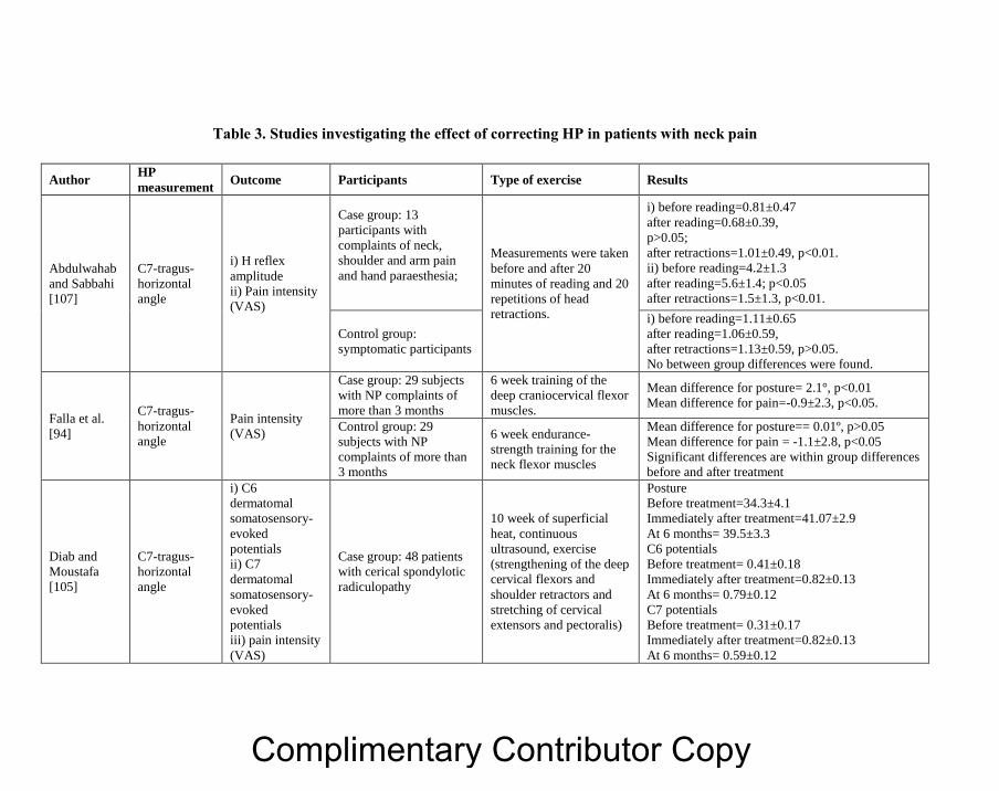
Table 3. Studies investigating the effect of correcting HP in patients with neck pain
Author HP
measurement Outcome Participants Type of exercise Results
Abdulwahab
and Sabbahi
[107]
C7-tragus-
horizontal
angle
i) H reflex
amplitude
ii) Pain intensity
(VAS)
Case group: 13
participants with
complaints of neck,
shoulder and arm pain
and hand paraesthesia;
Measurements were taken
before and after 20
minutes of reading and 20
repetitions of head
retractions.
i) before reading=0.81±0.47
after reading=0.68±0.39,
p>0.05;
after retractions=1.01±0.49, p<0.01.
ii) before reading=4.2±1.3
after reading=5.6±1.4; p<0.05
after retractions=1.5±1.3, p<0.01.
Control group:
symptomatic participants
i) before reading=1.11±0.65
after reading=1.06±0.59,
after retractions=1.13±0.59, p>0.05.
No between group differences were found.
Falla et al.
[94]
C7-tragus-
horizontal
angle
Pain intensity
(VAS)
Case group: 29 subjects
with NP complaints of
more than 3 months
6 week training of the
deep craniocervical flexor
muscles.
Mean difference for posture= 2.1°, p<0.01
Mean difference for pain=-0.9±2.3, p<0.05.
Control group: 29
subjects with NP
complaints of more than
3 months
6 week endurance-
strength training for the
neck flexor muscles
Mean difference for posture== 0.01º, p>0.05
Mean difference for pain = -1.1±2.8, p<0.05
Significant differences are within group differences
before and after treatment
Diab and
Moustafa
[105]
C7-tragus-
horizontal
angle
i) C6
dermatomal
somatosensory-
evoked
potentials
ii) C7
dermatomal
somatosensory-
evoked
potentials
iii) pain intensity
(VAS)
Case group: 48 patients
with cerical spondylotic
radiculopathy
10 week of superficial
heat, continuous
ultrasound, exercise
(strengthening of the deep
cervical flexors and
shoulder retractors and
stretching of cervical
extensors and pectoralis)
Posture
Before treatment=34.3±4.1
Immediately after treatment=41.07±2.9
At 6 months= 39.5±3.3
C6 potentials
Before treatment= 0.41±0.18
Immediately after treatment=0.82±0.13
At 6 months= 0.79±0.12
C7 potentials
Before treatment= 0.31±0.17
Immediately after treatment=0.82±0.13
At 6 months= 0.59±0.12
Complimentary Contributor Copy

Author HP
measurement Outcome Participants Type of exercise Results
Control group: 48
patients with cervical
spondylotic
radiculopathy
10 week of superficial
heat, continuous
ultrasound
Posture
Before treatment=35.8±6.3
Immediately after treatment=34.8±3.3
At 6 months=34.5±3.4
C6 potentials
Before treatment=0.42±0.21
Immediately after treatment=0.56±0.19
At 6 months=0.41±0.17
C7 potentials
Before treatment=0.32±0.21
Immediately after treatment=0.43±0.19
At 6 months= 0.28±0.18
All differences between case and control groups
were significant at p>0.05
Complimentary Contributor Copy

A. G. Silva, T. D. Punt and M. I. Johnson 26
All these potential changes allegedly associated with abnormal HP are believed to lead to
pathology and pain. [2,13,28,29] Whether existing research supports the assumption that HP
deviations from the norm result in biomechanical and physiological changes is addressed
below.
Head Posture and Muscles Moment Arms
Evidence exists that supports the claim that HP deviations affect the length of the muscle
moment arm, defined as the distance from the muscle's line of action to the joint's centre of
rotation. [30] Vasavada et al. [31] developed a biomechanical model of the head and neck to
evaluate changes on the length of neck muscle moment arms in different HPs. They reported
the length of the muscle moment arms of most extensor muscles, including the suboccipital
muscles, to decrease by 1 cm from extended to flexed HPs. Przybyla et al. [32] measured the
length of muscles moment arms in vivo using magnetic resonance imaging scans and also
reported a decrease in the length of extensors moment arms when the head was in flexion
compared to when it was in extension. This decrease in the length of the neck extensor
muscles moment arm results in a mechanical disadvantage during flexion, i.e., an increase in
muscle activity will be needed to counteract the head‘s tendency to tip forward.
Head Posture, Muscle Activity and Force/Strength
When comparing muscle activity in a simulated ideal HP against participants‘ usual HP
and a simulated forward HP, studies show conflicting results. McLean [16] compared muscle
activity in 18 asymptomatic participants and found increased muscle activity when
individuals stood in an ideal posture compared to their habitual forward HP. In contrast,
Enwemeka et al. [33] measured trapezius muscle activity in 10 asymptomatic participants and
reported a statistically significant increase in forward HP when compared to habitual or ideal
HP. However, studies fail to report whether simulated HPs are mid-range postures or end-of-
range postures and this may influence the muscular activity reported. Studies on other parts of
the body have shown that muscles play a more relevant role in the stabilization of mid-range
postures, while end-of-range postures seem to be mainly stabilized by inert structures such as
the ligaments and joint capsules. [34,35]
In addition, research also seems to support an association between HP, muscle activity
and muscle ability to produce force. Kumar et al. [36] studied the electromyography (EMG)
and isometric force of sternocleidomastoid, splenii and trapezii muscles of 40 asymptomatic
participants. They found that globally, EMG activity was approximately 66% higher in head
flexion than in extension, while force output was approximately 30% less in flexion than in
extension. This indicates that more muscle activity was required in flexion than extension to
generate the same force. Similar results were reported by Mayoux-Benhamou and Revel [37]
who investigated the influence of HP on muscular efficiency (calculated as the ratio between
muscular activation and the force produced). They found that less extensor muscle activity
was needed in neutral HP comparatively to extension and flexion to produce more force and
attributed the differences found to differences in muscle length and muscle lever arm length.
Complimentary Contributor Copy

The Clinical Usefulness of Head Posture Assessment for Patients with Neck Pain 27
Watson and Trott [38] measured the angle C7-tragus-horizontal (used as a surrogate measure
for forward HP) in 30 participants with headache and 30 asymptomatic participants and the
strength and endurance of the upper cervical flexors. They found lower endurance values of
the upper cervical flexors corresponded to lower values of the C7-tragus-horizontal angle.
Lower values of this angle indicate a more forward HP. This may suggest that the stabilizing
role of the deep flexors is compromised as forward HP increases. Suryanarayana [39]
quantified isometric cervical strength at neutral, 25%, 50% and 75% of flexion and extension
in 39 volunteers without pain. They found that cervical strength was highest in the neutral
posture for both flexion and extension and significantly decreased with an increasing angular
deviation of the neck, indicating a mechanical advantage for the cervical musculature when
the head and neck are in a neutral alignment. A recent study conducted by Peolsson et al. [40]
used ultrasound to evaluate the effect of three different HPs (neutral, flexed and forward HP)
on the activity of the dorsal neck muscles when resting and when performing a lifting task
with the upper limb in healthy subjects. Results demonstrated significantly greater muscle
deformation (indicative of greater muscle activity) induced by flexed and forward HPs,
compared to the neutral HP for all dorsal neck muscles at rest (muscles assessed were: upper
trapezius, splenius, semispinalis capitis, semispinalis cervicis and multifidus) and a
significant interaction between HP condition and activity (rest or lifting). When comparing
muscle deformation between rest and lifting, significant changes in muscle deformation were
only evident when lifting was performed in the neutral HP. Study authors, suggest that these
findings could be explained by high levels of muscle deformation in flexed and forward HP
even before the lifting task was initiated. This suggests that HP might modulate loading on
the neck structures when upper limb activities are performed. These results are in agreement
with those of Elliot [41] and Nimbarte et al. [42] who found that HP influences dorsal neck
muscle activity and patterns of recruitment in supine lying with the head loaded and
unsupported, during upper limb lifting tasks, respectively.
In addition, HP seems to influence the activity of scapular muscles. Weon [43] assessed
the effects of simulated forward HP on the activity of the scapular upward rotators during
loaded isometric shoulder flexion in the sagittal plane, in asymptomatic participants.
Participants performed shoulder flexion in both simulated forward HP and neutral HP.
Significantly increased EMG activity in the upper trapezius and lower trapezius, and
significantly decreased EMG activity in the serratus anterior were found during loaded
isometric shoulder flexion with forward HP.
If HP deviations from the norm contribute to increase muscle activity while decreasing
the ability to produce force and disturbing motor control, this may increase muscle fatigue as
well as the internal forces acting on neck sensitive structures leaving individuals vulnerable to
injury and pain.
Head Posture and Compressive Loading
The weight of the head and muscle forces create compressive loading that is supported by
the cervical spine. [44] It is believed that this force and the consequent intra-disc pressure are
affected by different HPs [45] and this is supported by the findings of studies described in the
previous section (head posture and muscle activity and force/strength). Bonney and Corlet
Complimentary Contributor Copy

A. G. Silva, T. D. Punt and M. I. Johnson 28
[46] investigated the effect of one hour sitting with the head at 0°, 20° and 40° of flexion and
found significant differences in the length of the spine between 0° and 20° of flexion (a
decrease of 0.88 mm) and between 0° and 40° of flexion (a decrease of 1.65 mm), concluding
that flexed HPs load the cervical spine more. This is consistent with the results of the studies
described previously reporting that HP deviations from the norm generally increase muscle
activity. [36-42] The increase in muscle activity may result in an increase in the compressive
forces acting on the neck, possibly explaining the decrease in length of the cervical spine
when in flexion. The shrinkage of the spine is usually due to a decrease in intervertebral disc
height, which results in an increase in the amount of load supported by the zygapophyseal
joints, [47] believed to accelerate inter-vertebral disc and zygapophyseal joints degeneration.
[47,48] Zygapophyseal joints have been considered to be the most common source of
idiopathic NP [49] although there is little evidence on the consequences of HP deviations on
the load supported by intervertebral discs, zygapophyseal joints or ligaments in the neck.
Studies using mainly in vitro or biomechanical modeling approaches provide some insight on
the effect of head flexion on neck internal loads, showing, in general, an increase in the load
supported by the anatomical structures in flexion. Goel and Clausen [50] used a
biomechanical model to investigate load sharing among anatomical components of a C5-C6
segment. The authors reported that the inter-spinous and capsular ligaments sustained the
most strains in flexion and axial rotation respectively and maximum intradiscal pressure
reached the highest values under combined compression and flexion. The quantity of load
shared by the disc varied from 14% in compression and extension, 68% in right lateral
bending, 75% in left axial rotation, 88% in compression, and 113% in compression and
flexion. Snijders et al. [51] used a biomechanical computer model, reporting that during
flexion, muscle forces and joint reaction forces increase, except the force between the
odontoid and the ligamentum transversum atlantis; the joint reaction forces on the levels C0-
C1, C1-C2 and C7-T1 reach minimum values in extension. It has been shown that stretch or
load applied to ligaments and discs can change their mechanical properties, affect their
function and result in micro-damage in the lumbar spine. [52,53] It can be speculated that
changes in tissue load induced by HP deviations may have the same consequences for neck
structures contributing to the onset or maintenance of NP.
Head Posture and Degenerative Changes
in the Neck
Despite evidence that mechanical changes relate to HP as outlined above, there appears to
be no clinical studies exploring whether there is a relationship between HP deviations, an
increase in loading and the consequent development of pathology in the neck. However,
studies investigating the effect of cervical spine fusion on adjacent-level intradiscal pressure
found a marked increase in intradiscal pressure and also an increase in the rates of disc
degeneration in segments adjacent to fusion. [48,54] Additionally, evidence from studies
investigating the effect of forces on biologic tissues seems to indicate that a variety of
changes occur to discs in response to joint loading, including: i) volumetric changes, ii) fluid
flow changes, iii) pressure changes, iv) electrokinetic changes and v) deformations in tension,
compression or shearing configurations. These factors are considered to be important
Complimentary Contributor Copy
The Clinical Usefulness of Head Posture Assessment for Patients with Neck Pain 29
regulators of cellular metabolism in the disc [55] and therefore, it is likely that overload
resulting from daily life activities may adversely affect the nutrition and remodeling changes
occurring in the intervertebral discs and consequently contribute to degenerative changes.
[55,56] Stokes and Iatridis [57] conducted a review examining the relationship between
mechanical loading and spinal disc degeneration. Reviewing evidence from biomechanics,
epidemiology, animal models and intervertebral disc physiology, it was concluded that any
abnormal loading conditions (including overload and immobilization) could probably produce
tissue trauma and/or adaptive changes, resulting in pain. It is unknown whether HP deviations
affect the biologic processes occurring in the disc or other anatomical structures. However it
can be speculated that if it has the potential to affect the internal forces acting in the neck
structures, it is likely to affect their nutrition and remodeling, leading to changes that may
facilitate tissue injury and/or prevent complete healing of an existing injury.
Head Posture, Changes in Muscle Blood Flow
and the Accumulation of Metabolites
No studies investigating the effect of HP on muscle blood flow or on the concentration of
algesic substances were found. However, there is some evidence to support that low level
muscle contractions of the trapezius may lead to the accumulation of algesic substances. Boix
et al. [58] demonstrated that sustained contractions of the trapezius to maintain shoulder
abduction in asymptomatic individuals results in increased bradykinin and pain intensity.
Therefore, studies are needed to ascertain whether changes in muscle activity associated with
HP can affect muscle blood flow and accumulation of metabolites.
Head Posture and Cervical Range of Motion
There is evidence to suggest that HP deviations from the norm may affect the range of
motion available in the cervical spine. Walmsley et al. [59] investigated the effect of five
different initial HPs on rotation range of movement in 60 asymptomatic participants,
reporting a significant decrease on rotation range of motion when the movement was initiated
in flexion, extension, retraction or protraction when compared to neutral. Similar results were
obtained by Edmondston et al. [60] who reported right and left rotation and right and left
side-flexions to be significantly decreased when the movement was initiated in retraction and
forward HP compared to neutral HP. The stretch of the ligaments induced by HP deviations,
in addition to anatomy, is likely to limit the movement when compared to the neutral HP. [60]
Similar results were found by De-la-Llave-Rincón [61] and Quek et al. [62] The first authors
found that the smaller C7-tragus-horizontal angle (reflective of a greater forward HP), the
smaller the range of motion in women with carpal tunnel syndrome and 25 matched healthy
women. The latter explored the mediating effects of forward HP on the relationship between
thoracic kyphosis and cervical mobility in 51 older adults with neck pain with or without
referred pain, numbness or paraesthesia. Thoracic kyphosis was measured using a flexicurve
and forward HP was assessed via the C7-tragus-horizontal angle. They found that greater
Complimentary Contributor Copy
A. G. Silva, T. D. Punt and M. I. Johnson 30
thoracic kyphosis was significantly associated with greater forward HP and greater forward
HP was associated with less neck flexion and rotation. Interestingly, participants with
idiopathic NP were also found to have a decrease in neck range of motion when compared to
asymptomatic individuals. [63]
Head Posture and Proprioception
No studies investigating whether HP affects neck proprioception were found. However,
investigations into the effect of HP on postural control and the effect of lumbar spine posture
on lumbar position sense have been conducted. Kang et al. [64] compared 2 groups of healthy
and young computer based workers and found that the group with more pronounced forward
HP had their centre of gravity in a relatively anterior location and had lower balance scores
than the group with more normal HP. This suggests that forward HP may affect the position
of the centre of gravity and contribute to some disturbance in the balance of healthy adults.
However, Silva and Johnson [65] when comparing postural control in a simulated forward HP
with postural control in normal HP in healthy young individuals found that simulated forward
HP did not disrupt postural control. However, authors hypothesised that this might be due to
an increase in muscle co-contraction throughout the body in forward HP conditions to avoid
falling as participants commented that balance was harder to maintain in these conditions.
However, muscle co-contraction was not measured. It was also hypothesised that a longer
period in forward HP is required before postural control is affected as Dolan and Green [66]
found a significant increase in lumbar joint repositioning error after 5 minutes in a slouched
posture, but no change was noted after 3 seconds in the same posture, suggesting that the
effects of a non-optimal posture could be time-dependent. This hypothesis needs to be
investigated. While initially, the deviation in HP might contribute to impaired proprioception,
[67]
Edmondston et al. [68] suggested that subsequently, proprioceptive impairment may
make correction of HP deviation problematic, possibly perpetuating pain as individuals adopt
postures that place greater load on the neck structures. Studies have also found that
individuals with idiopathic neck pain present impairment in neck proprioception when
compared to asymptomatic participants. [69,70]
Taken together, the evidence suggests that HP deviations from the norm contribute to
increased neck and scapular muscle activity, disturb normal patterns of recruitment, decrease
force production either in static postures or during activities of the upper limb, increase joint
compressive loading and decrease neck range of motion. These changes suggest that
maintaining normal HP might be advantageous and that it is likely that HP deviations from
the norm increase the probability of having neck pain or perpetuate existing NP. Interestingly,
studies comparing patients with chronic idiopathic NP and asymptomatic participants have
also reported that patients show decreased cervical range of motion, [63,71-73] impaired
proprioception, [69,70] reduced neck muscle strength, [71,74,75] reduced neck muscle
endurance, [76,77] greater neck muscle fatigue, [78] reduced neuromuscular efficiency [79]
and altered muscle control [80,81] when compared to asymptomatic participants. This raises
the question as to whether HP might be associated with these impairments.
Complimentary Contributor Copy

The Clinical Usefulness of Head Posture Assessment for Patients with Neck Pain 31
Measurement of Head Posture
Head Posture Assessment in Clinical Practice
Clinicians seem to use HP assessment through observation for patients with neck pain. In
particular, in a survey with 278 physiotherapists, 276 reported to assess HP for patients with
neck pain. Of the 276 respondents that reported to assess HP for patients with neck pain, 130
(47.1%) reported to ‗always‘ assess HP for these patients and an additional 80 respondents
(29.0%) reported to assess HP for more than 67% of their patients. [82] All respondents that
assessed HP reported to do it by observation and the use of qualitative descriptors such as:
normal HP, mild deviation, moderate deviation and severe deviation. However, a variety of
different procedures seem to be used indiscriminately for HP assessment. HP is assessed with
the patient standing, sitting, lying down with face up, in the most painful HP, walking,
dressing and undressing from lateral, frontal and posterior viewpoints. Instructions given to
patients also vary, including asking patients to adopt as natural a posture as possible, keeping
a horizontal look or to look straight ahead. [6] The lack of a standardized protocol for HP
assessment may account for these discrepancies, leading physiotherapists to do what they
perceive as being optimal for the patient based on their beliefs and clinical experience.
However, these inconsistencies among physiotherapists clearly threaten the reliability of HP
assessment and therefore hinder teamwork between colleagues. For example, Solow and
Tallgren [83] found that different instructions can lead to significantly different HPs and
Ferrario et al. [84] showed that HP differs between standing and seated positions. In addition,
Edmondston et al. [85] found that different sitting postures induce a different pattern of
muscle activation and a different posture of the uppermost segments. In this study, having the
lumbo-pelvic region in a neutral position promotes greater activation of the thoracic extensor
muscles and significantly reduces the levels of cervical extensor muscle activity associated
with a decreased forward HP when compared to sitting in a slouched posture.
Furthermore, in clinical practice, HP is assessed by observation without the aid of a
measurement instrument. However, visual inspection of the head has been shown to be
neither reliable nor valid. [86-88] We asked 10 physiotherapists to assess head posture of 40
individuals using a 4-point scale: 1) normal, 2) slightly deviated, 3) moderately deviated and
4) severely deviated in two separate occasions. [87] Inter-rater agreement was very poor.
When matching the rates attributed to the individuals against objective measures of posture
we found that individuals with the same objective measurement were attributed quite different
rates and individuals with different objective measures were attributed the same rate. Figure 2
shows a graphical presentation of 3 physiotherapist‘s rates (scale categories) plotted against
the objective measurement (the angle between C7, the tragus of the ear and the horizontal) for
the assessment of forward HP. It is unlikely that lack of previous experience using the scale
influenced the results as 3 of the 10 physiotherapists reported to assess HP in clinical practice
using the same scale. These findings compromise the usefulness of HP assessment for
patients with neck pain in clinical practice. [87]
Despite the lack of reliability and validity of HP assessment by observation, HP itself
seems to be consistent over time. [89] Therefore, it can be used as part of patients‘ assessment
as long as reliable and valid procedures are used. As previously reported, experimental
studies have measured distances and angles between anatomical landmarks as surrogate
Complimentary Contributor Copy

A. G. Silva, T. D. Punt and M. I. Johnson 32
measures for HP. [14,15,24,26,90,91] The aspect of HP most commonly assessed is forward
HP and its surrogate measure most commonly used is the angle between C7, the tragus of the
ear and the horizontal. [27] This angle can be measured using a goniometer. This is a cheap
and easy to use instrument providing a means to use objective measures of HP in clinical
practice [14,92]. Alternatively, clinicians can also measure distances between anatomical
landmarks and external references, such as a wall, using a metric ruler as previously
described. [25] However, the reliability and validity of cheap, simple and easy to use
assessments needs further investigation.
Figure 2. Rates attributed to participants for forward HP plotted against angular values used as a
surrogate of forward HP in clinical practice.
The Relationship between Head Posture and Neck Pain
A systematic review from our team aiming to determine whether there were differences
in angles or linear distances between anatomical landmarks used as surrogates for HP
between individuals with and without neck pain found 13 studies. [19] Of these, 11 studies
measured surrogates for forward HP, 6 studies measured surrogates for head
extension/flexion and 1 study measured surrogates of head side flexion and rotation. Patients
with neck pain were found to have greater forward HP in 4 of 11 studies, greater head
extension/flexion in 2 of 6 studies and less side-flexion and less rotation in 1 of 1 study
(Table 2). We concluded that there was insufficient good quality evidence to determine
whether forward HP, head extension, side-flexion and rotation differ between participants
Complimentary Contributor Copy
The Clinical Usefulness of Head Posture Assessment for Patients with Neck Pain 33
with neck pain and asymptomatic participants. Another study conducted by our team and not
included in this systematic review, compared 40 asymptomatic participants with 40 patients
with chronic idiopathic neck pain and found a significant difference for forward HP, but not
head extension and side flexion, between patients with neck pain and asymptomatic
participants. [19] Furthermore, when subgrouping the sample using the sample age mean, a
significant difference was found between participants with and without pain aged 50 years or
less, but not between those aged more than 50 years (Table 2). These results suggest that age
may be relevant to the relationship between HP and neck pain and suggest limitations in the
value of measuring HP in older patients presenting with chronic NP. Further research should
help clarify this. In addition, the absence of a significant difference between asymptomatic
participants and patients with neck pain for head extension and side-flexion suggest that it
might not be relevant to assess these aspects of HP for patients with neck pain.
Considering the studies found in our systematic review [27] and our own study [19] there
were 6 studies that compared forward HP between patients with neck pain and asymptomatic
participants using the angle between C7, the tragus of the ear and the horizontal. [14,19,93–
96] Of these 6 studies, 4 found a mean difference between groups [19,93,94,96] that varied
between a minimum of 2.2º in seating while playing a computer [94] and a maximum of 6.7º
in static standing, [93] consistent with patients with neck pain having a more forward HP than
asymptomatic participants. The two studies unable to find a difference used small sample
sizes (n=10) at least in one group, raising questions as to whether their results are false
negatives. [14,95]
Taken together, these results seem to suggest that measurement procedures, in particular
the surrogate measure of forward HP used, might influence study results. Additionally, the
assessment of forward HP for patients with NP might be of value as there seems to be
differences in the measurements of the angle C7-tragus-horizontal between participants with
and without NP. However, the size of the differences are too small, reinforcing the previous
suggestion of using objective measures to assess HP in clinical practice as it is highly unlikely
that clinicians are able to detect differences of this size either between or within patients.
Head Posture Correction
Is It Possible to Correct Head Posture?
Studies investigating techniques to correct HP have reported statistically significant
changes in surrogate measures of forward HP taken before and after exercises regimens in
asymptomatic participants, but not for measurements of surrogates of head extension
[103,104] (Table 3). Pearson and Walmsley [103] measured the effects of 10, 20, and 30
repeated neck retraction movements on HP in asymptomatic subjects and found a significant
change (mean change=4.0º±?mm) in the position and orientation of the tragus of the ear, C2,
C5 and T1 consistent with a less forward HP. Harman et al. [104] investigated the effect of a
10-week home exercise program on improving forward HP and extension between a group of
asymptomatic participants that received an exercise regimen and a control group that did not
receive any intervention. The exercise program consisted of neck extensor and pectoralis
major stretches and deep neck flexor and shoulder retractor strengthening exercises. They
Complimentary Contributor Copy

A. G. Silva, T. D. Punt and M. I. Johnson 34
found a significant mean increase in the angle C7, tragus, horizontal of 2.3º immediately after
the exercise program, indicating less forward HP after the treatment. No difference was found
for head extension. These studies used healthy individuals so the results might not apply to
patients with neck pain and whether the changes reported remain or were reversed after the
exercise programme had finished was also not investigated.
Falla et al., [94] Diab and Moustafa [105] and Diab [106] investigated forward HP
correction in patients with chronic non severe neck pain, cervical radiculopathy and scoliosis,
respectively. The results of all three studies suggest that it is also possible to correct HP in
patients. In addition, Diab [106] and Diab and Moustafa [105] showed that corrections are
still present at 3 and 6 months, respectively. Falla et al. [94] compared the change in HP
measured though the angle C7-tragus-horizontal between subjects with chronic neck pain and
control subjects while performing a 10 minute computer task. One group received a 6-week
endurance-strength training of the cervical flexor muscles and the other training of the
craniocervical flexor muscles. Only the group that received the training of the craniocervical
flexor muscles demonstrated an increase in the angle C7-tragus-horizontal (mean=4.4°; 95%
CI=3.3º–5.4º), indicative of a decrease in the degree of forward HP.
Diab and Moustafa [105] compared the C7, tragus, horizontal angle in two groups of
patients with cervical spondylotic radiculopathy before and after a 10 week intervention.
Patients in both the experimental and the control group received superficial heat on the neck
for 10 minutes followed by continuous ultrasound on upper trapezius for 10 minutes. The
exercise group additionally received a posture corrective exercise programme in the form of
strengthening of the deep cervical flexors and shoulder retractors and stretching of cervical
extensors and pectoralis. Measurements were taken before starting the treatment, at the end of
10 weeks, and at follow-up period of six months. They found a significant increase in the
angle C7, tragus, horizontal in the group that received the exercise regimen, (mean difference
after treatment=6.8º; mean difference at 6 months=5.2º), but not in the control group (mean
difference after treatment=1.0º; mean difference at 6 months=1.3º).
Diab [106] investigated the effectiveness of forward HP correction in adolescents with
idiopathic scoliosis after a 10 weeks regimen of exercises and at three-month follow-up. All
the patients received traditional treatment in the form of stretching and strengthening
exercises. In addition, patients in the experimental group received a forward HP corrective
exercise programme as described previously for Diab and Moustafa [105]. They found a
significant difference in the angle C7, tragus, horizontal immediately after treatment (mean
difference =7.7º), and at 6 month follow-up (mean difference =7.7º). No significant
differences were found for the control group (mean difference after treatment=0.3º; mean
difference at 3 months= 0.6º).
Taken together these results suggest that it is possible to correct forward HP and that
correction is maintained after the exercise programme has finished. Training of the deep
cervical flexors was present in the exercise regimens of all studies that found a significant
difference in forward HP after exercise, [94,103–106] suggesting that it might be an essential
component of an exercise regimen aiming at correcting forward HP. However, the within
subject HP changes induced by the exercise regimens were small.
This is in accordance with the small differences in HP between participants with and
without neck pain. Assuming that asymptomatic participants have a normal HP and
considering that differences between participants with and without neck pain are small
(maximum mean difference in studies that measured the angle between C7, the tragus and the
Complimentary Contributor Copy

The Clinical Usefulness of Head Posture Assessment for Patients with Neck Pain 35
horizontal is 6.7º) it is conceivable to expect similar differences for the within subject forward
HP changes after an exercise regimen. Whether it is possible to correct the other components
of HP is unknown.
Benefits to the Patient with Neck Pain from Head Posture Correction
In the previous section we discussed evidence that suggest that it is possible to correct
forward HP. However, this is relevant to the patients with neck pain only if it results in any
benefit, namely in an improvement in signs and symptoms associated with neck pain. Studies
comparing pain and nerve root function in patients with neck pain before and after exercises
showed conflicting results [94,105,107] (Table 3). Abdulwahab and Sabbahi [107] found that
20 repetitions of head retractions resulted in a significant improvement in the amplitude of the
H reflex (a monosynaptic reflex indicative of the reflex arc integrity) and pain intensity of
patients with complaints of neck, shoulder and arm pain and hand paraesthesia. Diab and
Moustafa [105] compared C6 and C7 amplitude of dermatomal somatosensory-evoked
potentials (as an indicative of nerve root function) and pain between two groups of patients
with spondylotic radiculopathy after a treatment intervention. Both groups received a similar
intervention, except for forward HP corrective exercises (as described in the previous
section). They reported a significant mean difference between the exercise and control groups
for measured variables immediately after treatment and at 3-month follow-up, consistent with
a greater improvement in the group that received the corrective exercises. These results
suggest that correcting forward HP in patients with cervical radiculopathy results in short and
long term benefits for the patients in terms of pain and root function improvement. In
contrast, Falla et al. [94] found a statistically significant improvement in the pain intensity
reported in two different groups of patients with chronic neck pain after two different exercise
regimens (endurance strength training of the neck flexors and training of the craniocervical
flexors). However, only the group that received the training of the craniocervical flexor
muscles demonstrated an increase in the angle between C7, the tragus and the horizontal
(indicative of a decrease in the degree of forward HP). The contradictory results from Falla et
al. [94] and Abdulwahab and Sabbahi [107] and Diab and Moustafa [105] suggest that HP
correction may be more important in certain subgroups of patients with neck pain. Further
research is needed to ascertain this; for example, comparing traumatic vs non-traumatic neck
pain, acute vs chronic neck pain or neck pain vs pain in the neck and arm.
Conclusion
Taken together the evidence reviewed in this chapter support the usefulness of forward
HP assessment for patients with neck pain, but not for the other components of HP. This lack
of support for head extension/flexion, side-flexion and rotation is not due to evidence of their
lack of utility but rather to the lack of studies investigating these components.
The arguments in favor of the clinical usefulness of forward HP assessment for patients
with neck pain are diverse:
Complimentary Contributor Copy

A. G. Silva, T. D. Punt and M. I. Johnson 36
There is biomechanical and physiological evidence in favor of normal HP vs.
abnormal HP;
Forward HP is reliable for over time comparisons;
There are differences between patients with neck pain and asymptomatic
participants, consistent with patients having a more forward HP;
It is possible to correct forward HP;
Correcting forward HP results in benefits to the patient.
However, the small size of the differences found between patients with neck pain and
asymptomatic participants and before and after exercise regimens aiming at correct forward
HP, measurement procedures must be objective, valid and reliable. Otherwise, clinicians will
not be able to assess forward HP changes within or between patients. Additionally,
assessment procedures must follow a standardized protocol as the posture of other segments
or the indications given to patients affect the degree of forward HP. To our knowledge there
is not a standardized protocol for forward HP assessment in clinical practice. Therefore, and
based on the available literature and on studies conducted by our team, we suggest that the
following procedures should be used when assessing HP for patients with neck pain as a
means to standardize the assessment:
1. The patient should be adequately free from clothes;
2. Standing without shoes in a firm surface;
3. Feet slightly apart (approximately shoulder width);
4. Patients are asked to flex and extend their head, decreasing the amplitude of
movement in a progressive manner until they stop in what they feel is their
natural/usual HP;
5. When they stop moving their head, measurements with an objective tool, such as a
goniometer, are taken for the angle C7, tragus, horizontal;
6. Measurement procedures should be taken 3 times and the average used for posterior
comparisons;
7. Additionally and using the same procedures from points 1 to 5, the effect of
correcting HP towards what is perceived as being a more optimal HP can also be
assessed.
Despite the growing body of evidence on HP and neck pain there is much that needs to be
undertaken:
To investigate whether the usefulness of forward HP assessment differs between
subgroups of neck pain;
To develop and validate a protocol for forward HP assessment in clinical practice
with objective, inexpensive and easy to use instruments;
To develop normative data for forward HP for age and sex;
To identify cut of points above or below which forward HP correction might be
beneficial;
To test several regimens of exercises for forward HP correction in order to identify
the most effective;
Complimentary Contributor Copy

The Clinical Usefulness of Head Posture Assessment for Patients with Neck Pain 37
To explore the clinical usefulness of the other components of HP (head
flexion/extension, side-flexion and rotation).
References
[1] Schwartz L. A résumé, with comments, of the available literature relating to posture.
Public Health Rep. 1927;27:1219-48.
[2] Kendall F, McCreary E, Provance P, Rodgers MM, Romani WA. Muscles testing and
function, with posture and pain . Philadelphia: Lippincott Williams & Wilkins; 2005.
[3] Petty NJ. Neuromusculoskeletal examination and assessment. A handbook for
therapists. Bath: Churchill Livingstone; 2011.
[4] McKenzie R, May S. The cervical and thoracic spine. Mechanical diagnosis and
therapy. New Zealand: Spinal Publications; 2006.
[5] American Physical Therapy Association. Guide to physical therapist practice. Phys
Ther. 2001;81:9-746.
[6] Silva A, Punt D, Johnson M. Using head posture assessment to inform the management
of neck pain: a pilot focus group study. Int J Ther Rehabil. 2012;19:98-105.
[7] Kesson M, Atkins E. Orthopaedic medicine. A practical approach. Oxford: Butterworth
Heinemann; 2001.
[8] Magee DJ. Orthopaedic physical assessment. Philadelphia: Saunders; 2008.
[9] Fejer R, Kyvik KO, Hartvigsen J. The prevalence of neck pain in the world population:
a systematic critical review of the literature. Eur Spine J. 2006;15:834-48.
[10] Basmajian J V, De Luca CJ. Muscles alive. Their functions revealed by
electromyography. Baltimore: Williams and Wilkins; 1985.
[11] Silva AG, Punt TD, Sharples P, Vilas-Boas JP, Johnson MI. Head posture assessment
for patients with neck pain: Is it useful? Int J Ther Rehabil. 2009;16:43-53.
[12] Silva AG, Sharples P, Punt D, Johnson M. Systematic review of studies comparing
head posture between participants with neck pain and asymptomatic participants. Pain
Eur. Congr. Lisbon: European federation of chapters of the international association for
the study of pain; 2009.
[13] Bryden L, Fitzgerald D. The influence of posture and alteration of function upon the
craniocervical and craniofacial regions. In: von Piekartz H, Bryden L, (eds).
Craniofacial dysfunction pain. Manual therapy assessment and management. Oxford:
Butterworth-Heinemann; 2001. p. 163-87.
[14] Harrison AL, Barry-Greb T, Wojtowicz G. Clinical measurement of head and shoulder
posture variables. JOSPT. 1996;23:353-61.
[15] Braun BL. Postural differences between asymptomatic men and women and
craniofacial pain patients. Arch Phys Med. Rehabil. 1991;72:653-6.
[16] McLean L. The effect of postural correction on muscle activation amplitudes recorded
from the cervicobrachial region. J Electromyogr Kinesiol. 2005;15:527-35.
[17] Woodhull AM, Maltrud K, Mello BL. Alignment of the human body in standing. Eur J
Appl Physiol. 1985;54:109-15.
Complimentary Contributor Copy

A. G. Silva, T. D. Punt and M. I. Johnson 38
[18] Szeto GP, Straker LM, O‘Sullivan PB. A comparison of symptomatic and
asymptomatic office workers performing monotonous keyboard work-2: neck and
shoulder kinematics. Man Ther. 2005;10:281-91.
[19] Silva AG, Punt TD, Sharples P, Vilas-Boas JP, Johnson MI. Head posture and neck
pain of chronic nontraumatic origin: a comparison between patients and pain-free
persons. Arch Phys Med Rehabil. 2009;90:669-74.
[20] Binnie C, Cooper R, Mauguiere F, Osselton J, Prior P, Tedman B. Clinical
neurophysiology. New York: Elsevier Science Publishers; 2003.
[21] Rokkedal-Lausch T, Lykke M, Hansen MS, Nielsen RO. Normative values for the foot
posture index between right and left foot: A descriptive study. Gait Posture;38:843-6.
[22] Redmond AC, Crane YZ, Menz HB. Normative values for the Foot Posture Index. J
Foot Ankle Res. 2008;1:6.
[23] The Association of Faculties of Medicine of Canada Public Health Educators‘ Network.
Establishing cut-points what is a normal value [Internet]. Primer. 2013 [cited 2013 Oct
28]. Available from: The association of faculties of medicine of Canada public health
educators' network.
[24] Dalton M, Coutts A. The effect of age on cervical posture in a normal population. In:
Boyling JD, Palastanga N (eds). Grieve‘s modern manual therapy of the vertebral
column. New York: Churchill Livingstone; 1994.
[25] Hanten WP, Lucio RM, Russel JL, Brunt D. Assessment of total head excursion and
resting head posture. Arch Phys Med Rehabil. 1991;72:877-80.
[26] Raine S, Twomey LT. Head and shoulder posture variations in 160 asymptomatic
women and men. Arch Phys Med Rehabil. 1997;78:1215-23.
[27] Silva AG, Sharples P, Johnson MI. Studies comparing surrogate measures for head
posture in individuals with and without neck pain. Phys Ther Rev. 2010;15:12-22.
[28] Taniguchi A, Ogita K, Murata T, Kuzuhara S, Tomimoto H. Painful neck on rotation:
diagnostic significance for crowned dens syndrome. J Neurol. 2010;257:132-5.
[29] Oatis CA. Kinesiology: the mechanics and pathomechanics of human movement.
Philadelphia: Lippincott Williams & Wilkins; 2004.
[30] Watkins J. Structure and function of the musculoskeletal system. Champaign, IL:
Human Kinetics; 1999.
[31] Vasavada AN, Li S, Delp SL. Influence of muscle morphometry and moment arms on
the moment-generating capacity of human neck muscles. Spine. 1998;23:412-22.
[32] Przybyla A, Mohite A, Blease S, Adams MA. How does posture affect lever arms of
neck flexor and extensor muscles? J Biomech. 2006;39 S49.
[33] Enwemeka CS, Bonet IM, Ingle JA, Prudhithumrong S, Ogbahon FE, Gbenedio NA.
Postural correction in persons with neck pain. Part II: Integrated electromyography of
the upper trapezius in three simulated neck positions. JOSPT. 1986;8:240-2.
[34] McGill SM, Brown S. Creep response of the lumbar spine to prolonged full flexion.
Clin Biomech. 1992;7:43-6.
[35] Terry GC, Hammon D, France P, Norwood LA. The stabilizing function of passive
shoulder restraints. Am J Sport Med. 1991;19:26-34.
[36] Kumar S, Narayan Y, Amell T, Ferrari R. Electromyography of superficial cervical
muscles with exertion in the sagittal, coronal and oblique planes. Eur Spine J.
2002;11:27-37.
Complimentary Contributor Copy

The Clinical Usefulness of Head Posture Assessment for Patients with Neck Pain 39
[37] Mayoux-Benhamou MA, Revel M. Influence of head position on dorsal neck muscle
efficiency. Electromyogr Clin Neurophysiol. 1993;33:161-6.
[38] Watson DH, Trott PH. Cervical headache: an investigation of natural head posture and
upper cervical flexor muscle performance. Cephalalgia. 1993;13:272-84.
[39] Suryanarayana L, Kumar S. Quantification of isometric cervical strength at different
ranges of flexion and extension. Clin Biomech. 2005;20:138-44.
[40] Peolsson M, Larsson B, Brodin LA, Gerdle B. A pilot study using tissue Velocity
ultrasound imaging (TVI) to assess muscle activity pattern in patients with chronic
trapezius myalgia. BMC Musculoskelet Disord. 2008;9:127.
[41] Elliott JM, O‘Leary SP, Cagnie B, Durbridge G, Danneels L, Jull G. Craniocervical
orientation affects muscle activation when exercising the cervical extensors in healthy
subjects. Arch Phys Med Rehabil. 2010;91:1418-22.
[42] Nimbarte A, Aghazadeh F, Ikuma L, Harvey C. Neck disorders among reconstruction
workers: Understanding the physical loads on the cervical spine in static lifting tasks.
Ind Health. 2010;145-53.
[43] Weon J-H, Oh J-S, Cynn H-S, Kim Y-W, Kwon O-Y, Yi C-H. Influence of forward
head posture on scapular upward rotators during isometric shoulder flexion. J Bodyw
Mov Ther. 2010;14:367-74.
[44] Miura T, Panjabi MM, Cripton PA. A method to simulate in vivo cervical spine
kinematics using in vitro compressive preload. Spine (Phila. Pa. 1976). 2002;27:43-8.
[45] Borestein DG, Wiesel SW, Boden SD. Low back and neck pain. Comprehensive
diagnosis and management. Philadelphia: Saunders; 2004.
[46] Bonney RA, Corlett EN. Head posture and loading of the cervical spine. Appl Ergon.
2002;33:415-7.
[47] Adams MA, Freeman BJ, Morrison HP, Nelson IW, Dolan P. Mechanical initiation of
intervertebral disc degeneration. Spine. 2000;25:1625-36.
[48] Pospiech J, Stolke D, Wilke HJ, Claes LE. Intradiscal pressure recordings in the
cervical spine. Neurosurgery. 1999;44:379-84.
[49] Manchikanti L, Cash K, Pampati V, Wargo B, Malla Y. Cervical epidural injections in
chronic discogenic neck pain without disc herniation or radiculitis: preliminary results
of a randomized, double-blind, controlled trial. Pain Physician. 2010;13:E265-78.
[50] Goel VK, Clausen JD. Prediction of load sharing among spinal components of a C5-C6
motion segment using finite element approach. Spine. 1998;23:684-91.
[51] Snijders CJ, van Dijke GAH, Roosch ER. A biomechanical analysis of the cervical
spine in static postures. J Biomech. 1991;24:783-92.
[52] Solomonow M, Baratta R V, Banks A, Freudenberger C, Zhou BH. Flexion-relaxation
response to static lumbar flexion in males and females. Clin Biomech. 2003;18:273-9.
[53] Solomonow M, Baratta R V, Zhou BH, Burger E, Zieske A, Gedalia A. Muscular
dysfunction elicited by creep of lumbar viscoelastic tissue. J Electromyogr Kinesiol.
2003;13:381-96.
[54] Eck JC, Humphreys SC, Lim TH, Jeong ST, Kim JG, Hodges SD, et al. Biomechanical
study on the effect of cervical spine fusion on adjacent-level intradiscal pressure and
segmental motion. Spine. 2002;27:2431-4.
[55] Setton L, Chen J. Mechanobiology of the intervertebral disc and relevance to disc
degeneration. J Bone Jt Surg. 2006;88:52-7.
[56] Adams MA, Dolan P. Spine biomechanics. J Biomech. 2005;38:1972-83.
Complimentary Contributor Copy

A. G. Silva, T. D. Punt and M. I. Johnson 40
[57] Stokes IAF, Iatridis JC. Mechanical conditions that accelerate intervertebral disc
degeneration: overload versus immobilization. Spine. 2004;29:2724-32.
[58] Boix F, Roe C, Rosenborg L, Knardahl S. Kinin peptides in human trapezius muscle
during sustained isometric contraction and their relation to pain. J Appl Physiol.
2005;98:534-40.
[59] Walmsley RP, Kimber P, Culham E. The effect of initial headposition on active
cervical axial rotation range of motion in two age populations. Spine. 1996;21:2435-42.
[60] Edmondston SJ, Henne S-E, Loh W, Østvold E. Influence of cranio-cervical posture on
three-dimensional motion of the cervical spine. Man Ther. 2005;10:44-51.
[61] De-la-Llave-Rincón AI, Fernández-de-las-Peñas C, Palacios-Ceña D, Cleland J a.
Increased forward head posture and restricted cervical range of motion in patients with
carpal tunnel syndrome. JOSPT. 2009;39:658-64.
[62] Quek J, Pua Y-H, Clark R a, Bryant AL. Effects of thoracic kyphosis and forward head
posture on cervical range of motion in older adults. Man Ther. 2013;18:65-71.
[63] Woodhouse A, Vasseljen O. Altered motor control patterns in whiplash and chronic
neck pain. BMC Musculoskelet Disord. 2008;20:90-100.
[64] Kang J-H, Park R-Y, Lee S-J, Kim J-Y, Yoon S-R, Jung K-I. The effect of the forward
head posture on postural balance in long time computer based worker. Ann Rehabil
Med. 2012;36:98-104.
[65] Silva AG, Johnson MI. Gait and posture: Does forward head posture affect postural
control in human healthy volunteers ? Gait Posture. 2013;38:352-3.
[66] Dolan KJ, Green A. Lumbar spine reposition sense: The effect of a ―slouched‖ posture.
Man Ther. 2006;11:202-7.
[67] Paris S V. Cervical symptoms of forward head posture. Top Geriatr Rehabil.
1990;5:11-9.
[68] Edmondston SJ, Chan HY, Ngai GC, Warren ML, Williams JM, Glennon S, et al.
Postural neck pain: an investigation of habitual sitting posture, perception of ―good‖
posture and cervicothoracic kinaesthesia. Man Ther. 2007;12:363-71.
[69] Kristjansson E, Dall‘Alba P, Jull G. A study of five cervicocephalic relocation tests in
three different subject groups. Clin Rehabil. 2003;17:767-74.
[70] Rix GD, Bagust J. Cervicocephalic kinesthetic sensibility in patients with chronic,
nontraumatic cervical spine pain. Arch Phys Med Rehabil. 2001;82:911-9.
[71] Chiu TT, Sing KL. Evaluation of cervical range of motion and isometric neck muscle
strength: reliability and validity. Clin Rehabil. 2002;16:851-8.
[72] Vogt L, Segieth C, Banzer W, Himmelreich H. Movement behaviour in patients with
chronic neck pain. Physiother Res Int. 2007;12:206-12.
[73] Wallis J a, Webster KE, Levinger P, Taylor NF. What proportion of people with hip
and knee osteoarthritis meet physical activity guidelines? A systematic review and
meta-analysis. Osteoarthritis Cartilage. 2013; 21:1648-59.
[74] Jordan K. Assessment of published reliability studies for cervical range-of-motion
measurement tools. J Manipulative Physiol Ther. 2000;23:180-95.
[75] Cagnie B, Cools A, De Loose V, Cambier D, Danneels L. Differences in isometric neck
muscle strength between healthy controls and women with chronic neck pain: the use of
a reliable measurement. Arch Phys Med Rehabil. 2007;88:1441-5.
Complimentary Contributor Copy

The Clinical Usefulness of Head Posture Assessment for Patients with Neck Pain 41
[76] Lee H, Nicholoson LL, Adams RD, Bae SS. Body chart pain location and side specific
physical impairment in subclinical neck pain. J Manipulative Physiol Ther.
2005;28:479-86.
[77] Harris KD, Heer DM, Roy TC, Santos DM, Whitman JM, Wainner RS. Reliability of a
measurement of neck flexor muscle endurance. Phys Ther. 2005;85:1349-55.
[78] Falla D, Rainoldi A, Merletti R, Jull G. Myoelectric manifestations of
sternocleidomastoid and anterior scalene muscle fatigue in chronic neck pain patients.
Clin Neurophysiol. 2003;114:488-95.
[79] Falla D, Jull G, Edwards S, Koh K, Rainoldi A. Neuromuscular efficiency of the
sternocleidomastoid and anterior scalene muscles in patients with chronic neck pain.
Disabil Rehabil. 2004;26:712-7.
[80] Falla D, Jull G, Hodges P. Feedforward activity of the cervical flexor muscles during
voluntary arm movements is delayed in chronic neck pain. Exp Brain Res.
2004;157:43-8.
[81] Johnston V, Jull G, Souvlis T, Jimmieson NL. Neck movement and muscle activity
characteristics in female office workers with neck pain. Spine (Phila. Pa. 1976).
2008;33:555-63.
[82] Silva AG, Punt D, Johnson M. A postal survey gathering information about
physiotherapists‘ assessment of head posture for patients with chronic idiopathic neck
pain. Pain europe. Congr. Lisbon: European federation of chapters of the international
association for the study of pain; 2009.
[83] Solow B, Tallgren A. Natural head position in standing subjects. Acta Odont Scand.
1971;29:591-607.
[84] Ferrario VF, Sforza C, Miani A, Tartaglia G. Craniofacial morphometry by
photographic evaluations. Am J Orthod Dentofac Orthop. 1993;103:327-37.
[85] Edmondston SJ, Sharp M, Symes A, Alhabib N, Allison GT. Changes in mechanical
load and extensor muscle activity in the cervico-thoracic spine induced by sitting
posture modification. Ergonomics. 2011;54:179-86.
[86] Cleland J, Childs J, Fritz J, Whitman J. Interrater reliability of the history and physical
examination in patients with mechanical neck pain. Arch Phys Med Rehabil.
2006;87:1388-95.
[87] Silva AG, Punt TD, Johnson MI. Reliability and validity of head posture assessment by
observation and a four-category scale. Man Ther. 2010;15:490-5.
[88] Passier LN, Nasciemento MP, Gesch JM, Haines TP. Physiotherapist observation of
head and neck alignment. Physiother Theory Pract. 2010;26:416-23.
[89] Silva A, Punt TD, Johnson MI. Variability of angular measurements of head posture
within a session, within a day, and over a 7-day period in healthy participants.
Physiother Theory Pr. 2011;27:503-11.
[90] Raine S, Twomey LT. Posture of the head, shoulders and thoracic spine in comfortable
sitting. Aust J Physiother. 1994;40:25-32.
[91] Shiau YY, Chai HM. Body posture and hand strength of patients with
temporomandibular disorders. Cranio. 1990;8:244-51.
[92] Teixeira Z, Lã B, Silva A. Head and Shoulder Functional Changes in Flutists. Med
Probl Perform Art. 2013;28:145-51.
Complimentary Contributor Copy

A. G. Silva, T. D. Punt and M. I. Johnson 42
[93] Lau HMC, Chiu TTW, Lam TH. Clinical measurement of craniovertebral angle by
electronic head posture instrument: a test of reliability and validity. Man Ther.
2009;14:363-8.
[94] Falla D, Jull G, Russell T, Vicenzino B, Hodges P. Effect of neck exercise on sitting
posture in patients with chronic neck pain. Phys Ther. 2007;87:408-17.
[95] Visscher CM, De Boer W, Lobbezoo F, Habets L. Is there any relationship between
head posture and craniomandibular pain? J Oral Rehabil. 2002;29:1030-6.
[96] Yip CH, Chiu TT, Poon AT. The relationship between head posture and severity and
disability of patients with neck pain. Man Ther. 2008;13:148-54.
[97] Hanten WP, Olson SL, Russel JL, Lucio RM, Campbell AH. Total head excursion and
resting head posture: normal and patients comparisons. Arch Phys Med Rehabil.
2000;81:62-6.
[98] Szeto GP, Straker L, Raine S. A field comparison of neck and shoulder postures in
symptomatic and asymptomatic office workers. Appl Erg. 2002;33:75-84.
[99] Lee H, Nicholson LL, Adams RD. Cervical range of motion associations with
subclinical neck pain. Spine (Phila. Pa. 1976). 2003;29:33-40.
[100] Nilsson BM, Soderlund A. Head posture in patients with whiplash-associated disorders
and the measurement method‘s reliability - a comparison to health subjects. Adv
Physiother. 2005;7:13-9.
[101] Arvidsson I, Hansson G-Å, Mathiassen SE, Skerfving S. Neck postures in air traffic
controllers with and without neck/shoulder disorders. Appl Erg. 2008;39:255-60.
[102] Straker LM, O'Sullivan PB, Smith AJ, Perry MC, Coleman J. Sitting spinal posture in
adolescents differs between genders, but is not clearly related to neck/shoulder pain: an
observational study. Aust J Physiother. 2008;54:127-33.
[103] Pearson ND, Walmsley RP. Trial into the effects of repeated neck retractions in normal
subjects. Spine. 1995;20:1245-51.
[104] Harman K, Hubley-Kozey CL, Butler H. Effectiveness of an exercise program to
improve forward head posture in normal adults: a randomized, controlled 10-week trial.
J Man Manip Ther. 2005;13:163-76.
[105] Diab A a, Moustafa IM. The efficacy of forward head correction on nerve root function
and pain in cervical spondylotic radiculopathy: a randomized trial. Clin Rehabil.
2012;26:351-61.
[106] Diab A. The role of forward head correction in management of adolescent idiopathic
scoliotic patients: a randomized controlled trial. Clin Rehabil. 2012;26:1123-32.
[107] Abdulwahab SS, Sabbahi M. Neck retractions, cervical root decompression, and
radicular pain. JOSPT. 2000;30:4-9.
Complimentary Contributor Copy
In: Posture: Types, Exercises and Health Effects ISBN: 978-1-63117-252-6
Editor: Sarah A. Curran © 2014 Nova Science Publishers, Inc.
Chapter 3
Head and Neck Posture and Upper
Spine Morphology in Relation
to the Craniofacial Profile
and Orofacial Function
Liselotte Sonnesen,* DDS, PhD
Dr. Odont., Specialist in Orthodontics, Head of Section for Oral Surgery,
Orthodontics and Oral Radiology, Department of Odontology,
Faculty of Health and Medical Sciences,
University of Copenhagen, Denmark
Abstract
Associations between posture of the head and neck, morphology of the craniofacial
profile, dysfunction of the masticatory muscles and jaws and obstruction of the upper
airway have been previously described. More recently, associations between upper spine
morphology and the morphology of the craniofacial profile including the cranial base and
associations between posture of the head and neck, and the upper spine morphology have
been described. These findings indicate that posture of the head and neck and the upper
spine morphology is a factor in the development of the craniofacial profile and orofacial
function in terms of dysfunction of the masticatory muscles and jaws and obstruction of
the upper airways. This knowledge is clinically valuable not only for the dentist and the
orthodontist, but also for the physician and physiotherapist.
Keywords: Head, neck, craniofacial profile, dysfunction, upper spine, morphology
* Author email: [email protected]
Complimentary Contributor Copy

Liselotte Sonnesen 44
Introduction
Previously it has been described that posture of the head and neck was related to the
morphology of the craniofacial profile, dysfunction of the masticatory muscles and jaws and
obstruction of the upper airway. Recent findings have been added concerning associations
between upper spine morphology and the morphology of the craniofacial profile including the
cranial base. In addition, new findings have shown the link between posture of the head and
neck and the upper spine morphology. Accordingly, it is suggested that upper spine
morphology and posture of the head and neck are associated with the development of the
craniofacial profile and orofacial function.
The main aim of this chapter is to define a standard procedure to record the head and
neck posture and to define upper spine morphology. It also aims to discuss the role of upper
spine morphology and head and neck posture in the development of the craniofacial profile
and orofacial function.
Standardised Upright Posture
The posture of the head and neck can be evaluated from photographs or from
cephalometric radiographs and it is important to have a standard procedure that is reliable and
can be reproduced. One method that can fulfil this requirement is lateral cephalometric
radiographs recorded in the standardised upright posture. [1-5] This method is defined as ―a
posture of the head and neck that is determined by the subjects‘ own postural control system‖.
Accordingly, the posture of the head and neck can be defined in two ways: with or without
external reference. The ―self-balance position‖ is without external reference (proprioceptive
system) and the ―mirror position‖, with external reference (proprioceptive and visual system).
This method is reproducible without systematic error and with a method error that is
insignificantly small. [6]
To record the lateral cephalometric radiograph in the standardised upright posture, it
needs only a few additional requirements from the basic conventional requirement of the
cephalometric radiographs. These additional requirements are rehearsal outside the
cephalometer and the actual positioning in the cephalometer. The rehearsal outside the
cephalometer consists of the positioning of the body, the orthoposition, the positioning of the
head, and the self-balance position. The orthoposition, a position of transition from standing
to walking, is rehearsed by walking on the spot, raise and lower shoulders, heels together and
let the arms hang. The self-balance position is rehearsed by tilting the head up and down with
decreasing amplitude (Figure 1).
The actual positioning in the cephalometer consists of positioning of the feet, body and
head in the mirror position or self-balance position. The operator places a foot in front or
behind the patient‘s feet. The patient is then instructed to move slightly forwards or
backwards to contact the operator‘s feet. Do not grab or push the patient‘s head or neck with
the hands (Figure 2). Finally, ear rods are gently inserted into the external part of the meatus
in order to enable the subject to stabilize the head posture during exposure and a check is
made for possible lateral tilt or rotation of the head by means of a light beam cross-projected
onto the face (Figure 3).
Complimentary Contributor Copy

Head and Neck Posture and Upper Spine Morphology … 45
Figure 1. Positioning of the body and head. If necessary the rehearsal outside the cephalometer is
repeated by tilting the head up and down with decreasing amplitude.
Figure 2. Positioning of the feet in the cephalometer. The operator places a foot in front or behind the
patient‘s feet. The patient is then instructed to move slightly forwards or backwards to contact the
operator‘s feet. Do NOT grab or push the patient‘s head or neck with the hands.
Figure 3. Subject standing in orthoposition in the self-balanced position. Ear rods are gently inserted
into the external part of the meatus in order to stabilize the head posture during exposure.
Complimentary Contributor Copy

Liselotte Sonnesen 46
Figure 4. Variables describing the posture of the head and neck.
The variables that describe the posture of the head and neck consist of three main
categories [1-3] (Figure 4):
1) Posture of the head related to an environmentally determined vertical or horizontal
line, i.e., the cranio-vertical angles (NSL/VER, NL/VER);
2) Posture of the head related to a line representing the upper spine i.e., the cranio-
cervical angles (NSL/OPT, NL/OPT, NSL/CVT, NL/CVT);
3) The upper spine inclination expressed in relation to the environmentally determined
true horizontal, i.e., the cervico-horizontal angles (OPT/HOR, CVT/HOR).
Terms that are related to these variables:
a) Extension of the head means a raised position of the head in relation to the upper
spine or true vertical, i.e., large cranio-cervical angle and cranio-vertical angle
respectively;
b) Forward inclination of the upper spine means a small cervico-horizontal angle.
Upper Spine Morphology
The upper spine morphology can be obtained from conventional 2D cephalometric
radiographs or from 3D cone beam computed tomography (CBCT). One method to describe
the upper spine morphology on either cephalometric radiographs or on CBCT is by visual
assessment of the first five cervical vertebral units. The morphological deviations are divided
into two categories ―Posterior arch deficiency‖ and ―fusion anomalies‖ [7] (Figure 5):
1) Posterior arch deficiency consisted of partial cleft: failure of the posterior part of the
neural arch to fuse and dehiscence: failure of part of a vertebral unit to develop
(Figure 5).
2) Fusion anomalies consisted of fusion: fusion of one unit with another at the
articulation facets, neural arch or transverse processes, block fusion: fusion of more
than 2 units at the vertebral bodies, articulation facets, neural arch or transverse
Complimentary Contributor Copy
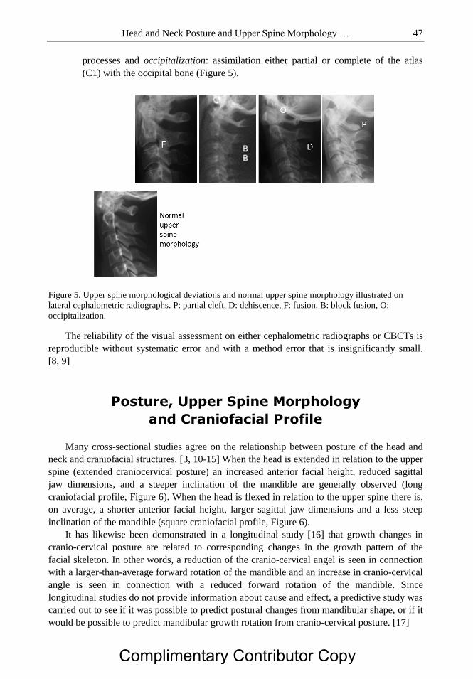
Head and Neck Posture and Upper Spine Morphology … 47
processes and occipitalization: assimilation either partial or complete of the atlas
(C1) with the occipital bone (Figure 5).
Figure 5. Upper spine morphological deviations and normal upper spine morphology illustrated on
lateral cephalometric radiographs. P: partial cleft, D: dehiscence, F: fusion, B: block fusion, O:
occipitalization.
The reliability of the visual assessment on either cephalometric radiographs or CBCTs is
reproducible without systematic error and with a method error that is insignificantly small.
[8, 9]
Posture, Upper Spine Morphology
and Craniofacial Profile
Many cross-sectional studies agree on the relationship between posture of the head and
neck and craniofacial structures. [3, 10-15] When the head is extended in relation to the upper
spine (extended craniocervical posture) an increased anterior facial height, reduced sagittal
jaw dimensions, and a steeper inclination of the mandible are generally observed (long
craniofacial profile, Figure 6). When the head is flexed in relation to the upper spine there is,
on average, a shorter anterior facial height, larger sagittal jaw dimensions and a less steep
inclination of the mandible (square craniofacial profile, Figure 6).
It has likewise been demonstrated in a longitudinal study [16] that growth changes in
cranio-cervical posture are related to corresponding changes in the growth pattern of the
facial skeleton. In other words, a reduction of the cranio-cervical angel is seen in connection
with a larger-than-average forward rotation of the mandible and an increase in cranio-cervical
angle is seen in connection with a reduced forward rotation of the mandible. Since
longitudinal studies do not provide information about cause and effect, a predictive study was
carried out to see if it was possible to predict postural changes from mandibular shape, or if it
would be possible to predict mandibular growth rotation from cranio-cervical posture. [17]
Complimentary Contributor Copy

Liselotte Sonnesen 48
Figure 6. Illustration on lateral cephalometric radiographs of the head and neck posture in relation to the
craniofacial profile.
It was found that prediction of the development in the individual child was possible for
children with extreme cranio-cervical postural relations. This means that an extreme small
cranio-cervical angle (< 79 degrees) was followed by an larger-than-average forward true
rotation of the mandible and on average a more horizontal facial growth pattern, where as an
extreme large cranio-cervical angle (> 113 degrees) was followed by a less-than-average
forward true rotation of the mandible and on average a vertical facial development. These
findings do indicate that posture, or factors determining posture, influences the direction of
the growth of the face to same extent.
Recently associations between posture of the head and neck and upper spine morphology
have been found. [18, 19] The studies showed that the cervical lordosis was significantly
more curved in subject with fusion than subjects without fusion (Figure 7). Furthermore, the
inclination of the upper spine was more backwards (the cervico-horizontal posture) in
subjects with fusion than subjects without fusion (Figure 7) and the head in relation to the
upper spine (the craniocervical posture) was more extended in subjects with occipitalization.
Figure 7. Illustration of associations between head and neck posture and fusion of the upper spine.
Complimentary Contributor Copy

Head and Neck Posture and Upper Spine Morphology … 49
Figure 8. Illustrations on lateral cephalometric radiographs of associations between upper spine
morphological deviations severe skeletal malocclsion traits and craniofacial profile. P: Partial cleft, B:
Block fusion, F: Fusion between two cervical vertebrae.
Furthermore, a series of studies have determined associations between upper spine
morphology and the craniofacial profile in adult patients with severe skeletal malocclusion
traits [8, 20-24] (Figure 8). In these studies, it was revealed that fusion of the upper spine was
associated with the craniofacial profile. A significant association between fusion and a large
cranial base angle, between fusion and retrognathia of the jaws and between fusion and
inclination of the jaws was found in patients with severe skeletal malocclusions. This
indicates that there might be an association between upper spine morphology, posture of the
head and neck and the development of the craniofacial profile including the cranial base.
Two main explanations have been given for the associations between the upper spine, the
cranial base and the craniofacial profile: a functional and an embryological approach. The
functional approach is the ―soft-tissue stretching hypothesis‖. [25] This hypothesis proposes
that a larger cranio-cervical angle (extension of the head) results in a larger distance between
the mandible and the sternum, and thus in a stretching of the soft-tissue envelope of the face
and neck. The increased tension in the soft-tissue layer will exert slightly higher forces on the
facial skeleton. [26] When active over a long period of time during growth, the forces will
restrict the forward growth of the maxilla and the mandible, and redirect in a more caudal
direction. Such a mechanism would explain the effect of extension of the cranio-cervical
posture on the development of the facial skeleton and in particular, of the mandible.
The other approach is the embryological hypothesis regarding the shared origin of the
spine and the posterior part of the cranial base to which the jaws are attached. In the early
embryogenesis, the notochord develops in the human germ disc and determines the
development of the spine, especially the vertebral bodies, and also the basilar part of the
occipital bone in the cranial base (the posterior part of the cranial base). [27-31] The para-
axial mesoderm forming the vertebral arches and remaining parts of the occipital bone are
also formed from notochordal inductions. Therefore, a deviation in the development of the
notochord may influence the surrounding bone tissue in the spine as well as in the posterior
part of the cranial base. On cephalometric radiographs and CBCTs it can be observed that the
bone tissue formed around the notochord are the vertebral bodies and the basilar part of the
occipital bone. The shared origin of the spine and posterior part of the cranial base is the basis
for the new hypothesis of associations between the spine and the cranial base to which the
jaws are attached. [32, 33] From early studies we also know that the angulation of the cranial
Complimentary Contributor Copy
Liselotte Sonnesen 50
base influences the growth direction of the jaws. [34, 35] Children with a large cranial base
angle develop a larger inclination of the jaws and retrognathia of the jaws (long craniofacial
profile). [34, 35] These findings correspond to the craniofacial profile seen in subjects with
fusion of the upper spine and an extended posture of the head. [23, 24]
Posture, Upper Spine Morphology
and Orofacial Function
The upper spine posture and morphology is not only related to the craniofacial profile,
but also to the orofacial function. In this chapter, two aspects of the orofacial function are
discussed: Dysfunction of the masticatory system in terms of temporomandibular disorders
(TMD) and obstruction of the upper airways. The term of TMD refers to symptoms and signs
associated with pain and functional and structural disturbances of the masticatory system,
especially the temporomandibular joints (TMJ) and the masticatory muscles. Textbooks and
clinicians generally agree that the most important symptoms and signs of TMD are headache,
tenderness in the masticatory muscles and the TMJ, reduced or impaired mobility of the
mandible, and TMJ sounds. [36] Symptoms and signs of TMD to a certain extent overlap
symptoms and signs of upper spine disorders, and clinical observations of a forward head
posture in subjects with TMD have already been reported in the 1950s. [37] In more recent
studies, a forward head posture (FHP) has been defined as a small value of the angle between
a horizontal line and a line from the tragus or the corner of the eye to the spinal process of
CV7, assessed clinically or measured on lateral photographs. Studies of FHP have so far
reported conflicting results of associations between posture of the head and neck and TMD.
[38-42] This lack of consistency of the findings may to some extent have been due to the fact
that skin surface measurements of posture do not reflect the actual postural relationship
between the bony components of the head and neck. [43] By using a more accurate technique
of cephalometric radiographs recorded in the standardized upright posture, it was possible to
confirm the Physiotherapists‘ clinical observations of relationships between TMD and the
posture of the head and neck. [5] Subjects with difficulty in jaw opening, clicking and locking
of the jaw and asymmetric jaw opening movement showed a marked forward inclination of
the upper spine. Furthermore, subjects with locking of the jaw and asymmetric jaw opening
movement also had an extended cranio-cervical posture (Figure 9).
Various explanatory models for this relationship have been proposed, but so far no
studies have documented whether the symptoms and signs of TMD are the results or the
causes of a forward and extended posture of the head and neck, or whether both are triggered
by other factors. In subjects with obstruction of the upper airways as in patients with
obstructive sleep apnoea (OSA) studies on posture of the head and neck and upper spine
morphology have been performed. The prevalence of upper spine morphological deviations
ranged between 32% to 46% and the morphological deviations occurred significantly more
often in patients with OSA than in subjects with no history of upper airway obstruction and
normal craniofacial morphology. [9, 44, 45] Upper spine morphological deviations in OSA
patients were described as fusions between two cervical vertebrae, block fusions,
occipitalization, partial cleft of the first cervical vertebra (C1) or dehiscence of the third
cervical vertebra (C3) and the fourth cervical vertebra (C4) [9, 44, 45] (Figure 10).
Complimentary Contributor Copy

Head and Neck Posture and Upper Spine Morphology … 51
Figure 9. Mean diagrams of lateral cephalometric radiographs of the children with (---) and without (-)
locking of the jaw. Superimposition on OPT (cv2ip).
Figure 10. Illustration on lateral cephalometric radiograph of upper spine morphological deviations in
patients with obstructive sleep apnoea. O: Occipitalization, B: Block fusion, D: Dehiscence.
In addition, the craniofacial profile of OSA, patients with block fusions in the cervical
vertebrae and fusion of two vertebrae differed significantly from the craniofacial profile of
other OSA patients. [45] The study suggests different craniofacial phenotypes in patients with
sleep apnea based on a new sub-classification of the patients according to the upper spine.
This may be a valuable basis for analysis of etiology, for diagnostics and treatment of sleep
apnoea patients. Furthermore, the head in relation to the upper spine (cranio-cervical posture)
was found to be extremely extended in patients with OSA. [46, 47] The findings were
considered to reflect a compensatory physiological postural mechanism that serves to
maintain airway adequacy in OSA patients in the awake erect posture. [47]
It would appear that the results indicate that there is an association between the posture of
the head and neck, upper spine morphology and orofacial function. So far however, the
explanations for the associations are still unknown. Thus, the extended posture of the head in
relation to the upper spine in patients with obstructive sleep apnoea is considered to be due to
a compensatory postural mechanism that serves to maintain airway adequacy.
Complimentary Contributor Copy

Liselotte Sonnesen 52
Conclusion
Posture of the head and neck and upper spine morphology can be evaluated on
cephalometric radiographs and CBCTs recorded in the standardised upright posture. On
average, an extended posture of the head in relation to the upper spine as well as fusion
between two cervical vertebrae was associated with a long craniofacial profile. On average,
TMD were seen in connection with a marked forward inclination of the neck and an extended
cranio-cervical posture. On average, an extended posture of the head and a significantly larger
occurrence of upper spine morphological deviations were seen in patients with obstruction of
the upper airways as in patients with obstructive sleep apnoea. The results indicate that
posture of the head and neck and upper spine morphology is a factor in the development of
the craniofacial profile and orofacial function in terms of TMD and obstruction of the upper
airways. This knowledge is clinically valuable, not only for the dentist and the orthodontist,
but also for the physician and physiotherapist.
References
[1] Solow B, Tallgren A Natural head position in standing subjects. Acta Odontologica
Scandinavica. 1971;29:591-607.
[2] Solow B, Tallgren A. Craniocervical posture in relation to skeletal and dentoalveolar
morphology. Transactions of the European Orthodontic Society. 1975; p. 231-35.
[3] Solow B, Tallgren A. Head posture and craniofacial morphology. Am J Physical
Anthropology. 1976;44:417-35.
[4] Solow B, Sonnesen L. Head posture and malocclusion. Eur J Orthod dontics. 1998;20:
685-93.
[5] Sonnesen L, Bakke M, Solow B. Temporomandibular disorders in relation to
craniofacial dimensions, head posture and bite force in children selected for orthodontic
treatment. Eur J Orthod. 2001;23:179-92.
[6] Siersbaek-Nielsen S, Solow B. Intra- and interexaminer variability in head posture
recorded by dental auxiliaries. Am J Orthod. 1982; 82:50-7.
[7] Sandham A. Cervical vertebral anomalies in cleft lip and palate. Cleft Palate J. 1986;
23:206-14.
[8] Sonnesen L, Kjær I. Cervical body fusions in patients with skeletal deep bite. Eur J
Orthod. 2007; 29:464-70.
[9] Sonnesen L, Jensen KE, Petersson AR, Petri N, Berg S, Svanholt P. Cervical vertebral
column morphology in patients with obstructive sleep apnoea assessed using lateral
cephalograms and cone beam CT. A comparative study. Dentomaxillofac Radiol. 2013;
42:20130060.
[10] Opdebeek H, Bell WH, Eisenfeld J, Mishelevich D. Comparative study between the
SFS and LFS rotation as a possible morphogenetic mechanism. Am J Orthodontics.
1978;74:509-21.
[11] Marcotte MR. Head posture and dentofacial proportions. Angle Orthodontics. 1981;
51:208-13.
Complimentary Contributor Copy

Head and Neck Posture and Upper Spine Morphology … 53
[12] Von Treuenfels H. Die Relation der Atlasposition bei prognather und progener
Kieferanomalie. Fortschr Kieferorthop. 1981;42:482-91.
[13] Solow B, Siersbæk-Nielsen S, Greve E. Airway adequacy, head posture, and
craniofacial morhpology. Am J Orthodontics. 1984;86:214-23.
[14] Hellsing E, McWilliam J, Reigo T, Spangfort E. The relation between craniofacial
morphology, head posture and spinal curvature in 8, 11 and 14-year-old children. Eur J
Orthod. 1987; 9:254-64.
[15] Huggare J. Head posture and craniofacial morphology in adults from Northern Finland.
Proc Finn Dent Soc.1989; 82:199-208.
[16] Solow B, Siersbæk-Nielsen S. Growth changes in head posture related to craniofacial
development. Am J Orthodontics. 1986;89:132-40.
[17] Solow B, Siersbæk-Nielsen S. Cervical and craniofacial posture as predictors of
craniofacial growth. Am J Orthodontics Dentofacial Orthopedics. 1992;101:449-58.
[18] Sonnesen L, Pedersen CE, Kjær I. Cervical column morphology related to head posture,
cranial base angle, and condylar malformation. Eur J Orthod. 2007;29:398-403.
[19] Arntsen T, Sonnesen L. Cervical vertebral column morphology related to craniofacial
morphology and head posture in preorthodontic children with Class II malocclusion and
horizontal maxillary overjet. Am J Orthod Dentofacial Orthop.2011; 140:e1-7.
[20] Sonnesen L, Kjær I. Cervical column morphology in patients with skeletal Class III
malocclusion and mandibular overjet. Am J Orthod. Dentofacial Orthop. 2007;132:
427.e7-12.
[21] Sonnesen L, Kjær I. Anomalies of the cervical vertebrae in patients with skeletal Class
II malocclusions and horizontal maxillary overjet. Am J Orthod Dentofac Orthop. 2008;
133: 198.e15-20.
[22] Sonnesen L, Kjær I. Cervical column morphology in patients with skeletal open bite.
Orthod. Craniofac. Res. 2008; 11:17-23.
[23] Sonnesen L. Association between the Cervical Vertebral Column and Craniofacial
Morphology. Int J Dent. 2010;295728.
[24] Sonnesen L. Cervical vertebral column morphology associated with head posture and
craniofacial morphology. Semin Orthod. 2012; 18:118-25.
[25] Solow B, Kreiborg S. Soft-tissue stretching: A possible control factor in craniofacial
morphogenesis. Scandinavian J Dental Res. 1977;85:505-7.
[26] Hellsing E, L´Estrange P. Changes in lip pressure following extension and flexion of
the head and a changed mode of breathing. Am J Orthod Dentofac Orthop. 1987;91:
286-94.
[27] Müller F, O‘Rahilly R. The early development of the nervous system in staged
insectivore and primate embryos. J Comp Neurol. 1980; 193:741-51.
[28] Kjær I. Human prenatal craniofacial development related to brain development under
normal and pathologic conditions. Acta Odontol Scand. 1995; 53:135-43.
[29] Kjær I. Neuro-osteology. Crit Rev Oral Biol Med. 1998;9:224-44.
[30] Kjær I, Hansen BF. The adenohypophysis and the cranial base in early human
development. J Craniofac Gen Dev Biol. 1995;15:157-61.
[31] Sadler TW. Embryology of the neural tube development. Am J Med Gen. 2005;135C:
2-8.
Complimentary Contributor Copy

Liselotte Sonnesen 54
[32] Sonnesen L, Nolting, Kjær KW, Kjær I. Associations between the development of the
body axis and the craniofacial skeleton studied by immunohistochemical analyses using
Collagen II, Pax 9, Pax 1, and Noggin antibodies. Spine. 2008; 33:1622-26.
[33] Sonnesen L, Nolting D, Engel U, Kjær I Cervical vertebrae, cranial base, and
mandibular retrognathia in human triploid fetuses. Am J Med Genet. Part A. 2009;
149A:177-87.
[34] Björk A. The face in profile. Sven Tandlak Tidskr. 1947;40:1.
[35] Björk A. Kæbernes relation til det øvrige cranium. In: Lundstrøm A (ed). Nordisk
Lärobok I Ortodonti. Stockholm: Sveriges Tandläkarforbunds Förlagsförening. 1975;
69-110.
[36] Bush FM, Dolwick MF (eds). Signs and symptoms. In: The temporo-mandibular joint
and related orofacial disorders. J B Lippincott Company, Philadelphia. 1995. p. 35-36.
[37] Perry HT Jr. Facial, cranial and cervical pain associated with dysfunction of the
occlusion and articulations of the teeth. Angle Orthodontist. 1956; 26:121-28.
[38] Kritsineli M, Shim YS. Malocclusion, body posture, and temporomandibular disorders
in children with primary and mixed dentition. J Clinical Pediatric Dentistry. 1992;16:
86-93.
[39] Hackney J, Bade D, Clawson A. Relationship between forward head posture and
diagnosed internal derangement of the temporomandibular joint. J Orofacial Pain.
1993;7:386-90.
[40] Watson DH, Trott PH. Cervical headache: an investigation of natural head posture and
upper cervical flexor muscle performance. Cephalalgia. 1993;13:272-84.
[41] Lee WY, Okeson JP, Lindroth J. The relationship between forward head posture and
temporomandibular disorders. J Orofacial Pain. 1995; 9: 161-67.
[42] Visscher CM, de Boer W, Lobbezoo F, Habets LLMH, Naeije M. Is there a relationship
between head posture and craniomandibular pain? J Oral Rehab. 2002; 29:1030-36.
[43] Johnson GM. The correlation between surface measurement of head and neck posture
and the anatomic position of the upper cervical vertebrae. Spine. 1998; 23:921-27.
[44] Sonnesen L, Petri N, Kjær I, Svanholt P. Cervical column morphology related to head
posture, cranial base angle, and condylar malformation. Eur J Orthod. 2007; 29:398-
403.
[45] Svanholt P, Petri N, Wildschiødtz G, Sonnesen L, Kjær I. Associations between
craniofacial morphology, head posture, and cervical vertebral body fusions in men with
sleep apnoea. Am J Orthod Dentofac Orthop. 2009; 135:702. e1-9.
[46] Solow B, Ovesen J, Nielsen PW, Wildschiødtz G, Tallgren A. Head posture in
obstructive sleep apnoea. Eur J Orthod. 1993; 15:107-14.
[47] Solow B, Skov S, Ovesen J, Norup PW, Wildschiødtz G. Airway dimensions and head
posture in obstructive sleep apnoea. Eur J Orthod. 1996;18:571-9.
Complimentary Contributor Copy

In: Posture: Types, Exercises and Health Effects ISBN: 978-1-63117-252-6
Editor: Sarah A. Curran © 2014 Nova Science Publishers, Inc.
Chapter 4
Emotion: The Missing Link in Posture
José Luís Pimentel do Rosário,* PT, PhD
Federal University of São Paulo, São Paulo, Brazil
Abstract
Posture is defined as the way we stand and how we ambulate from one point to the
next. It is associated with many aspects of human life including our political, emotional
and ethical positions. Despite this importance, the study of human posture is relatively
new compared to other areas of medical science. Certain deviations in posture can be
unsightly and can adversely affect muscular efficiency, as well as predisposing
individuals to musculoskeletal pathology. Posture can also alter or be altered by certain
psychological conditions.
The following factors can influence posture: biomechanics and the relation of the
body with gravity; the vestibular system and balance; the visual system; proprioception,
among others. The management of just one of these components in isolation could be
perceived as a clinical mistake, since there are many variables that can be present each of
which can include the role emotions. Pain or pleasure are described as opposite forms of
emotions which in turn can cause changes in action and decisions. The influence of pain
in particular has been linked to changes in emotion, and recent studies have confirmed
the connection between posture and the human emotional system. Awareness and
intervention to address the influence of emotions could assist with postural problems that
remain resistant to traditional postural management. The aim of this chapter is to review
the aspects of biomechanics and neurophysiology of posture and explore how they can be
related to emotions experienced by individuals.
Keywords: Emotion, posture, pain, postural problems, posterior chain
* Contact email: [email protected]
Complimentary Contributor Copy

José Luís Pimentel do Rosário 56
Introduction
The adoption of upright posture occurred relatively early in human evolutionary history.
Three million years ago, the pelvis was transformed from being adapted primarily to arboreal
quadrupedality as seen in living apes, to being tailored to terrestrial bipedality as seen in
modern humans, with the whole body supported by the feet. [1] An ancient riddle, dating
back over 2,500 years, describes postural changes in man: ―what is it that walks on four feet
in the morning, two at noon, and three at night?‖ Those who could not answer this riddle to
the sphinx were put to death. Greek mythology provided the following answer: ―Man, who
crawls on four limbs as a baby, walks upright on two as an adult, and walks with the aid of a
stick in old age.‖ [2]
This riddle demonstrates how posture is influenced by many aspects of human life. Our
posture is characterized by how we behave in life, how we relate to other people, the way we
stand and how we move from one point to the next. Our political, emotional or ethical
positions are evident in our posture and stance, so too are our viewpoint, standpoint, position,
stance, condition, state, mood, and attitude in various situations. In spite of this importance,
the study of human posture is relatively new compared to other areas of medical science.
Certain deviations in posture can be unsightly and can adversely affect muscular efficiency,
as well as predisposing individuals to musculoskeletal pathology. It is also acknowledged that
posture can be influenced by some psychological conditions such as sadness and depression.
[3-7] For these reasons, posture has been studied by various professionals such as medical
practitioners, psychologists and physical therapists, architects, podiatrists, sports scientists as
well as others.
In the nineteenth century, based on his observations, Charles Darwin wrote about the role
of posture. [8] This study however, only began to emerge in 1927, when Wilhelm Reich, an
Austrian physician, paid attention to the fact that body language could be more revealing than
the words a patient used. He further acknowledged that body posture was closely related to
muscle contractions, which he referred to as ‗body armor‘ that in turn was provoked by
emotions. [9] By 1930, the Australian author Frederick Alexander, after curing his own
hoarseness and correcting his cervical spine, went on to defend the idea that a better posture
would prevent the onset of diseases and body aches. His studies on the alignment of the
cervical and lumbar spine to facilitate an organic general improvement were called ―The
Alexander technique‖, [10] and is used to the present day by various clinicians (e.g.,
osteopaths, physical therapists).
In 1947, the kinesiotherapist Françoise Mézières stated that human muscles are
completely inter-related and demonstrated that we do not have a single muscle that causes
poor posture, but rather chains of muscles (Figure 1) that can create pathology in a specific
location. This can be from a generalized tension, whereby the root of the problem can be
distant from where the patient feels pain. [11] In the next decade, Ida Rolf began to teach her
recently developed technique based on the fascia instead of muscles. [12] It is interesting to
note that the patterns of the fascia treated by Ida Rolf and further explained by her students
follow a very similar pattern of the Mézières‘ chains. [13]
The first edition of “Muscles: Testing and Function” was released in 1967. New
chapters, specifically about posture, were added to later editions and the words ‗with Posture
and Pain‘ were also added to the title. The core textbook for many involved in
Complimentary Contributor Copy

Emotion: The Missing Link in Posture 57
musculoskeletal assessment and rehabilitation and is now in its 5th
edition. The prime feature
of this textbook focused on the development of an established standard assessment of posture
(Figure 2) and drew attention to the fact that only correct posture relieves certain spine, feet
and leg pain. [14] In the seventies, Mosche Feldenkrais, an Israeli physicist and martial arts
master, explained that body posture can only be improved when there is conscience of the
movement. According to Feldenkrais, the improvement of kinesthetic and proprioceptive self-
awareness of functional movement and body position could lead to increased function,
reduced pain, and greater ease and pleasure of movement. [15]
Figure 1. The posterior chain according to the Muscular Chain Therapy (based on the Mézières
method).
Figure 2. A bad posture alters the relation between the body segments, which may alter the joint
mechanics and produce pain (A). This specific alteration is pushing the body forward, which can also
move the center of pressure of the body in the same direction (B).
Complimentary Contributor Copy

José Luís Pimentel do Rosário 58
During this period and thereafter, many schools and authors showed an interest in
researching human posture. [3,5,6,16-29] In the beginning however, only Reich [9] associated
posture and emotions. The aim of this chapter is to review the aspects of biomechanics and
neurophysiology of posture and explore how they can be related to emotions experienced by
individuals.
Biomechanics
Ideal postural balance exists when there is a perfect distribution of body mass around the
center of gravity, with the compressive forces on spinal discs balanced by ligamentous
tension. [30] This position creates the least amount of joint stress, requires the least amount of
muscular activity and, therefore, is the position of maximum effectiveness. A deviation from
this optimum position should be compensated by changes in joint position which, in turn,
must be maintained by an increase in muscle activity. Therefore, postural imbalance results in
excessive energy consumption. [31]
Ideal posture should not deviate from a plumb line passing sagittally in an anterior or
posterior view. In the lateral view, the plumb line should pass slightly anterior to the lateral
malleolus, slightly anterior to the axis of the knee joint, and slightly posterior to the axis of
the hip joint, the bodies of lumbar vertebrae, the shoulder joint, the bodies of most cervical
vertebrae as well as the external auditory canal and slightly posterior to the apex of the
coronal suture. [14]
Present day orthopaedic assessment uses the assumption that referred pain in a joint may
have its cause in adjacent joints. [32] In terms of posture, this concept can be taken beyond
adjacent segments. The misalignment of a body segment will influence the next, which will
have a further influence, and is described as a chain reaction. This can occur in the following
circumstances:
When a muscle is in a shortened position to distribute the tension generated in a joint
to other muscles;
When tension is distributed in the fascia and connective tissue; when the position of
the center of gravity is altered and compensated at an opposite change of equal
magnitude in one or more segments above or below the unaligned segment.
Thus, a deformity that occurs anywhere in the body can be compensated many times
throughout the body segments, and the last segment, which cannot be compensated in any
other point, is usually the location of the pain. [33]
Spine
Much of the muscle tension generated will be transmitted to the spine, which is a central
region with great capacity for compensation at all levels. This tension can arise in the lower
limbs (ascendant), or in the head (descendant). [34] It is considered a very important region
once it holds the spinal cord. Postural misalignments on the spine can generate pain all over
Complimentary Contributor Copy

Emotion: The Missing Link in Posture 59
the body and even visceral problems. [35] All postural methods highlight the importance of
the spine for the treatment of postural deviations and overall health. Due to its many
articulations the spine can show several different alterations in posture, which can be small.
These include inclinations or rotations in one or more vertebras, affecting many spinal
segments such as hyperlordosis or scoliosis.
Diaphragm and Inspiratory Muscles
The respiratory pump consists of breathing muscles which affect the ribs, scapula,
clavicle, sternum and thoracic spine. The fact that breathing can be altered by changes in
position, emotional state, activity level, level of alert or sleep stage, illness, posture, muscle
strength, airflow resistance, compliance of the lungs and chest wall and even tight clothing
means that there are numerous varieties in breathing patterns. [14,36] When posture is altered,
the body tends to maintain the chest in an inspiratory position rather than an expiratory
position, for survival purposes. [37]
In fact, the shortening of the diaphragm produces postural changes such as elevated ribs
and hyperlordosis at L2 level and, with the help of other inspiratory muscles, could cause the
elevation of the entire rib cage. [38]
Lower Limbs
When one limb is significantly shorter than the other, the result is referred to as a
functional scoliosis. [39] Several idiopathic scoliosis types are caused by a greater torsion in
one of the lower limbs, causing the same effect as a smaller limb. This rotation of the lower
limb, in turn causes rotation of the hips. When these alterations reach the spine they may
produce compensations in all planes, vertebral inclination, rotation and even listesis.
Inclinations and rotations are the root cause of scoliosis. [40] Any disturbance at the
foundational level of the feet (feet are the interface between the body and supporting surface)
can, depending on its features, affect the knee (valgus or varus misalignment or asymmetric
rotations) provoking in many cases, spinal alterations such as hyperlordosis, straightening of
the spine or scoliosis. [41]
Upper Limbs
Compared to the lower limbs, the upper limbs do not have a marked an effect on posture,
but a bundle of tonic muscle connects the forearm to the arm, the shoulder girdle and the
neck. All the tension of the upper limbs can be transmitted to the body through the neck,
while the opposite can also happen. [37,38]
Complimentary Contributor Copy

José Luís Pimentel do Rosário 60
Temporomandibular Joint
The position of the cervical spine has a close relationship with the temporomandibular
joint (TMJ). [42] When the atlanto-occipital joint moves toward flexion, an associated sliding
backwards movement occurs in the same joint. When the movement is characterized by
extension, it is associated with a sliding forward movement. [43] Forces generated by anterior
translation of the atlas with the occipital (extension) lead to a retropulsion of the mandible. In
the opposite case, cervical flexion provokes a translation and consequently a jaw antepulsion.
[44]
Neurophysiology
Posture is peripherally produced by alpha motor neurons and commonly involves the
antigravity muscles. Virtually all systems involved in the regulation of posture and the
production of movement exert a direct (monosynaptic) or indirect (via segmental
interneurons) influence on the somatic efferent neurons. [45]
Three sensory systems provide information about the body position in relation to gravity
and the environment. These are known as the vestibular, proprioceptive and visual systems.
The vestibular system provides information about head position in relation to gravity and
linear and rotational head movements. Proprioceptors, particularly those associated with the
axial joints and muscles (muscle spindles and Golgi tendon organs), provide information
about the movement of individual body segments. The visual system provides information
about the body position in relation to the external environment. [41,45]
The muscle spindles monitor changes in muscle length and the speed of these changes
and play a vital role in sensing position and movement. The spindle may be the cause of a
muscle spasm, which is sometimes strong and painful, in the case of cramp, and is sometimes
caused by the central nervous system, known as spasticity. [46] These spasms can affect
posture even when light and permanent. [47] The Golgi tendon organs monitor intramuscular
tension and inhibit muscle contraction with an exaggerated protective function. They are also
responsible for muscular relaxation after stretching. [48]
The vestibular system maintains body balance by regulating the antigravity muscles of
the neck, trunk and limbs and preventing the body's center of mass from moving away from
the support base. It aids by coordinating movements of the head and body, and maintaining
the eyes fixed while the head is moving. Balance abnormalities caused by an injury to the
labyrinth or vestibular nerve (peripheral vestibular syndrome) usually broaden the support
base and can cause the head and trunk to tilt to the side of the injury. [49]
The visual system may be the cause of serious deviations of the neck and trunk. A
strabismus, caused by hypofunction of the right lateral rectus muscle in the right eye, can
cause a head rotation to the left. This pure example however is rare. Rotation and side
bending caused by more than one muscle usually compensate for the problem. Bilateral
palpebral ptosis (eyelid closure) requires the carrier to maintain the head in extension so that
they can see through the remaining opening. [50]
Complimentary Contributor Copy

Emotion: The Missing Link in Posture 61
Body Image
Another important factor is body image, which is defined as a visual and mental image,
including feelings of one‘s own body. Body schema is slightly different and refers to a type of
body posture, including the relationship of body parts with each other, and the relationship of
the body with the environment. This phenomenon derives from the integration of the tactile,
proprioceptive and interoceptive functions. [51,52] Asomatognosia is a disorder which causes
a lack of awareness of body structure and the relationship between body parts in the
individual. Unilateral spatial neglect is the failure to register and integrate stimuli and
perceptions on one side of the body. [53] Common disorders such as anorexia nervosa
(characterized by significant weight loss, distorted body image and the pursuit of thinness) are
linked to body image. [54-56] Therefore, even small disturbances may affect posture in some
way.
Sacks [57] reported a case of a 93-year-old man with Parkinson's disease who exhibited a
significant trunk tilt to one side that was almost on the verge of collapse, without realizing it.
The author commented that the proprioceptive and visual systems of this patient had affected
the individual body image and labyrinthine.
Emotions
Emotions are brief psychophysiological phenomena that have an adaptive aspect in
relation to a changing environment. They alter attention, change certain behaviors and
activate relevant associative networks in memory. Emotions directly influence perception
through the senses, affecting information processing and subsequent actions. [58] Human
behavior is strongly determined by emotions, [59] and it is possible to consider them as the
primary motivational system of human beings. [60]
According to Aristotle, [61] emotions are reactions accompanied by pain or pleasure that
lead to alterations in people, causing them to change their mind. An emotion can also be
defined as the integration of particular sets of neurochemical, motor and mental processes,
with a number of structural characteristics that distinguishes it from other emotions. [60,62]
There is evidence [59,63] of distinct activity patterns in the Autonomic Nervous System
(ANS) for anger, fear and sadness. [64] An emotion can quickly organize the physiological
responses of different biological systems such as facial expressions, muscle tone, the voice,
ANS activity and endocrine activity.
These alterations are intended to produce the best conditions for an effective physical
response. [60,64] Siegel [65] stated that moods change the physical state by means of the
central nervous system, the endocrine system and the immune system. According to the same
author, every tissue and organ is influenced by a complex interaction between hormones that
obey the hypothalamus and the pituitary. The hypothalamus regulates the majority of the
body‘s unconscious life processes, such as heartbeat, respiration and blood pressure, among
others. Nerve fibers enter the hypothalamus from almost all regions of the brain. Thus, the
emotional and intellectual processes which occur in the brain affect the body. This may be
one way that emotions influence the physical state and vice versa. Emotions, as well as
posture and facial expressions are usually momentary. [66] However, it is notable that people
often repeat certain emotional and postural patterns.
Complimentary Contributor Copy
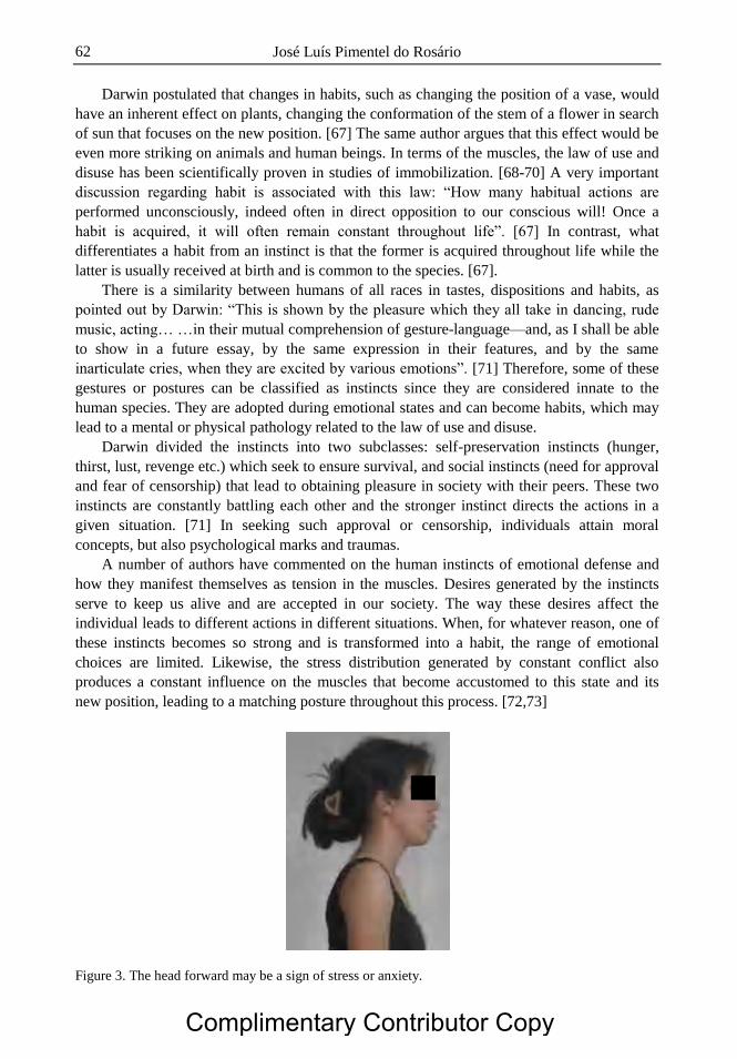
José Luís Pimentel do Rosário 62
Darwin postulated that changes in habits, such as changing the position of a vase, would
have an inherent effect on plants, changing the conformation of the stem of a flower in search
of sun that focuses on the new position. [67] The same author argues that this effect would be
even more striking on animals and human beings. In terms of the muscles, the law of use and
disuse has been scientifically proven in studies of immobilization. [68-70] A very important
discussion regarding habit is associated with this law: ―How many habitual actions are
performed unconsciously, indeed often in direct opposition to our conscious will! Once a
habit is acquired, it will often remain constant throughout life‖. [67] In contrast, what
differentiates a habit from an instinct is that the former is acquired throughout life while the
latter is usually received at birth and is common to the species. [67].
There is a similarity between humans of all races in tastes, dispositions and habits, as
pointed out by Darwin: ―This is shown by the pleasure which they all take in dancing, rude
music, acting… …in their mutual comprehension of gesture-language—and, as I shall be able
to show in a future essay, by the same expression in their features, and by the same
inarticulate cries, when they are excited by various emotions‖. [71] Therefore, some of these
gestures or postures can be classified as instincts since they are considered innate to the
human species. They are adopted during emotional states and can become habits, which may
lead to a mental or physical pathology related to the law of use and disuse.
Darwin divided the instincts into two subclasses: self-preservation instincts (hunger,
thirst, lust, revenge etc.) which seek to ensure survival, and social instincts (need for approval
and fear of censorship) that lead to obtaining pleasure in society with their peers. These two
instincts are constantly battling each other and the stronger instinct directs the actions in a
given situation. [71] In seeking such approval or censorship, individuals attain moral
concepts, but also psychological marks and traumas.
A number of authors have commented on the human instincts of emotional defense and
how they manifest themselves as tension in the muscles. Desires generated by the instincts
serve to keep us alive and are accepted in our society. The way these desires affect the
individual leads to different actions in different situations. When, for whatever reason, one of
these instincts becomes so strong and is transformed into a habit, the range of emotional
choices are limited. Likewise, the stress distribution generated by constant conflict also
produces a constant influence on the muscles that become accustomed to this state and its
new position, leading to a matching posture throughout this process. [72,73]
Figure 3. The head forward may be a sign of stress or anxiety.
Complimentary Contributor Copy

Emotion: The Missing Link in Posture 63
The effects of emotional tension on posture can be confirmed by the increased
electromyographic muscle activity during a condition of mental stress [74] and the increased
difficulty of maintaining posture control with the threat caused by increased height. [75]
Another study states the there is a relation between physical or psychological stress with neck
pain caused by an increased contraction of the upper trapezius, verified by electromyography.
[76] It means that stress has an effect on the posture of the shoulder and head. Another author
states that anxiety and forward head seems to be related as well [77] (Figure 3).
Experiences with Posture
It is assumed that if the body segments are kept out of alignment for extended periods,
the muscles are used in a shortened position and become stronger than their antagonists,
which are elongated and weak. [14,78] Strengthening exercises have been prescribed in an
attempt to correct postural deviations. In theory, the strengthening of weak and overly
stretched muscles results in an adaptive shortening. Together with the stretch of their short
antagonists, the body segment would be repositioned. [14,79] However, a number of authors
have found no significant association between lumbar lordosis and the strength of the trunk
flexors. [80-82] In order to maintain posture for a long time, resistance exercises may be more
important than maximal strength exercises. Associations between the resistance of the
abdominal muscles and lumbar posture have been sought, but no significant relationship was
found. [3,83]
The definition of what classifies as a strong muscle is interesting. A weightlifting
champion may have very strong extensors of the trunk with an absence of lower back pain or
shortened muscles. It is possible that an investigation into the relationship between flexors
and extensors could aid in an improved understanding, although there is difficulty in relating
muscle groups with different demands of strength.
Itoi and Sinaki undertook a two-year prospective study to test the hypothesis that
increasing the strength of trunk extensors could reduce a hyperkyphosis. [84] The group that
performed the trunk extension in prone position ten times, five days per week, increased in
strength significantly more than the control group who did not exercise at all. There was no
radiographic difference for kyphosis and only a minimal difference for lordosis, which was
lower in the group that did exercises. In fact, the trunk extension exercise had a greater effect
on the lower back. The eccentric phase of the exercise should have been slower than the
positive phase in order to prevent an accident in front of the trunk. There was an emphasis on
eccentric muscle contraction, which may have generated a strong stretch of the lower back.
No association was found between the horizontal extension range of motion of the shoulder
and the abducted scapula. [85] No significant correlation was found between lumbar lordosis
and the radiographically measured range of motion in flexion and trunk extension and hip
extension in women. [80] There was also no significant association between the length of the
back muscles and lumbar lordosis, although there was a significant association between the
length of the abdominal muscles and lumbar lordosis. [81]
The effect of the combination of stretching and strengthening on lumbar lordosis has
been studied. A small difference in the decrease of lumbar lordosis was found in an
experimental group who performed the lumbar spine and stretching exercises for the hip
extensor and flexor muscles of the trunk three times a week for a period of four weeks. [87]
Complimentary Contributor Copy

José Luís Pimentel do Rosário 64
The three groups that performed exercises with a vest and electrical stimulation were
compared in terms of alterations in idiopathic scoliosis. After three months all groups showed
a trend towards a reduction of scoliosis, although there were no significant differences
between the groups.
Wang et al. investigated the influence of a program of strengthening and stretching for
scapula posture. Exercises were performed three times per week and six weeks after any
change in scapular resting posture was noted. [88] In a review of intervention in scoliosis and
kyphosis, White and Panjabi questioned the effectiveness of these exercises to correct
postural deviations. They suggested that the forces generated by corrective exercises are
usually of small amplitude, frequency and duration and thus, are not sufficient to promote
permanent changes in muscle length. [89] One possible benefit of an exercise program could
be the reeducation of the patient to adopt better posture during daily activities. [90] In a more
recent review, Rosário questioned whether resistance training alone actually produces the
adaptive shortening of the muscles that causes postural changes. The author concluded that
the frequency and duration of exercise programs were ineffective to produce an adaptive
shortening of muscles. [91]
It is clear that existing knowledge about posture is insufficient. Moreover, it is not
recommended to analyze a single aspect of posture in isolation. For example, it is difficult to
assume that lumbar lordosis is only caused by a weakness of the abdominal muscles or short
paravertebral muscles.
Other factors may be influencing the adoption of a certain posture. Some of the
associated problems include shortening of the diaphragm and other muscles, body image
issues and adjustment problems of body segments in relation to the equilibrium point.
Techniques involving the Muscular Chain theory have been successful in treating postural
problems. [91]
The number of studies that have associated posture and emotion is very small. McHugh
and Newell [92] reported the possibility of determining the emotions of groups of people,
whose faces were hidden, thereby confirming that emotions can only be understood by
studying posture and movements. In a similar study, Kret and Gelder [93] instructed the
subject to describe the emotion of a single person among a group. Grammer et al. [94]
analyzed the movement of a number of subjects and investigated which movements, such as
elbow flexion or external rotation for example, were more commonly repeated. The subjects
completed a questionnaire in which they reported their emotional state (confidence or
irritation) at the time.
Depression, anger, sadness and other emotions have recorded statistical significance
when correlated with certain postural deviations. [95] However, until now, there has been no
research about the possibility of emotional treatment for postural problems or postural
treatment for emotional problems. Exceptions include the empirical studies based on the work
of Reich [72] and the contemporary followers of his philosophy. Nevertheless, it seems clear
that stretching and strengthening the muscles of a depressed patient, in order to improve their
forward and flexed head, will not be enough if this psychological state continues and this
same posture is adopted in life.
Complimentary Contributor Copy

Emotion: The Missing Link in Posture 65
Conclusion
Posture is a challenging subject to study. It is difficult to assess quantitatively and from a
qualitative perspective it can be open to misinterpretations. It is not possible to consider one
body segment in isolation, which is one of the most common mistakes in this area. The
human body is completely interconnected and relationships in one segment cannot be as
reductionist as studying an agonist and its antagonist. The eyes, TMJ and the length of the
limbs are, among other factors, all linked to posture. Likewise, it makes no sense to correct a
scoliosis without treating the rotations of the lower limb or correcting the difference of the
members if any.
Whilst emotions represent further challenges in terms of study, there does appear to be a
correlation with postural deviations which should be further investigated. Postural work
should aim not only to stretch and strengthen muscles, but also to deal with muscular chains,
and to improve associated aspects of the nervous system, as well as factors related to
psychological and body image self-perception.
References
[1] Lovejoy OC. The natural history of human gait and posture Part 1 Spine and pelvis.
Gait Posture. 2005;21:95-112.
[2] Dudeney HE. The Canterbury puzzles and other curious problems. Project: Gutenberg
(ebook); 2008.
[3] Rosário JLP, Marques AP, Maluf SA. Aspectos Clínicos do Alongamento: uma revisão
de literatura. Revista Brasileira de Fisioterapia. 2004;8:83-88.
[4] Liebenson C. Postural correction. J Bodyw Movement Ther. 2008; 12:318-9.
[5] Wallden M. The neutral spine principle. J Bodyw Movement Ther. 2009; 13:350-61.
[6] Rosário JLP, Diógenes MSB, Mattei R, Leite JR. Can sadness alter the posture? J
Bodyw Movement Ther. 2013;17:328-31.
[7] James H, Castaneda L, Miller ME, Findley T. Rolfing structural integration treatment
of cervical spine dysfunction. J Bodyw Movement Ther. 2009;13:229-38.
[8] Darwin CR. The descent of man, and selection in relation to sex. London: John Murray;
1871.
[9] Reich W. The function of the orgasm. New York: Orgone Institute Press; 1942.
[10] Brown R. The authorized summaries of F.M. Alexander's four books. London: STAT
Books; 1992.
[11] Bertherat T, Bernstein C. O corpo tem suas razões – Antiginástica e a consciência de si.
São Paulo: Martins Fontes; 1987.
[12] Feitis R, Schultz L. Remembering Ida Rolf. North Atlantic Books: Berkeley; 1997.
[13] Myers T. Anatomy trains. 2nd
ed. Edinburgh: Elsevier; 2009.
[14] Kendall FP, Mccreary EK, Provance PG, Rodgers M, Romani W. Muscles: testing and
function, with posture and pain. Baltimore: Lippincott Williams & Wilkins; 2010.
[15] Feldenkrais M. Consciência pelo movimento. São Paulo: Summus; 1977.
Complimentary Contributor Copy

José Luís Pimentel do Rosário 66
[16] Falconer MA, Mcgeorge M, Begg AC. Obsevations on the course and mechanism os
syntom-production in sciatica and low back pain. J Neurol Neurosurg Psychiatry. 1948;
2:13.
[17] Jones LH. Spontaneus release by positioning. Doctor Osteopathy. 1964;4:109.
[18] Caillet R. Neck and arm pain. Philadelphia: F. A. Davis; 1964.
[19] Caillet R. Soft tissue pain and disability. Philadelphia: F. A. Davis; 1977.
[20] Maitland GD. Peripheral manipulation. London: Butterworth;1970.
[21] Maitland GD. Vertebral manipulation. London: Butterworth;1977.
[22] Magora A. Investigation of the relationship between low back pain and occupation, age,
sex, community, education and other factors. Industr Med Surg. 1971;39:465-71.
[23] Farfan HF. Mechanical disorders of the low back. Philadelphia: Lea and Febiger; 1973.
[24] Rosário JLP, Sousa A, Cabral CMN, João SMA, Marques AP. Reeducação postural
global e alongamento estático segmentar na melhora da flexibilidade, força muscular e
amplitude de movimento: um estudo comparativo. Fisioterapia e Pesquisa. 2008;15:
12-8.
[25] Sweeting K, Mock M. Gait and posture - assessment in general practice. Austral Family
Physician. 2007; 36:398-401, 404-5.
[26] Murley GS, Landorf KB, Menz HB, Bird AR. Effect of foot posture, foot orthoses and
footwear on lower limb muscle activity during walking and running: a systematic
review. Gait Posture. 2009;29:172-87.
[27] Bötzel K, Kraft E. Strategies for treatment of gait and posture associated deficits in
movement disorders: the impact of deep brain stimulation. Restorative Neurology
Neuroscience. 2010; 28:115-22.
[28] Grilley P. Working posture and the fascial structures of the lower spine J Bodyw
Movement Ther. 2008;12:257-66.
[29] Curnow D, Cobbin D, Wyndham J, Choy B. Altered motor control, posture and the
Pilates method of exercise prescription. J Bodyw Movement Ther. 2009;13:104-11.
[30] Kappler RE. Postural balance and motion patterns. J Am Osteopathic Assoc. 1982;81:
69-77.
[31] Lee D. Princípios e práticas da força muscular e das técnicas funcionais. In: Moderna
terapia manual da coluna vertebral, Grieve GP. Editorial Médica Panamericana: São
Paulo; 1994.
[32] Hoppenfeld S. Propedêutica ortopédica coluna e extremidades. São Paulo: Atheneu.
1980.
[33] Bienfait M. Fáscias e pompagens. São Paulo: Summus Editorial; 1999.
[34] Bienfait M. Fisiologia da terapia manual. São Paulo: Summus Editorial; 1989.
[35] Barral JP. Visceral manipulation II. Seattle: Eastland Press;1988.
[36] Shneerson J. Disorders of ventilation. London: Blackwell Scientific Publications. 1988.
[37] Souchard PE. O stretching global ativo - A reeducação postural global a serviço do
esporte. São Paulo: Manole; 1996.
[38] Marques AP. Cadeias musculares – um programa para ensinar avaliação
fisioterapêutica global. São Paulo: Manole; 2000.
[39] Gurney B. Leg length discrepancy. Gait Posture. 2002;15:195-206.
[40] Souchard PE, Ollier M. As escolioses. São Paulo: É realizações; 2001.
[41] Bricot B. Posturologia, São Paulo: Ícone; 1999.
Complimentary Contributor Copy

Emotion: The Missing Link in Posture 67
[42] Huggare JA, Raustia AM. Head posture and cervicovertebral and craniofacial
morphology in patients with craniomandibular dysfunction. J Craniomandib Pract.
1992;10: 173-9.
[43] Kapandj AI. Fisiología articular. Tronco y raquis. Madrid: Editorial médica
panamericana; 1999.
[44] Makofsky HW. The effect of head posture on muscle contact position: the sliding
cranium theory. J Craniomandib Pract. 1989;7:286-92.
[45] Jewell MJ. Visão geral da estrutura e função do sistema nervoso central. In: Umphred
DA, Schmitz TJ. Fisioterapia neurológica. São Paulo: Manole;1994.
[46] Knight KL. Crioterapia no tratamento das lesões esportivas. São Paulo: Manole; 2000.
[47] Ge W, Long CR, Pickar GJ. Vertebral position alters paraspinal muscle spindle
responsiveness in the feline spine: effect of positioning duration. J Physiol. 2005; 569:2
655-65.
[48] Schmitz TJ. Examination of Sensory Function. In: Umphred DA & Schmitz TJ.
Physical rehabilitation. Philadelphia: F.A. Davis; 2007. p.121-158.
[49] Berne RM, Levy MN. Fisiologia. Rio de Janeiro: Editora Guanabara Koogan; 1988.
[50] Bicas HEA. Torcicolo. Posição viciosa de cabeça. Medicina, Ribeirão Preto. 2000;
33:64-72.
[51] Halperin E, Cohen BS. Perceptual-motor dysfunction. Stumbling block to
rehabilitation. Maryland Med J. 1971;20:139.
[52] Rumsey N, Clarke A, Musa M. Altered body image: the psychosocial needs of patients.
Br J Community Nurs. 2002;7:563-6.
[53] Lamm-Warburg C. Estratégias para avaliação e planejamento do tratamento das
deficiências da percepção. In: O‘Sullivan SB, Schimitz TJ. Fisioterapia avaliação e
tratamento. São Paulo: Manole; 1993. p. 93-120.
[54] Matusevich D, Garcia A, Gutt S, de la Parra I, Finkelsztein C. Hospitalization of
patients with anorexia nervosa: a therapeutic proposal. Eat Weight Disord. 2002;7:196-
201.
[55] Farrington A, Waller G, Neiderman M, Sutton V, Chopping J, Lask B. Dissociation in
adolescent girls with anorexia: relationship to comorbid psychopathology. J Nerv Ment
Dis. 2002; 190:746-51.
[56] Tamburrino MB, McGinnis RA. Anorexia nervosa. A review. Panminerva Med.
2002;44:301-11.
[57] Sacks OW. O homem que confundiu sua mulher com um chapéu e outras histórias
clínicas. São Paulo: Companhia das letras; 1997.
[58] Damásio A. O Erro de descartes: Emoção, razão E cérebro humano. Lisboa:
Publicações Europa-América; 1995.
[59] Levenson RW, Ekman P, Friesen WV. Voluntary facial expression generates emotion-
specific nervous system activity. Psychophysiology. 1990; 27: 363-84.
[60] Izard CE. The psychology of emotions. New York: Plenum Press; 1991.
[61] Aristóteles. Arte Retórica e Arte Poética. São Paulo, Difusão Européia do Livro. 1959.
[62] Izard CE. Emotion-cognition relationship and human development. In CE Izard, J
Kagan, RB Zajonc (Eds.), Emotions, cognition, and behavior. New York: Cambridge
University Press; 1984. p.17-37.
[63] Ekman P, Levenson RW, Friesen WV. Autonomic nervous system activity
distinguishes between emotions. Science. 1983; 221, 1208-10.
Complimentary Contributor Copy

José Luís Pimentel do Rosário 68
[64] Levenson RW, Carstensen LL, Friesen WV, Ekman P. Emotion, physiology, and
expression in old age. Psychol Aging. 1991; 6, 28-35.
[65] Siegel BS. Amor, Medicina e Milagres - A cura espontânea de doentes graves, Segundo
a experiência de um Famoso cirurgião Norte-Americano. São Paulo: Best Seller; 1989.
[66] Frijda NH. Moods, emotion episodes, and emotions. In: Lewis M, Haviland IM (eds).
Handbook of emotions. New York: Guilford Press; 1993.p. 381-403.
[67] Darwin CR. A origem das espécies e a seleção natural. Curitiba: Hemus. 1989.
[68] Goldspink G. Sarcomere length during post-natal growth in mammalian muscle fibers.
J Cell Sci. 1968; 3:539-48.
[69] Williams PE, Goldspink G. Longitudinal growth of striated muscle fibers. J Cell Sci.
1971; 9: 751-67.
[70] Williams PE. Use of intermittent stretch in the prevention of serial sarcomere loss in
immobilized muscle. Ann Rheum Dis. 1990; 49: 316-7.
[71] Darwin CR. The expression of the emotions in man and animals. New York: d.
Appleton and company; 1899.
[72] Reich WA função do orgasmo. São Paulo: Brasiliense. 1995.
[73] Keleman S. Embodying experience: Forming a personal life. Berkeley: Center Press.
1987.
[74] Loon E, Van Masters RSW, Ring C, Mcintyre DB. Changes in limb stiffness under
conditions of mental stress. J Mot Behav. 2001; 33: 153-64.
[75] Adkin AL, Frank JS, Carpenter MG, Peysar GW. Postural control is scaled to level of
postural threat. Gait Posture. 2000; 12:87-93.
[76] Shahidi B, Haight A, Maluf K. Differential effects of mental concentration and acute
psychosocial stress on cervical muscle activity and posture. J Electromyogr Kinesiol.
2013;23:1082-9.
[77] Rosario JLP. Avaliação do corpo para tratamento emocional. Ed. Barauna. São Paulo;
2011.
[78] Novak CB, Mackinnon SE. Repetitive use and static postures: a source of nerve
compression and pain. J Hand Ther. 1997; 10:151-9.
[79] Alter MJ. Ciência da flexibilidade. Artmed. 1996.
[80] Flint MM. Lumbar posture: a study roentogenographic measurement and the influence
of flexibility and strenght. Res Q. 1962; 34:15-21.
[81] Walker ML, Rothstein SD, Finucane SD, Lamb RL. Relationships between lumbar
lordosis, pelvic tilt, and abdominal muscle performance. Phys Ther. 1987;67:512-16.
[82] Youdas JW, Garret TR, Harmsen S, Sauman VJ, Carey JR. Lumbar lordosis and pelvic
inclination of asymptomatic adults. Phys Ther. 1996; 76:1066-81.
[83] Mulhearn S. George K. Abdominal muscle endurance and its association with posture
and low back pain. Physiotherapy. 1999; 85:210-16.
[84] Itoi E, Sinaki M. Effect of back strengthening exercise on posture in healthy women 49
to 65 years of age. Mayo Clin Proc. 1994; 69:1054-9.
[85] Coppock DE. Relationship of tightness of pectoral muscles to round shoulders in
college women. Res Q. 1958; 29:146-153.
[86] Alizadeh MH. Standring J. The effect of an exercise regime on lumbar spine curve. In:
The engineering of sport. Haake S editor. Rotterdam: A.A. Balkemia; 1996.
[87] El-Sayyad M, Conine TA. Effect of exercise, bracing and electrical surface stimulation
on idiopathic scoliosis: a preliminary study. Int J Rehabil Res. 1994;17:70-4.
Complimentary Contributor Copy

Emotion: The Missing Link in Posture 69
[88] Wang CH, Mcclure P, Pratt NE, Nobilini R. Stretching and strengthening exercises:
their effect on dimensional scapular kinematics. Arch Phys Med Rehabil. 1999;80:923-
29.
[89] White AA, Panjabi MM. Practical biomechanics of the spine. 2nd ed. New York:
Springer-Verlag; 1990.
[90] Hrysomallis C, Goodman C. A review of resistance exercise and posture realignment. J
Strength Cond Res. 2001;15:385-90.
[91] Rosário JLP. Efficiency of modified yoga positions to treat postural pathologies
associated pain: A literature review. J Yoga Phys Ther. 2012;2:128.
[92] McHugh JE, McDonnell R, O‘Sullivan C, Newell FN. Perceiving emotion in crowds:
the role of dynamic body postures on the perception of emotion in crowded scenes.
Experimental Brain Research. 2009; 204:361-72.
[93] Kret ME, Gelder B. Social context influences recognition of bodily expressions.
Experimental Brain Research. 2010; 203:169-180.
[94] Grammer K, Fink B, Oberzaucher E, Atzmüller M, Blantar I, Mitteroecker P. The
representation of self reported affect in body posture and body posture simulation.
Collegium Antropologicum. 2004;28:159-173.
[95] Rosário JLP, Nakashima IY, Rizopoulos K, Kostopoulos D, Marques AP. Improving
posture: Comparing segmental stretch and muscular chains therapy. Clin Chiropractic.
2012; 15:121:128.
Complimentary Contributor Copy

Complimentary Contributor Copy
In: Posture: Types, Exercises and Health Effects ISBN: 978-1-63117-252-6
Editor: Sarah A. Curran © 2014 Nova Science Publishers, Inc.
Chapter 5
The Influence of Fear, Happiness
and Concern on Posture
José Luís Pimentel do Rosário,* PT, PhD
Federal University of São Paulo, São Paulo, Brazil
Abstract
Emotion can be described as a complex set of interactions mediated by
neurohormonal systems, which can give rise to affective experiences, generate cognitive
processes, activate widespread physiological adjustments to elicit conditions, affect
behaviour, attention and responses to events. Posture is defined as the way the body
segments relate with each other in a determined position, and is a behaviour that has
connections with emotions. This chapter presents a study which investigated the
existence of a relationship between body posture and subjective fear, concern and
happiness in 28 women (range 20 - 39 years) who had a normal body mass index range
(or were underweight) and an absence of neurological, psychiatric or musculoskeletal
disorders. The degree of each emotion was rated by analogue scales representing current
and usual fear, concern and happiness. The results indicated that a relationship existed
between current fear and protrusion of the shoulders (p = 0.05). Current and usual
concern related to angle of tales, both with the same p value (p = 0.02). Current
happiness is related to inclination of the shoulder (p=0.04 and r=0.387) and usual
happiness is related to lateral elevation of the shoulder (p=0.05 and r=0.367). However,
there was no relationship between usual fear and the postural alterations assessed. In
conclusion, fear, concern and happiness may lead to postural alterations.
Keywords: Posture, fear, concern, happiness, emotion, postural assessment, analogue scales
* Contact email: [email protected]
Complimentary Contributor Copy

José Luís Pimentel do Rosário 72
Introduction
Emotions, Fear, Concern and Happiness
Emotion is a complex set of interactions mediated by neurohormonal systems, which can
give rise to affective experiences, generate cognitive processes, activate widespread
physiological adjustments to elicit conditions and affect behaviour. [1] Emotions can also
affect attention and responses to events, as well as activating relevant associative networks in
memory. [2] A painful or traumatic experience is encoded in the central nervous system by
the amygdala as a non-verbal memory and stored in patterns of muscle contractions and
neurovegetative sensations by the autonomic nervous system, which connects emotions to
organic functions. [3] It is important to consider that each emotion is the integration of
specific neurochemical and motor processes, and it can be considered as the primary
motivational system in humans. [2] The emotion of sadness can be seen as a reaction to the
failure to achieve or maintain a goal. [4]
Emotion comes from the Latin emotionem, which means in motion. Thus, when we speak
of emotions, as the Greeks and Romans already knew, we talk of what drives us to take an
action or not. Fear is an emotion induced by a perceived threat. It is a basic survival
mechanism, the ability to recognize danger leading to a fight-or-flight response. [5] All
people have an instinctual response to potential danger, which is in fact important to the
survival of all species. The reactions elicited from fear are seen through advantages in
evolution. [6]
Concern is related to fear and is characterized by a mental attempt to avoid anticipated
potential threat, accompanied by a negative emotional charge of worry or anxiety. A
moderate amount of worrying may even have positive effects, if it prompts an individual to
take precautions or avoid risky behaviours. However, excessive worry is the main component
of generalized anxiety disorder.
Happiness is more difficult to define because it has a broad definition and can mean
many different things. Many biological, psychological, religious, and philosophical
approaches have attempted to define this emotion. From a minimalist point of view,
happiness can be described as emotional state of well-being characterized by positive or
pleasant emotions ranging from contentment to intense joy. It is an emotion of fundamental
importance that acts like a reward for achieving a goal or motivation (pursuit of happiness).
Happiness is not solely derived from external, momentary pleasures. It seems that there
are five main sources of happiness for humans: 1. Pleasure (e.g.: tasty food, warm baths.);
2. Engagement (the absorption of an enjoyed yet challenging activity); 3. Relationships
(social ties); 4. Meaning (a perceived quest or belonging to something bigger); and
5. Accomplishments (having realized tangible goals). [7]
Posture
Posture and emotions, empirically have a strong connection. However, scientific
evidence for a relationship between these two human characteristics has not yet been
reported. Emotions affect the muscles in many ways including the following: the effect of
Complimentary Contributor Copy

The Influence of Fear, Happiness and Concern on Posture 73
anxiety on heart rate and its rhythm generating a tachycardia; the diaphragm and respiratory
rate and hyperventilation; and the skeletal muscles in general. Another example is the effect
of long breathing to reduce anxiety. [8,9]
The word posture (from the Latin placement) is used to describe how we stand in space,
and also emotions: ―His sunken posture conveys defeat.‖ Or for a meaning of attitude: ―Our
posture toward the new government is still the same.‖ It is also important to note that the
range of our posture varies with the time of day (we're all shorter in the afternoon, for
instance) as well as our inner attitude (fear, eagerness, fatigue). [10]
The study of human posture is relatively new compared to other areas of medical science,
and refers to the alignment and maintenance of body segments in certain positions. [11] Some
postural deviations may adversely affect muscular efficiency, predispose individuals to pain
and pathological musculoskeletal conditions and provoke unaesthetic alterations.
[11,12,13,14] The specific focus on posture is very important since it is related to quality of
life, a fact that has stimulated interest in different areas.
Wilhelm Reich was a pioneer in correlating body posture and muscle contraction
(muscular armor), which in turn is related to emotional aspects. [15] A program could exist in
humans, at the level of the nervous system, which establishes a connection between specific
emotions and certain muscle movements. This correspondence would be invariable. [16]
Based on a review of the literature, emotional changes can, with proper stimulation,
affect any human being and cause muscle changes coherent with the emotional state. In this
context, posture can be an important tool in terms of the diagnosis and treatment of emotional
problems. However, there are still very few studies in the literature that have investigated this
correlation. This chapter presents a study which set out to investigate the existence of a
relationship between the posture and the emotions fear, concern and happiness.
Methods
Twenty-eight women, aged between 20 and 39 years, who had a normal body mass
indices between 16 to 24.9 kg/m2 were assessed. The women could not be in their menstrual
period at the time of the assessment. The exclusion criteria included any psychiatric,
neurological or musculoskeletal disorders. The present study received approval from the
Human Research Ethics Committee of the Federal University of São Paulo (UNIFESP) under
protocol number 1391/05 and the participants signed a statement of informed consent.
The volunteers were subjected to the same assessment protocol which included
demographic data (age, weight in kilograms and height in meters). A digital camera (Canon
Power Shot A400) was used for the documentation of the subject's standing right lateral view.
The image was transferred to an Intel Core 2 Duo computer and the angle of protrusion of the
shoulder was examined by Corel Draw, (Figure 1) as described by Munhoz et al. [17] The
first step was to draw a parallel line to the ground. Then, another line was drawn
perpendicular to the first line, which had the same function as a plumb line. This line was
positioned in the photo, at the very back of the heel of the subject. Another line was drawn
from this heel point to the most anterior part of the shoulder. The angle between these two
lines, with the heel as the fulcrum, revealed the shoulder‘s protrusion angle. The temperature
in the assessment room was kept at a constant 25°C to avoid possible changes in posture.
Complimentary Contributor Copy

José Luís Pimentel do Rosário 74
The following angles were determined from the lateral view (Figure 1):
Protrusion of the head (A): Another line was drawn from the heel point to the
intertragic notch. The angle between this and the plumb line, with the heel as the
fulcrum, revealed the protrusion angle of the head.
Protrusion of the shoulder (B): Another line was drawn from the heel point to the
most anterior part of the shoulder. The angle between this and the plumb line, with
the heel as the fulcrum, revealed the shoulder‘s protrusion angle.
Lateral elevation of the shoulder (C): an exact copy of the line parallel to the floor
(in red) was taken to the suprasternal notch. Another line was drawn from this point
towards the upper portion of the shoulder. The supra-sternal notch worked as the
fulcrum.
Lumbar lordosis (D): a line was drawn from the most posterior part of the calcaneus
(fulcrum) going toward the most anterior point of the lumbar spine, around L2 or L3.
The angle was obtained with the line plumb line.
Extension of knee (E): the fulcrum was placed on the popliteal crease. A line was
drawn from this point toward the most posterior part of the hamstrings and the other
to the most posterior part of the gastrocnemius.
The following angles were involved in the anterior view (Figure 1):
Inclination of the head (F): an exact copy of the line parallel to the floor (in red)
crossed the center of the lowest pupil. Another line was drawn leaving this point
towards the center of the other pupil, with the center of the lower pupil as the
fulcrum.
Inclination of the shoulders (G): an exact copy of the line parallel to the floor (in
red) crossed the uppermost part of the acromion. Another line was drawn leaving this
point towards the same point on the opposite acromion, with the highest acromion as
the fulcrum.
Elevation of shoulders (H): the fulcrum was placed on the suprasternal notch. From
this point a line was drawn toward the highest part of the acromion on both sides.
Angle of tales (I): It is used as a marker of scoliosis. It is the angle that arises at the
level of the last ribs, because from them up the chest begins to expand and from them
down the body expands to reach the hip. When the angles of both sides are equal it
indicates that there is not a thoracolumbar scoliosis. The greater the difference
between them the greater and more severe the scoliosis is.
Valgus knee (J): the fulcrum was placed on the more medialized point of the knees.
A line was drawn from this place towards the medial malleolus on both sides. When
the knees were not touching each other to generate the fulcrum point, a copy of both
lines was brought towards the center until they touched, creating the angle.
Analogue scales were used to assess the degree of subjective depression, [18] which
facilitated an assessment of usual depression (a chronic feeling) and current depression
(momentary feeling present at the time of assessment). The scales were characterized by a 10
centimeter line, with the words: “no depression at all” to the extreme left and “Utter
Complimentary Contributor Copy

The Influence of Fear, Happiness and Concern on Posture 75
depression” to the extreme right, numbered from 0 to 10 (Figure 2). The volunteers were
instructed to make a dash at the place of the line that best described their emotional state. The
usual depression scale sheet, designed to help the subjects to understand, said “you always
feel” right above the numbers. The current depression scale sheet reads “How you are feeling
at this exact moment.”
The linear regression technique and the effect of sample size were calculated using the
'Statistical Package for the Social Sciences' (SPSS) software version 20. The level of
significance for the linear regression was set at p < 0.05. The power of the sample had a small
effect if r = 0.10 (explained 1% of the total variance), a medium effect if r = 0.30 (explained
9% of the total variance) and a large effect if r = 0.50 (explained 25% of the total variance).
Figure 1. Angle measurements of the lateral and anterior views.
Figure 2. Example of the analogue scales used in the present study. A value of 0 signifies an absence of
the emotion studied whereas a value of 10 would be the strongest possible feeling related to that
emotion.
Complimentary Contributor Copy

José Luís Pimentel do Rosário 76
Table 1. Correlation index between the degree of self-analyzed depression
(analogue scales) both current and usual; and the postural parameters assessed
(angle of tales, inclination of the shoulder, protrusion of the shoulder and lateral
elevation of the shoulder) with the correlation degree (r) and the statistical
significance for each correlation (p < 0.05)
Postural
Variables
Fear Concern Happiness
Current Usual Current Usual Current Usual
p r p r p r p r p r p r
Protrusion of
the shoulder
0.02 0.431 N.S* N.S* N.S* N.S* N.S*
Angle of
Tales
N.S* N.S* 0.02 0.409 0.02 0.430 N.S* N.S*
Inclination of
the shoulder
N.S* N.S* N.S* N.S* 0.04 0.387 N.S*
Lateral
elevation of
the shoulder
N.S* N.S* N.S* N.S* N.S* 0.05 0.367
* N.S – non-significant values.
Results
Table 1 displays the data obtained from the calculation of the linear regression between
the postural and emotional variables. It is possible to note the correlation between Current
fear and protrusion of the shoulders (p = 0.05). The power of the sample value was r = 0.431,
which indicated a medium sample size effect. Current and usual concern related to angle of
tales, both with the same p value (p = 0.02) and similar sample size effect (0.409 and 0.430
respectively). Current happiness is related to Inclination of the shoulder (p=0.04 and r=0.387)
and usual happiness is related to lateral elevation of the shoulder (p=0.05 and r=0.367). Usual
fear did not show a correlation with any of the studied variables. All other postural variables
assessed did not show any correlation with the emotions studied.
Discussion
It was possible to confirm a statistically significant association between protrusion of the
shoulder and usual subjective sadness. These results are similar to those of other authors who
have stated that emotions are related to patterns of contraction of the facial muscles [16,19]
and body posture. [20] These results demonstrated that there are postures taken by the body in
response to the experience of a specific emotion. There were no statistically significant
associations between protrusion of the shoulder and current subjective sadness at the time of
testing.
Based on these results it is possible to observe relationships between the variables current
happiness and inclination shoulder (p = 0.04). The inclination of the shoulders is a disorder
seen in the frontal plane, meaning that one shoulder is higher than the other. It may be related
to scoliosis or a tenser trapezius in one side. Since the angle of tales is also an indicator of
scoliosis and is not associated with happiness, it makes sense to imagine that this emotion has
Complimentary Contributor Copy

The Influence of Fear, Happiness and Concern on Posture 77
a connection with a trapezius muscle tension. A relation was found among usual happiness
the shoulder elevation in lateral view (p = 0.05). As the elevation of the shoulders in the
lateral view may remain unilateral, it is possible to be the same alteration found in current
happiness, shoulder inclination. Still, the possibility of a bilateral elevation of the shoulder
cannot be ruled out. However, there is a significant result of this emotion lifting the shoulders,
which would indicate the potential for bilateral elevation of the shoulder girdle.
The relationships found for current concern had significant results with variable angle of
tales (p = 0.02) indicating a scoliosis. The same alteration happened for usual concern (p =
0.03) and suggested that worries have a tendency of twisting the body.
Current fear had relations with protrusion of the shoulders (p = 0.02), which shows a
pattern of closing like a shell in self-protection. The shoulders forward are a component of the
fetal position. There were no relations of usual fear with any of the postural variables studied.
The most likely reason is that usual fear is related to a postural trait not studied in this
research. Also, the absence of associations between usual fear and a postural alteration may
have occurred because of the environment where the assessments took place. In a neutral
situation, an event that causes fear to the subject may not have as strong an influence at the
time of assessment. If this fear was not experienced with the relevant intensity when the photo
was taken, this could explain why the posture does not alter significantly in response.
In a previous study by Rosário et al., the protrusion of the shoulders was related to
sadness. [21] It is interesting to note how the shoulders are communicative. It would appear
that if you are sad, you may have the tendency of protruding the shoulders in an introspective
posture. The fear have the same result, however, it would be a protective movement.
Even though, both have the same meaning: “leave me alone!” Totton and Edmondson [22]
stated that a caved chest with the shoulders slumped down and forward is an image of defeat
and that people who are stuck in this posture, protecting the heart, have generally given up,
probably due to constant frustration.
The same authors suggest some exercises which free the shoulders and can lead to an
emotional improvement. In contrast, happiness has the tendency of raising the shoulder in an
almost opposite movement. Chinese Medicine states that every emotion is good and bad at
the same time and that they are all necessary. However, too much or too few of a specific
emotion may cause harm. [23]That is why even happiness, the most desired emotion, in
excess can cause postural alterations.
Some postures had no significant relations with any emotion. Although, it does not mean
that a relationship does not exist just because it was not found. Lumbar lordosis changes are
very difficult to analyze through photos: the volunteers frequently cover part of the lumbar
spine with elbows during the picture. This may be the reason why it had no significant results.
Other changes involving rotations are very difficult to measure. Rotations can make big
differences with small angles, which facilitate mistakes finding the landmarks.
The aim of the present study was not to find an emotional treatment for posture problems
or a postural treatment based on cultivating positive emotions. However, since a correlation
was detected between emotions and postural deviations, further studies of this treatment are
suggested.
Complimentary Contributor Copy
José Luís Pimentel do Rosário 78
Conclusion
A significant association was found in the present study between protraction of the
shoulder and current fear. Current and usual concern related to angle of tales. Current
happiness is related to inclination of the shoulder while usual happiness is related to lateral
elevation of the shoulder. No associations were found with usual fear.
The findings presented may be useful for clinical practice by helping to identify a
patient's emotional condition even without verbalization. They also possibly suggest a new
method for treating a sadness component of depression through postural correction, or vice
versa, by helping to correct posture in the case of an emotional improvement. Further studies
on this topic should be conducted in order to map and trace postural changes related to
emotional states and mood disorders, with proper application in clinical practice.
References
[1] Kleinginna PR, Kleinginna AM. A categorized list of emotion definitions with
suggestions for a consensual definition. Motivation Emotion. 1981;5:345-79.
[2] Izard CE. The psychology of emotions. New York: Plenum Press; 1991.
[3] Schulz ML. Despertando a intuição - usando a sintonia entre mente e corpo para o
entendimento e a cura. Objetiva, Rio de Janeiro; 1998.
[4] Stein NL, Trabasso T. The organization of emotional experience: creating links among
emotion, thinking and intentional action. Cognition Emotion. 1992;6:225-44.
[5] Ohman A. Fear and anxiety: Evolutionary, cognitive, and clinical perspectives. In:
Lewis M, Haviland-Jones JM (eds.). Handbook of emotions. New York: The Guilford
Press; 2000. p. 573-593.
[6] Olsson A. Phelps EA. Social learning of fear. Nature Neuroscience. 2007; 10:1095-102.
[7] Seligman MEP. Can happiness be taught? Daedalus J. Spring; 2004.
[8] Darwin CR. The expression of the emotions in man and animals. New York: Appleton
and company; 1899.
[9] Garbossa A, Maldaner E, Mortari DM, Biasi J, Leguisamo CP. Effects of
physiotherapeutic instructions on anxiety of CABG patients. Revista Brasileira de
Cirurgia Cardiovasc. 2009;24:359-66.
[10] Myers T. Acture! Posture in action. Massage & Bodywork magazine; 2006.
[11] Rosário JLP, Marques AP, Maluf SA. Aspectos clínicos do alongamento: uma revisão
de literatura. Revista Brasileira de Fisioterapia. 2004;8:83-8.
[12] Liebenson C. Postural correction. J Bodyw Move Ther.2008; 12:318-19.
[13] James H, Castaneda L, Miller ME, Findley T. Rolfing structural integration treatment
of cervical spine dysfunction. J Bodyw Move Ther.2009;13:229-38.
[14] Wallden M. The neutral spine principle. J Bodyw Move Ther. 2009;13:350-61.
[15] Reich W. The function of the orgasm. New York: Orgone Institute Press; 1942.
[16] Ekman P, Levenson RWE, Friesen WV. Autonomic nervous system activity
distinguishes between emotions. Science. 1983;221:1208-10.
Complimentary Contributor Copy

The Influence of Fear, Happiness and Concern on Posture 79
[17] Munhoz WC, Marques AP, Siqueira JTT. Evaluation of global body posture in
individuals with internal temporomandibular joint derangement. J Craniomandibular
Practice. 2005;23:1-9.
[18] Williams VSL, Morlock RJ, Feltner D. Psychometric evaluation of a visual analog scale
for the assessment of anxiety. Health Quality Life Outcomes. 2010; 8:57-63.
[19] Ekman P, Sorenson ER, Friesen WV. Pan-cultural elements in facial displays of
emotion. Science. 1969;164: 86-8.
[20] Grammer K, Fink B, Oberzaucher E, Atzmüller M, Blantar I, Mitteroecker P. The
representation of self reported affect in body posture and body posture simulation.
Collegium Antropologicum. 2004;28:159-73.
[21] Rosário JLP, Diógenes MSB, Mattei R, Leite JR. Can sadness alter posture? J Bodyw
Move Ther. 2013;17:328-31.
[22] Totton N, Edmondson E. Reichian growth work – melting the blocks to life and love.
Ross-on-Wye: PCCS Books; 2009.
[23] Veith I (translated). The yellow emperor's classic of internal medicine translated by Ilza
Veith. Berkley and Los Angeles: University of California Press; 2002.
Complimentary Contributor Copy

Complimentary Contributor Copy

In: Posture: Types, Exercises and Health Effects ISBN: 978-1-63117-252-6
Editor: Sarah A. Curran © 2014 Nova Science Publishers, Inc.
Chapter 6
Influence of Hamstring Extensibility
on Spinal and Pelvic Postures
in Highly Trained Athletes
Pedro A. López-Miñarro,* PhD, José M. Muyor, PhD, MSc
2,
Fernando Alacid, PhD, MSc3 and Raquel Vaquero, MSc
4
1Department of Physical Education. University of Murcia
Main Researcher of Physical Exercise and Health Group 2Faculty of Education Sciences, Nursing and Physiotherapy,
Laboratory of Kinesiology, Biomechanics and Ergonomics (KIBIOMER),
University of Almería, Almería, Spain 3Department of Sport Science at the Catholic University of San Antonio of Murcia,
Murcia, Spain 4Sport Traumatology at the Catholic University of San Antonio of Murcia,
Murcia, Spain
Abstract
Hamstring muscle extensibility is an important component of health-related fitness.
Because the hamstring muscles originate in the ischial tuberosity of the pelvis, it is
logical that tension in the hamstring muscles will have some effect on movement and
posture of the pelvis. Some studies have compared the spinal and pelvic postures between
subjects in relation to their hamstring extensibility. Reduced hamstring muscle
extensibility has been associated with decreased flexion range of motion of the hip and
lumbar spine and increased thoracic flexion when trunk flexion movements are
performed. However, most of these studies have analyzed non-athlete populations. This
chapter presents a study which aimed to compare spinal and pelvic postures in relation to
hamstring muscle extensibility in highly-trained athletes. A total of 260 highly trained
* Corresponding author: Email: [email protected]
Complimentary Contributor Copy

Pedro A. López-Miñarro, José M. Muyor, Fernando Alacid et al. 82
athletes (mean age: 20.56 5.86 years) were recruited. Hamstring muscle extensibility
was determined by the passive straight leg raise test (PSLR) in both legs. Thoracic and
lumbar curvatures and pelvic position were evaluated with a Spinal Mouse system in
standing position and maximal trunk flexion with knees extended (sit-and-reach test) and
flexed (Macrae & Wright test position). The sample was divided into three groups
according to the straight leg raise angle (Group 1: PSLR < 73º, n=84; Group 2: PSLR ≥
73º and < 84º, n=90; and Group 3: PSLR ≥ 84º, n=86). The mean values (± SD) of PSLR
angle were 63.15 ± 7.01º for Group 1; 77.77 ± 7.41º for Group 2; and 92.06 ± 7.99º for
Group 3 (p < 0.001). No significant differences were detected between groups when
standing. In the sit-and-reach test, the athletes with lower hamstring extensibility
presented significantly higher thoracic angles (Group 1: 65.75 ± 10.83º; Group 2: 61.99 ±
9.57º; Group 3: 57.51 ± 10.81º, p < 0.001) and a more posterior pelvic tilt (Group 1: -
16.81 ± 9.88º; Group 2: -12.80 ± 8.62º; Group 3: -6.78 ± 8.35º, p < 0.001). For the
Macrae & Wright test, the group with more reduced hamstring extensibility showed
higher thoracic angles (Group 1: 59.04 ± 19.54º; Group 2: 44.83 ± 18.29º; Group 3: 42.26
± 17.19º), lower lumbar flexion (Group 1: 48.72 ± 25.75º; Group 2: 66.02 ± 27.48º;
Group 3: 71.62 ± 26.42º) and lower anterior pelvic tilt (Group 1: 52.92 ± 12.42º; Group
2: 58.42 ± 13.81º; Group 3: 59.64 ± 13.03º). Hamstring extensibility has a direct
influence on pelvic tilt during flexion movements, especially when knees are extended.
The hamstring muscles may influence the spinal and pelvic postures only when the trunk
is moderately or maximally flexed and the hamstrings are under tension. Lower
hamstring extensibility is related to increased thoracic curve and more posterior pelvic tilt
when maximal trunk flexion is performed, which can overload the spine. Since it appears
that lower hamstring extensibility is related with poor thoracic and pelvic postures, a
systematic stretching programme to improve hamstring extensibility is recommended.
Keywords: thoracic, lumbar, pelvic, spine, straight leg raise, athletes, hamstring, extensibility
Introduction
Sagittal spinal curvatures are geometric parameters which influence mechanical
properties. [1, 2] Sagittal alignment influences postural loading and load balance of the
intervertebral disc in healthy male and female subjects. [1, 3, 4] Abnormal spinal curvatures
cause greater forces to act on the intervertebral discs. Alterations in spinal angles may
influence the development of lower back pain. [5, 6]
The sagittal spinal curvatures are considered to influenced by several factors. [7]
Hamstring muscle extensibility has been associated with changes in lumbopelvic rhythm [8]
and spinal posture. [9-14] Since the hamstring muscles originate at the ischial tuberosity of
the pelvis (except for the short head of biceps femoris), the tension in the hamstring muscles
influences pelvic posture. [8, 15] The pelvis is considered to be the base for the spine, and its
anteroposterior orientation affects the sagittal curves of the spine. For this reason, a change in
hamstring extensibility should have some influence in pelvic and spinal posture when the
hamstring muscles are subjected to moderate or high tension.
Some studies have suggested that hamstring muscle extensibility influences pelvic and
spinal postures in maximal trunk flexion with knees extended. Lower hamstring muscle
extensibility has been associated with decreased flexion range of motion of the hip and
lumbar spine and increased thoracic flexion. [15-17] Other researchers [18, 19] report an
Complimentary Contributor Copy

Influence of Hamstring Extensibility on Spinal and Pelvic Postures … 83
association between greater lumbar spinal flexion and lower hamstring extensibility when the
sit-and-reach test is performed.
Some studies have compared the spinal and pelvic postures between subjects in relation
to hamstring extensibility. Tully and Stillman [20] observed differences in spinal posture
between successful and unsuccessful toe-touchers. Differences in hip (pelvis) flexion between
males with or without reduced hamstring extensibility were reported by Gajdosik et al., [16]
whilst Carregaro and Coury [21] stated that individuals with decreased hamstring
extensibility adopted higher spinal angles and a limitation on pelvic movements during
handling tasks. Other studies [22, 23] have suggested differences in pelvic movement in
individuals with short and long hamstring during an active straight leg raise. López-Miñarro
et al. [24] found that static stretching of the hamstring muscles was associated to immediate
changes in the sagittal spinal curvatures and pelvic position. This in turn facilitated greater
lumbar flexion and anterior pelvic tilt, as well as lower thoracic kyphosis when performing
maximum trunk flexion with the knees extended. Whilst these studies show clear evidence of
a relationship between altered spinal and pelvic postures and hamstring extensibility, the data
was based on a non-athletic population.
In recent years, several studies have focused on the relationship between hamstring
extensibility and spinal posture in athletes. In paddlers, López-Miñarro and Alacid [9] and
Muyor et al. [17] found that hamstring muscle extensibility influenced the thoracic and pelvic
postures when the sit-and-reach test was performed. In another study, López-Miñarro et al.
[25] reported that paddlers with a lower straight leg raise angle adopted a more flexed posture
and greater posterior pelvic tilt when sitting in their boat. In cyclists, Muyor et al. [26]
observed that hamstring muscle extensibility had a significant relationship in maximal trunk
flexion postures with knees flexed and extended. No relationship was noted whilst standing or
in the bicycle postures with different handlebars.
Whilst it is evident that a relationship exists between hamstring muscle extensibility,
spinal and pelvic posture, there is a need to explore this link in various dynamic positions in
an athletic population. Therefore, the aim of this chapter was to present a study that compared
spinal and pelvic postures between athletes in relation to their hamstring muscle extensibility.
Material and Methods
A total of 260 highly trained paddlers (mean age: 20.56 5.86 years; height: 172.16
8.70 cm; body mass: 64.01 9.21 kg) were recruited for this study. The inclusion criteria
were more than 4 years' paddling experience and training at least six times per week. Paddlers
were excluded if they presented with pain induced or exacerbated by the test procedures,
injury preventing participation in paddling training before testing, or known structural spinal
pathology.
Procedures
An Institutional Ethical Committee approved the study and all subjects or parents signed
an informed consent form before participation. The Spinal Mouse system, a hand-held,
Complimentary Contributor Copy

Pedro A. López-Miñarro, José M. Muyor, Fernando Alacid et al. 84
computer-assisted electromechanical-based device was used to measure sagittal spinal
curvatures and pelvic inclination in relaxed standing, maximal trunk flexion with knees
extended (sit-and-reach test) and with knees flexed (Macrae & Wright test position). The
Spinal Mouse is an electronic computer-aided measuring device, which measures sagittal
spinal range of motion and intersegmental angles in a non-invasive way, a so-called surface-
based technique. The device is connected radiographically via an analog-digital converter to a
standard PC. The Spinal Mouse is a valid and reliable device for global spinal angles. [27-29]
Hamstring muscle extensibility was determined in both legs by the passive straight leg
raise test. The measurements were made in a randomized order. No warm-up or stretching
exercises were performed by the subjects prior to the test measurements. The subjects were
allowed to rest briefly, standing for 5 minutes between measures. All measurements were
made during the same testing session and were administered under the same environmental
conditions. The measurements lasted a total of 40 minutes. Participants were instructed not to
undertake a weight-training session or strenuous exercise the day before testing to ensure
consistent test conditions.
Prior to measurements, the principal researcher determined by palpation and marked on
the skin surface with a pencil the spinous process of C7 (starting point) and the top of the anal
crease (end point). The Spinal Mouse was guided along the midline of the spine (or slightly
paravertebrally in particularly thin individuals with prominent processus spinous) starting at
the processus spinous of C7 and finishing at the top of the anal crease (approximately S3).
For each testing position, the thoracic (T1-2 to T11-12) and lumbar (T12-L1 to the sacrum)
spine and the position of the sacrum and the hips (difference between the sacral angle and the
vertical) were recorded (Figure 1). In the lumbar curve, negative values corresponded to
lumbar lordosis (posterior concavity). With respect to the pelvic position, a value of 0º
represented the vertical position. Thus, a greater angle reflected an anterior pelvic tilt while a
lower angle (negative values) reflected a posterior pelvic tilt.
Figure 1. Data obtained from the Spinal Mouse.
Complimentary Contributor Copy

Influence of Hamstring Extensibility on Spinal and Pelvic Postures … 85
Standing
The subject assumed a relaxed position, with the head looking forward, the arms hanging
by the side, the knees normally extended, and the feet shoulder-width apart (Figure 2).
Maximal Trunk Flexion with Knees Extended
Spinal angles and pelvic tilt were measured in the sit-and-reach test when the subjects
reached the maximal trunk flexion with knees extended. The subjects were required to sit
with knees straight and legs together so that the soles of the feet were flat against the end of a
constructed box (ACUFLEX I Flexibility tester, height: 32 cm). With palms down, placing
one hand on top of the other, the subjects slowly reached forward as far as possible (Figure 3)
sliding the hands along the box with the knees as straight as possible and held the position for
approximately five seconds while the spinal curvatures and pelvic tilt were measured.
Figure 2. Standing position with the Spinal Mouse applied at the top of the thoracic curve.
Figure 3. Sit-and-reach test with the Spinal Mouse applied to the thoracic curve.
Complimentary Contributor Copy

Pedro A. López-Miñarro, José M. Muyor, Fernando Alacid et al. 86
Maximal Trunk Flexion with Knees Flexed in Sitting
Spinal and pelvic angles were measured when the subjects reached the maximal trunk
flexion with knees flexed in sitting position (Figure 4). The subjects sitting with knees flexed
(90º) were asked to bend forward maximally. When maximal trunk flexion was achieved the
spinal posture was measured.
Figure 4. Maximal trunk flexion in sitting with the Spinal Mouse applied in the lower part of thoracic
curve.
Hamstring Muscle Extensibility
The criterion measure of hamstring extensibility was determined by performing a passive
straight leg raise (PSLR) on each limb in counterbalanced order. While the participant was in
the supine position, a Uni-level inclinometer (ISOMED, Inc., Portland, OR) was placed on
the distal tibia. The participant‘s leg was lifted passively by the tester into hip flexion. The
knee remained straight during the leg raise. The ankle of the tested leg was restrained in
plantar flexion. Moreover, the pelvis was fixed to avoid the posterior pelvic tilt and an
auxiliary tester kept the contralateral leg straight to avoid external rotation. [30] The criterion
score of hamstring extensibility was the maximum angle (degree) read from the inclinometer
at the point of maximum hip flexion. Angles were recorded to the nearest degree for each leg.
Two trials were given for each leg and the average of the two trials on each side was used for
subsequent analysis.
Statistical Analysis
Only subjects with PSLR difference between right and left sides lower or equal to 5
degrees were included in the analysis. Eighteen subjects were excluded. The left and right
PSLR measurements were then averaged. After this, the sample was divided into three groups
based on the straight leg raise angle (Group 1: PSLR < 73º, n=84; Group 2: PSLR ≥ 73º and
<84º, n=90; and Group 3: PSLR ≥ 84º, n=86).
Complimentary Contributor Copy
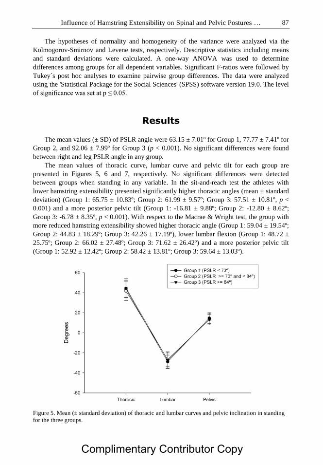
Influence of Hamstring Extensibility on Spinal and Pelvic Postures … 87
The hypotheses of normality and homogeneity of the variance were analyzed via the
Kolmogorov-Smirnov and Levene tests, respectively. Descriptive statistics including means
and standard deviations were calculated. A one-way ANOVA was used to determine
differences among groups for all dependent variables. Significant F-ratios were followed by
Tukey´s post hoc analyses to examine pairwise group differences. The data were analyzed
using the 'Statistical Package for the Social Sciences' (SPSS) software version 19.0. The level
of significance was set at p ≤ 0.05.
Results
The mean values (± SD) of PSLR angle were 63.15 ± 7.01º for Group 1, 77.77 ± 7.41º for
Group 2, and 92.06 ± 7.99º for Group 3 (p < 0.001). No significant differences were found
between right and leg PSLR angle in any group.
The mean values of thoracic curve, lumbar curve and pelvic tilt for each group are
presented in Figures 5, 6 and 7, respectively. No significant differences were detected
between groups when standing in any variable. In the sit-and-reach test the athletes with
lower hamstring extensibility presented significantly higher thoracic angles (mean ± standard
deviation) (Group 1: 65.75 ± 10.83º; Group 2: 61.99 ± 9.57º; Group 3: 57.51 ± 10.81º, p <
0.001) and a more posterior pelvic tilt (Group 1: -16.81 ± 9.88º; Group 2: -12.80 ± 8.62º;
Group 3: -6.78 ± 8.35º, p < 0.001). With respect to the Macrae & Wright test, the group with
more reduced hamstring extensibility showed higher thoracic angle (Group 1: 59.04 ± 19.54º;
Group 2: 44.83 ± 18.29º; Group 3: 42.26 ± 17.19º), lower lumbar flexion (Group 1: 48.72 ±
25.75º; Group 2: 66.02 ± 27.48º; Group 3: 71.62 ± 26.42º) and a more posterior pelvic tilt
(Group 1: 52.92 ± 12.42º; Group 2: 58.42 ± 13.81º; Group 3: 59.64 ± 13.03º).
Figure 5. Mean (± standard deviation) of thoracic and lumbar curves and pelvic inclination in standing
for the three groups.
Complimentary Contributor Copy

Pedro A. López-Miñarro, José M. Muyor, Fernando Alacid et al. 88
Figure 6. Mean (± standard deviation) of thoracic and lumbar curves and pelvic inclination in maximal
trunk flexion with knees flexed for the three groups. * p < 0.01 with respect to group 1.
Figure 7. Mean (± standard deviation) of thoracic and lumbar curves and pelvic inclination in maximal
trunk flexion with knees extended for the three groups. *p < 0.01 between groups 1 and 3; † p < 0.05
between groups 2 and 3.
Complimentary Contributor Copy

Influence of Hamstring Extensibility on Spinal and Pelvic Postures … 89
Discussion
The aim of this chapter was to present a study which analyzed the relationship between
hamstring muscle extensibility and spinal and pelvic postures in several positions. The main
findings showed that lower hamstring extensibility is associated with greater thoracic angles
and more posterior pelvic tilt when maximal trunk flexion is performed. However, no
differences were found between groups in standing. These results are in agreement with
previous studies in younger paddlers, [7, 17] cyclists [26, 31] and young adults. [15] For
example, Muyor et al. [26] found a relationship between hamstring extensibility and trunk
flexion postures in cyclists. This relationship was stronger when performed a maximal trunk
flexion with knees extended. Several studies have found that females reach lower thoracic
angles when sit-and-reach tests are performed. [9, 10, 12-14] This posture has been associated
to higher hamstring extensibility of females compared to males. [32] However, no association
was detected between hamstring extensibility and spinal posture in standing and slumped
sitting. In agreement with previous cross-sectional studies [9, 15, 33] the thoracic and lumbar
angles and pelvic tilt in standing are not influenced by hamstring extensibility. This may be
due to the hamstring muscles are slightly extended, with little passive tension. [7, 34]
Differences between groups were higher in the sit-and-reach test than maximal trunk
flexion with knees flexed. The higher thoracic angles during the flexion movements in
subjects with lower extensibility could indicate that they were compensating for their pelvic
restriction. Tully and Stillman [20] state that subjects with greater capacity to flex the hips
with extended knees can reach their toes without needing to use the full available thoracic
motion. Decreased hamstring extensibility has been previously associated with poor hip
mobility. [35] Hip range of motion influences lumbo-sacral posture due to the muscles‘ direct
attachment to the ischial tuberosities. [15, 21, 22] As the subject bends, the pelvis freely
rotates forward until the passive tension in the hamstrings begins to influence pelvic rotation.
[34] The hamstring muscles may only influence the spinal and pelvic postures when the trunk
is moderately or maximally flexed and the hamstrings are under tension. When the knees are
flexed (Macrae & Wright test), the tension in the hamstrings is less and the hamstring
extensibility has less influence on pelvic and spinal posture. Indeed, when trunk flexion is
performed with knees flexed (90º), the pelvis achieves a larger anterior pelvic tilt.
Hamstring extensibility appears not to affect the lumbar curve in any position. Several
studies report a weak and nonsignificant correlation between lumbar curve and hamstring
extensibility in bending postures. [12, 36, 37] However, Gajdosik et al. [16] found that
lumbar flexion was influenced by hamstring extensibility during maximal trunk flexion, but
that it had no effect on standing. In contrast, Kendall et al. [18] and Sahrmann [19] observed
an association between excessive lumbar spinal flexion and reduced hamstring extensibility
when forward bending or touching the toes.
Previous studies have analyzed the influence of hamstring extensibility in sport specific
postures. López-Miñarro et al. [25] evaluated pelvic and spinal postures in kayakers when
sitting on their boat. Paddlers with lower extensibility values in the straight leg raise test
presented higher thoracic and lumbar flexion and a more posterior pelvic tilt in the kayak in
all evaluated positions. However, no significant differences were found between both groups
when standing. In a recent study, Kang et al. [38] found a significant increase in hip flexion as
well as decreases in both lumbar flexion and the lumbar/hip flexion ratio during the
Complimentary Contributor Copy
Pedro A. López-Miñarro, José M. Muyor, Fernando Alacid et al. 90
preparation phase of stoop lifting after an acute bout of hamstring-stretching exercises.
Hamstring-stretching exercises may be useful for decreasing lumbar flexion and increasing
hip flexion in people who perform repetitive stoop lifting in the workplace or during daily
activities. [38] López-Miñarro et al. [24] find that a protocol of static stretching of the
hamstring muscles was associated to greater lumbar flexion and anterior pelvic tilt as well as
lower thoracic kyphosis when performing the sit-and-reach test. However, improved
hamstring extensibility had no effect on spinal curvatures and pelvic tilt in either the standing
position or maximal trunk flexion in sitting with knees flexed (MacRae & Wright position).
Borman et al. [39] found no changes in lumbar flexion range of motion or in lumbar curvature
in adults with short hamstring muscles after a 4-week program of hamstring stretching,
although hamstring muscle extensibility increased.
The spinal curvatures influence intradiscal pressures, compressive and shear forces in the
intervertebral discs. [1-4] Reduced hamstring extensibility is related to increased thoracic
angles and posterior pelvic tilt, which can overload the spine during sport and daily activities.
The restriction of pelvic movement is considered to be a predisposing factor for low back
pain. Thus, if the pelvic tilting is limited, the more lax spinal tissues will be stressed. [40] In
fact, it has been recognized that, during trunk flexion, the flexed position of the lumbar spine
produces larger shear forces. [41] Greater spinal angles also produce larger contributions
from passive components and greater intradiscal pressures on the thoracic and lumbar tissues.
This is of special concern in young athletes, because an increased thoracic angle or kyphotic
lumbar postures impose great demands on the immature spine, altering the spine‘s exposure
to mechanical loadings during growth.
Stretching can provide a range of health-related motion benefits. Clinicians have
generally considered hamstring flexibility training to be an integral component in the
prevention and rehabilitation of injuries, as well as a method of improving performance in
daily activities and sports. Systematic stretching of the hamstring muscles should be included
in the training program of athletes to reduce the thoracic intervertebral flexion and improve
anterior pelvic tilt during trunk flexion movements. Li et al. [33] found that hip motion during
late and total forward bending was increased after a stretching program. López-Miñarro et al.
[24] recommended perform hamstring stretching previously to sport activities involving trunk
flexion with the knees straight. Stretching exercises for hamstring muscles should be
performed maintaining an appropriate spinal alignment. Some studies have found
improvements on hamstring extensibility after a stretching program. [42-44] However,
stretching may be beneficial only if the technique employed and the stretch holding times are
suitable. Recommendations for duration of hamstrings stretching range from 5 to 60 seconds.
Bandy and Irion [45] determined that stretching durations of 30 and 60 seconds resulted in
significantly greater range of motion improvements than a 15-second stretch. In the study
conducted by Cipriani et al. [46] stretching for 10 seconds 6 times was shown to be equally as
effective as stretching 2 times for 30 seconds over 3- and 6-week periods. Roberts and Wilson
[47] demonstrated that a 15-second stretch repeated 3 times was more effective than a 5-
second stretch repeated 9 times.
Complimentary Contributor Copy

Influence of Hamstring Extensibility on Spinal and Pelvic Postures … 91
Conclusion
The paddlers with lower hamstring extensibility showed a greater thoracic and more
posterior pelvic tilt when maximum trunk flexion is performed with knees flexed and
extended. Since there appears to be a relationship between lower hamstring extensibility and
poor thoracic and pelvic postures, a systematic stretching programme to improved hamstring
extensibility is recommended.
References
[1] Keller TS, Colloca CJ, Harrison DE, Harrison DD, Janik TJ. Influence of spine
morphology on intervertebral disc loads and stresses in asymptomatic adults:
implications for the ideal spine. Spine J. 2005;5:297-300.
[2] McGill SM. Low back disorders. Evidence-Based prevention and rehabilitation.
Champaign, IL: Human Kinetics; 2002. [3] Polga DJ, Beaubien BP, Kallemeier PM, Schellhas KP, Lee WD, Buttermann GR, et al.
Measurement of in vivo intradiscal pressure in healthy thoracic intervertebral discs.
Spine J. 2004;29:1320-4.
[4] Wilke HJ, Neef P, Hinz B, Seidel H,Claes, LE. Intradiscal pressure together with
anthropometric data - a data set for the validation of models. Clin Biomech. 2001;1:
S111-26. [5] Harrison DE, Colloca CJ, Harrison DD, Janik TJ, Haas JW, Keller TS. Anterior
thoracic posture increases thoracolumbar disc loading. Eur Spine J. 2005;14:234-42.
[6] Smith A, O´Sullivan P,Strajer L. Classification of sagittal thoraco-lumbo-pelvic
alignment of the adolescence spine in standing and its relationship to low back pain.
Spine J. 2008;33:2101-7.
[7] López-Miñarro PA, Muyor JM, Alacid F, Rodríguez PL. Influence of sport training on
sagittal spinal curvatures. In: Wright AM, Rothenberg SP, editors. Posture: Types,
assessment and control. New York: Nova Publishers Pub Inc; 2011. P. 63-98. [8] Esola MA, McClure PW, Fitzgerald GK, Siegler S. Analysis of lumbar spine and hip
motion during forward bending in subjects with and without a history of low back pain.
Spine J. 1996;21:71-8.
[9] López-Miñarro PA, Alacid F. Influence of hamstring muscle extensibility on spinal
curvatures in young athletes. Sci Sports. 2010;25:188-93.
[10] López-Miñarro PA, Rodríguez PL. Hamstring muscle extensibility influences the
criterion-related validity of sit-and-reach and toe-touch tests. J Strength Cond Res.
2010;24:1013-8.
[11] López-Miñarro PA, Alacid F, Muyor JM. Comparación del morfotipo raquídeo y
extensibilidad isquiosural entre piragüistas y corredores. Int J Med Sci Phys Act Sport.
2009;36:379-92.
[12] López-Miñarro PA, Sáinz de Baranda P, Rodríguez-García PL. A comparison of the sit-
and-reach test and the back-saver sit-and-reach test in university students. J Sports Sci
Med. 2009;8:116-22.
Complimentary Contributor Copy
Pedro A. López-Miñarro, José M. Muyor, Fernando Alacid et al. 92
[13] López-Miñarro PA, Sáinz de Baranda P, Rodríguez-García PL, Yuste JL. Comparison
between sit-and-reach test and V sit-and-reach test in young adults. Gaz Med Ital.
2008;167:135-42.
[14] Rodríguez-García PL, López-Miñarro PA, Yuste JL, Sáinz de Baranda P. Comparison
of hamstring criterion-related validity, sagittal spinal curvatures, pelvic tilt and score
between sit-and-reach and toe-touch tests in athletes. Med Sport. 2008;61:11-20.
[15] Gajdosik RL, Albert CR, Mitman JJ. Influence of hamstring length on the standing
position and flexion range of motion of the pelvic angle, lumbar angle, and thoracic
angle. JOSPT. 1994;20:213-9.
[16] Gajdosik RL, Hatcher CK, Whitsell S. Influence of short hamstring muscles on the
pelvis and lumbar spine in standing and during the toe-touch test. Clin Biomech.
1992;1:38-42.
[17] Muyor JM, Alacid F, Rodríguez-García PL, López-Miñarro PA. Influencia de la
extensibilidad isquiosural en la morfología sagital del raquis e inclinación pélvica en
deportistas. Int J Morphol. 2012;30:176-81. [18] Kendall FP, McCreary EK, Provance PG, Rodgers MM, Romani WA. Muscles: testing
and function with posture and pain (5th ed.). Baltimore: Lippincott Williams &
Wilkins; 2005.
[19] Sahrmann SA. Diagnosis and treatment of movement impairment syndromes. St. Louis:
Mosby; 2002.
[20] Tully EA, Stillman BC. Computer-aided video analysis of vertebrofemoral motion
during toe touching in healthy subjects. Arch Phys Med Rehabil. 1997;78:59-66.
[21] Carregaro RL, Coury HJC. Does reduced hamstring flexibility affect trunk and pelvic
movement strategies during manual handling? Int J Ind Ergon. 2009;39:115-20.
[22] Congdon R, Bohannon R, Tiberio D. Intrinsic and imposed hamstring length influence
posterior pelvic rotation during hip flexion. Clin Biomech. 2005;20:947-51.
[23] Dewberry MJ, Bohannon RW, Tiberio D, Murray R, Zannotti CM. Pelvic and femoral
contributions to bilateral hip flexion by subjects suspended from a bar. Clin Biomech.
2003;18:1067-76.
[24] López-Miñarro PA, Muyor JM, Belmonte F, Alacid F. Acute effects of hamstring
stretching on sagittal spinal curvatures and pelvic tilt. J Human Kinet. 2012;31:69-78.
[25] López-Miñarro PA, Muyor JM, Alacid F. Influence of hamstring extensibility on
sagittal spinal curvatures and pelvic tilt in high-trained young kayakers. Eur J Sports
Sci. 2012;12:469-74.
[26] Muyor JM, López-Miñarro PA, Alacid F. The relationship between hamstring muscle
extensibility and spinal postures varies with the degree of knee extension. J Applied
Biomech. 2013, in press.
[27] Guermazi M, Ghroubi S, Kassis M,Jaziri O, Keskes H, Kessomtini W, et al. Validity
and reliability of Spinal Mouse® to assess lumbar flexion. Ann Readapt Med Phys.
2006;49:172-7.
[28] Mannion AF, Knecht K, Balaban G, Dvorak J, Grob D. A new skin-surface device for
measuring the curvature and global and segmental ranges of motion of the spine:
reliability of measurements and comparison with data reviewed from the literature. Eur
Spine J. 2004;13:122-36.
[29] Post RB, Leferink VJ. Spinal mobility: sagittal range of motion measured with the
SpinalMouse, a new non-invasive device. Arch Orthop Trauma Surg. 2004;124:187-92.
Complimentary Contributor Copy
Influence of Hamstring Extensibility on Spinal and Pelvic Postures … 93
[30] Santonja Medina FM, Sainz de Baranda Andújar P, Rodríguez García PL, López
Miñarro PA, Canteras Jornada M. Effects of frequency of static stretching on straight-
leg raise in elementary school children. J Sports Med Phys Fitness. 2007;47:304-8.
[31] Muyor JM, López-Miñarro PA, Alacid F. Influence of hamstring muscles extensibility
on spinal curvatures and pelvic tilt in highly trained cyclists. J Hum Kinet. 2011;29:15-
23.
[32] Miñarro PA, Andújar PS, García PL, Toro EO. A comparison of the spine posture
among several sit-and-reach test protocols. J Sci Med Sport. 2007;10:456-62.
[33] Li Y, McClure PW,Pratt N. The effect of hamstring muscle stretching on standing
posture and on lumbar and hip motions during forward bending. Phys Ther.
1996;76:836-49.
[34] Shin G, Shu Y, Li Z, Jiang Z, Mirka G. Influence of knee angle and individual
flexibility on the flexion-relaxation response of the low back musculature. J
Electromyogr Kinesiol. 2004;14:485-94. [35] Sjolie AN. Associations between activities and low back pain in adolescent. Scand J
Med Sci Sports. 2004;14:352-9.
[36] Hui SS, Yuen PY. Validity of the modified back-saver sit-and-reach test: a comparison
with other protocols. Med Sci Sports Exerc. 2000;32:1655-9.
[37] Liemohn W, Sharpe GL, Wasserman JF. Criterion related validity of the sit-and-reach
test. J Strength Cond Res. 1994;8:91-4.
[38] Kang MH, Jung DH, AnDH, Yoo WG, Oh JS. Acute effects of hamstring-stretching
exercises on the kinematics of the lumbar spine and hip during stoop lifting. J Back
Musculoskelet Rehabil. 2013;26:329-36.
[39] Borman NP, Trudelle-Jackson E, Smith SS. Effect of stretch positions on hamstring
muscle length, lumbar flexion range of motion, and lumbar curvature in healthy adults.
Physio Theory Pract. 2011;27:146-54.
[40] Norris CM. Back stability. Champaign, IL: Human Kinetics; 2000.
[41] Potvin JR, McGill SM, Norman RW. Trunk muscle and lumbar ligament contributions
to dynamic lifts with varying degrees of trunk flexion. Spine J. 1991;16:1099-107.
[42] Nelson RT, Bandy WD. Eccentric training and static stretching improve hamstring
flexibility of high school males. J Athl Train. 2004;39:254-8.
[43] Reid DA, McNair PJ. Passive force, angle, and stiffness changes after stretching of
hamstring muscles. Med Sci Sports Exerc. 2004;36:1944-8.
[44] Rodríguez PL, Santonja F, López-Miñarro PA, Sáinz de Baranda P, Yuste JL. Effect of
physical education programme on sit-and-reach score in schoolchildren. Sci Sports.
2008;23:170-5.
[45] Bandy WD, Irion JM. The effect of time on static stretch on the flexibility of the
hamstring muscles. Phys Ther.1994;74:845-50.
[46] Cipriani D, Abel B, Pirrwitz D. A comparison of two stretching protocols on hip range
of motion: implications for total daily stretch duration. J Strength Cond Res.
2003;17:274-8.
[47] Roberts JM, Wilson K. Effect of stretching duration on active and passive range of
motion in the lower extremity. Br J Sports Med. 1999;33:259-63.
Complimentary Contributor Copy

Complimentary Contributor Copy

In: Posture: Types, Exercises and Health Effects ISBN: 978-1-63117-252-6
Editor: Sarah A. Curran © 2014 Nova Science Publishers, Inc.
Chapter 7
Spinal Posture in Cycling
José M. Muyor, 1,*
PhD, MSc, Pedro A. López-Miñarro,2 PhD,
Fernando Alacid,3 PhD, MSc and Raquel Vaquero-Cristóbal,
4 MSc
1Faculty of Education Sciences, Nursing and Physiotherapy,
Laboratory of Kinesiology, Biomechanics and Ergonomics (KIBIOMER Lab)
University of Almería, Almería, Spain 2Department of Physical Education, University of Murcia, Murcia, Spain
Main Researcher of Physical Exercise and Health Group 3Department of Sport Science at the Catholic University of San Antonio of Murcia,
Murcia, Spain 4Sport Traumatology at the Catholic University of San Antonio of Murcia,
Murcia, Spain
Abstract
Currently, cycling is a popular sport, in spite of its demanding physical requirements,
the interaction with motor vehicles and the need to adopt an unnatural position on the
bicycle for better aerodynamics. The analysis of posture in cyclists on the bicycle and
their spinal adaptations in different positions has been studied in the field of sports
medicine. This chapter describes the results of major research on the position adopted in
cycling, the spinal adaptations generated by the systematic practice of this sport, and the
variables that influence the posture of cyclists. The studies reviewed suggest that the
morphology of the thoracic curve of the cyclist is characterized by a standing
hyperkyphotic posture. This condition, however, would be more associated with a lack of
postural awareness than with the position on the bicycle because thoracic kyphosis
significantly decreases due to the support of the hands on the handlebars. Instead, the
lumbar spine modifies its lordosis in the standing position toward a lumbar kyphosis
(inversion) on the bicycle, with greater lumbar flexion, as the grip on the handlebars is
lower and farther away relative to the seat. This posture, held on the bicycle, has been
associated with the ability of the cyclist to adopt greater lumbar flexion in positions of
* Corresponding author: Email: [email protected]
Complimentary Contributor Copy

José M. Muyor, Pedro A. López-Miñarro, Fernando Alacid et al. 96
maximum trunk flexion with bent or extended knees compared with subjects who do not
practice this sport. It has also been observed that the degree of hamstring extensibility
does not influence the position of the spine and pelvis on the bicycle even though it does
influence the spinal position in movements of maximum trunk flexion with extended
knees. In conclusion, cycling posture is characterized by maintaining better alignment of
the thoracic spine on the bicycle than in the standing position. In contrast, the lumbar
spine maintains an inverted position, which leads to spinal adaptations in positions of
maximum trunk flexion and could possibly cause low back pain as a result. Due to the
characteristics and specific posture requirements of this sport, core strengthening
exercises and improvements in the body schema of cyclists are recommended.
Keywords: Cycling, spine, posture, pelvis, core strengthening, lumbar, kyphosis
Introduction
In the standing position, the spine has different physiological curves in the sagittal plane
(cervical lordosis, thoracic kyphosis and lumbar lordosis) that are determined by the
morphology of the vertebral bodies, the functionality of the intervertebral discs and
ligamentous structures, and the anatomical-physiological muscle integrity. [1, 2] Sports
practice involves, in many cases, adopting postures considered to be unnatural to execute a
precise technical movement or to maintain a particular body position in pursuit of increased
athletic performance. Systematically maintaining these postures for sustained periods has
been associated with adaptations of sagittal spine alignment. [3] For example, a greater
degree of thoracic kyphosis has been found in athletes who participate in sports in which
optimum technique is characterized by the maintenance of trunk flexion postures, including
skiing, rowing or wrestling, compared with subjects who do not practice these sports. [4-6]
However, positions that hold the spine in flexion have been associated with increased
intradiscal pressure in the thoracic [7] and lumbar spine, [8] greater distortion of the spinal
tissues [9] and high spinal stress; [10] all of which increase the likelihood of suffering, pain,
and/or spinal injury.
Currently, cycling is one of the most popular sports both from a competitive standpoint
and for amateurs and hobbyists, even though it is considered as a discipline in which several
different factors are involved in performance, including high physical demands, weather
conditions and interactions with motor vehicles. [11] The predominant position is the seated
position on the bicycle with an anterior inclination of the trunk and lumbar flexion to reach
the handlebars with the hands. Because this position is considered unnatural, [12] several
studies have analyzed the position adopted by the spine during cycling, how different
variables affect the spinal position, and whether cycling modifies spinal curvatures in the
sagittal plane.
Cycling and Low Back Pain
Low back pain is one of the most common overuse injuries in cycling. [13] Clarsen et al.
[14] interviewed 7 professional teams with a total of 116 cyclists and found that 45% had
Complimentary Contributor Copy

Spinal Posture in Cycling 97
suffered low back pain in the past 12 months and that 41% had received medical care for the
pain. Similar research has shown that bicycle fit, improper equipment, training errors, and
individual anatomical factors are important variables affecting the occurrence of low back
pain in these athletes. [15]
Some authors have tried to explain how changes in different bicycle components would
affect the posture of the cyclist and either cause or decrease low back pain. In this regard,
Salai et al. [16] analyzed the effect of changing the seat tilt on the incidence of low back pain
in recreational cyclists. The authors found that the incidence and severity of low back pain
was significantly reduced with an anterior tilt of the seat between 10°-15°. In comparison,
Fanucci et al. [17] reported that the incidence and importance of low back pain in cyclists
could be reduced if the bicycle‘s crankset axis was placed behind the seat axis. This
arrangement allowed riders to maintain the lumbar spine in a more physiological position
compared with the classical design with the bicycle‘s crankset axis in front of the seat axis.
Although the design of different prototype bicycles and the analysis of how modification
of their components affect low back pain or cyclist's posture may be of scientific interest, the
reality is that the Union Cycliste Internationale (UCI) currently sets forth regulations that
specify the dimensions and characteristics of bicycles that can be certified and therefore used
in official cycling competitions. Thus, several authors have evaluated the spinal morphotypes
and the postures adopted by cyclists on bicycles certified for training and/or competition.
Spinal Posture on the Bicycle
Few studies in the scientific literature have evaluated the position of the cervical spine in
cyclists. Kolehmainen et al. [1] analyzed the influence of handlebar height on the position of
the cervical and thoracic spine using a cycle ergometer in 3 handlebar positions (high,
medium and low). The results showed that there were no major changes in thoracic kyphosis
and lumbar lordosis in any of the handlebar grips evaluated. However, with a low grip, there
was cervical spine extension that tripled load moments compared with the vertically aligned
position. These authors recommended the use of a high grip on the handlebar, particularly if
the cyclist experiences discomfort in the cervical spine.
Usabiaga et al. [18] conducted a radiological and electromyographic study to evaluate
changes in the lumbar spine with different handlebar grips using cyclists‘ own racing
bicycles. The authors found that the lumbar spine changed from lordosis in the standing
position to kyphosis in the seated cycling position. Low back muscle contraction was
proportional to pedaling intensity and decreased with improved aerodynamic positioning. The
tone of the thoracic paravertebral muscles depended on the degree of cervical hyperextension.
However, the abdominal muscles remained relaxed for all pedaling positions and intensities.
The authors concluded that observed changes in the lumbar spine could modify the normal
biomechanics of this spinal region, although mechanical load would be reduced by the weight
distribution of the trunk on the handlebar grip. Moreover, the imbalance found between the
activity of the flexor and extensor muscles of the trunk could cause low back pain in cyclists
without adequate physical preparation.
Burnett et al. [19] evaluated whether there were differences between spine kinematics
and muscle activity in cyclists with and without low back pain. The results showed that
cyclists with nonspecific low back pain suffered a motor control disorder of the lumbo-pelvic
Complimentary Contributor Copy
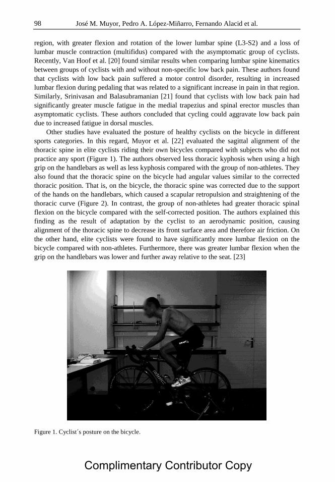
José M. Muyor, Pedro A. López-Miñarro, Fernando Alacid et al. 98
region, with greater flexion and rotation of the lower lumbar spine (L3-S2) and a loss of
lumbar muscle contraction (multifidus) compared with the asymptomatic group of cyclists.
Recently, Van Hoof et al. [20] found similar results when comparing lumbar spine kinematics
between groups of cyclists with and without non-specific low back pain. These authors found
that cyclists with low back pain suffered a motor control disorder, resulting in increased
lumbar flexion during pedaling that was related to a significant increase in pain in that region.
Similarly, Srinivasan and Balasubramanian [21] found that cyclists with low back pain had
significantly greater muscle fatigue in the medial trapezius and spinal erector muscles than
asymptomatic cyclists. These authors concluded that cycling could aggravate low back pain
due to increased fatigue in dorsal muscles.
Other studies have evaluated the posture of healthy cyclists on the bicycle in different
sports categories. In this regard, Muyor et al. [22] evaluated the sagittal alignment of the
thoracic spine in elite cyclists riding their own bicycles compared with subjects who did not
practice any sport (Figure 1). The authors observed less thoracic kyphosis when using a high
grip on the handlebars as well as less kyphosis compared with the group of non-athletes. They
also found that the thoracic spine on the bicycle had angular values similar to the corrected
thoracic position. That is, on the bicycle, the thoracic spine was corrected due to the support
of the hands on the handlebars, which caused a scapular retropulsion and straightening of the
thoracic curve (Figure 2). In contrast, the group of non-athletes had greater thoracic spinal
flexion on the bicycle compared with the self-corrected position. The authors explained this
finding as the result of adaptation by the cyclist to an aerodynamic position, causing
alignment of the thoracic spine to decrease its front surface area and therefore air friction. On
the other hand, elite cyclists were found to have significantly more lumbar flexion on the
bicycle compared with non-athletes. Furthermore, there was greater lumbar flexion when the
grip on the handlebars was lower and further away relative to the seat. [23]
Figure 1. Cyclist´s posture on the bicycle.
Complimentary Contributor Copy

Spinal Posture in Cycling 99
Figure 2. Cyclist´s scapular retropulsion maintained to support of the hands on the handlebars.
Muyor et al. [24] compared spinal postures and pelvic tilts on the bicycle between elite
cyclists and Masters 30 category cyclists. The authors found that elite cyclists had lower
thoracic flexion and increased lumbar flexion and pelvic tilt compared with Masters 30
cyclists. The differences in the postures adopted on the bicycle were related to age. That is,
the younger cyclists (elite) had less thoracic kyphosis in the standing position than the older
cyclists (Masters 30). The elite cyclists therefore maintained lower thoracic spine angle
values on the bicycle.
Spine Morphology of the Cyclist in the Standing
and Trunk Flexion Positions
Several studies have analyzed the spine morphologies of cyclists in different sport
categories to determine whether cycling alters the physiological curvature of the spine in the
standing position or if there are spinal adaptations in trunk flexion movements. Rajabi et al.
[25] compared the thoracic spine angular values in the standing position in a group of cyclists
with a minimum cycling training experience of 5 years and a group of non-athletes. The
results showed significantly greater thoracic kyphosis in the cyclists compared with the non-
athletes. The authors attributed these findings to the specific posture of the cyclist on the
bicycle, which significantly increased stress on the spine and thereby increased thoracic
kyphosis.
In this regard, Muyor et al. [22] also found significantly higher thoracic angular values in
elite cyclists compared with non-athletes in the standing position. However, these authors,
unlike Rajabi et al., [25] also evaluated the thoracic spine position on the bicycle and found
that both the cyclists and the non-athletes showed a significant decrease in thoracic curvature
Complimentary Contributor Copy

José M. Muyor, Pedro A. López-Miñarro, Fernando Alacid et al. 100
on the bicycle compared with the standing position. Similar results with respect to decreased
thoracic kyphosis on the bicycle have been found in other sport categories in Masters 30 [24]
and Masters 40 cyclists. [26]
The categories proposed by Mejia et al. [27] are frequently used to classify thoracic
kyphosis: neutral thoracic kyphosis is between 20° and 45°; thoracic hyperkyphosis is > 45°;
and thoracic hypokyphosis is < 20°. A high percentage of cyclists have thoracic
hyperkyphosis in the relaxed standing position (Figure 3). Specifically, 58.3% of elite
cyclists, 53.3% of Masters 30 cyclists [24] and 68% of Masters 40 cyclists. [26] Researchers
have suggested that the high percentage of thoracic hyperkyphosis found in cyclists is due to
factors such as an imbalance in the body schema or postural attitude rather than the position
taken on the bicycle, where the spine is better aligned than in the standing position and
therefore is not in a kyphosed position. Later, Muyor et al., [28] after evaluating spinal
morphology in the standing position in different age groups of cyclists with the same number
of years of cycling practice and training volume experience, found a significant increase in
thoracic kyphosis with increasing age in cyclists. These authors concluded that age was a
major factor in the angular alignment of the thoracic spine in these athletes.
Similarly, the lumbar spine has also been studied in cyclists. Maintaining an inverted
lumbar spine position on the bicycle can cause spinal adaptations in trunk flexion positions.
In this sense, Muyor et al. [29] found a significantly higher capacity for lumbar flexion in
elite cyclists compared with non-athletes in postures of maximum trunk flexion in seated
positions with bent knees and with extended legs. In contrast, no significant differences were
found when comparing lumbar flexion in seated positions between elite and Masters 30
cyclists. [30]
Figure 3. Angle references for spinal curvatures in standing posture.
Complimentary Contributor Copy
Spinal Posture in Cycling 101
As discussed earlier in this chapter, a higher frequency of thoracic hyperkyphosis has
been found in cyclists in the standing position. The classification proposed by Tüzün et al.
[31] is frequently used to classify lumbar spine morphology: neutral lumbar lordosis is
between 20° and 40°; lumbar lordosis is > 40°; and lumbar hypolordosis is < 20°. (Figure 3)
Most cyclists have a neutral lumbar lordosis in the standing position. This is true for 88.3% of
elite cyclists, 76.7% of Masters 30 cyclists, [24] and 68% of Masters 40 cyclists. [26]
Regarding the pelvis, McEvoy et al. [32] compared the degree of the anterior pelvic tilt in
the seated position with extended knees (long sitting posture) in a group of elite cyclists with
another group of non-athletes. The authors found that cyclists had a significantly greater
anterior pelvic tilt than non-athletes. The authors attributed their results to greater adaptation
to trunk flexion postures by pelvic tilt in cyclists because that is their normal position on the
bicycle. However, these authors did not assess the degree of hamstring extensibility in both
groups.
Other studies on athletes of other sports [33] as well as on cyclists [34, 35] have found
that the degree of hamstring extensibility influences pelvic movement in positions of
maximum trunk flexion with extended knees. Since the hamstring muscles have their
anatomical attachment on the ischial tuberosity, when maximal trunk flexion with the knees
extended is performed, the pelvis rotates forward until the passive tension in the hamstrings
increase significantly. However, in cyclists, the degree of hamstring extensibility does not
influence spinal posture [33] or pelvic tilt [36] adopted on the bicycle. This difference is most
likely because cyclists‘ knees never reach their maximum extension on the bicycle during
pedaling and therefore the degree of hamstring extensibility would have no effect on the
spinal posture adopted on the bicycle.
Core Stability in Cycling
"Core" stability has been defined as the ability to control the position and movement of
the trunk on the pelvis, allowing an optimum production, transfer and control of force and
motion in the integration of athletic activities. [37] Spine stability refers to the combined and
coordinated work of the passive components of the spinal column (bones and ligaments), of
the active components (spinal and abdominal muscles), and neuromuscular control (spinal
cord and central nervous system).
Asplund and Ross [38] have suggested that core stability is critical for the efficient
transfer of power from the lower to the upper extremities and vice versa. Less energy is lost in
movement with good core training, which enables the trunk to be more stable, avoiding
flexion or rotation movements. Additionally, having a trained core allows cyclists to
maintaining their trunk alignment longer, thereby delaying postural fatigue.
In the same vein, core strengthening has also been used as a therapeutic exercise in the
treatment of low back pain. [39] In this regard, some studies have reported that the position
on the bicycle is related to exercise intensity, ventilatory thresholds, the power capacity, and
the performance achieved. [36, 40, 41] Therefore, core training could improve both the
posture on the bicycle and prevent the low back pain suffered by these athletes after
constantly maintaining the lumbar spine in flexion.
Complimentary Contributor Copy

José M. Muyor, Pedro A. López-Miñarro, Fernando Alacid et al. 102
Conclusion
The cyclist's spinal column morphotype in the standing position is characterized by
thoracic hyperkyphosis, which is more related to alterations in the body schema than to the
position maintained on the bicycle. On the bicycle, the thoracic spine decreases its kyphosis
due to the support of the hands on the handlebars and retropulsion of the scapulohumeral
girdle. Instead, the lumbar spine changes from lumbar lordosis in the standing position to
lumbar kyphosis (inversion) on the bicycle, with greater lumbar flexion when the grip on the
handlebar is lower and farther away from the seat.
Whilst maintaining an inverted lumbar spine on the bicycle does not generate alterations
in lumbar lordosis in the standing position, it predisposes to greater lumbar flexion in postures
of maximum trunk flexion. The degree of hamstring extensibility in cyclists influences the
anterior pelvic tilt in positions of maximum trunk flexion with extended knees but has no
influence on the spinal position adopted on the bicycle. Due to the characteristics and specific
postural requirements of this sport, exercises to strengthen the core and improve the body
schema in cyclists are recommended.
References
[1] Kolehmainen I, Harms-Ringdahl K, Lanshammar H. Cervical spine positions and load
moments during bicycling with different handlebar position. Clin Biomech. 1989;4:105-
10.
[2] White AA, Panjabi MM. Clinical biomechanics of the spine. 2 ed. Philadelphia: J. B.
Lippincott Company; 1990.
[3] Wojtys EM, Ashton-Miller JA, Huston LJ, Moga PJ. The association between athletic
training time and the sagittal curvature of the immature spine. Am J Sports Medicine.
2000;28:490-8.
[4] Alricsson M, Werner S. Young elite cross-country skiers and low back pain. A 5-year
study. Phys Ther Sport. 2006;7:181-4.
[5] Rajabi R, Doherty P, Goodarzi M, Hemayattalab R. Comparison of thoracic kyphosis
in two groups of elite Greco-Roman and freestyle wrestlers and a group of non-athletic
participants. Brit J Sports Med. 2008;42:229-32.
[6] Stuchfield BM, Coleman S. The relationships between hamstring flexibility, lumbar
flexion, and low back pain in rowers. Euro J Sports Science. 2006;6:255-60.
[7] Polga DJ, Beaubien BP, Kallemeier PM, Schelhas KP, Lee WD, Buttermann GR, et al.
Measurement of in vivo intradiscal pressure in healthy thoracic intervertebral disc.
Spine. 2004;29:1320-4.
[8] Wilke HJ, Neef P, Caimi M, Hoogland T, Claes LE. New in vivo measurements of
pressures in the intervertebral disc in daily life. Spine. 1999;24:755-62.
[9] Solomonow M, Zhou BH, Baratta RV, Burger E. Biomechanics and electromyography
of a cumulative lumbar disorder: response to static flexion. Clin Biomech. 2003;18:
890-8.
[10] Beach TA, Parkinson RJ, Stothart JP, Callaghan JP. Effects of prolonged sitting on the
passive flexion stiffness of the in vivo lumbar spine. Spine J. 2005;5:145-54.
Complimentary Contributor Copy

Spinal Posture in Cycling 103
[11] Winters M, Davidson G, Kao D, Teschke K. Motivators and deterrents of bicycing:
comparing influences on decisions to ride. Transportation. 2011;38:153-68.
[12] de Vey Mestdagh K. Personal perspective: in search of an optimum cycling posture.
Applied Ergonomics. 1998;29:325-34.
[13] Marsden M, Schwellnus M. Lower back pain in cyclists: A review of epidemiology,
pathomechanics and risk factors. Int Sport Med J. 2010;11:216-25.
[14] Clarsen B, Krosshaug T, Bahr R. Overuse injuries in professional road cyclists. Am J
Sports Med. 2010;38:2494-501.
[15] Asplund C, Webb C, Barkdull T. Neck and back pain in bicycling. Current Sports Med
Reports. 2005;4:271-4.
[16] Salai M, Brosh T, Blankstein A, Oran A, Chechik A. Effect of changing the saddle
angle on the incidence of low back pain in recreational bicyclists. Brit Sports Med.
1999;33:398-400.
[17] Fanucci E, Masala S, Fasoli F, Cammarata R, Squillaci E, Simonetti G.
Cineradiographic study of spine during cycling: effects of changing the pedal unit
position on the dorso-lumbar spine angle. Radiologia Medica. 2002;104:472-6.
[18] Usabiaga J, Crespo R, Iza I, Aramendi J, Terrados N, Poza JJ. Adaptation of the lumbar
spine to different positions in bicycle racing. Spine. 1997;22:1965-9.
[19] Burnett AF, Cornelius MW, Dankaerts W, O´Sullivan PB. Spinal kinematics and trunk
muscle activity in cyclists: a comparison between healthy controls and non-specific
chronic low back pain subjects -a pilot investigation. Manual Therapy. 2004;9:211.
[20] Van Hoof W, Volkaerts K, O´Sullivan K, Verschueren S, Dankaerts W. Comparing
lower lumbar kinematics in cyclists with low back pain (flexion pattern) versus
asymptomatic controls - field study using a wireless posture monitoring system.
Manual Therapy. 2012;17:312-7.
[21] Srinivasan J, Balasubramanian V. Low back pain and muscle fatigue due to road
cycling - An sEMG study. J Bodyw Move Ther. 2007;11:260-6.
[22] Muyor JM, López-Miñarro PA, Alacid F. A comparison of the thoracic spine in the
sagittal plane between elite cyclists and non-athlete subjects. J Back Musculo Rehab.
2011;24:129-35.
[23] Muyor JM, López-Miñarro PA, Alacid F. Comparison of sagittal lumbar curvature
between elite cyclists and non-athletes. Science & Sports, in press.
[24] Muyor JM, López-Miñarro PA, Alacid F. Spinal posture of thoracic and lumbar spine
and pelvic tilt in highly trained cyclists. J Sports Science Med. 2011;10:355-61.
[25] Rajabi R, Freemont A, Doherty P. The investigation of cycling position on thoracic
spine. A novel method of measuring thoracic kyphosis in the standing position. Arch
Physiology Biochemistry. 2000;1:142.
[26] Muyor JM, Alacid F, López-Miñarro PA. Spinal posture of thoracic and lumbar spine
in master 40 cyclists. Int J Morphology. 2011;29:727-32.
[27] Mejia EA, Hennrikus WL, Schwend RM, Emans JB. A prospective evaluation of
idiopathic left thoracic scoliosis with magnetic resonance imaging. J Pediatric
Orthopaedics. 1996;16:354-8.
[28] Muyor JM, Alacid F, López-Miñarro PA, Casimiro AJ. Evolution of spinal morphology
and pelvic tilt in cyclists of different ages. A cross sectional study. Int J Morphology.
2012;30:199-204.
Complimentary Contributor Copy

José M. Muyor, Pedro A. López-Miñarro, Fernando Alacid et al. 104
[29] Muyor JM, López-Miñarro PA, Alacid F. Sagittal lumbar curvature in elite cyclists and
non-athletic subjects. Revista Internacional de Medicina y Ciencias de la Actividad
Física y del Deporte. 2012;12:219-31.
[30] Muyor JM, López-Miñarro PA, Alacid F, Casimiro AJ. Sagittal spinal curvatures and
pelvic tilt in cyclists: A comparison between two master cyclists categories. Int
SportMed J. 2012;13:122-32.
[31] Tüzün Ç, Yorulmaz I, Cindaş A, Vatan S. Low back pain and posture. Clin
Rheumatology. 1999;18:308-12.
[32] McEvoy M, Wilkie K, Williams MT. Anterior pelvic tilt in elite cyclists - A
comparative matched pairs study. Phys Ther Sport. 2007;8:22-9.
[33] Muyor JM, Alacid F, López-Miñarro PA. Influence of hamstring muscles extensibility
on spinal curvatures and pelvic tilt in highly trained cyclists. J Human Kinetics.
2011;29:15-23.
[34] Muyor JM, Alacid F, Rodríguez-García PL, López-Miñarro PA. Influence of hamstring
extensibility on sagittal spinal curvatures and pelvic inclination in athletes. Int J
Morphology. 2012;30:176-81.
[35] Muyor JM, López-Miñarro PA, Alacid F. The relationship between hamstring muscle
extensibility and spinal postures varies with the degree of knee extension. J Appl
Biomechanics. 2013, in press.
[36] Sauer JL, Potter JJ, Weisshaar CL, Ploeg HL, Thelen DG. Influence of gender, power,
and hand position on pelvic motion during seated cycling. Med Sci Sports Exer.
2007;39:2204-11.
[37] Kibler WB, Press J, Sciascia A. The role of core stability in athletic function. Sports
Medicine. 2006;36:189-98.
[38] Asplund C, Ross M. Core stability and bicycling. Curent Sports Medicine Reports.
2010;9:155-60.
[39] Akuthota V, Ferreiro A, Moore T, Fredericson M. Core stability exercise principles.
Current Sports Medicine Reports. 2008;7:39-44.
[40] Egaña M, Smith S, Green S. Revisiting the effect of posture on high-intensity constant-
load cycling performance in men and women. Euro J Appl Physiology. 2007;99:495-
501.
[41] Ashe MC, Scroop GC, Frisken PI, Amery CA, Wilkins MA, Khan KM. Body position
affects performance in untrained cyclists. Brit J Sports Med. 2003;37:441-4.
Complimentary Contributor Copy

In: Posture: Types, Exercises and Health Effects ISBN: 978-1-63117-252-6
Editor: Sarah A. Curran © 2014 Nova Science Publishers, Inc.
Chapter 8
Effects of Physical and Sporting
Activities on Postural Stability
in Children
Sonia Sahli,1,2,*
PhD, Rym Baccouch,2 MSc
and Haithem Rebai,3 PhD
1Research Unit Neurophysiology of Vigilance, Attention and Performance,
Functional Explorations of the Nervous System Department,
University Hospital Center Sahloul, Sousse University, Tunisia 2High Institute of Sport and Physical Education, Sfax University, Tunisia
3Research Unit Cardio Circulatory, Respiratory, Metabolic and Hormonal
Adaptations to Exercise, Faculty of Medicine, Sousse University, Tunisia
Abstract
Concerns have been raised about the effects of physical and sporting activities on
postural stability because it is an important determinant of both sports level and
susceptibility to injuries. It may be also useful for clinicians interested in designing new
rehabilitation therapies. While the sensorimotor control of posture and how physical and
sport training affects it, have been widely studied in adults, surprisingly little is known
about these effects in children‘s upright postural control. With this consideration, this
chapter starts with a summary of the major results established in research undertaken on
adults. It moves on to provide detailed analysis of the few available studies on the effects
of physical activities on postural stability in children. In adults, several papers have
shown that training may improve postural control by acting on the somatosensory, visual
and vestibular systems involved in the control of a posture; or on the motor response
through an increase of muscular strength. It has been observed that the nature of the
movements involved in the practice of different sports would influence postural
adaptation because training in a specific activity develops specific modalities of postural
* Corresponding author: [email protected]
Complimentary Contributor Copy

Sonia Sahli, Rym Baccouch and Haithem Rebai 106
control. Moreover, there is a relationship between competition level and postural control
level. It is well known that postural control is not yet fully developed at birth. Indeed, it
takes children almost a year to stand independently and many years thereafter to develop
adult-like postural control. It has been suggested that postural control development in
children is a result of how children integrate sensory information to achieve or maintain a
desired posture. Recent studies showed that children before 12 years of age use sensory
information different from adults to maintain an upright standing posture, and they do not
compensate for sensory cue quality and magnitude changes as adults do. It was suggested
that the practice of a physical exercise could modify the use of sensory information in
children. To our best knowledge, there are 3 scientific works that investigate the relation
between physical exercise training and postural stability in children. The performance of
the postural control system improved in 5-6 year old children who practiced gymnastics
and circus activities, whereas expertise in gymnastics did not seem to improve postural
stability of 9-11 year old children. For older children aged 13 years of age, soccer
training was efficient in improving the medial-lateral postural control. More studies are
needed to gain a better understanding of how sensory organization develops in children
and how physical and sporting activities affect their control of posture.
Keywords: Physical activities, sports, training, children, posture, postural stability
Introduction
Posture serves as a reference frame for the production of accurate movements that are
vital for the activities of daily living like walking, stepping up the stairs, or standing in a bus.
The most important function of it is to ensure the maintenance of equilibrium during the
initiation and continuation of movement. [1] Beside appropriate strength and endurance
capacity, the control of posture is considered to be an important indicator of musculoskeletal
health and therefore could be of importance in view of clinical issues. [2] Postural stability
refers to the inherent ability of a person to maintain, achieve or restore a specific state of
balance under varied types of external disturbance to prevent falling while performing a range
of activities from quite stance to complex athletic tasks. [3] Maintaining an upright standing
posture results from an interaction between sensory information and motor action, [4] with
visual, vestibular, and somatosensory systems as the main sources of sensory information. [5-
9]
Several studies have shown that sport training may improve postural control by acting on
these balance sensors [10-15] or on the motor response through an increase of muscular
strength. [16] While the sensorimotor control of posture and how physical and sport training
affects it, have been widely studied in adults, surprisingly little is known about these effects
in children upright postural control. The nature of the development of postural control has
intrigued researchers for many years.
A better understanding of how sensory organization develops in children and how
physical and sporting activities affect their control of posture is important for many reasons.
This knowledge would enable earlier detection of atypical postural development in children,
provide better understanding and appreciation of the differences seen between individual and
groups of children, and might also lead to improved interventions for children with
pathological balance impairments.
Complimentary Contributor Copy

Effects of Physical and Sporting Activities on Postural Stability in Children 107
Therefore, the aims of this chapter are:
(i) to provide a summary of the major results established in research undertaken on
adults;
(ii) to determine if children integrate sensory information like adults to achieve or
maintain a desired posture;
(iii) and to investigate the relation between physical exercise training and postural
stability in children.
Physical and Sport Training Effects on Adult
Postural Control
Training has widely been reported to be one of the best ways to improve balance. [10, 14,
15, 17-21] To understand how physical and sport training affects the control of posture in
adults, several authors have attempted to assess differences in postural control between
athletes and normal participants, [15, 22] athletes of different levels of sports expertise, [21-
23] and athletes from different sports [15, 24-27] using only cross-sectional studies.
It seems that athletes, through years of sport experience, would have a different model of
sensory integration, as compared to the non-athletes. [28] Previous studies have shown that
training of each of the three levels of the sensorimotor chain may improve the maintenance of
balance in complex conditions [i.e., with sensory deprivation] [29-31] indicating a positive
effect of training on sensorimotor adaptability. [12-15] Differences in knee and ankle
proprioception between trained athletes and non-athletes suggest that sport participation, by
challenging sensorimotor systems, may improve balance. [13, 32] Some evidence in the
literature suggested that superior balance among athletes is largely due to the repetitive
training experiences that influence motor responses rather than to a greater sensitivity of the
vestibular system. [33] Others argue that superior balance is the result of training experiences
that influence a person‘s ability to attend to relevant proprioceptive and visual cues. [3]
Although these experts may not agree on the mechanism, research suggested that changes in
both sensory and motor systems influence balance performance. [26] Training experiences
improving muscular strength of the lower limbs, [35] neuromuscular coordination, joint
strength, and range of motion are also probable mechanisms that lead to improved balance.
[13, 33, 36]
Compared to the non-athletes, athletes seem to have a special role of the visual vestibular
interaction, when proprioception is altered. [28] They are coping more effectively than non-
athletes with the multisensory integration challenges of the sway amplitude, while standing
on an unstable support. [28] In addition, they seem to have a superior kinesthetic awareness,
[37] increased safety margins [38] and an increased automaticity level. [39,40] Moreover, it
has been suggested that compared to normal subjects, sportsmen adopted the reaction most
appropriate to the task, using both past knowledge and short term memory very efficiently.
[31, 41].
It has been asserted that the improvement of postural control seems to depend on the
sport or the activity practiced. [15, 26, 42-45] The motor programs required are inevitably
different from one sport to another. For example, individual sports like rhythmic gymnastics,
Complimentary Contributor Copy

Sonia Sahli, Rym Baccouch and Haithem Rebai 108
dance, and artistic gymnastics require fine postural control especially in the one-legged
stance. [24, 27, 46-49] In addition, many of gymnasts‘ skills require great strength and
sometimes exaggerated joint range of motion. [50] Postural control performance of adult
gymnasts has been widely investigated and compared with adult non-gymnasts using various
physical exercises. [26, 24, 37, 47, 51, 52] In general, adult gymnasts present better
performance of postural control than adult non-gymnasts, [24, 37, 47, 52] especially, during
specific conditions such as the unipedal stance and on unstable bases of support. [24]
However, Vuillerme et al. [24] showed that expert artistic gymnasts were not more stable than
expert athletes in other sports while maintaining a bipedal and unipedal unperturbed stance
suggesting that the eloquent sense of balance demonstrated by the gymnasts during their
acrobatic movements does not provide any benefit to the achievement of more simple tasks.
The good performances of judoists found in unusual situations, when compared to dancers
and controls, could be due to the difference of balance modalities involved in judo and dance.
[15] In fact, dancers train for long hours in a very stable environment and generate their own
imbalance during their complex chained dynamic choreographic figures. Conversely, both in
training and in competition, judoists are constantly subjected to unexpected movements
imposed by their opponent in order to make them fall on the tatami. While ballet dancers
often perform static postures, [15] modern dancers often perform dynamic movements [53]
like other athletes; therefore the benefits of modern dance practice appear to be similar to
those of athletics for improving balance. [54] A better balance control in static conditions has
been observed in shooters compared to fencers. Shooting is a static activity based on a strict
control of body posture where shooters are specifically trained to maintain a bipedal posture,
as still as possible, in order to stabilize a weapon [28] or a rifle [55] in a stable environment;
whereas, fencing is based on unceasingly moving situations of high-speed attacks forcing
opponents to make constant spatial and temporal adjustments. [28]
Some team sports such as soccer or basketball demand a high degree of postural balance
and quick movement changes. In fact, soccer players often perform single-leg reaching
movements outside their base of support during passing, receiving, dribbling and shooting.
[56] Basketball is particularly an activity of jumps, upper extremity passing, shooting, and
dribbling skills requiring a great number of accelerations and decelerations, and direction
changes. [57] Due to the training of these specific motor programs, the soccer players may
have acquired balance ability much earlier than the controls [58] and made greater use of the
somatosensory system during one-legged stance than basketball players and non-athletes. [27]
It is expected that athletes participating in other land-based sports [such as volleyball and
athletics], and who rarely support their body weight on one leg while playing, should have
similar balance ability to basketball players. [27] However, when gymnasts were compared to
basketball players, they develop less balance; whereas compared to soccer players they may
have similar or better one-legged stance ability. [26, 27] Even though swimmers rarely use
their anti-gravity muscles during training, no difference in stance stability between basketball
players, swimmers, and non-athletes has been found. [27] This suggests that swimmers use
their anti-gravity muscles to some extent when standing upright and walking in daily life,
which may be sufficient to negate any negative influence of swimming training on their
stance stability. [27] Highly skilled sportsmen were examined after long-term training with
various kinds of sport and results showed that postural stability decreased in the following
order: biathlon<judo<boat racing<water polo. [42]
Complimentary Contributor Copy

Effects of Physical and Sporting Activities on Postural Stability in Children 109
Regarding the sensory organization of the standing balance, training in a specific activity
develops specific postural adaptations [15, 28, 59] and lead to the discipline-oriented [37]
optimal use of sensorimotor modalities responsible for body balance. [60] In this context, it
has been asserted that postural stability in some sports such as in soccer, ballet dance,
gymnastic, pistol shooting, cycling and surfing have a strong visual dependence, whereas, in
judo, acrobatics, fencing, mountain bike training and triathlon have proprioceptive
dependence. [15, 22, 43, 61-64] The vestibular input can be developed by Tai Chi, trampoline
and some acrobatic activities. [65] It has been reported that team sport experts display
improved perceptual abilities thanks to a relevant use of the visual information. [66] It has
been documented that proprioceptive activities practice helps achieve better management of
the conflict between visual and vestibular afferences, by improving particularly the
proprioceptive afferences integration. [65] Perrin et al. [15] reported that judo and acrobatics
training strengthens the accuracy of proprioceptive inputs better than dance that shifts with
difficulty sensorimotor dominance from vision to proprioception. They also seem to be faster
in switching between sensory inputs which suggests a better central integration. [14] Fencers
and shooters exhibit more efficient balance control than the controls, both in eyes open and
eyes closed conditions by developing particularly proprioceptive sensitivity. Because the role
of vision is mainly attributed to the management of the action in both sports their vision
cannot be used preferentially to control postural stability. [28] It has been reported that Tai
Chi practitioners have better sensory organization ability as a result of using their vestibular
inputs to mediate postural control than their non–Tai Chi counterparts. [67]
Due to the use of specific sensory cues to postural control, rhythmic gymnasts, as well as
dancers and artistic gymnasts were more influenced by vision removing during an upright
stance than adult non-gymnasts that practiced other physical exercises. [19, 46, 48, 52, 68]
However, Roberston and Elliot [69] found that expert gymnasts are not influenced by vision.
Vuillerme et al. [47] suggested that in challenging postures, gymnasts present the particularity
of being less dependent on visual cues than other sportsmen for maintaining balance and
explained this result by the fact that gymnasts can switch between visual and other sensory
systems more efficiently and / or they have a more sensitive sensory system as compared to
other sports experts. The expert soccer players developed the ability to shift the sensorimotor
dominance from vision to proprioception [61] and built up a better internal representation of
erect posture. [70] It has been argued that these specific postural adaptations or strategies of
soccer players resulted from their experience in changing environment where vision is mainly
used to collaborate with partner, to anticipate pass destination, and to fixate on peripheral
aspects of the match, such as the positions and movements of other players. [23, 61, 71]
Because ballet dancers use the visual afference as a major input to achieve a better balance
regulation, [46] they are likely to do less well than controls in daily life situations where this
afferent is missing. Conversely, judo training leads to the best performances in terms of
maintaining a stable stance in all circumstances because they use proprioceptive afferences as
an essential component of balance control. [72-75] These results confirm the hypothesis of a
redistribution of postural control processing developed by judo practice [72-74] as in Tai Chi.
[76]
A review of the literature [77] has provided support suggesting that there is a close
relationship between the competition level and the postural control level in athletes. [18, 19,
23, 28, 37, 52, 55, 61, 70, 78] In general, a higher level of competition was associated with
higher level of postural balance. This result was verified in soccer [61, 79] suggesting that
Complimentary Contributor Copy

Sonia Sahli, Rym Baccouch and Haithem Rebai 110
professional soccer players may have improved their proprioceptive capacities to control the
ball while maintaining balance [61] and would have a greater tone of the posterior leg
muscles compared with the amateur soccer players. [80] The same results have been shown in
golf [81], rifle shooting, [18] surfing [78] and combative sports. [72] It has been asserted that
the more that the postural condition is specific for a certain sport and difficult, the greater is
the difference in postural ability between expert sportsmen and non-expert sportsmen. [28,
52] Furthermore, the relationship between the postural ability level and the competition level
should be stronger for the sport activities that involve maintaining balance with unstable
support. [58]
The better postural abilities in the national and international levels than in the local level
in surfing have been linked to better sensorimotor adaptability to training or better genetic
disposition [78] or to the greater number of practice sessions and of hours spent in training
that may have facilitated the improved postural adaptations. It seems that intensive sports
training involves learning automatic movements that become stereotyped and reproducible.
[57]
It appears that expert sportsmen use predominantly certain sensorial information to
regulate their posture according to the requirement of their discipline. [14, 47] The
relationship between the contribution of vision in postural control and the expertise level is
specific to disciplines characterized by a strong sensorimotor dominance of vision. [15, 82]
For example, in expert dancers [19] the visual contribution to postural maintenance increased
as the level of competition increases; whereas with soccer players [61] and surfing, [78] the
participation of visual cues in postural regulation decreases as the level of competition
increases suggesting that the lower dependence on vision for postural control is attributed to
the acquisition of this ability to control the ball without watching it in professional soccer
players [61] and to a greater competence to perform technical figures while maintaining
balance in national and international surfers. [78] Chapman et al. [22] have previously
suggested that expert surfers could shift the sensorimotor dominance from vision to
proprioception for postural maintenance. A better efficiency of proprioceptive inputs in high
level sportsmen than in lower level sportsmen have been reported and attributed either to a
higher sensitivity of sensory receptors [15] and / or a better knowledge of the orientation of
body axis and verticality. [83]
Postural Control Development in Children
Significant concerns about the development of balance control in humans during the life
have been raised. [84-87] Standing upright and maintaining balance is one of the major
milestones that characterize the motor development of early childhood. To control a posture
requires an accurate and reliable relationship between sensation and action which is not yet
fully developed at birth. Indeed, it takes infants almost a year to stand independently and
many years thereafter to develop adult-like postural control. [88]
Although, several studies asserted that young children exhibit a greater magnitude of
postural sway than adults during a quiet standing position [89-93] and that postural control
improvement during childhood is characterized by a decreasing magnitude [94, 95] and
frequency [96] of postural sway with a non-linear rate, [97] there is a controversy concerning
Complimentary Contributor Copy
Effects of Physical and Sporting Activities on Postural Stability in Children 111
when adult-like begin to appear. It has been reported that it is around the age of 7–8 years that
adult-like balance control strategies and the ability to resolve sensory conflict begin to appear.
[85, 87] Based on data from large sample sizes and specific age ranges, many studies
demonstrated that mature responses do not appear until much later in childhood or
adolescence, whether considering postural sway with quiet stance or when evaluating the use
of sensory information. Rival et al. [98] found that children until 10-years-old are less
efficient than adults in the control of either static or dynamic balance. Similarly, Wu et al.
[99] conclude that it takes more than 10 years for children to refine the variability partitioning
strategy and achieve an adult-like capability. Findings of other studies showed that children
aged 9–12 years of age presented postural control performance similar to adults, [100] and
achieve adult-like sensory integration during stance at 12-years-old. [89] Peterka and Black
[101] found that children younger than 15 years old demonstrated increased postural sway
compared to adults when all sensory information was available and accurate; Hirabayashi and
Iwasaki [102] proposed that generalized postural stability had not reached adult level by age
15 years, nor had vestibular function for resolving sensory conflict. Garcia et al. [103]
concluded that adult-like performance in children‘s postural control is task-dependent and
might vary as a function of sensory manipulation.
The logical explanation for the postural stability differences between children and adults
seems to be the immaturity of the sensory systems. From a physiological viewpoint, the
visual [104, 105] and vestibular [106, 107] systems become mature well before balance
performance is adult-like. Therefore, differences in postural control between children and
adults seem to be a result of how children integrate visual [94, 108], vestibular [109] and
somatosensory [110] information into motor action to achieve or maintain a desired posture.
[89]
Some studies have shown that visual information affects [90, 98] postural control of
children while others have not. [93, 100, 111] Whatever the result considered, the integration
of visual information seems to improve during childhood. From age 4 to 6 years, the visual
information is considered as the dominant sensory information for postural control [84] where
children use a ballistic strategy (open-loop control) with large and fast corrections which may
be accounted for by the predominance of feedback responses. [110]
Critical changes around the 7–8 years of age occur when children seem to have some
difficulties in minimizing the magnitude of the centre of pressure (COP) displacements.
Whereas the amount of activity required decreased as result of the increase of the range and
the decrease of the speed. [98, 108] These critical changes in postural control characteristics
have been reported earlier in childhood (4–6 years) in other studies. [112] After the critical
stage, a progress from a ballistic strategy to an integrated open-loop and closed-loop mode of
control occurs resulting in reduced both the range and the speed of COP displacements. [98]
This pattern has been interpreted as a change from a speed to an accuracy strategy, and then
to a combination of these two constraints; [85] and as a change from a reactive strategy
[feedback responses] to an effective feedforward postural adjustments. [110] Then, through
upright posture experience in standing and walking, infants may learn to refine the
sensorimotor relationship and thus to better incorporate sensory information in the postural
control system. [89] Moreover, it has been not only shown that the integration of sensory
information in children before 12 years old is different from adults, [89] but also that these
children do not compensate for sensory cue quality [113] and magnitude changes [114] as
adults do. For vestibular perception several studies reported that vestibular maturity for
Complimentary Contributor Copy
Sonia Sahli, Rym Baccouch and Haithem Rebai 112
resolving sensory conflict occurs at a later age of 15–16 years. [89, 101, 115, 116] In further
efforts to explain the nature of postural control development, the influence of anthropometric
characteristics has been investigated; and results of these investigations vary from no
correlation between sway parameters and anthropometric characteristics when children were
asked to maintain static stance with or without visual feedback [117] to approximately 20% of
contribution of these characteristics to the variability in equilibrium scores. [89, 95]
According to Assaiante and Amblard [84] postural control development in children is
linked to the interaction of mechanical and sensory development: the first step for children
consists of building a repertoire of postural strategies; the second step consists of learning to
select the most appropriate postural strategy depending on the ability to anticipate on the
consequence of the movement in order to maintain balance control and the efficiency of the
task. [1]
Another explanation of postural control improvement during childhood has been
presented: a gradual refinement of both the localization and the level of muscle activity as
electromyographic activity involved in static postural task performance decreased with
increasing age. [118] Furthermore, as leg muscles contract in an effort to control a stable
upright posture, proper muscular strength and architecture might be beneficial for postural
control. [119] Thus, the poor postural control in young children could be explained by the
lack of the skeletal musculature development. [120] In this context, it has been reported that
deficits in postural control and muscle strength have been observed in children compared with
that in young healthy adults. [121, 122] Conversely, no significant associations were found
between variables of postural control and muscle strength in children. [123]
Physical and Sport Training Effects on Postural
Stability in Children
Peterson et al. [89] suggested that age alone accounted for only 16% of the total postural
stability variance in children, with anthropometric characteristics and gender adding another
4% contribution. Stambolieva et al., [28] believe that a fair part of the remaining 80% comes
from physical activity. On the other hand, it has been asserted that postural control
development requires not only the maturation of the nervous and musculoskeletal systems,
but also the coordination between these systems and the experience of practicing postural
tasks. [124] If these developmental changes are related to experience, then specific physical
and sporting activities training could provide a support to develop postural control.
Unfortunately, few studies on the effects of physical and sporting activities on postural
stability in healthy children are available and surprisingly only 3 activities have been studied:
gymnastics, circus activities and soccer.
The influence of gymnastics training on postural control have been investigated in
children aged 5–7 and 9–11 years-old with and without the use of visual information. Results
showed that younger gymnasts were more stable than younger nongymnasts indicating that
regular and systematic training of physical exercise, such as gymnastics, might improve
postural control approximately during the fifth year of life. Such improvements have been
related to the use of sensory cues that children have available to maintain a desired
posture.[103] Moreover, the controversial observations regarding the visual information
Complimentary Contributor Copy

Effects of Physical and Sporting Activities on Postural Stability in Children 113
effects on postural control reported in children could be attributed to some uncontrolled
factors that may significantly affect postural control such as regular physical training. It is
interesting to observe that training effects for the younger children seem to occur even with
relatively short period (an average of 1.65 hour) of gymnastics experience. [103]
In the case of older children, it seems that even long periods (an average of 3.95 hour) of
gymnastics experience does not affect postural performance. [103] In fact, the benefits of
gymnastics training on postural control of the older gymnasts (9–11 year-olds) were not
observed suggesting that older gymnasts do not gain any advantage from their specific
training or that maintaining a bipedal upright stance may not challenge the postural control
system enough in order to discriminate the performance of postural control between gymnasts
and non-gymnasts, especially at this age. [103] Therefore, it has been concluded that postural
control development in older children may be age-related and also task-dependent, [89, 103]
similar to adults. [125] Postural control of children under different stances should be
investigated in future studies to confirm this conclusion.
The effect of circus activity training on postural control has been investigated in children
aged between 5 and 6 years, critical stage of postural development according to Woollacott et
al. [112] Postural control results showed that circus group children had better performances
than control group children suggesting that training in circus activities seems to have an effect
on children postural control. In fact, performing acrobatics and dynamic equilibrium tasks in
challenging conditions similar to those involved in circus activities, places great demand on
the postural control system. Even if their sport consists essentially to control body movement,
they are also trained to stand still for a certain period in various postures. The better postural
performance demonstrated in circus group has been explained by a better ability to use
proprioceptive and somesthesic information. [126]
By showing a better postural control for both the unspecific (static balance condition) and
specific (dynamic condition) postures in circus group children, these results argue in favor of
the general motor ability hypothesis, [127] predicting that any human skill should be
observed among various test conditions [128] and indicate that circus training can develop
sensorimotor adaptabilities transferable to general postural control. [126] Similarly, adult elite
gymnasts have been shown to have better dynamic and static balance than novices. [129, 130]
However, in support of the Henry‘s specificity hypothesis stipulating the idea that
improvement of performance due to learning is specific to the task and not directly
transferable, [31] the adult ballet dancers have been shown to perform the bipedal posture
similarly to controls; but when performing a posture consistent with their training, the
unipedal one, they had significantly better postural performances suggesting that these
athletes develop specific modalities of balance that are not transferable to posture control in
daily activities. [132]
Results of posture‘s difficulty classification showed a main effect of postural condition
on postural control of both groups indicating that balance control depends on the difficulty of
the task being performed and that circus group children could not counteract the difficulty
induced by the task‘s characteristics similarly to controls. [126] The difficulty in maintaining
the dynamic conditions is essentially due to biomechanical constraints and sensorial
characteristics. [133] It has been previously shown that postural sway increased similarly for
adult expert gymnasts and adult experts in other non-gymnastics sports as the posture
difficulty increased. [24, 128] It would be possible that the control group balance improved
Complimentary Contributor Copy

Sonia Sahli, Rym Baccouch and Haithem Rebai 114
through the play activities and games they engaged in over the previous 2 years and
transferred to their performance of the involved postures. [126]
Removal of vision decreased the postural control of both groups of children in all
postures [126] which is in agreement with previous studies investigating adult postural
balance. [19, 61, 82] However, studies analyzing children postural balance provided
conflicting evidence about the role of vision in the control of posture. These conflicting
findings are probably due to the fact that these studies used different dependent variables in
different motion planes with a variety of task conditions over a wide age ranges. [108]
Furthermore, removal of vision affected circus group children and controls similarly
suggesting that circus training could not nullify the common effect of vision on postural
control of children [126] as reported for adult gymnasts [52, 68] and skiers. [21] In contrast,
gymnasts were found to be less dependent on visual cues than untrained subjects. [130, 134]
Moreover, it has been suggested that postural control of a circus group of children and
controls became progressively altered by the removal of vision as the difficulty of the posture
increased. [126] In accordance with previous adult studies, [24, 135] this suggested that the
role of vision increased when the difficulty of the stance increased. [126]
The influence of soccer training has also been studied; but for older children (pre-
adolescents) aged 13 years-old. [58] Results of this study showed that: (1) soccer training was
efficient in improving the medial-lateral postural control in young boys; (2) athletes
developed specific postural strategies characterized by decreased COP frequency and lower
reliance on vision.
Postural differences between soccer players and non-athletes in the medial-lateral plane
probably indicates that playing soccer and the specific exercises improve medial-lateral
stability more than anterior-posterior stability. It is also possible that postural control in
children matures faster in the anterior-posterior than in the medial-lateral plane. This suggests
that when a need for stability improvement is considered, priority be given to exercises which
reinforce the medial-lateral stability. [58]
To identify possible neurophysiologic changes or sensorimotor modalities which might
have emerged as a result of soccer, several explanation have been advanced. The lower sway
frequency that accompanied the better postural stability in preadolescent soccer players is a
good indicator of postural strategies development due to maturation and / or training. [58] As
explained above, very young children have been shown to use higher rate of postural
corrections [111] which may be accounted for by the predominance of feedback responses.
The effective feedforward postural adjustments turn up later and partially replace this reactive
strategy which results in a decrease of the COP frequency. [110] Due to the specific training,
the soccer players may have acquired this ability much earlier than the controls. [58] In
addition, experience in dynamic standing tasks improves postural stability by increasing the
safety margins [38] which allow for smoother sway excursions probably resulting in slowing
down the rate of postural corrections. [58] Increased automaticity level due to learning of new
postural activity may also leads to better performance. [39, 40] Thus, the decreased COP
frequency in soccer players may be accounted for by eliminating some ‗‗noise‘‘ [136] which
was introduced by redundant motor units involved in postural regulation at the early stage of
motor learning. [58]
Nevertheless, it is possible that there are many other factors related to physical activity
such as its type, intensity, frequency, and timing that may significantly modify and optimize
the children postural control long before their biological maturation. [58] Therefore, further
Complimentary Contributor Copy

Effects of Physical and Sporting Activities on Postural Stability in Children 115
investigation of these factors effects on postural stability development in children is needed as
it may offer new inspirations in physiotherapy and in sports requiring special balance
abilities. Moreover, the beneficial effects of these three activities (gymnastics, circus and
soccer) on postural stability of children have only been demonstrated by simple comparisons
with sedentary subjects. It has been asserted in adults that the improvement of postural
control due to sports training seems to depend on the sport or the activity practiced. [15, 26,
42-45] Thus, to better understand how physical and sport training affects the control of
posture in children, further comparisons between younger athletes from different sports
should be conducted as they may offer a support for determining which of these sports
appeared to mostly improve balance control of children in unexpected situations. It may be
also inspiring for clinicians interested in designing new rehabilitation therapies for injured
children or nonactive children with balance instability.
Conclusion
Training has widely been reported to be one of the best ways to improve balance as it has
a positive effect on sensorimotor adaptability. Compared to the non-athletes, athletes seem to
have a special role of the visual vestibular interaction, when proprioception is altered,
increased safety margins, improve muscular strength of the lower limbs and a better
neuromuscular coordination. The improvement of postural control due to training seems to
depend on the sport or the activity practiced.
There is a close relationship between the competition level and the postural control level
in athletes. In general, a higher level of competition has been associated with higher level of
postural balance and linked either to better sensorimotor adaptability to training or better
genetic disposition or to the intensive sports training involving learning automatic
movements.
Significant concerns about the development of balance control in humans during life have
been raised. Improvement of postural control during childhood has been characterized by a
decreasing magnitude and frequency of postural sway and a non-linear rate of decrease.
Differences in postural control between children and adults seem to be a result of how
children integrate sensory information to achieve or maintain a desired posture. There is
controversy concerning when adult-like begin to appear. Some studies reported that it is
around the age of 7–8 years of age that adult-like balance control strategies become apparent,
whereas, others demonstrated that mature responses do not appear until much later in
childhood or adolescence.
Through upright posture experience in standing and walking, children may learn to better
incorporate sensory information in the postural control system; and to compensate for sensory
cue quality and magnitude changes as adults do. Postural control development in children has
also been linked to the interaction of mechanical and sensory development or to a gradual
refinement of both the localization and level of muscle activity, or to anthropometric
characteristics.
Few studies on the effects of physical and sporting activities on postural stability in
healthy children are available. Results indicated that regular and systematic training of
physical exercise, such as gymnastics, might improve postural control approximately during
Complimentary Contributor Copy
Sonia Sahli, Rym Baccouch and Haithem Rebai 116
the fifth year of life. Whereas, older gymnasts do not gain any advantage from their specific
training to better perform a bipedal upright stance. Circus activity training at a rate of twice a
week improves postural control of children aged between 5 and 6 years old. As the difficulty
of the postural task increases, children having this experience in circus training do not control
their balance better than controls. Circus training could not nullify the common effect of
removing vision on postural control. Moreover, this effect increased when the difficulty of the
postural task increased. Soccer training was found to be efficient in improving the medial-
lateral postural control in young boys aged 13-years-old. Pre-adolescent soccer players also
seem to develop specific postural strategies characterized by decreased COP frequency and
lower reliance on vision.
To better understand how physical and sport training affects the control of posture in
children, further studies investigating the effects of many other factors related to the physical
or sports activity training such as type, intensity, frequency, and timing are needed as they
may offer a support for determining which sports activity could be incorporated into
rehabilitative programs for injured children or nonactive children with balance instability.
References
[1] Assaiante C, Mallau S, Viel S, Jover M, Schmitz C. Development of postural control in
healthy children: a functional approach. Neural Plast. 2005;12:109-18.
[2] Geldhof E, Cardon G, De Bourdeaudhuij I, Danneels L, Coorevits P, Vanderstraeten G
et al. Static and dynamic standing balance: test-retest reliability and reference values in
9 to 10 year old children. Eur J Pediatr. 2006;65:779-86.
[3] Pollock AS, Durward BR, Rowe PJ, Paul JP. What is balance? Clin Rehabil.
2000;14:402-06.
[4] Horak FB, Macpherson JM. Postural orientation and equilibrium. In: Rowell LB,
Shepard JT (eds). Handbook of Physiology, Oxford University Press, New York; 1996.
p. 255-92.
[5] Nashner LM. Analysis of stance posture in humans. In: Towe AL, Luschei ES (eds).
Motor coordination. Handbook of behavioral neurology. Vol 5. New York: Plenum
Press NY; 1981. p. 527-565.
[6] Horak FB, Shupert CL, Dietz V, Horstmann G. Vestibular and somatosensory
contributions to responses to head and body displacements in stance. Exp Brain Res.
1994;100:93-106.
[7] Massion J. Postural control system. Curr Opin Neurobiol. 1994;4:877–87.
[8] Winter D. Human balance and posture control during standing and walking. Gait
Posture. 2000;3:193-214.
[9] Vuillerme N, Pinsault N, Vaillant J. Postural control during quiet standing following
cervical muscular fatigue: effects of changes in sensory inputs. Neurosci Lett.
2005;378:135-9.
[10] Hu MH, Woollacott MH. Multi-sensory training of standing balance in older adults:
kinetic and electromyographic postural responses. J Gerontol. 1994;9:69-71.
[11] Perrin Ph, Gauchard GC, Perrot C, Jeandel C. Effects of physical and sporting activities
on balance control in elderly people. Br J Sports Med. 1999;33:121-26.
Complimentary Contributor Copy

Effects of Physical and Sporting Activities on Postural Stability in Children 117
[12] Crotts D, Thompson B, Nahom M, Ryan S, Newton RA. Balance abilities of
professional dancers on select balance tests. JOSPT. 1996;23:12-7.
[13] Lephart SM, Giraldo JL, Borsa PA, Fu FH. Knee joint proprioception: a comparison
between female intercollegiate gymnasts and controls. Knee Surg Sports Traumatol
Arthrosc. 1996;4:121-4.
[14] Perrin Ph, Schneider D, Deviterne D, Perrot C, Constantinescu L. Training improves
the adaptation to changing visual conditions in maintaining human postural control in a
test of sinusoidal oscillation of the support. Neurosci Lett. 1998;245:155-8.
[15] Perrin P, Deviterne D, Hugel F, Perrot C. Judo, better than dance, develops
sensorimotor adaptabilities involved in balance control. Gait Posture. 2002;15:187-94.
[16] Wolfson L, Whipple R, Derby C, Judge J, King M, Amerman P, et al. Balance and
strength training in older adults: intervention gains and Tai Chi maintenance. J Am
Geriatr Soc. 1996;44:498-506.
[17] Campbell AJ, Robertson MC, Gardner MM, Norton RN, Tilyard MW, Buchner
DM.Randomised controlled trial of a general practice programme of home based
exercise to prevent falls in elderly women. Br Med J. 1997;315:1065-9.
[18] Era P, Konttinen N, Mehto P, Saarelas P, Lyytinen H. Postural stability and skilled
performance: a study on top-level and naïve rifle shooters. J Biomech. 1996;29:301-6.
[19] Golomer E, Cremieux J, Dupui P, Isableu B, Ohlmann T. Visual contribution to self-
induced body sway frequencies and visual perception of male professional dancers.
Neurosci Lett. 1999;267:189-92.
[20] Bringoux L, Marin V, Nougier V, Barraud PA, Raphel C. Effects of gymnastics
expertise on the perception of body orientation in the pitch dimension. J Vestib Res.
2000;10:251-8.
[21] Noé F, Paillard T. Is postural control affected by expertise in alpine skiing? Br J Sports
Med. 2005;39:835-7.
[22] Chapman DW, Needham KJ, Allison GT, Lay B, Edwards DJ. Effects of experience in
dynamic environment on postural control. Br J Sports Med. 2008;42:16-21.
[23] Paillard T, Noé F. Effect of expertise and visual contribution on postural control in
soccer. Scand J Med Sci Sports. 2006;16:345-8.
[24] Vuillerme N, Danion F, Marin L, Boyadjian A, Prieur JM, Weise I et al. The effect of
expertise in gymnastics on postural control. Neurosci Lett. 2001;303:83-6.
[25] Vuillerme N, Nougier V. Attentional demand for regulating postural sway: the effect of
expertise in gymnastics. Brain Res Bull. 2004;63:161-5.
[26] Bressel E, Yonker JC, Kras J, Heath EM. Comparison of static and dynamic balance in
female collegiate soccer, basketball, and gymnastics athletes. J Athl Train. 2007;42:
42-46.
[27] Matsuda S, Demura S, Uchiyama M. Centre of pressure sway characteristics during
static one-legged stance of athletes from different sports. J Sports Sci. 2008;26:775-9.
[28] Stambolieva K, Diafas V, Bachev V, Christova L, Gatev P. Postural stability of
canoeing and kayaking young male athletes during quiet stance. Eur J Appl Physiol.
2012;112:1807-15.
[29] Hlavacka F, Mergner T, Schweigart G. Intervention of vestibular and proprioceptive
inputs for human selfmotion perception. Neurosci Lett. 1992;138:161-164.
[30] Perrin Ph, Béné MC, Perrin C, Durupt D. Ankle trauma significantly impairs posture
control: a study in basketball players and controls. Int J Sports Med. 1997;18:387-92.
Complimentary Contributor Copy

Sonia Sahli, Rym Baccouch and Haithem Rebai 118
[31] Vidal PP, Berthoz A, Millavoye M. Difference between eye closure and visual
stabilization in the control of posture in man. Aviat Space Environ Med. 1982;53:
166-78.
[32] Aydin T, Yildiz Y, Yildiz C, Atesalp S, Kalyon TA. Proprioception of the ankle: a
comparison between female teenaged gymnasts and controls. Foot Ankle Int.
2002;23:123-9.
[33] Balter SGT, Stokroos RJ, Akkermans E, Kingma H. Habituation to galvanic vestibular
stimulation for analysis of postural control abilities in gymnasts. Neurosci Lett.
2004;366:71-5.
[34] Ashton-Miller JA, Wojtys EM, Huston LJ, Fry-Welch D. Can proprioception really be
improved by exercises? Knee Surg Sports Traumatol Arthrosc. 2001;9:128-136.
[35] Gauchard GC, Jeandel C, Tessier A, Perrin PP. Beneficial effect of proprioceptive
physical activities on balance control in elderly human subjects. Neurosci Lett.
1999;273:81-4.
[36] Paterno MV, Myer GD, Ford KR, Hewett TE. Neuromuscular training improves single-
limb stability in young female athletes. JOPST. 2004;34:305-16.
[37] Davlin CD. Dynamic balance in high level athletes. Percept Mot Skills. 2004;98:
1171-6.
[38] Patton JL, Lee WA, Pai YC. Relative stability improves with experience in a dynamic
standing task. Exp Brain Res. 2000;135:117-26.
[39] Logan GD. Automaticity, resources, and memory: theoretical controversies and
practical implications. Hum Factors.1988;30:583-98.
[40] Milton JG, Small SS, Solodkin A. On the road to automatic: dynamics aspects in the
development of expertise. J Clin Neurophysiol. 2004;21:134-43.
[41] Thomson JA. Is continuous visual monitoring necessary in visually guided locomotion?
J Exp. Psychol Hum Percept Perform. 1983;9:427-43.
[42] Arkov VV, Abramova TF, Nikitina TM, Ivanov VV, Suprun DV, Shkurnikov MU et al.
Comparative study of stabilometric parameters in sportsmen of various disciplines.
Bulletin of Experimental Biology Medicine. 2009;147:233-35.
[43] Nagy E, Toth K, Janositz G, Kovacs G, Feher-Kiss A, Angyan L et al. Postural control
in athletes participating in an ironman triathlon. Eur J Appl Physiol. 2004;92:407-13.
[44] Gerbino P, Griffin E, Zurakowski D. Comparison of standing balance between female
collegiate dancers and soccer players. Gait Posture. 2007;26:501-7.
[45] Ford KR, Hewett TE, Paterno MV, Myer GD. Neuromuscular training improves single-
limb stability in young female athletes. JOSPT. 2004;34:305-16.
[46] Hugel F, Cadopi M, Kohler F, Perrin Ph. Postural control of ballet dancers: a specific
use of visual input for artistic purposes. Int J Sports Med. 1999;20:86-92.
[47] Vuillerme N, Teasdale N, Nougier V. The effect of expertise in gymnastics on
proprioceptive sensory integration in human subjects. Neurosci Lett. 2001;311:73-6.
[48] Calavalle AR, Sisti D, Rocchi MBL, Panebianco R, Del Sal M, Stocchi V. Postural
trials: expertise in rythmic gymanastics increases control in lateral directions. Eur J
Appl Physiol. 2008;104:643-9.
[49] Lamoth CJC, Van Lummel RC, Beek PJ. Athletic skill level is reflected in body sway:
a test case for accelometry in combination with stochastic dynamics. Gait Posture.
2009;29:546-51.
Complimentary Contributor Copy

Effects of Physical and Sporting Activities on Postural Stability in Children 119
[50] Hay JG. The Biomechanics of Sports Techniques. 4th ed. Englewood Cliffs, NJ:
Prentice Hall; 1993. p. 528.
[51] Gautier G, Thouvarecq R, Vuillerme N. Postural control and perceptive configuration:
influence of expertise in gymnastics. Gait Posture. 2008;28:46-51.
[52] Asseman F, Caron O, Crémieux J. Are there specific conditions for which expertise in
gymnastics could have an effect on postural control and performance? Gait Posture.
2008;27:76-81.
[53] Sides SN, Ambegaonkar JP, Caswell SV. High incidence of shoulder injuries in
collegiate modern dance students. Athletic Therapy Today. 2009;14:43-6.
[54] Ambegaonkar JP, Caswell SV, Winchester JB, Shimokochi Y, Cortes N, Caswell AM.
Balance comparisons between female dancers and active nondancers. Research
Quarterly Exercise Sport. 2013;84:24-9.
[55] Aalto H, Pyykkö I, Ilmarinen R, Kähkönen E, Starck J. Postural stability in shooters.
ORL J Otorhinolaryngol Relat Spec. 1990;52:232-8.
[56] Golomer E, Vandewalle H, Lefevre P, Pérès G. Equilibre et pied d'appui du footballeur.
In: Doin, editor. Les troubles de l'équilibre. Paris; 1992. p. 137-41.
[57] Leroy D, Polin D, Tourny-Chollet C, Weber J.Spatial and temporal gait variable
differences between basketball, swimming and soccer players. Int J Sports Med.
2000;21:158-62.
[58] Biec E, Kuczynski M. Postural control in 13-year-old soccer players. Eur J Appl
Physiol. 2010;110:703-8.
[59] Asseman F, Caron O, Crémieux J. Is there a transfer of postural ability from specific to
unspecific postures in elite gymnasts? Neurosci Lett. 2004;358:83-6.
[60] Rose DJ. Motor control and learning. Boston: Allyn & Bacon;1997.
[61] Paillard T, Noé F. Effect of expertise and visual contribution on postural control in
soccer. Scand J Med Sci Sports. 2006;16:345-8.
[62] Schmit JM, Regis DI, Riley MA. Dynamic patterns of postural sway in ballet dancers
and track athletes. Exp Brain Res. 2005;163:370-8.
[63] Lion A, Gauchard GC, Deviterne D, Perrin PhP. Differentiated influence of off-road
and on-road cycling practice on balance control and the related-neurosensory
organization. J Electromyogr Kinesiol. 2009;19:623-30.
[64] Herpin G, Gauchard GC, Lion A, Collet P, Keller D, Perrin PP. Sensorimotor
specificities in balance control of expert fencers and pistol shooters. J Electromyogr
Kinesiol. 2010;20:162-9.
[65] Caillet G, Bosser G, Gauchard GC, Chau N, Benamghar L, Perrin PP. Effect of sporting
activity practice on susceptibility to motion sickness. Brain Res Bull. 2006;69:288-93.
[66] Kioumourtzoglou E, Derri V, Tzetzis G, Theodorakis Y. Cognitive, perceptual, and
motor abilities in skilled basketball performance. Percept Mot Skills. 1998;86:771-86.
[67] Fong SM, Ng GY. The effects on sensorimotor performance and balance with Tai Chi
training. Arch Phys Med Rehabil. 2006;87:82-7.
[68] Bardy BG, Laurent M. How is body orientation controlled during somersaulting? J Exp
Psychol Hum Percept Perform. 1998;24:963-77.
[69] Robertson S, Elliott D. Specificity of learning and dynamic balance. Res Q Exerc Sport.
1996;67:69-75.
Complimentary Contributor Copy

Sonia Sahli, Rym Baccouch and Haithem Rebai 120
[70] Paillard T, Montoya R, Dupui P. Specific postural adaptations according to the
throwing techniques practiced in competition level judoists. J Electromyogr Kinesiol.
2007;17:241-4.
[71] Williams AM, Davids K, Burwitz L, Williams JG. Visual search strategies in
experienced and inexperienced soccer players. Res Q Exerc Sport. 1994;65:127-235.
[72] Perrot C, Deviterne D, Perrin Ph. Influence of training on postural and motor control in
a combative sport. J Hum Mov Studies. 1998;35:119-36.
[73] Mesure S, Crémieux J. The effect of judo training on postural control assessed by
accelerometry. In: Brandt Th, editor. Proceeding of the Xth international symposium on
disorders of posture and gait. New York: Stuttgart; 1992. p. 302-6.
[74] Barrault D, Brondani JC, Rousseau D. Médecine du Judo. Paris:Masson; 1991.
[75] Perrot C, Mur JM, Mainard D, Barrault D, Perrin PP. Influence of trauma induced by
judo practice on postural control. Scand J Med Sci Sports. 2000;10:292-7.
[76] Hain TC, Fuller L, Weil L, Kotsias J. Effects of Tai Chi on balance. Arch Otolaryngol
Head Neck Surg. 1999;125:1191-5.
[77] Hrysomallis C. Balance ability and athletic performance. Sports Med. 2011;41:221-232.
[78] Paillard T, Margnes E, PortetM. Arnaud Breucq A. Postural ability reflects the athletic
skill level of surfers. Eur J Appl Physiol. 2011;111:1619-23.
[79] Butler RJ, Southers C, Gorman PP, Kiesel KB, Plisky PJ. Differences in soccer players'
dynamic balance across levels of competition. J Athl Train. 2012;47:616-20.
[80] Gagey PM, Weber B. Posturologie. Paris: Masson, 1999.
[81] Sell TC, Tsai YS, Smoliga JM, Myers JB, Lephart SM. Strength, flexibility, and
balance characteristics of highly proficient golfers. J Strength Cond Res. 2007;21:
1166-71.
[82] Paillard T, Costes-Salon MC, Lafont C, Dupui P. Are there differences in postural
regulation according to the level of competition in judoists? Br J Sports Med
2002;36:304-5.
[83] Paillard T, Bizid R, Dupui P. Do sensorial manipulations affect subjects differently
depending on their postural abilities? Br J Sports Med. 2007;41:435-8.
[84] Assaiante C, Amblard B, An ontogenetic model for the sensorimotor organization of
balance control in humans. Human Mov. Sci. 1995;14:13-43.
[85] Kirshenbaum N, Riach CL, Starkes JL. Non-linear development of postural control and
strategy use in young children: a longitudinal study. Exp Brain Res. 2001;140:420-431.
[86] Nougier V, Bard C, Fleury M, Teasdale N. Contribution of central and peripheral vision
to the regulation of stance: developmental aspects. J Exp Child Psychol. 1998; 68:202-
215.
[87] Shumway-Cook A, Woollacott M. The growth of stability: postural control from a
developmental perspective. J Mot Behav. 1985;17:131-47.
[88] Chen LC, Metcalfe JS, Chang TY, Jeka JJ, Clark Je. The development of infant upright
posture: sway less or sway differently? Exp Brain Res. 2008;186:293-303.
[89] Peterson ML, Christou E, Rosengren KS. Children achieve adult-like sensory
integration during stance at 12-years-old. Gait Posture. 2006;23:455-63.
[90] Riach C, Starkes J. Velocity of centre of pressure excursions as an indicator of postural
control systems in children. Gait Posture. 1994;2:167-72.
[91] Riach C, Starkes J. Visual fixation and postural sway in children. J Mot Behav.
1989;21:265-76.
Complimentary Contributor Copy

Effects of Physical and Sporting Activities on Postural Stability in Children 121
[92] Usui N, Maekawa K, Hirasawa Y. Development of the upright postural sway of
children. Dev Med Child Neurol. 1995;37:985-96.
[93] Zernicke RF, Gregor RJ, Cratty BJ. Balance and visual proprioception in children. J
Hum Mov Stud. 1982;8:1-13.
[94] Hatzitaki V, Zisi V, Kollias I, Kioumourtzoglou E. Perceptual motor contributions to
static and dynamic balance control in children. J Mot Behav. 2002; 34:161-170.
[95] Odendrick P, Sandstedt P. Development of postural sway in the normal child. Human
Neurobiol. 1984;3:241-4.
[96] Figura F, Cama G, Capranica L, Guidetti L, Pulejo C. Assessment of static balance in
children. J Sports Med Phys Fitness. 1991;31:235-42.
[97] Forssberg H, Nashner LM. Ontogenetic development of postural control in man:
adaptation to altered support and visual conditions during stance. J Neurosci.
1982;2:545-52.
[98] Rival C, Ceyte H, Olivier I. Developmental changes of static standing balance in
children. Neuroscience Letters. 2005;376:133-6.
[99] Wu J, McKay S, Angulo-Barroso R. Center of mass control and multi-segment
coordination in children during quiet stance. Exp Brain Res. 2009;196:329-39.
[100] Taguchi K, Tada C. Change in body sway with growth of children. In: Amblard B,
Berthoz A, Clarac F, editors. Posture and gait: development, adaptation, and
modulation. Amsterdam: Elsevier; 1988. p. 59-65.
[101] Peterka RJ, Black FO. Age-related changes in human posture control: sensory
organization tests. J Vest Res. 1990;1:73-85.
[102] 1023. Hirabayashi S, Iwasaki Y. Developmental perspective of sensory organization on
postural control. Brain Dev. 1995;17:111-3.
[103] Garcia C, Barela JA, Viana AR, Barela AM. Influence of gymnastics training on the
development of postural control. Neurosci Lett. 2011;492:29-32.
[104] Neuringer M, Jeffrey BG. Visual development: neural basis and new assessment
methods. J Pediatr. 2003;87-95.
[105] Brecelj J. From immature to mature pattern ERG and VEP. Doc Opthalmol.
2003;107:215-24.
[106] Dayal VS, Farkashidy J, Kokshanian A. Embryology of the ear. Can J Otolaryngol.
1973;2:136-42.
[107] Bergstrom B. Morphology of the vestibular nerve. Part I. Anatomical studies of the
vestibular nerve in man. Acta Otolaryngol. 1973;76:162-72.
[108] Riach C, Starkes J. Stability limits of quiet standing postural control in children and
adults. Gait Posture. 1993;1:105-11.
[109] Perrin P, Perrin C. Sensory afferences and motor control of equilibrium using static and
dynamic posture tests. Ann Otolaryngol Chir Cervicofac. 1996;113:133-46.
[110] Haas G, Diener HC, Rapp H, Dichgans J. Development of feedback and feedforward
control of upright stance. Dev Med Child Neurol. 1989;31:481-8.
[111] Riach CL, Hayes KC. Maturation of postural sway in young children. Dev Med Child
Neurol. 1987;29:650-658.
[112] Woollacott M, Debu B, Mowatt M. Neuromuscular control of posture in the infant and
child: is vision dominant? J Mot Behav. 1987;19:167-86.
[113] Godoi D, Barela JA. Body sway and sensory motor coupling adaptation in children:
effects of distance manipulation. Dev Psychobiol. 2008;50:77-87.
Complimentary Contributor Copy
Sonia Sahli, Rym Baccouch and Haithem Rebai 122
[114] Rinaldi NM, Polastri PF, Barela JA. Age-related changes in postural control sensory
reweighting. Neurosci Lett. 2009;467:225-229.
[115] Rine RM, Rubish K, Feeney C. Measurement of sensory system effectiveness and
maturational changes in postural control in young children. Pediatr Phys Ther.
1998;10:16-22.
[116] Steindl R, Kunz K, Schrott-Fischer A, Scholtz AW. Effect of age and sex on maturation
of sensory systems and balance control. Dev Med Child Neurol. 2006;48:477-482.
[117] Lebiedowska MK, Syczewska M. Invariant sway properties in children. Gait Posture.
2000;12:200-4.
[118] Williams HG, Fisher JM, Tritschler KA. Descriptive analysis of static postural control
in 4, 6, and 8 year old normal and motorically awkward children. Am J Phys Med.
1983; 62:12–26.
[119] Heitkamp HC, Horstmann T, Mayer F, Weller J, Dickhuth HH. Gain in strength and
muscular balance after balance training. Int J Sports Medicine.2001; 22:285-90.
[120] Liao CH, Yang YH, Chiang BL. Systemic lupus erythematosus with presentation as
vertigo and vertical nystagmus: report of one case. Acta Paediatr Taiwanica.
2003;44:158-60.
[121] Bosco C, Komi PV. Influence of aging on themechanical behavior of leg extensor
muscles. Eur J Appl Physiol. 1980;45:209-19.
[122] Hytonen M, Pyykko I, Aalto H, Starck J. Postural control and age. Acta Otolaryngol.
1993;113:119-22.
[123] Granacher U, Gollhofer A. Is there an association between variabes of postural control
and strength in prepubertal children? J Strength Cond Res. 2012;26:210-6.
[124] Massion J. Movement, posture and equilibrium: interaction and coordination. Prog
Neurobiol. 1992;38:35-56.
[125] Prioli AC, Cardozo AS, de Freitas Júnior PB, Barela JA. Task demand effects on
postural control in older adults. Hum Mov Sci. 2006;25:435-46.
[126] Sahli S, Ghroubi S, Rebai H, Chaâbane M, Yahia A, Pérennou D, Elleuch MH. The
effects of circus activity training on postural control of 5-6-year-old chikdren. Science
& Sports. 2013;28:11-16.
[127] Adams JA. Historical review and appraisal of research on the learning, retention, and
transfer of human motor skills. Psychol Bull.1987:101:41-74.
[128] Vuillerme N, Nougier V. Attentional demand for regulating postural sway: the effect of
expertise in gymnastics. Brain Res Bull. 2004;63:161-65.
[129] Kioumourtzoglou E, Derri V, Mertzanidou O, Tzetzis G. Experience with perceptual
and motor skills in rhythmic gymnastics. Percept Mot Skills.1997;84:1363-72.
[130] Robertson S, Collins J, Elliot D, Starkes J. The influence of skill and intermittent vision
on dynamic balance. J Mot Behav. 1994;26:333-9.
[131] Henry FM. Specifcity versus generality in learning motor skill. In: Brown RC, Kenyon
GS (eds). Classical studies on physical activity. Englewood Cliffs (NJ): Prentice Hall;
1968. p. 330-41.
[132] Simmons RW. Sensory organization determinants of postural stability in trained ballet
dancers. Int J Neurol. 2005;115:87-97.
[133] Winter DA, Patla AE, Prince F, Ischac M, Gielo-Perczak F. Stiffness control of balance
in quiet standing. J Neurophysiol. 1998;80:1211-21.
Complimentary Contributor Copy

Effects of Physical and Sporting Activities on Postural Stability in Children 123
[134] Golomer E, Dupui P, Monod H. The effects of maturation on self-induced dynamic
body sway frequencies of girls performing acrobatics or classical dance. Eur J Appl
Physiol Occup Physiol. 1997;76:140-4.
[135] Crémieux J, Mesure S, Amblard B. Does the role of vision increase with the difficulty
of the postural task? In: Taguchi K, Igarashi M, Mori S, editors. Vestibular and neural
front. Pro-ceedings of the 12th international symposium on posture and gait,
international congress series V, 1070. Amsterdam: Elsevier Science; 1994. p. 267-70.
[136] Olivier I, Palluel E, Nougier V. Effects of attentional focus on postural sway in children
and adults. Exp Brain Res. 2008;185:341-5.
Complimentary Contributor Copy

Complimentary Contributor Copy
In: Posture: Types, Exercises and Health Effects ISBN: 978-1-63117-252-6
Editor: Sarah A. Curran © 2014 Nova Science Publishers, Inc.
Chapter 9
The Role of Unstable Shoe
Constructions for the Improvement
of Postural Control
Andreia S. P. Sousa1, PhD
and João Manuel R. S. Tavares,2,*
PhD 1Escola Superior da Tecnologia de Saúde do Porto, Área Científica de Fisioterapia,
Centro de Estudos de Movimento e Actividade Humana, Vila Nova de Gaia, Portugal 2Instituto de Engenharia Mecânica e Gestão Industrial,
Departamento de Engenharia Mecânica, Faculdade de Engenharia,
Universidade do Porto, Porto, Portugal
Abstract
Postural control has been defined as the control of the body‘s position in space for
the purposes of balance and orientation. Given the mechanical instability of the human
body system, the neural process involved in stability organization and body orientation in
space is necessary almost all motor actions. To manage movement variability, the
postural control system presents a high adaptability in response to changing task and
environment demands. The main sensory systems contribute to the development of an
internal representation of body posture that is continuously updated based on
multisensory feedback and is used to forward commands to control body position in
space. For example, understanding the importance of proprioceptive information, and
how ankle muscles can influence changes of support and stability that could improve
postural control is of significant relevance. The purpose of this chapter is to review and
discuss the short and long term influence of wearing an unstable shoe construction on
postural control. The review provides instinctive knowledge that can be used during
rehabilitation to improve motor performance. It also aims to provide a significant insight
* Corresponding author: [email protected]
Complimentary Contributor Copy

Andreia S. P. Sousa and João Manuel R. S. Tavares 126
into areas that have been dedicated to the implementation of preventive measures, such as
ergonomy.
Keywords: Postural control, unstable shoe construction, reorganization, ankle muscles
Introduction
Postural control is an essential function in daily activities, which varies as a result of the
task-individual-environment interaction. [1, 2] Indeed, considering that the body centre of
mass (CoM) is located at two-thirds of body height above the ground, the human body is an
inherently unstable system that provides a particularly challenging balance task to the central
nervous system (CNS). [3] In turn, the CNS has to manage the redundancy of degrees of
freedom resulting from the large number of muscles and joints involved to create flexible
synergies according to task specificities. [4-9] The neural process involved in postural control
is necessary for all dynamic motor actions. [10] In fact, most voluntary movement induces a
postural perturbation because of dynamic, inter-segmental forces, and also shifts of the CoM.
Therefore, voluntary movements may be considered self-inflicted postural perturbations that
may be predicted, to a certain degree, by the CNS, which adjusts the activity of postural
muscles both prior to the actual perturbation and in response to it. [11]
Studies on balance and posture during quiet or perturbed standing have identified the
dominance of ankle muscles in the anteroposterior direction (Figure 1) and hip
abductor/adductor muscles in the mediolateral direction. [3, 12-15] During quiet standing,
postural sway results from a combination of inherent fluctuations in the musculoskeletal
system, cardiac and respiratory variations, and neural activity. However, it has also been
suggested that postural sway serves as an exploratory behavior for the stimulation of
somatosensory and vestibular pathways to provide sensory information for increased postural
control. [16, 17] By reconciling these two points of view, it is likely that sway characteristics
result from an interaction between physiological states, the environment, and the implicit and
explicit goals of the current task. [1, 18]
Under normal conditions during standing on a rigid floor, the postural control system
elaborates the reference position using information about the relative positions of body
segments, muscular torques and interaction with the base of support, taking into account the
energy cost of standing and demands for stability and orientation. [19-21] Body alignment
can minimise the effect of gravitational forces that tend to pull-off the CoM from the base of
support, and muscle tone, i.e., intrinsic stiffness of the muscles, background muscle tone and
postural tone, keeps the body from collapsing due to gravity. [1] When postural conditions
change, the CNS must identify and selectively focus the most reliable sensory inputs to
provide optimal control. As a result of this weighting of afferent input, muscle forces can be
produced to control the CoM efficiently to maintain a good equilibrium. [22] Much of the
research has focused in quantifying the human response to balance system perturbation in a
different number of ways, such as displacement of support surface, predictable and
unpredictable external perturbations and internal perturbations. Several factors have been
shown to influence postural control responses, but only at an immediate level. Taking into
account the high adaptability of the CNS [23] in response to changing task and environment
Complimentary Contributor Copy

The Role of Unstable Shoe Constructions for the Improvement of Postural Control 127
demands, [1] it is important to understand the long-term influence of these changes of afferent
information that could be beneficial to postural control. Given the importance of the ankle
joint in upright postural control, [24] the influence of an unstable support base in postural
control is of significant relevance.
Figure 1. Muscle activity of ankle plantarflexors (soleus and gastrocnemius medialis muscles) and tibial
anterior muscle during 60s of upright standing.
Neuromuscular Changes When Standing on an Unstable Support Base
When standing on an unstable support base the new postural requirements lead to
postural control reorganisation through increased central drive [25, 26] associated with
augmented gamamotoneuron activity leading to higher sensitivity of the muscle spindles [27]
and higher muscle co-contraction. [28] However, anticipatory postural adjustments (APA)
have been shown to decrease not only in very stable conditions, [29] but also in very unstable
conditions. [30] Since the need for stabilising posture is diminished in stable conditions, the
requirement for APA is also reduced. [29] Also, APA could themselves be a potential source
of perturbation in case of unstable posture, and as such they are also reduced not to
additionally destabilise posture. [30] Hence, it seems that the permanence in an unstable
support base could, up to a certain level of instability, improve postural control.
Unstable Shoe Construction and Postural Control
Short-Term Postural Control Reorganisation in Response to an Unstable
Shoe Construction
During quiet stance on a flat, stable platform individuals sway slightly, and the body
oscillates around the ankle-joint axis like an inverted pendulum. [31] When standing on a see-
saw (Figure 2), humans project the CoM onto the see-saw´s point of contact with the floor.
[32]
There is an increased postural sway when wearing rocking shoes, [33] Table 1, expressed
by changes in most of the representative centre of pressure (CoP) displacement parameters,
[34] reflecting a situation of high postural demand, which associated to a higher active control
at the ankle level [33, 35] would lead to an increased proprioceptive acuity provided by ankle
muscles [36] as a result of a higher fusimotor drive. [27, 37, 38] This aspect is supported by
Complimentary Contributor Copy

Andreia S. P. Sousa and João Manuel R. S. Tavares 128
recent studies that demonstrated a decrease of onset timing of ankle plantarflexors in response
to an external forward perturbation under the condition of higher support instability. [39, 40]
The relation between an increase of proprioceptive acuity and the higher fusimotor drive of
ankle plantarflexors (gastrocnemius and soleus) during standing in an unstable support base is
acceptable as length signals coming from the less adaptable spindles secondaries provide the
CNS with an appropriate input for detecting low-frequency displacements occurring mainly
about the ankle [21] and for assisting ankle muscle reflex responses. [41] This interpretation
is valid if we consider the studies arguing that ankle plantarflexors are the main responsible
for proprioceptive information signaling changes in body position. [42] Recently, based on
the fact that during quiet standing reciprocal inhibition could be more important than
autogenic stretch reflex, importance has been given to the role of un-modulated muscles
crossing the joint in parallel with the active agonist. [43] Under this perspective, the use of
unstable shoe construction (Figure 2), not affecting tibialis anterior muscle activity, [35, 39]
would be also associated with increased acuity of proprioceptive information provided by the
un-modulated muscles, since no differences were observed in antagonist muscles activity in
response to the unstable support base condition. In summary, it can be suggested that the
higher performance of the postural system when using unstable shoes is associated with an
increased proprioceptive acuity related to higher motor drive of ankle plantarflexors spindles
and/or to a consequent lower motor drive over muscle spindles of the antagonist.
Table 1. Representation of the variation of CoP displacement parameters obtained
during standing with an unstable shoe construction in relation to a stable condition
Unstable shoe condition vs stable condition
CoP displacement CoP root mean square CoP mean velocity CoP area
↑ ↑ ↑ ↑
Figure 2. Illustration of a kind of unstable shoe construction and a schematic representation of the see-
saw adopted in various studies to induce an unstable support base.
Studies about postural control when standing on a see-saw have devaluated the role of the
information provided by muscle spindles, arguing that the higher changes in orientation
provided by this kind of support surfaces makes it difficult to use proprioceptive information
about the relative positions of successive links of the kinematic chain for the reconstruction of
a body‘s position internal representation. [32, 44] As a consequence, the authors gave more
Complimentary Contributor Copy

The Role of Unstable Shoe Constructions for the Improvement of Postural Control 129
importance to the information provided by the Golgi organ tendon. [32] However, it should
be noted that the format of the unstable shoes commercialized is different from the see-saw
used in previous studies, leading to lower levels of changes in segment orientation at the
ankle joint, justifying the maintenance of the role of muscle spindles in this condition. The
influence of the information provided by cutaneous afferents of the feet should be also
questioned, considering its importance in standing postural control. [45, 46] However, to date,
there is no evidence supporting this possibility.
In association with the previous paragraph, standing in an unstable support base is
associated with increased reciprocal activation at the leg, thigh segments and muscle group
levels, and lower co-activation levels. [39] Reciprocal activation of the agonist-antagonist set
is present when the subject is uncertain of the task to be performed, when a quick
compensatory force contraction is perceived to be required, [47] and in joints involving joint
movement. [48] Studies using see-saws and continuous rotating platforms have indicated
higher joint movement associated with the need of making see-saw rotation to place the
support under the CoM. [32, 44] However, because the degree of perturbation induced by
commercial unstable shoes is lower than that applied in the studies mentioned, it would be
expected higher co-activation levels in the unstable support base condition, as co-activation
has been described as the most robust strategy to counteract perturbations [49-52] by
increasing joint stiffness. [53-57] However, the results obtained in recent studies [33, 39]
indicate that wearing unstable shoes leads the postural control system to rely more on reflex
feedback more than on stiffness increase to compensate for the decrease of stability, which
has been demonstrated to be more efficient and accurate, but also more challenging for the
postural control system. [58-64] This synergy pattern selection in association with the
changes occurred in the magnitude of ankle muscles activity only [35] indicates that this kind
of unstable shoe leads to instability levels that are perfectly managed by the CNS as a low
perturbation. [65, 66] In fact, the postural performance is not modified when exposed to an
external perturbation. [39, 40]
The higher demand over postural control in an unstable support base led to changes in the
neuromusculoskeletal system, mainly at the ankle joint, leading also to positive effects over
the venous return. [35] This could be related to the increased activity of the gastrocnemius
muscle, to the kind of contraction imposed, as well as to the increased reciprocal activation.
Long-Term Postural Control Reorganisation in Response
to an Unstable Shoe Construction
After prolonged standing in an unstable support base, the individual muscle activity, as
well as the relation between agonist and antagonist, are close to the necessary for a stable
support base condition. [33, 35, 40] However, exceptions were observed at the thigh level as
reciprocal activation remained higher in the unstable condition [33, 40] associated to an
increased biceps femoris activity in automatic and voluntary compensatory response and a
decrease of gastrocnemius medialis activity. [40] These findings suggest a transfer of postural
control synergy for the thigh which has been reported as more beneficial to optimise postural
stability. [13, 19, 67-69] In fact, CoP parameters indicate higher performance by the postural
control system in compensatory responses after prolonged wearing of unstable shoes, [40]
and the relation between CoP and CoM [33] indicates a higher performance and efficiency
[70, 71] of the supraspinal process, as well as of the action of spinal reflexes and/or of the
intrinsic mechanical properties of muscles and joints. [72] Bearing in mind the important role
Complimentary Contributor Copy

Andreia S. P. Sousa and João Manuel R. S. Tavares 130
of the information provided by group II fibers in postural control during standing, [41, 73-77]
it can be hypothesised, based on the findings presented by Sousa et al., [40] that decreased
gastrocnemius activity after prolonged wearing of unstable shoes could also be related to a
possible decrease of medium latency responses. This reduction could be because of the
relationship to group II afferences, being more influenced by the postural set and being the
only that have a stabilising effect during perturbation of stance. [78, 79] The maintenance of
decreased values of ankle muscle latency, even after prolonged unstable shoe wearing, [40]
can be related to a remaining instability effect expressed by the higher activity of total agonist
activity and higher values of CoP displacement in the unstable support base condition. [33]
From the findings and observations presented in this chapter, [33, 40] it is evident that
prolonged wearing of unstable shoes leads to improved postural performance and efficiency.
Also, even after adaptation by the postural control system, the venous return is higher in the
unstable support base condition than in the control condition. [35] The results obtained by
Sousa et al. [40] suggest that the main factor responsible for increased venous return is related
to the kind of muscle contraction of plantarflexors, since dynamic muscle contractions favour
venous circulation. This conjecture is sustained by higher CoP displacement values observed
after prolonged wearing of unstable shoe [33] and by studies on see-saws. [32, 44]
The impact of postural control strategies on venous return is of significant relevance in
healthy subjects that are in risk of developing venous insufficiency, but also in subjects with
venous insufficiency.
Chronic venous insufficiency explains those manifestations of venous disease resulting
from ambulatory venous hypertension, which is associated with failure of the lower extremity
muscle pumps due to outflow obstruction, musculo-fascial weakness, loss of joint motion or
valvular failure. [80-83] Although the effect of an unstable support base has been explored
only in healthy subjects, beneficial results would be expected in subjects with venous
insufficiency, as balance training has been demonstrated to promote improved postural
control performance both in healthy subjects [84-90] and in subjects with ankle muscles
impairment. [87, 91] This is of significant relevance since it has been demonstrated that calf
muscle strengthening exercises restore the pumping ability of the calf muscle and improve the
haemodynamic performance in limbs with active ulceration subsequent to severe venous
valvular and calf muscle pump impairment. [92-94]
Conclusion
This review chapter has discussed the role of instability imposed by rocking (unstable)
shoes and how it may lead to positive effects over the postural control system. This effect
appears to be related to higher acuity muscle spindles at the immediate level and after a long-
term exposure. The changes occurred in terms of ankle muscle activity in response to an
unstable support base favour venous circulation even after training adaptation by the
neuromuscular system. Considering the review presented in this chapter, the use of unstable
shoe constructions should be encouraged in rehabilitation as a strategy to improve postural
control performance.
Complimentary Contributor Copy

The Role of Unstable Shoe Constructions for the Improvement of Postural Control 131
Acknowledgments
The first author would like to thank the PhD grant and the support and contribution from
Instituto Politécnico do Porto (IPP) and Escola Superior de Tecnologia da Saúde (ESTSP), in
Portugal.
References
[1] Shumway-Cook A, Woolacott M. Motor control: translating research into clinical
practice. 3rd ed. Philadelphia: Lippincott Williams & Wilkins; 2007.
[2] Allum JHJ, Bloem BR, Carpenter MG, Hulliger M, Hadders-Algra M. Proprioceptive
control of posture: a review of new concepts. Gait Posture. 1998;8:214-42.
[3] Winter DA. Human balance and posture control during standing and walking. Gait
Posture. 1995;3:193-214.
[4] Allum JHJ, Honegger F, Schicks H. Vestibular and proprioceptive modulation of
postural synergies in normal subjects. J Vestibular Research. 1993;3:59-85.
[5] Allum JHJ, Honegger F, Schicks H. The influence of a bilateral peripheral vestibular
deficit on postural synergies. J Vestibular Research. 1994;4:49-70.
[6] Diener HH, FB; Nashner, LM. Influence of stimulus parameters on human postural
responses. J Neurophysiology. 1988;59:1888-905.
[7] Horak B, Nashner L. Central programming of postural movements: adaptation to
altered support-surface configurations. J Neurophysiology. 1986;55:1369-81.
[8] Keshner EW, MH; Debu, B. Neck, trunk and limb muscle responses during postural
perturbations in humans. Experimental Brain Research. 1988;71:455-66.
[9] Macpherson J. Changes in postural strategy with inter-paw distance. J
Neurophysiology. 1994;71:931-40.
[10] Massion J. Postural control systems in developmental perspective. Neuroscience
Biobehavioral Review. 1998;22:465-72.
[11] Arruin A. The organization of anticipatory postural adjustments. J Automatic Control.
2002;12:31-7.
[12] Fitzpatrick R, Taylor J, McCloskey D. Ankle stiffness of standing humans in response
to imperceptible perturbation: reflex and task-dependent components. J Physiology.
1992;454:533-47.
[13] Kuo AZ, FE. Human standing posture: multi-joint movement strategies based on
biomechanical constraints. Progress in Brain Research. 1993;97:349-58.
[14] Johanson R. System modeling and identification. Englewood Cliffs: Prentice Hall;
1993.
[15] Winter DA, Prince F, Frank J, Powell C, Zabjek K. Unified theory regarding A/P and
M/L balance in quiet stance. J Neurophysiology. 1996;75:2334-43.
[16] Riley MA, Balasubramaniam R, Turvey MT. Recurrence quantification analysis of
postural fluctuations. Gait & Amp; Posture. 1999;9:65-78.
[17] Riley MA, Wong S, Mitra S, Turvey MT. Common effects of touch and vision on
postural parameters. Experimental Brain Research. 1997;117:165-70.
Complimentary Contributor Copy

Andreia S. P. Sousa and João Manuel R. S. Tavares 132
[18] Rothwell J. Control of human voluntary movement. 2nd ed. London: Chapman & Hall;
1994.
[19] Horak F, Nashner L, Diener H. Postural strategies associated with somatosensory and
vestibular loss. Experimental Brain Research. 1990;82:167-77.
[20] Dietz V. Human neuronal control of automatic functional movements: interaction
between central programs and afferent input. Physiological Reviews. 1992;72:33-69.
[21] Gurfinkel VS, Ivanenko YP, Levik YS, Babakova IA. Kinesthetic reference for human
orthograde posture. Neuroscience. 1995;68:229-43.
[22] Carver S, Kiemel T, Jeka J. Modeling the dynamics of sensory reweighting. Biological
Cybernetics. 2006;95:123-34.
[23] Winter DA. Kinematic and kinetic patterns in human gait: Variability and
compensating effects. Human Movement Sci. 1984;3:51-76.
[24] Fitzpatrick R, Douglas K, McCloskey D. Stable human standing with lower-limb
muscle afferents providing the only sensory input. J Physiology. 1994;2536:395-403.
[25] Ivanenko YP, Talis VL, Kazennikov OV. Support stability influences postural
responses to muscle vibration in humans. European J Neuroscience. 1999;11:647-54.
[26] Gavrilenko T, Gatev N, Dimitrova D. Somatosensory evoked potentials during standing
posture on different support conditions. Homeostasis. 1995;33:39-46.
[27] Ribot-Ciscar E, Hospod V, Roll J-P, Aimonetti J-M. Fusimotor drive may adjust
muscle spindle feedback to task requirements in humans. J Neurophysiology.
2009;101:633-40.
[28] Dietz V, Mauritz K, Dichgans J. Body oscillations in balancing due to segmental stretch
reflex activity. Experimental Brain Research. 1980;40:89-95.
[29] Nardone A, Schieppati M. Postural adjustments associated with voluntary contraction
of the leg muscles in standing man. Experimental Brain Research. 1988;69:469-80.
[30] Arruin A, Forrest W, Latash M. Anticipatory postural adjustments in conditions of
postural instability. Electroencephalography Clinical Neurophysiology. 1998;109:
350-9.
[31] Winter DA, Patla A, Prince F, Ishac M, Gielo-Perczak K. Stiffness control of balance in
quiet standing. J Neurophysiology. 1998;80:1211-21.
[32] Ivanenko Y, Levik Y, Talis V, Gurfinkel V. Human equilibrium on unstable support:
the importance of feet-support interaction. Neuroscience Letters. 1997;235:109-12.
[33] Sousa ASP, Macedo R, Santos R, Silva A, Tavares JMR. The influence of wearing
unstable shoes on upright standing postural control. Medical Engineering & Physics.
2013;(submitted).
[34] Rocchi L, Chiari L, Cappello A. Feature selection of stabilometric parameters based on
principal component analysis. Med Biol Eng Comput. 2004;42:71-9.
[35] Sousa ASP, Tavares JMRS, Macedo R, Rodrigues AM, Santos R. Influence of wearing
an unstable shoe on thigh and leg muscle activity and venous response in upright
standing. Applied Ergonomics. 2012;43:933-9.
[36] Gandevia SC, McCloskey DI, Burke D. Kinaesthetic signals and muscle contraction.
Trends in Neurosciences. 1992;15:62-5.
[37] Gorassini M, Prochazka A, Taylor JL. Cerebellar ataxia and muscle spindle sensitivity.
J Neurophysiology. 1993;70:1853-62.
Complimentary Contributor Copy

The Role of Unstable Shoe Constructions for the Improvement of Postural Control 133
[38] Gurfinkel VS, Popov KK, B.N. S. The support input as a reference for postural control.
In: Woolacott M, Horak F (eds). Posture and gait: Control mechanisms. Oregon:
University of Oregon Books; 1992. p. 182-6.
[39] Sousa ASP, Macedo R, Santos R, Tavares JMR. The influence of wearing unstable
shoes on compensatory control of posture. Human Movement Sci. 2013; in press.
[40] Sousa ASP, Macedo R, Silva A, Santos R, Tavares JMR. Influence of long-term
wearing of unstable shoes on compensatory control of posture: An electromyography-
based analysis. Gait Posture. 2014;39:98-104.
[41] Schieppati M, Nardone A, Siliotto R, Grasso M. Early and late stretch responses of
human foot muscles induced by perturbation of stance. Experimental Brain Research.
1995;105:411-22.
[42] Loram I, Maganaris C, Lakie M. Active, non-spring-like muscle movements in human
postural sway: how might paradoxical changes in muscle length be produced? J
Physiology. 2005;564:283-93.
[43] Di Giulio I, Maganaris C, Baltzopoulos V, Loram I. The proprioceptive and agonist
roles of gastrocnemius, soleus and tibialis anterior muscles in maintaining human
upright posture. J Physiology. 2009;587:2399-416.
[44] Ivanenko Y, Talis V. Effect of surface support stability on the postural vibration
reactions in man. Human Physiology. 1995;21:116-24.
[45] Roll R, Kavounoudias A, Roll J-P. Cutaneous afferents from human plantar sole
contribute to body posture awareness. NeuroReport. 2002;13:1957-61.
[46] Wright W, Ivanenko Y, Gurfinkel V. Foot anatomy specialization for postural sensation
and control. J Neurophysiology. 2012;107:1513-21.
[47] De Luca C, Mambrito B. Voluntary control of motor units in human antagonist
muscles: coactivation and reciprocal activation. J Neurophysiology. 1987;58:525-42.
[48] Lavoie B, Devanne H, Capaday C. Differential control of reciprocal inhibition during
walking versus postural and voluntary motor tasks in humans. J Neurophysiology.
1997;78:429-38.
[49] Milner T. Adaptation to destabilizing dynamics by means of muscle cocontraction.
Experimental Brain Research. 2002;143:406-16.
[50] Osu R, Gomi H. Multijoint muscle regulation mechanisms examined by measured
human arm stiffness and EMG signals. J Neurophysiology. 1999;81:1458-68.
[51] Humphrey D, Reed D. Separate cortical systems for control of joint movement and joint
stiffness: reciprocal activation and coactivation of antagonist muscles. Advances
Neurology. 1983;39:347-72.
[52] Damm L, McIntyre J. Physiological basis of limb-impedance modulation during free
and constrained movements. J Neurophysiology. 2008;100:2577-88.
[53] Feldman A. Superposition of motor programs: 1. Rhythmic forearm flexion in man.
Neuroscience. 1980;5:81-90.
[54] Joyce GC, Rack PMH, Westbury DR. The mechanical properties of cat soleus muscle
during controlled lengthening and shortening movements. J Physiology. 1969;204:461-
74.
[55] Nichols TR, Houk JC. Improvement in linearity and regulation of stiffness that results
from actions of stretch reflex. J Neurophysiology. 1976;39:119-42.
[56] Serres SJ, Milner TE. Wrist muscle activation patterns and stiffness associated with
stable and unstable mechanical loads. Experimental Brain Research. 1991;86:451-8.
Complimentary Contributor Copy

Andreia S. P. Sousa and João Manuel R. S. Tavares 134
[57] Milner T, Cloutier C, Leger A, Franklin D. Inability to activate muscles maximally
during cocontraction and the effect on joint stiffness. Experimental Brain Research.
1995;107:293-305.
[58] Friedli WG, Hallett M, Simon S. Postural adjustments associated with rapid voluntary
arm movements 1. Electromyographic data. J Neurology, Neurosurgery & Psychiatry.
1984;47:611-22.
[59] Hong D, Corcos D, Gottlieb G. Task dependent patterns of muscle activation at the
shoulder and elbow for unconstrained arm movements. J Neurophysiology.
1994;71:1261-5.
[60] Latash M, Aruin A, Neyman I, Nicholas J. Anticipatory postural adjustments during
self-inflicted and predictable perturbations in Parkinson's disease. J Neurology,
Neurosurgery & Psychiatry. 1995;58:326-34.
[61] Hogan N. Adaptive control of mechanical impedance by coactivation of antagonist
muscles. IEEE Trans Autom Control. 1984;29:681-90.
[62] Aruin A, Almeida G. A coactivation strategy in anticipatory postural adjustment in
persons with Down syndrome. Motor Control. 1997;2:178-97.
[63] Garland SJ, Stevenson TJ, Ivanova T. Postural responses to unilateral arm perturbation
in young, eldery, and hemiplegic subjects. Arch Physical Med Rehab. 1997;78:1072-7.
[64] Massion J, Ioffe M, Schmitz C, Viallet F, Gantcheva R. Acquisition of anticipatory
postural adjustments in bimanual load-lifting task: normal and pathological aspects.
Experimental Brain Research. 1999;128:229-35.
[65] Santos M, Aruin A. Effects of lateral perturbations and changing stance conditions on
anticipatory postural adjustment. J Electromyography Kinesiology. 2009;19:532-41.
[66] Adkin A, Frank J, Carpenter M, Peysar G. Postural control is scaled to level of postural
threat. Gait Posture. 2000;12:87-93.
[67] Day BL, Steiger MJ, Thompson PD, Marsden CD. Effect of vision and stance width on
human body motion when standing: implications for afferent control of lateral sway. J
Physiology. 1993;469:479-99.
[68] Runge C, Shupert C, Horak F, Zajac F. Ankle and hip postural strategies defined by
joint torques. Gait Posture. 1999;10:161-70.
[69] Yang JF, Winter DA, Wells RP. Postural dynamics in standing human. Biological
Cybernetics. 1990;62:309-20.
[70] Prieto T, Myklebust J, Hoffmann R, Lovett E, Myklebust B. Measures of postural
steadiness: differences between healthy young and elderly adults. Biomedical
Engineering, IEEE Transactions on. 1996;43:956-66.
[71] Bennell K, Goldie P. The differential effects of external ankle support on postural
control. JOSPT. 1994;20:287-95.
[72] Zatsiorsky VM, Duarte M. Instant equilibrium point and its migration in standing tasks:
rambling and trembling components of the stabilogram. Motor Control. 1999;3:28.
[73] Corna S, Galante M, Grasso M, Nardone A, Schieppati M. Unilateral displacement of
lower limb evokes bilateral EMG responses in leg and foot muscles in standing
humans. Experimental Brain Research. 1996;109:83-91.
[74] Marchand-Pauvert V, Nicolas G, Marque P, Iglesias C, Pierrot-Deseilligny E. Increase
in group II excitation from ankle muscles to thigh motoneurones during human
standing. Journal Physiology. 2005;566:257-71.
Complimentary Contributor Copy

The Role of Unstable Shoe Constructions for the Improvement of Postural Control 135
[75] Morasso PG, Schieppati M. Can muscle stiffness alone stabilize upright standing?
Journal of Neurophysiology.1999;82:1622-6.
[76] Nardone A, Grasso M, Giordano A, Schieppati M. Different effect of height on latency
of leg and foot short- and medium-latency EMG responses to perturbation of stance in
humans. Neuroscience Letters. 1996;206:89-92.
[77] Schieppati M, Nardone A. Medium-latency stretch reflexes of foot and leg muscles
analysed by cooling the lower limb in standing humans. J Physiology. 1997;503:691-8.
[78] Nardone A, Giordano A, Corrá T, Schieppati M. Responses of leg muscles in humans
displaced while standing. Effects of types of perturbation and of postural set. Brain.
1990;113:65-84.
[79] Jacobs JV, Horak FB. Cortical control of postural responses. J Neural Transm. 2007
2007/10/01;114:1339-48.
[80] Araki C, Back T, Padberg F, Thompson P, Jamil Z, Lee B. The significance of calf
muscle pump function in venous ulceration. J Vasc Surg. 1994;20:872-7.
[81] Plate G, Brudin L, Eklof B, Jensen R, Ohlin P. Congenital vein valve aplasia. World J
Surg. 1986;10:929-34.
[82] Nicolaides A, Hussein M, Szendro G, Christopoulos D, Vasdekis S, Clarke H. The
relationship of venous ulceration with ambulatory venous pressure measurements. J
Vasc Surg. 1993;17:414-9.
[83] Stewart J, Medow M, Montgomery L, McLeod K. Decreased skeletal muscle pump
activity in patients with postural tachycardia syndrome and low peripheral blood flow.
Am J Heart Circulatory Physiology. 2004;286:1216-22.
[84] Balogun J, Adesinasi C, Marzouk D. The effects of a woble board exercise training
program on static balance performance and strength of lower extremity muscles.
Physiotherapy Canada. 1992;44:23-30.
[85] Heitkamp H, Horstmann T, Mayer F, Weller J, Dickhurth H. Gain in strength and
muscular balance after balance training. Int J Sports Med. 2001;22:285-90.
[86] Hoffman M, Payne V. The effect of proprioceptive ankle disk training on healthy
subjects. JOSPT. 1995;21:90-3.
[87] Rozzi S, Lephart S, Sterner R, Kuligowski L. Balance training for persons with
functionally unstable ankles. JOSPT. 1999;29:478-86.
[88] Waddington G, Adams R. The effect of a 5-week wobble-board exercise intervention
on ability to discriminate different degrees of ankle inversion, barefoot and wearing
shoes: a study in healthy eldery. J Am Geriatrics Society. 2004;52:573-6.
[89] Waddington G, Seward H, Wrigley T, Lacey N, Adams R. Comparing wobble board
and jump-landing training effects on knee and ankle movement discrimination. J Sci
Med Sport. 2000;3:449-59.
[90] Wester J, Jesperson S, Nielsen K, Neumann L. Wobble board training after partial
sprains of the lateral ligaments of the ankle: a prospective randomized study. JOSPT.
1996;23:332-6.
[91] Mattacola C, Lloyd J. Effects of a 6-week strength and proprioceptive training program
on measures of dynamic balance: a single-case design. J Athletic Training.
1997;32:127-35.
[92] Yang D, Vandongen Y, Stacey M. Effect of exercise on calf muscle pump function in
patients with chronic venous disease. Brit J Surg. 1999;86:338-41.
Complimentary Contributor Copy
Andreia S. P. Sousa and João Manuel R. S. Tavares 136
[93] Kan Y, Delis K. Hemodynamic effects of supervised calf muscle exercise in patients
with venous leg ulceration. A prospective controlled study. Arch Surg. 2001;136:1364-
9.
[94] Padberg F, Johnston M, Sisto S. Structured exercise improves calf muscle pump
function in chronic venous insufficiency: a randomized trial. J Vasc Surg. 2004;39:79-
87.
Complimentary Contributor Copy

In: Posture: Types, Exercises and Health Effects ISBN: 978-1-63117-252-6
Editor: Sarah A. Curran © 2014 Nova Science Publishers, Inc.
Chapter 10
Efficacy of Modified Yoga Positions
and Postural Chains Therapy
for Spinal Pain Treatment
José Luís Pimentel do Rosário,* PT, PhD
Federal University of São Paulo, São Paulo, Brazil
Abstract
Several types of musculoskeletal pathologies can be related to poor posture. This
may occur because of joint stress and the consequent increase in muscle energy
expenditure, thereby creating tensional stress in muscles, joints, ligaments, fascia, bones
and even arteries and peripheral nerves. Postural chains therapy (PCT) is a technique that
uses modified yoga positions to treat postural pathologies, among others, fitting these
yoga positions into the Mézières concept of muscular chains. The body works as a whole
chain and these chains can be subdivided into the following: posterior chain; inspiratory
chain; hip adductor chain; arm internal rotator chain; arm adductor chain and arm
elevator-abductor chain. This chapter presents a study exploring the efficiency of a single
session of two modified yoga positions with 200 subjects and their 235 spinal pain-
related complaints. The subjects were divided into two groups: The yoga group that
received a treatment of two 15 minutes postures and the control group that received a
placebo treatment of two 15 minutes relaxation in the supine position. All volunteers
experienced some pain before treatment and were assessed before and after treatment
using the analog pain scale. A score of 0 indicated no pain whereas 10 was the maximum
degree of pain on the scale. The average analog pain scale values drop from 5.55 to 1.79
in the yoga group and from 5.42 to 4.64 in the control group. The difference before and
after treatment was compared between the groups with a p-value = 0.0001, as measured
by the Student's t-test. It is possible to conclude that one therapy session is effective in
the treatment of various musculoskeletal problems.
* Contact email: [email protected]
Complimentary Contributor Copy

José Luís Pimentel do Rosário 138
Keywords: Manual therapy, muscular chain, posture, yoga, analog scale, musculoskeletal,
pain, treatment
Introduction
The word posture refers to the alignment and maintenance of body segments in certain
positions such as standing or seated against gravity action. [1] In order to achieve this goal,
some tonic muscles, which can maintain a certain level of contraction over a long period of
time, use the bones as levers. When there is an unbalance between these muscles, poor
posture takes over. If body segments are kept out of alignment for extended periods, some of
the muscles involved are used in a shortened position as a consequence. [2, 3] These muscles
are usually seen as strong, while their antagonists are taken to be elongated and weak, and this
is one of the effects of poor posture.
Poor posture can compromise more than just muscles. In comparison, good posture
creates the least amount of joint stress and requires the least amount of muscle activity.
Consequently, it is the position of maximum efficiency. [4, 5] However, a deviation from
optimal positioning is compensated by changes in the joint position which, in turn, must be
maintained by an increase in muscle activity. [6, 7] Therefore, postural instability results in an
excess of power consumption, muscle tension and joint stress. This change in joint position
described by Lee, [4] in reference to the spine, is very similar to the concept of chiropractic
subluxation. Generations of chiropractors have claimed that a large percentage of all diseases
are caused by subluxation, involving not only the musculoskeletal system, but the neural
system as well [9].
Focusing on muscle imbalances, these postural deviations can adversely affect muscular
efficiency, predisposing individuals to pathologic musculoskeletal conditions. One of the
major symptoms of postural change is pain. [9] In support of this observation, Moreira et al.
[10] investigated the use of physical therapy to correct their shoulder posture and to reduce
the pain caused by postural abnormalities in a group of adult females. The patients were
radiographed one week after therapeutic discharge. The postural improvement was evidenced
by the retraction of the shoulder and a reduction in the pain experienced.
In 1947, kinesiotherapist Françoise Mézières, the originator of therapies based in
muscular chain [11, 12] stated that human muscles are completely inter-related and
demonstrated that there is not just a single muscle that causes a poor posture. Instead, it is
related to chains of muscles that can end up causing a pathology in a specific place from a
generalized tension. Therefore, a localized muscular action provokes reactions at a distance,
underlining that the root of the problem can be distant from where the patient feels pain. [13]
More recently, Hoppenfeld (14) in accordance with Mézières theories, highlighted in his book
on orthopedic evaluation that referred pain in a joint can have its cause in adjacent joints.
The postural chains therapy (PCT) uses this postural concept observed and elaborated by
Mézières in order to understand the posture. For the evaluation, PCT uses two yoga positions
and five for the treatment. These yoga positions were modified aiming to stretch at the same
time all the muscles of the chain to be worked. A number of existing scientific studies have
approached PCT or similar techniques based on Meziérès muscular chains and modified yoga
postures with assistance of a therapist. These studies have produced results in treating various
Complimentary Contributor Copy

Efficacy of Modified Yoga Positions and Postural Chains Therapy … 139
musculoskeletal conditions. [15-23]. Considering the fact that poor posture is associated with
joint positioning changes, and that this malposition can cause pain, the origin of which is far
from the location.
The aim of this chapter is to present a study which set out to determine the efficiency of a
single application of modified yoga positions. These yoga positions were modified aiming to
stretch at the same time all the muscles of the posterior and anterior chains based on the work
of Mézières. [24]
Methods
In total, 200 volunteers between 20 and 39-years-old participated in the study. The
inclusion criterion for the subjects was to be currently experiencing some spinal
musculoskeletal pain. Each subject could complain of more than one spinal pain. For this
reason, the total number of complaints was 235. The exclusion criteria included any
psychiatric or neurological disorders. Volunteers signed a statement of informed consent and
were assessed and treated with muscular chain therapy (MCT), as described by Rosário, [7] or
placed in a placebo group. The present study received approval from the Human Research
Ethics Committee of the State University of the Center-West (UNICENTRO), under protocol
number 289/2011.
The subjects were divided into two groups of 100 participants each: The MCT group that
received MCT and the control group that received a placebo treatment, which consisted in
relaxing while laying down in supine. The name of this position in sanskrit is shavasana,
which means “corpse pose”. Both groups were treated by a physical therapist with MCT
certification and knowledge in yoga with two postures during 15 minutes each, with a 2
minutes interval between them.
MCT Group Assessment and Comparison of Muscle Chains
The assessment of the anterior and posterior chains as described by Rosário [24] are
explained below, and were performed to find the most compromised muscular chain. The
chain presenting the highest compensation was the first to be treated.
MCT Treatment
Treatment consisted of two postures of 15 minutes each. Before treatment, the subjects
were taught how to separate breathing by region: apical; lower ribs and diaphragmatic
breathing, aimed at helping the maintenance of posture. The selection of posture was based on
the assessment described above. If the therapist found more postural alterations in the
posterior chain, two postures of the posterior chain were performed. If more deviations were
found in the anterior chain, two postures of the anterior chain were performed. If the two
chains exhibited similar deviations, the treatment included one posture for each chain. Both of
the supine postures were performed according to Rosário [24] and are explained below.
Complimentary Contributor Copy

José Luís Pimentel do Rosário 140
Postural Chains Therapy
Muscular chains are formed by gravitational muscles that work synergistically in the
same chain [very well explained by the theory of anatomy trains]. [24,25] The maintenance of
the standing position against the action of gravity shapes these muscles and their connective
tissue. [24-26] The concept of muscular chains is based on the observation that the shortening
of a muscle creates compensation in the adjacent and also distant muscles. In this study the
fascial treatment was not added in order to emphasize the muscles. Therefore, PCT based on
the muscular chains theory, is a method of global static stretching that uses postural positions
to stretch several muscle groups simultaneously rather than simply treating an isolated
muscle. [1-24] There are basically two main muscle chains: the anterior and the posterior,
which have strong similarities with the anatomy trains‘ superficial frontal line and superficial
back line, [25] respectively.
The posterior chain (Figure 1) includes the following muscles: gastrocnemius and soleus;
flexor hallucis brevis; flexor hallucis longus; short flexor of fingers and flexor digitorum
longus; adductor hallucis; abductor hallucis; hamstrings (semitendinosus; semimembranosus
and biceps femoris); popliteal; gluteus maximus. It also includes the following paraspinals:
iliocostalis lumborum; iliocostalis thoracis; iliocostalis cervicis; longissimus thoracis;
longissimus cervicis; longissimus capitis; spinalis thoracis; spinalis cervicis; spinalis capitis;
semispinalis dorsi; semispinalis cervicis; semispinalis capitis; multifidus; rotatores;
interspinales, and the intertransversarii. [26, 27]
Figure 1. Posterior chain.
Complimentary Contributor Copy

Efficacy of Modified Yoga Positions and Postural Chains Therapy … 141
The anterior chain is divided in two: the inspiratory chain (Figure 2) and the hip internal
rotator chain (Figure 3).
The inspiratory chain includes the following muscles: scalenus; sternocleidomastoid;
pectoralis minor; intercostals and the diaphragm. [26, 27] The hip internal rotator chain
includes the following muscles: iliacus; psoas minor; psoas major; adductor brevis; adductor
longus; adductor magnus; gracilis; and pectineus. [26, 27]
The arm chains can be divided in three: the shoulder adductor chain that may be
considered part of anterior chain; the shoulder elevator-abductor chain that may be
considered part of the posterior chain; and the arm internal rotator chain which is neutral,
because it is associated to both anterior and posterior chains and can also be stretched in
anterior and posterior postures.
The arm internal rotator chain (Figure 4) includes the following muscles: pectoralis
major; brachial biceps and brachialis; pronator teres and pronator quadratus; flexor digitorum
profundus; flexor digitorum superficialis; flexor pollicis longus and adductor pollicis;
abductor pollicis brevis; flexor pollicis brevis, and the opponens pollicis. [26, 27]
Figure 2. Inspiratory chain.
Complimentary Contributor Copy

José Luís Pimentel do Rosário 142
Figure 3. Hip internal rotator chain.
Figure 4. Arm internal rotator chain.
Complimentary Contributor Copy

Efficacy of Modified Yoga Positions and Postural Chains Therapy … 143
The shoulder elevator-adductor chain (Figure 5) includes the following muscles:
subscapularis; deltoid, and the upper trapezius. The shoulder adductor chain (Figure 6)
includes the following muscles: pectoral major; coracobraquialis, and the subscapularis. [26,
27]
Figure 5. Shoulder elevator-adductor chain.
Figure 6. Shoulder adductor chain.
Complimentary Contributor Copy

José Luís Pimentel do Rosário 144
This muscular chain concept differs from the segmental stretch which treats each
shortened muscle separately, usually those directly involved in the joint with decreased range
of motion. PCT applies a long duration stretch, which lasts approximately 15 minutes per
postural position while coupled with eccentric physical exertion for the posture maintenance,
featuring an active form of stretching. [1-27] Clinically, the MCT has been efficient treating
postural deviations and providing greater flexibility. [27]
Assessment with PCT is described according to Rosário et al. [24] (A – Assessment).
Examination of the Anterior Chain
The anterior muscles that can raise the lordosis are the diaphragm, the iliopsoas and the
pubic adductors. They pull the lumbar spine forward, placing the pelvis in anteversion.
Therefore, it was necessary to rectify the lumbar in order to test this chain. The tadasana –
mountain pose – is used to assess the anterior chain (Figure 4). Patients who have either
shortening or tension in this chain exhibit compensatory changes.
Assessment Steps
1) Place the individual standing with their heels together;
2) Rectify lumbar lordosis by retroversion;
3) Observe the compensation, which may be one of the following:
Leaning the torso back (Figure 7);
Bending the knees. A small flexion is normal (Figure 8);
Chest blocked in an inspiratory position;
Protrusion of the head and shoulders (Figure 9).
Figure 7. Compensation for the anterior chain test: leaning the torso back.
Complimentary Contributor Copy

Efficacy of Modified Yoga Positions and Postural Chains Therapy … 145
Figure 8. Compensation for the anterior chain test: bending the knees and inspiratory blocking.
Figure 9. Compensation for the anterior chain test: head protrusion.
Examination of the Posterior Chain
The posterior spinal muscles flatten and push the lumbar spine back, leading to pelvis
retroversion, with a tendency to keep the sacrum in a horizontal position. It is necessary to
Complimentary Contributor Copy

José Luís Pimentel do Rosário 146
flex the trunk in order to assess this chain. The flexion alone can provide some clues about
posture. Since this chain tends to invert the lumbar curve, causing a kyphosis, it is important
to request a small lumbar lordosis with the hip flexion in order to put this chain in full
tension. Consequently, the trunk will be raised but a loosening of hip flexion will be avoided.
When it exists, shortening becomes obvious at this stage. The shaktyasana – The shakty
goddess pose – is used to assess the posterior chain (Figure 10).
Assessment Steps
1) The subject must lean forward with the knees straight and the heels together. Note if
the curvature of the spine and the spinous processes are all clearly visible.
2) To complete the test, align the lumbar spine, requesting a small lumbar lordosis.
3) A shortened individual will extend the trunk opening the hip angle of flexion (Figure
11). Another possible abnormality is the opening of the ankle angle (Figure 12),
which is the angle formed between the foot and the tibia.
Figure 10. Shaktyasana - The shakty goddess pose.
Comparison of Chains
The most compromised chain is the one presenting the highest compensation in the test.
This chain is the first to be treated.
Complimentary Contributor Copy

Efficacy of Modified Yoga Positions and Postural Chains Therapy … 147
Figure 11. Compensation for the posterior chain test: opening the hip angle of flexion.
Figure 12. Compensation for the posterior chain test: opening the hip angle of flexion and opening of
the ankle angle.
Complimentary Contributor Copy

José Luís Pimentel do Rosário 148
Examination of the Arm Chains
If the anterior chain is the most compromised, this test should be done in a standing
(Figure 13) position, with rectification of the lumbar spine. If the posterior chain is the most
compromised, the test should be performed seated, holding a small lordosis (Figure 14). After
this positioning, it is essential to confirm that the thoracic curve is not flattened and to add
adduction and depression of the scapula, external rotation of the arm, forearm supination and
extension of the wrist and fingers.
After aligning the posture, it is necessary to observe the compensation generated by both
abduction and then adduction of the upper limbs. If abduction generated more compensation
than adduction of the upper limbs, it was given priority in terms of treatment. If adduction
compensated more, adduction was the priority.
Figure 13. Examination of the arm chains in the standing position (anterior chain shortened).
Complimentary Contributor Copy

Efficacy of Modified Yoga Positions and Postural Chains Therapy … 149
Figure 14. Examination of the arm chains in the seated position (posterior chain shortened).
Treatment
Treatment consists of two 15-minute postures. Before treatment, the patients were taught
how to separate their breathing by region: apical; lower ribs and diaphragmatic breathing, in
order to help the maintenance of posture. The selection of posture was based on the
assessment described above. If there were more alterations in the posterior chain, two
postures of the posterior chain were performed. If there were more alterations in the anterior
chain, two postures of the anterior chain were performed. If the two chains exhibited similar
alterations, treatment involved one posture of each chain. The therapist should choose a lie
down posture in the following circumstances: 1 - more work was needed on breathing; 2 -
Complimentary Contributor Copy
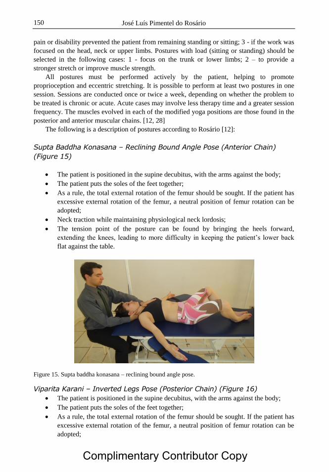
José Luís Pimentel do Rosário 150
pain or disability prevented the patient from remaining standing or sitting; 3 - if the work was
focused on the head, neck or upper limbs. Postures with load (sitting or standing) should be
selected in the following cases: 1 - focus on the trunk or lower limbs; 2 – to provide a
stronger stretch or improve muscle strength.
All postures must be performed actively by the patient, helping to promote
proprioception and eccentric stretching. It is possible to perform at least two postures in one
session. Sessions are conducted once or twice a week, depending on whether the problem to
be treated is chronic or acute. Acute cases may involve less therapy time and a greater session
frequency. The muscles evolved in each of the modified yoga positions are those found in the
posterior and anterior muscular chains. [12, 28]
The following is a description of postures according to Rosário [12]:
Supta Baddha Konasana – Reclining Bound Angle Pose (Anterior Chain)
(Figure 15)
The patient is positioned in the supine decubitus, with the arms against the body;
The patient puts the soles of the feet together;
As a rule, the total external rotation of the femur should be sought. If the patient has
excessive external rotation of the femur, a neutral position of femur rotation can be
adopted;
Neck traction while maintaining physiological neck lordosis;
The tension point of the posture can be found by bringing the heels forward,
extending the knees, leading to more difficulty in keeping the patient‘s lower back
flat against the table.
Figure 15. Supta baddha konasana – reclining bound angle pose.
Viparita Karani – Inverted Legs Pose (Posterior Chain) (Figure 16)
The patient is positioned in the supine decubitus, with the arms against the body;
The patient puts the soles of the feet together;
As a rule, the total external rotation of the femur should be sought. If the patient has
excessive external rotation of the femur, a neutral position of femur rotation can be
adopted;
Complimentary Contributor Copy

Efficacy of Modified Yoga Positions and Postural Chains Therapy … 151
Neck traction while maintaining physiological neck lordosis;
The therapist flexes the patient‘s hip holding them by the heels. Alternatively, the
therapist can use a support for the heels (Figure 16) in order to free the hands for
treatment;
The tension point of the posture can be found by bringing the heels forward,
extending the knees, leading to more difficulty in keeping the patient‘s lower back
flat against the table.
Figure 16. Viparita Karani – inverted legs pose.
The standing postures were not used in this study aiming a better control of the scientific
variables.
Postures Evolution
During the 15 minutes-posture period, the therapist must seek to maintain the symmetry
of the patient. When the patient maintains a posture for a certain time, muscular
viscoelasticity decreases. As a result, the posture becomes easier to sustain. Thus, the
difficulty of the posture must be gradually increased. The name of this process is posture
evolution. In other words, evolution involves finding a new tension point each time the
position starts to become easier. The following rules must be respected:
At no time is it permissible to reverse the spinal physiological curves;
Both anterior and posterior postures require gradual extension of the knees;
The posture of the posterior chain requires a gradual increase in hip flexion.
Pain Assessment
Pain is a symptom that accompanies the majority of pathological conditions that require
medical care transversely. Among the internationally validated scales for measuring the
Complimentary Contributor Copy

José Luís Pimentel do Rosário 152
intensity of pain, the analog scale is one of the most utilized. [29-31] In the present study, a
line, scored from 0 to 10, was shown to the subject, who verbally identified the degree of their
pain, with zero indicating no pain and ten indicating the most intense pain possible.
Statistical Analysis
Besides the descriptive analysis, the Student's t-test was used, with the significance level
set at p = 0.05. Paired Student‘s t-test was used when comparing the pain relief after the
treatment with the pain values before the treatment in the same group. Student‘s t-test for two
samples with equal variance was used when comparing the groups.
Results
Table 1 displays the number of times that each score (from zero to ten) was reported for
all of the 126 spinal pain complaints of the PCT group, before and after treatment.
Table 1. Number of times each value of the analog scale was reported in the 126
complaints, before and after one session of muscular chain therapy
Amount of occurrence of each value of the analog scale
Value on the analog
scale
Before treatment with PCT
(n = 126)
After treatment with PCT
(n = 126)
10 0 0
9 5 0
8 15 3
7 18 0
6 28 0
5 29 3
4 8 18
3 18 10
2 5 13
1 0 38
0 0 41
Before the PCT session, the greatest number of complaints was between the values 5 to 8.
After the session, the majority of values were between 0 and 1, with a significant number of
complaints also found at value 2, 3 and 4 of the scale. The no pain value (zero) increased
from zero reports to forty one, from the total of one hundred twenty six complaints.
The mean of the 126 values before treatment was 5.55 with a standard deviation of 1.62.
After treatment, these values dropped to a mean of 1.79 and a standard deviation of 1.79. The
number of complaints with some improvement was 122, which is equal to 97% of cases. PCT
had no effect in only 1 case (0.7%). In addition, there were also 3 cases (2.3%) that worsened.
The difference between the before and after averages is 3.93. The significance of these data in
Complimentary Contributor Copy

Efficacy of Modified Yoga Positions and Postural Chains Therapy … 153
the Student‘s t-test was p = 0.00001. Thus, it is possible to reject the equality between values
before and after treatment. Table 2 displays the values for 50 complaints of the control group.
Table 2. Number of times each value of the analog scale was reported in the 109
complaints, before and after one session of turned off ultrasound (placebo)
Amount of occurrence of each value of the analog scale
Value on the analog
scale
Before treatment with placebo
(n = 109)
After treatment with placebo
(n = 109)
10 0 1
9 5 3
8 12 9
7 14 8
6 23 15
5 18 16
4 15 18
3 20 25
2 2 12
1 0 0
0 0 2
Before the placebo session, the largest number of complaints was between the values 3 to
8. After the session, the majority of values were between 2 and 6. On the scale, the value of
zero (no pain) increased from zero reports to one.
The mean of the 109 values before treatment was 5.42 with a standard deviation of 1.82.
After treatment, these values dropped to a mean of 4.64 and a standard deviation of 2.07. The
number of complaints with some improvement was 40, which is equal to 36.7% of cases. The
placebo therapy had no effect in 64 cases (58.7%) and in 5 cases (4.5%) the pain worsened.
The difference between the before and after averages is 0.78.The significance of this data in
the Student‘s t-test was p = 0.0001.
Comparing the values before the treatment between groups with the Student t-test showed
a p value of 0.9. Therefore, it is not possible to reject the equality between the groups before
treatment. Comparing the difference before and after treatment between the groups with the
Student‘s t-test, the value of p was 0.0001 (Table 3).
Table 3. Mean and standard deviation before and after treatment.
Intergroup p value and p value between the groups
Mean and standard
deviation before
treatment
Mean and standard
deviation after
treatment
Intergroup
p value
p value
between the
groups
MCT Group 5.55 + 1.79 1.62 + 1.79 0.00001 0.0001
Control Group 5.42 + 1.82 4.64 + 2.07 0.0001
Complimentary Contributor Copy

José Luís Pimentel do Rosário 154
Discussion
Both Groups showed significant relief of undiagnosed spinal pain. The placebo exhibited
a p-value of 0.0001, whereas the value for yoga was 0.00001 after treatment. Comparing the
groups, yoga was significantly better with a p value of 0.0001. The control group had a mean
pain level of 5.42 before the treatment and 4.64 after (a difference of 0.78). The mean pain
level in the yoga group was 5.55 before treatment and 1.79 after (a difference of 3.93).
Treatment with yoga provided some sort of pain relief in 97% of cases, whereas this figure
was 36.7% in the control group. In a previous study, [32] the same technique was applied for
any type of musculoskeletal pain including the spine, but not restricted to this region, with
similar results.
Although there are relatively few scientific studies approaching these yoga postures or
similar techniques based on Meziérès stretching techniques [MST], they have produced
results in treating various musculoskeletal conditions. [15-23, 33]
The present study
corroborates these previous experiments.
Canto et al. [16] studied the efficiency of a MST in individuals with lower back pain, in
terms of the level of pain using visual analog pain at the time of the first and tenth treatment
session. In total, 85.7% of the participants reported a decrease in the level of pain at the end
of treatment and 77.1% of the subjects recorded a lower score on the Roland Morris
questionnaire. The result of 85% is close to the 97% found in the present study, although the
main difference is the number of sessions: while these authors used ten sessions, the present
study used only one. This brings into question the number of sessions necessary to perform
the treatment. Most MST therapists perform ten sessions as a basis for a treatment that
provides results. [12] Moreover, since Canto et al. [16] did not assess patients before the
eleventh session, but after the tenth, these researchers have assessed the pain with the acute
effect of the last session and not with the chronic effect of the ten sessions alone. The present
study also did not assess the effect of a single session after 24 or 48 hours. This form of data
would be very interesting, in terms of finding a more effective treatment.
However, if MST or PCT treat pain through postural improvement, it is possible that
these musculoskeletal pains will not return after full treatment. Moreira et al. [20] studied a
group of five women aged between 20 and 30 years. The women were submitted to physical
therapy to correct their posture and to reduce the pain caused by postural abnormalities. The
patients were radiographed one week after therapeutic discharge. The postural improvement
was evidenced by the retraction of the shoulder. Despite the small number of patients, the
radiographic image is a high point of the study. Based on this study, it is possible to see that
some of the complaints of musculoskeletal pain may be related to postural problems.
Another indication that postural correction may be the answer for the pain reduction is in
the work of Rossi et al. [23] who obtained the postural improvements with just one treatment
session using a similar technique. They assessed the effect of an application of the lying hip
extension posture on 11 photographic postural variables. Of these variables, only 4 recorded
significant improvements, and these four were all related to the head. The present study
recorded pain reduction with one session, which can be related to that better posture achieved
in the very first session. Rosário [24] obtained results in 3 of 6 variables with eight postural
reeducation sessions, and there was no improvement in the shoulders and head. This
highlights the fact that therapists place emphasis on postural correction, which may be more
Complimentary Contributor Copy

Efficacy of Modified Yoga Positions and Postural Chains Therapy … 155
influential in a chosen segment. These studies revive the discussion, with a focus on posture
and not on pain, about the time required for postural corrections and relief from the related
pain. Rossi et al. [23] obtained the postural result with one posture and the present study
recorded pain reduction with one session and two postures.
Marques et al. [19] assessed the effect of postural treatment on fibromyalgia. Twenty
patients that had been diagnosed with fibromyalgia were treated for six sessions on average.
Of the 20 patients, 18 reported some improvement, and 65% rated it as excellent or good,
whereas 25% reported it as fair and only 10% reported no improvement. Although
fibromyalgia is listed as a rheumatologic disease, these data are similar to the results of the
present study.
Basso et al. [15] decreased the pain of 20 patients with temporomandibular disorders,
using 10 muscular chain treatment sessions. In a further study, Gil et al. [17] reduced back
pain in pregnant women in 8 weeks. Using analog scales, Heredia and Rodrigues [34]
relieved the pain of patients with epidural fibrosis in post-operative lumbar disc herniation
with 15 sessions.
Using a pressure platform (Tekscan-Matscan), Teodori et al. [35] undertook an
interesting case study and noted changes in plantar pressure distribution and the location of
the center of force in a subject with a history of right ankle sprain, with free bipedal support
and with the eyes open. Asymmetry was found in the distribution of plantar pressure applied
to the subject in one postural reeducation session, and was followed by an assessment of the
pressure platform immediately after the intervention, and after 7, 14 and 30 days. The results
clearly showed a recovery of symmetry, which continued for 7 days. After this period, there
was a gradual recovery of asymmetry, although the initial values had not been attained after
30 days. Although a case study, if the data for this author is correct for the general population
and most musculoskeletal pain is related to posture, one MCT session, as performed in the
present study, would have the effect of pain resolution for seven days, and would remain in
effect for 30 days or more in cases of chronic pain.
Rosário et al. [27] argue that this type of postural treatment technique does not act on
posture simply by stretching, since a 15-minute posture provided similar results for hamstring
flexibility as a 30-second hamstring stretch. Body awareness and the active maintenance of
better joint positioning, reducing an existing subluxation, can exert their influence on postural
adjustment and consequently solve related pain. [36] Whatever the reason for the effect, this
study and previous studies have shown that MST causes a significant improvement in the
efficiency of a musculoskeletal pain source. The relief of 94.4% of the complaints, with a
complete absence of pain in 24 of the 71 complaints, demonstrated the efficiency of yoga
modified postures when properly applied.
An interesting result in the present study was the success of the placebo therapy, which
was inferior to the Yoga treatment but still provided some pain relief in 36.7% of cases. These
results were similar to the ones found by Rosário et al. [32] who used the same method to
treat any type of undiagnosed musculoskeletal pain and used a disconnected ultrasound as a
placebo therapy, which had a 36% rate of success. These data also support the findings of
Beecher [36] who suggested that the placebo effect occurs in 35% of the population. Placebo
analgesia for post-operative dental pain was effective in 39% of cases. [37] Grelotti and
Kaptchuk [38] discussed that the placebo effect comes from an emotional response and can be
so strong that not only the patient feels better, but members of the family can also think the
treatment is working, provoking a whole social effect. These results corroborate the findings
Complimentary Contributor Copy

José Luís Pimentel do Rosário 156
of other authors who suggested that the placebo effect occurs in 20% to 40% of the cases.
[39]
PCT was shown to reduce musculoskeletal pain in young adult patients immediately post
intervention. However, our study did not determine short term or the long term effects of
PCT. Further studies are required to verify the short and long-term effect evaluation and
optimal treatment duration for postural improvement and the decrease of pain. Objectives
measures are also recommended for future works.
Conclusion
This chapter showed that the PCT, using modified yoga positions, was shown to reduce
musculoskeletal pain in patients immediately after the intervention, probably because of a
postural improvement. However, it did not determine the short term or long term effects of
just one intervention with yoga. Further studies are required to understand these effects and
the disorders that could be treated effectively by this method, as well as those that prove
unresponsive. Moreover, identifying the optimal timeframe and frequency of application for
each disorder should be determined.
References
[1] Rosário JLP, Marques AP, Maluf SA. Aspectos clínicos do alongamento: uma revisão
de literatura. Revista Brasileira de Fisioterapia. 2004;8:83-88.
[2] Kendall FP, McCreary EK, Provance PG. Músculos – Provas e Funções. São Paulo:
Manole; 2010.
[3] Novak CB, Mackinnon SE Repetitive use and static postures: a source of nerve
compression and pain. J Hand Ther. 1997;10:151-9.
[4] Lee D. Principles and practices of muscle energy and functional techniques. In: Grieve
GP (ed.) Modern manual therapy of the vertebral column. New York: Churchill
Livingstone; 1986. p. 279-301.
[5] Kappler RE Postural balance and motion patterns. J Am Osteopatic Assoc. 1982; 81:69-
77.
[6] Orchard J, Marsden J, Lord S, Garlick D Preseason hamstring muscle weakness
associated with hamstring muscle injury in Australian footballers. Am J Sports
Medicine. 1997; 25:81-5.
[7] Bennell K, Wajswelner H, Lew P, Schall-Riaucour A, Leslie S, Plant D, et al.
Isokinetic strength testing does not predict hamstring injury in Australian Rules
footballers. Brit J Sports Medicine. 1998; 32:309-14.
[8] Mirtz TA, Morgan L, Wyatt LH, Greene L. An epidemiological examination of the
subluxation construct using Hill's criteria of causation. Chiropr Osteopat. 2009;
2;17:13.
[9] Rosário JLP, Marques AP, Maluf AS. Aspectos Clínicos do alongamento: uma revisão
de literatura. Revista Brasileira de Fisioterapia. 2004; 8:83-8.
Complimentary Contributor Copy

Efficacy of Modified Yoga Positions and Postural Chains Therapy … 157
[10] Moreira CMC, Soares DRL. Análise da efetividade da reeducação postural global na
protusão do ombro após a alta terapêutica. Fisioter Mov. 2007;20:93-9.
[11] Teodori RM, Negri JR, Cruz MC, Marques AP Reeducação postural global: uma
revisão da literatura. Rev Bras Fisioter. 2011;15: 185-9.
[12] Rosário JLP. Manual Prático de Reeducação Postural: o que você precisa saber para um
tratamento eficiente. São Paulo: Ed. Baraúna; 2011.
[13] Bertherat T, Bernstein C. O corpo tem suas razões – Antiginástica e a consciência de si.
São Paulo: Martins Fontes; 1987.
[14] Hoppenfeld S. Propedêutica ortopédica coluna e extremidades. São Paulo:
Atheneu;1999.
[15] Basso D, Corrêa E, Silva AM. Efeito da reeducação postural global no alinhamento
corporal e nas condições clínicas de indivíduos com disfunção temporomandibular
associada a desvios posturais. Fisioter Pesqui. 2010;17: 63-8.
[16] Canto CREM, Oliveira LF, Gobbi FCM, Theodoro MN. Estudo da eficácia do metódo
de reeduçação postural global em indivíduos com dor lombar com relação a dor e
incapacidade funcional. Ter Man. 2010;38: 292-7.
[17] Gil VFB, Osis MJD, Faúndes A Lombalgia durante a gestação: eficácia do tratamento
com Reeducação Postural Global (MCT). Fisioter Pesqui. 2011;18: 164-70.
[18] Luz, GCP, Cheik NC, Ferreira F, Pereira PAC, Vidal JS, Affonso F, Baraúna MA.
Tratamento da lombalgia através do dispositivo lombo abdominal e da reeducação
postural global. Ter Man. 2008; 6:287-92.
[19] Marques AP, Mendonça LLF, Cossermelli W. Alongamento muscular em pacientes
com fibromialgia a partir de um trabalho de reeducaçao postural global (RPG). Rev
Bras Reumatol. 1994;34: 232-4.
[20] Moreira CMC, Soares DRL. Análise da efetividade da reeducação postural global na
protusão do ombro após a alta terapêutica. Fisioter Mov. 2007;20: 93-9.
[21] Moreno MA, Catai AM, Teodori RM, Borges BLA, Cesar MC, Silva E, et al. Efeito de
um programa de alongamento muscular pelo método de Reeducação Postural Global
sobre a força muscular respiratória e a mobilidade toracoabdominal de homens jovens
sedentários. J Bras Pneumol. 2007; 33: 679-86.
[22] Moreno MA, Catai AM, Teodori, RM, Borges BLA, Zuttin RS, Silva E, et al.
Adaptações do sistema respiratório referentes à função pulmonar em resposta a um
programa de alongamento muscular pelo método de reeducação postural global.
Fisioter Pesqui. 2009;16: 11-5.
[23] Rossi LP, Brandalize M, Gomes ARS. Efeito agudo da técnica de reeducação postural
global na postura de mulheres com encurtamento da cadeia muscular anterior. Fisioter
Mov. 2011;24: 255-63.
[24] Rosário JLP, Nakashima IY, Rizopoulos K, Kostopoulos D, Marques AP. Improving
posture: Comparing segmental stretch and muscular chains therapy. Clin Chiropractic.
2012; 15: 121-8.
[25] Myers T. Anatomy Trains. 2nd ed. Edinburgh: Churchill Livingstone / Elsevier; 2009.
[26] Marques AP. Cadeias musculares – um programa para ensinar avaliação
fisioterapêutica global. Manole, São Paulo; 2005.
[27] Rosário JLP, Sousa A, Cabral CMN, João SMA, Marques AP. Reeducação postural
global e alongamento estático segmentar na melhora da flexibilidade, força muscular e
amplitude de movimento: um estudo comparativo. Fisioter Pesqui. 2008;15: 12-18.
Complimentary Contributor Copy

José Luís Pimentel do Rosário 158
[28] Karminoff L. Yoga anatomy. Champaign IL: Human Kinetics; 2007.
[29] Gracely RH, Price DD, Roberts WJ, Bennett GJ. Quantitative sensory testing in patients
with CRPS-I and -II. In Janig W, Stanton-Hicks M. (Eds.). Reflex sympathetic
dystrophy – A reappraisal. Seattle: IASP Press;1996. p.151-172.
[30] Aicher B, Peil H, Peil B, Diener HC. Pain measurement: Visual analogue scale (VAS)
and verbal rating scale (VRS) in clinical trials with OTC analgesics in headache.
Cephalalgia. 2012;32:185-97.
[31] Bailey B, Gravel J, Daoust R. Reliability of the visual analog scale in children with
acute pain in the emergency department. Pain. 2012;153:839-42.
[32] Rosário JLP, Orcesi LS, Kobayashi FN, Aun AN, Diolindo Assumpção IT, Blasioli GJ,
Hanada ES. The immediate effects of modified Yoga positions on musculoskeletal pain
relief. J Bodyw Mov Ther. 2013;17:469-74.
[33] Fozzatti MCM, Palma P, Herrmann V, Dambros M. Impacto da reeducação postural
global no tratamento da incontinência urinária de esforço feminina. Rev Assoc Med
Bras. 2008; 54: 17-22.
[34] Heredia EP, Rodrigues FF. O tratamento de pacientes com fibrose epidural pela
reeducação postural global. Rev Bras Neurol. 2008;44: 19-26.
[35] Teodori RM, Guirro ECO, Santos RM. Distribuição da pressão plantar e localização do
centro de força após intervenção pelo método de reeducação postural global: um estudo
de caso. Fisioter Mov. 2005;18: 27-35.
[36] Beecher HK. The powerful placebo. J Am Med Assoc. 1955; 159: 1602-6.
[37] Levine JD, Gordon NC, Bornstein JC, Fields HL. Role of pain in placebo analgesia.
Proc Natl Acad Sci USA. 1979; 76: 3528-31.
[38] Grelotti DJ, Kaptchuk TJ. Placebo by proxy. BMJ. 2011; 11:343-5.
[39] Verhulst J, Kramer D, Swann AC, Hale-Richlen B, Beahrs J. The medical alliance:
from placebo response to alliance effect. J Nerv Ment Dis. 2013; 201:546-52.
Complimentary Contributor Copy

In: Posture: Types, Exercises and Health Effects ISBN: 978-1-63117-252-6
Editor: Sarah A. Curran © 2014 Nova Science Publishers, Inc.
Chapter 11
Yoga Postures and Colon Cleanse
Vijaypal Arya, MD
Cornell University, New York, US
North Shore LIJ Health System, New York, US
Endoscopy Unit, WHMC, Brooklyn, New York, US
Abstract
This chapter presents a novel approach to colon cleanse. No harsh laxative chemicals
such as phosphates, sulphates, or citrates are used. The process emphasizes the basic tenet
of yoga (unifying mind, body and spirit). The innovative aspect involves a bolus drinking
of lukewarm saline in conjunction with sequential posture changes. In addition, deep
breathing, relaxation and meditation helps in bowel cleansing by parasympathetic
activation. This yoga based purification technique can be learned by following a DVD.
On an average, it takes less than two hours to complete the process and is ideal for
healthy men and women who have an open mind for yoga and are fit enough to do a little
exercise.
Keywords: Posture, saline, colon cleanse, yoga
Introduction
Physicians have been prescribing laxatives for centuries, but the need to cleanse the colon
was first recognized by the radiologist circa 1895 after the invention of radiographs and the
use of barium enema to study the large bowel. [1] Subsequently, surgeons realized that if the
colon is clean before abdominal surgeries, the incidence of post operative infections can be
reduced. [2, 3] Initially, a large volume (9-12 liters) of normal saline/balanced electrolyte
solutions were used to cleanse the bowel. [4, 5] Patients were either asked to drink at a rate of
Author contact: [email protected]
Complimentary Contributor Copy

Vijaypal Arya 160
25-30 ml/minute (usual sipping rate) or solutions were infused via naso-gastric tube for about
4-6 hours to complete the cleansing process. In 1957, the introduction of the first fiber optic
endoscope revolutionized the medical field and a new subspecialty emerged – gastrointestinal
endoscopy. [6] For a meticulous examination of the colonic mucosa, the lumen should be
completely devoid of waste material, particularly if the aim is to recognize and remove
abnormalities as small as a pin head (5-6 mm). These are called polyps and are of many
different histologic subtypes. The adenoma type is pre-cancerous and early removal can
prevent colon cancer. [7] Colon cancer is the third most common cancer in United States and
is the second leading cause of cancer deaths. [8] Screening and surveillance colonoscopy and
polypectomy has already reduced the incidence of colon cancer deaths, the poor bowel prep
(about 10-15%) remains a significant limiting factor in the success of this life saving
procedure. [9] Many patients dread the task of cleaning more than the procedure itself. The
large volume (4L) and unpalatable taste of the most commonly used polyethylene glycol
(PEG) based preps are a popular deterrent among patients. [10] An ideal preparation should
be effective, safe, palatable, fast and impose few dietary restrictions on the patients. While no
prep regimen can be ideal for all groups of patients, a novel approach has been explored,
which is based on yogic principles. [11, 12] A kriya (sanskrit word for process) known as
shankh prakshalana has been standardized as ―shudh‖ (pronounced ―should‖ and in sanskrit
means purity). This process involves drinking lukewarm saline in bolus form (large volumes,
240ml, taken intermittently) alternating with five yoga asanas or postures. This represents a
potential time saving alternative, which is economical as well.
The knowledge presented in this chapter highlights tremendous medical value of a
centuries-old practice. The process has been standardized so it can be used easily and without
supervision by a yoga teacher. The basic mechanism of action has been delineated.
Scope of Complementary and Alternative
Medicine (CAM)
More than one third of American adults use some form of CAM. Approximately 20
million individuals practice yoga, spending $10.3 billion a year. [13] Credible scientific
research is published in peer reviewed journals and added to the literature every day. The
medical value of CAM therapies is being recognized and is starting to be practiced. The
common CAM therapies include acupuncture, tai chi, recki, and yoga. The word "yoga"
comes from sanskrit, the most ancient Indian language and has two meanings. The first is
union (union of spirit, mind and body). The root of union is "yugir". The second meaning is
smadhi (The absolute knowledge, the highest state of mind and the final realization). The root
of samadhi is yuja.
The average person understands yoga as simply a physical practice – this is a huge
misconception. Yoga actually has three components: spiritual, mental, and physical. The goal
is to unify spirit, mind, and body (Figure1). In its most original form, described by Rishi
Patanjali in the yoga darshan, yoga has eight limbs and is called ashtanga ("asht-" meaning
eight, and "-anga" meaning limbs) yoga. The eight limbs are: yama (duties towards others, of
which there are five), niyama (duties towards oneself, of which there are also five), asana
(postures), pranayama (breathing exercises), pratyahara (introspection), dharana
Complimentary Contributor Copy

Yoga Postures and Colon Cleanse 161
(concentration), dhyana (meditation), and samadhi (state of super-consciousness). The last
three limbs describe advanced meditative practices through which the practitioner can achieve
autonomic nervous system control.
Figure 1. The union of spirit, mind and body is yoga.
The first niyama is saucha (pronounced shau-cha), which means cleanliness of mind and
body. Shankh-prakshalana has been defined in yoga literature as a means of cleansing the
digestive tract. In sanskrit, shankh means "conch." The sea shell, with its convoluted
chambers, is a metaphor for the gastrointestinal tract. Prakshalana means "to clean." In an
effort to make this millennia-old method more widely known, Arya et al. [11] have simplified
shankh-prakshalana to shudh. The average time to complete the cleanse process is less than
two hours, as compared to four to six hours with conventional methods.
Shudh Process
The first pre-requisite to practice yogic processes is willingness to perform. It is the
authors‘ advice to health practitioners to experience the shudh process themselves before
prescribing it to the patients. For someone with no prior knowledge and experience the only
way is to take a leap of faith and try. To practice shudh one should either watch the DVD
(www.shudhinc.com) and learn the poses or read up the instructions and practice the poses in
advance. All the poses are simple to practice except the fifth pose (demands squatting) and
can be modified as explained. The best time to perform the process is early morning after a
good night's sleep. The body is well rested and all the nutrients have passed from the small
bowel into the large bowel. The morning time is also the time for increased colonic motility
and natural bowel movement. It‘s advised to eat a light dinner the day before the process. If
the purpose of doing the shudh-process is cleansing the colon before colonoscopy, one should
Complimentary Contributor Copy
Vijaypal Arya 162
eat light lunch by 2.00 pm the day before and then stay on liquids for the rest of the day until
going to sleep. Clear broth, gelatin, apple juice, and white grape juice is fine, but nothing red
and nothing with pulp. It is also advised to avoid eating or drinking anything in the morning.
This dietary restriction is necessary to achieve high quality colon cleansing, so that the doctor
performing the colonoscopy will be able to perform a high quality examination. To begin,
wear comfortable and loosely fitting clothing and choose a quiet environment and firm
surface. A dedicated bathroom should be available for use. All distractions should be avoided
i.e., turn off phones and beepers, relax and practice mindfulness. To prepare the solution
dissolve one sachet of pre-packaged 9 gm of NaCl (www.shudhinc.com) in one liter of
lukewarm water. A twist of lemon/lime can be added according to palatability preferences.
1. The actual shudh process includes – drinking lukewarm saline in bolus forms (rather
than sipping) and then perform yoga poses (Table 1):
Drink one or two 8-ounce glasses of the solution. Do the upward stretch and the side
stretch;
Drink one or two 8-ounce glasses. Do the twist stretch and the push-up stretch;
Drink one or two 8-ounce glasses. Do the squat stretch;
Repeat the cycle.
The rapid drinking is what brings the rapid results. This works like a flush while the
stretching exercise works like a pump until it all goes through and one comes out clean. An
average person needs to drink 2-3 liters of solution and completes 2-3 cycles of yoga poses to
complete the process. Sometimes fullness in the stomach and feeling of nausea precludes the
required drinking. In this situation, skip the drinking; continue the yoga poses until ready to
drink again.
One can expect to have first bowel movement between 20 and 40 minutes. The stool
comes out in a rush; hence slightest urge to defecate should not be ignored. It‘s advised to
interrupt the process to use the toilet and avoid straining. The process should be continued
where left off. Individual results vary and sometimes a delayed response, up to few hours has
been noticed. The average time to complete the process is about 2 hours. At this point the
excreta would look like urine. Stop the process and take rest in supine position also called
shav-asana for 20-30 minutes.
Upward Stretch - Palm Tree Pose - Tad-asana (Taad’as’ana)
―Tad‖ pronounced – taad, means ―palm tree‖ and ―asana‖ means posture. This posture
resembles a palm tree. To begin, stand up straight, interlace fingers of both hands and put
them on your head with the palm facing up. While inhaling stretch the body up and stand on
your toes, like you're trying to reach the sky. Keep your knees straight. Look straight in the
infinity. Wait for 5 seconds, while exhaling come to the ready position. To practice
mindfulness and achieve concentration, try to feel your breath as it enters through your
nostrils and imagine that all positive energy is coming inside you. At the same time while
exhaling imagine that all toxins are leaving your body. Do not occupy your mind with any
Complimentary Contributor Copy

Yoga Postures and Colon Cleanse 163
thoughts but to achieve cleansing of your body. Relax your shoulders when you come down.
Repeat this pose ten times.
Side Stretch - Oblique Palm Tree Pose - Tiryak Tadasana
(Tir’yak’taad’as’ana)
To get ready, stand straight, move your feet 1 – 2 feet apart, interlace your fingers, stretch
arms upwards, palms facing up. While inhaling, bend to the right side; try to bend directly to
the side, not forward or backward. Try relaxing your neck and letting your head flop over
when you bend. After waiting for five seconds, exhale slowly and come back to the ready
position. Take a resting break and then bend to the left side. Repeat this stretch five times on
right and five times on left.
Twist Stretch - Waist Twisting Pose - Katichakrasana
(Khat - Ee’chak’ra’sana)
Start off with pulling feet apart and arms stretched out horizontally, taking up the position
of the famous DaVinci drawing. Put the left hand on your right shoulder and the right hand on
the small of your left back, the palm facing out. Inhale and start with the twist while looking
back at your left heel. Wait for five seconds, then exhale and come back to the ready position.
Now repeat the pose on other side. Repeat this stretch five times on right and five times on
left.
Turtle Stretch - Twisting Cobra - Tiryak Bhujangasana
(Tir’yak’bhuj’ang’asana)
Lie down on your stomach with the feet apart, like doing a push up. Push your upper
body off the floor leaving the hips on the ground. While inhaling push yourself up as you lift
your chest, and twist your upper body to the right side, try to look at your left heel. Wait for
five seconds the exhale and come to ready position. Repeat this stretch five times on right and
five times on left.
Squat Stretch - Abdominal Stretch Pose - Udarakarshanasana
(Ud’ara’kar’asana)
Grab onto your knees with your feet apart and gently lower yourself into a squat. Stand
on the balls of your feet if you can. This is the ready position. Inhale and try to touch your left
knee to the right toes. Rotate your rib cage around the spine, twisting your torso around
looking up and over the shoulder. Exhale as you come back to center. Your hands should help
with balance and steering your knees around. Repeat the pose on left side. You can hold on to
Complimentary Contributor Copy

Vijaypal Arya 164
a chair for support and squat as much as you can and just twist. Repeat this stretch five times
on right and five times on left.
Precautions
The process should be practiced by healthy people only. According to the yogic literature
the process can be done with plane lukewarm water in case of salt sensitivity and
contraindications to salt intake. Anybody with poor heart, liver and kidney function are
specifically at risk for salt and fluid overload and should not attempt this process. At the same
time inability to perform poses due to arthritis and joint diseases and pregnant females should
not practice this process. This also should not be considered as a treatment for severe
constipation. The process should not be practiced more than once in 2-3 months.
Adverse Effects
Failure of the process meaning no bowel movements after drinking 2-3 liters of
lukewarm saline has been experienced. This will also lead to benign swelling of the body
resolving in few days. Sometimes increased urination has been reported instead of defecation.
If feelings of nausea, dizziness, severe bloating, abdominal distension and pain persist or if
bowel movement is not initiated after consuming approximately two liters of solution, stop
the process. It is advised not to consume more than four liters of salt solution at a time.
Benefits - Detoxification
Modern medicine believes that colon / large intestine is a self-cleansing organ, hence it
does not need to be cleaned. The users of shudh, anecdotally have reported, the feeling of
rejuvenation and being full of energy after the process. This is in contrast to feeling weak
after using other bowel cleansing methods and harsh laxatives. Further research is needed to
prove this point. For those who believe in colon cleansing, shudh is the most natural,
economic and fast alternative. This can be learned by watching a DVD in the comfort of your
home, without the worry of inappropriate social constraints, like embarrassment from rectal
instrumentation during colonics.
Constipation is a very common problem worldwide. About 15% of Americans (60
million) suffer from this problem. [14] If left untreated chronic constipation can lead to
complications like hemorrhoids, anal fissures, solitary rectal ulcers, and overflow
incontinence. According to a recent report more than $700 million are spent on laxatives
yearly by Americans alone. Although no study has been done using this process to treat
constipation, intuitively the yogic cleansing process can be a very economical, simple and
faster alternative to treat mild constipation.
Complimentary Contributor Copy

Yoga Postures and Colon Cleanse 165
Science of Shudh Process: Millennia Old Process
and Modern Physiological Principles
Other than mindfulness, shudh process has two components: A - bolus (8oz-16oz in 1-2
minutes) drinking of lukewarm normal saline and B - yoga postures with deep breathing.
According to yoga philosophy water should be taken in bolus form especially in the morning
to generate an urge to defecate. The distension of duodenum initiates the gastro-colic reflex,
well recognized in modern physiology. [15] The signals from the duodenum reach to the right
colon, causing movement of content from right to left instantaneously, leading to defecation.
Thus another concept of modern physiology plays an important role in bolus drinking is first
order kinetics. [16] The isotonic saline empties from stomach based on the first order
kinetics- 50% of volume empties out in 8-18 minutes, meaning higher volume will empty out
by bolus drinking as compare to sipping. The dumping of large volume of isotonic saline in
intestine rushes through causing minimal time for absorption and no significant electrolyte
imbalance.
The conventional, PEG (polyethylene glycol) based colon preps are advised to drink
chilled partly to mask the unpalatable taste. Animal studies have shown that warmer liquids
cause esophageal and gastric relaxation, suggesting that practice of drinking lukewarm saline
might be beneficial. [17, 18] The common practice also dictates use of warmer temperature
for cleansing.
Role of Yoga Postures
Application of different body positions is well established in modern medicine. Some of
the well known positions are supine, prone, left and right lateral, Trendelenburg, reverse
Trendelenburg, and lithotomy. All these positions have specific use in clinical medicine.
Significant effect of body position on gastric emptying has been noticed on radio-nuclide
studies. [19] Simply lying on right side improves gastric emptying. [20] Position change
together with gravity changes the gastric configuration, leading to intra gastric re-distribution
and faster gastric emptying. [21, 22]
During the first pose, stretching of the body upwards (with deep breathing causing
diaphragmatic movements) results in change in gastric configuration. Arm elevation is known
to increase the tidal volume. [23]
The first pose resulting in linear acceleration of vestibular fluid of semicircular canals
increases parasympathetic tone. [24] The second, third and fourth poses will also lead to the
same effect. The twisting of spine will be associated with compression of abdominal organs
especially intestinal tract. Additionally, the effects on the vestibular system also might play a
role in faster gastric emptying. During, the fifth pose (squat stretch) – squatting is known to
increase intra-abdominal pressure and straightening of anorectal angle which facilitate
defecation. [25]
Complimentary Contributor Copy

Complimentary Contributor Copy

Complimentary Contributor Copy
Vijaypal Arya 168
Role of Exercise on Gastrointestinal Motility
Acute aerobic exercise decreases colonic phasic motor activity causing less resistance to
colonic flow. [26] The post exercise effect is increased amplitude of propagated waves
leading to enhance propulsion of colonic contents. Simple walking has been shown to
improve colonic preparation. [27] The exercise component of yoga postures are although mild
intensity (metabolic equivalent value of three) should have a complementary role.
Role of Deep Breathing
Deep breathing is the most potent natural action to stimulate parasympathetic tone. [28]
Deep breathing stimulates pulmonary stretch reflex, with the final effect of bradycardia,
relaxation of intestinal sphincters and increased intestinal motility. [29]
Role of Mindfulness
Psychological factors (stress and relaxation) are well known to influence gut motility.
[30] The role of turning inward - mindfulness, avoiding routine distractions -is difficult to
measure. The process is performed in a calm environment. The postures are motivated by
deep, rhythmic inhalation and exhalation with an aim to achieve relaxation of mind and body.
Mind - body interaction has been identified by modern biomedical research. [31]
Conclusion
Shudh is a modern version of shankh prakshalana, a yogic purification technique. A new
mechanism of colon cleansing has been explored. The process cleanses the colon naturally
using only lukewarm saline water and a series of five yoga poses. Instead of chemical action
the body‘s own reflexes do the work. The shudh process is ideal for healthy men and women
who have open mind for yoga and are fit enough to do a little exercise.
References
[1] Rooney A. History of medicine. New York: Rosen Pub Group; 2012.
[2] Ellis H. The first successful elective laparotomy. J Perioperative Pract. 2008;18:211.
[3] Hewitt J, Reeve J, Rigby J Cox JA. Whole-gut irrigation in preparation for large-bowl
surgery. Lancet. 1973;3:37-40.
[4] Levy AG, Benson JW, Hewlett EL, Herdt JR, Doppman JL, Gordon RS Jr. Saline
lavage: A rapid, effective, and acceptable method for cleansing the gastrointestinal
tract. Gastroenterology. 1976;70:157-61.
Complimentary Contributor Copy

Yoga Postures and Colon Cleanse 169
[5] Postuma R. Whole bowel irrigation in pediatric patients. J Pediatric Surgery.
1982;17:350-52.
[6] Irvin Modlin, A brief history of endoscopy. Milan: Multi Med; 2000.
[7] Winawer SJ, Zauber AG, Ho MN, O'Brien MJ, Gottlieb LS, Sternberg SS, et al.
Prevention of colorectal cancer by colonoscopic polypectomy. The National Polyp
Study Workgroup. New England J Medicine. 1993; 329:1977-81.
[8] Cummings LC. Colorectal cancer screening: update 2011. Seminar in Oncology. 2011:
38:483-489.
[9] Belsey J, Epstein O, Heresbach D. Systematic review: oral bowel preparation for
colonoscopy. Aliment Pharmacol Ther. 2007;15; 25:373-84.
[10] Harewood GC, Wiersema MJ, Melton LJ. A prospective, controlled assessment of
factors influencing acceptance of screening colonoscopy. Am J Gastroenterol.
2002;97:3186-94.
[11] Arya V, Gupta K, Arya S. Efficacy of bolus kukewarm saline and yoga postures as
colonoscopy preparation: A pilot study. J Altern Complementary Med. 2010; 16:1269-
77.
[12] Arya V, Gupta K, Valluri A, Arya SV, Lesser ML. Rapid colonoscopy preparation
using bolus lukewarm saline combined with sequential posture changes: A randomized
controlled trial. Digestive Diseases Sci. 2013;58:2156-66.
[13] Yoga Journal. ―Yoga journal releases 2012 yoga in America market study‖
http://www.prnewswire.com/news-releases/yoga-journal-releases-2012-yoga-in-
america-market-study-182263901.html. 5th December 2012. Retrieved on: 20th
September 2013.
[14] Consumer health information corporation. http://www.consumer-health.com/services/
LaxativesProceedwithCaution.php‖ Retrieved on: 29th October 2013.
[15] Tansy MF, Kendall FM. Experimental and clinical aspects of gastrocolic reflexes. Am J
Dig Dis. 1973; 18:521-31.
[16] Hunt JN, Macdonald I. The influence of volume on gastric emptying. J Physiol.
1954;126:459-74.
[17] Bateman DN. Effects of meal temperature and volume on the emptying of liquid from
the human stomach. J Physiol. 1982;331:461-67.
[18] McArthur KE, Feldman M. Intragastric temperature, gastric acid secretion, gastrin
release, and gastric emptying following ingestion of hot, warm, or cold coffee in
humans. Gastroenterology. 1984;90:1540.
[19] Moore JG, Datz FL, Christian PE, Greenberg E, Alazraki N. Effect of body posture on
radionuclide measurements of gastric emptying. Digestive Diseases Sci. 1988; 33:1592-
95.
[20] Burn-Murdoch R, Fisher MA, Hunt JH. Does lying on the right side increase the rate of
gastric emptying? J Physiol. 1980;302:395-98.
[21] Amidon GL, DeBrincat GA, Najib N. Effects of gravity on gastric emptying, intestinal
transit, and drug absorption. J Clinical Pharmacology. 1991; 31:968-73.
[22] Anvari M, Horowitz M, Fraser R, Maddox A, Myers J, Dent J, Jamieson GG. Effects of
posture on gastric emptying of nonnutrient liquids and antropyloroduodenal motility.
Am Physiological Society Am J Physiol. 1995;268:G868-71.
[23] Couser JI Jr, Martinez FJ, Celli BR. Respiratory response and ventilatory muscle
recruitment during arm elevation in normal subjects. Chest. 1992;101:336-40.
Complimentary Contributor Copy

Vijaypal Arya 170
[24] Carter JR, Ray CA. Sympathetic responses to vestibular activation in humans. Am J
Physiol Regul Integr Comp Physiol. 2008;294:R681-8.
[25] Bassotti G, Germani U, Morelli A. Human colonic motility: physiological aspects. Int J
Colorectal Dis. 1995;10:173-80.
[26] Rao SC, Beaty J, Chamberlain M, Lambert PG, Gisolfi C. Effects of acute graded
exercise on human colonic motility. Am Physiological Society. 1999;276: G1221-6.
[27] Kim HS, Park DH, Kim JW, Jee MG, Baik SK, Kwon SO, Lee DK. Effectiveness of
walking exercise as a bowel preparation for colonoscopy: A randomized controlled
trial. Am J Gastroenterology. 2005;19:43-50.
[28] Veerabhadrappa SG, Baljoshi VS, Khanapure S, Herur A, Patil S, Ankad RB,
Chinagudi S. Effect of yogic bellows on cardiovascular autonomic reactivity. J
Cardiovasc Dis Res. 2011;2:223-7.
[29] Jerath R, Edry JW, Barnes VA, Jerath V. Physiology of long pranayamic breathing:
neural respiratory elements may provide a mechanism that explains how slow deep
breathing shifts the autonomic nervous system. Med Hypotheses. 2006;67:566-71.
[30] Gaylord SA, Palsson OS, Garland EL, Faurot KR, Coble RS, Mann JD, Frey W, Leniek
K, Whitehead WE. Mindfulness training reduces the severity of irritable bowel
syndrome in women: results of a randomized controlled trial. Am J Gastroenterol.
2011;106:1678-88.
[31] Brower V. Mind-body research moves towards the mainstream. EMBO Rep.
2006;7:358-61.
Complimentary Contributor Copy
In: Posture: Types, Exercises and Health Effects ISBN: 978-1-63117-252-6
Editor: Sarah A. Curran © 2014 Nova Science Publishers, Inc.
Chapter 12
The Behavior Characteristics
and Postural Angles in Teenagers
Who Wear High-Heeled Shoes
and How Pilates Can Be Used
for Postural Control
Patricia Angélica de Oliveira Pezzan,1,
MSc
and Daniel Marcondes de Freitas Lopes,2 PT
1MSc. Pontifical Catholic University of Minas Gerais, PUC-MG, Brazil
2PT, Strength and Balance Studio, Brazil
Abstract
Body posture in childhood and adolescence undergo changes due to hormonal
influences that occur with the onset of puberty and skeletal muscle growth. The pre-
pubertal and pubertal phases are crucial to the development of good posture. Among the
factors that influence these variables include the use of high-heeled shoes, which are
becoming worn more frequently and at an earlier stage within these populations. This
chapter aims to explore and discuss the influence of high-heeled shoes on postural angles
among adolescents. Using biophotogrammetry, data were collected on 100 adolescents
(ages ranging from 13 to 20). Postural angles such as lordosis, horizontal alignment of the
pelvis, tibiofemoral angle, angle of the ankle, the Q angle, and angle of the rearfoot were
selected. It was perceived that these angles were important in relation to the influence of
high-heeled shoes on posture, as well as a reflection on the influence of behavior and
clothing characteristics. A secondary aim of this chapter is to present the Pilates method
as a focus of treatment and physical practice for patients with spinal disorders. This
particular technique describes and emphasizes the need to search for the perfect balance
between the body and the mind. It consists of controlling the whole body, whilst the mind
E-mail: [email protected]
Complimentary Contributor Copy
Patricia A. de Oliveira Pezzan and Daniel M. de Freitas Lopes 172
and breathing enables the focus of the method to be carried out with precision and
concentration. In particular, it requires balance and stability from the call center of the
body (powerhouse) to perform exercises and control over deep exhalation, with
contraction of the rectus abdominis, pelvic floor, and transversus abdominis, as well as
keeping the pelvis in a neutral position. This method provides strength and elongation of
all muscles of the body, and improves flexibility and coordination by providing
relaxation and body awareness. It establishes and enhances appropriate breathing,
stimulates circulation and provides correction of posture. For practitioners and clinicians,
this method creates a greater awareness of the influence of posture on the body. It also
highlights the principles necessary for rehabilitation and maintenance of an ideal posture,
functional performance and a healthy pain-free lifestyle.
Keywords: Posture, teenagers, biomechanics, gait, photogrammetry, high-heeled shoes,
Pilates
Introduction
Following the adaptation of man to use two limbs to get around and maintain an upright
bipedal posture, several physiological changes continued where static posture and spinal
muscular action overrode all gestural actions and dynamic membership. [1-3] The advantage
was achieved by the fact that with this assumed posture, the arms became free, and with eyes
far away from the ground, there was an increase in the field of vision. In addition, with the
release of the arms came the ability to carry and collect food, while the lower limbs began to
support the entire weight of body. [4-5]
These changes make a designation of posture, which is a term used to describe not only
the alignment of the different body segments, but also describes the orientation of the body
through daily life. Correct posture is described as a position, or attitude of the body: an
arrangement for the different parts of the body in the formation of a specific attitude, or a
specific way someone sustains their body. [6] Therefore, activities undertaken are using less
expenditure of energy, which in turn can reduce muscle function. To maintain posture
requires a complex neuromuscular system integrated with various proprioceptors in muscles,
tendons, joints, and vestibular receptors and visuals. [7]
According to Kendall et al. [8] good posture is the state of muscular and skeletal balance
which protects the supporting structures of the body against an injury, or a progressive
deformity irrespective of the attitude (i.e., erect, lying, squatting, bent) which these structures
are working, or resting.
Magge [9] comments that posture is the composition of the different position of the joints
of the body at a given moment and define ideal postural alignment as a straight line (line of
gravity), profile view which passes through the earlobe, the body of the cervical vertebrae, the
edge of the shoulder joint in the middle of the thoracic spine, the lumbar vertebral body,
slightly posterior to the hip joint, slightly above the axis of the knee and just above the lateral
malleolus. In this condition, minimum stress is applied to each joint and the least minimal
muscle activity is required to maintain the position.
However, many situations of daily life and work may lead to dysfunctions in the entire
vertebral column. [10] In addition to these changes, further demands and changes are placed
on other body structures. These changes may compromise the development during
Complimentary Contributor Copy
The Behavior Characteristics and Postural Angles in Teenagers … 173
adolescence and adulthood, as postural patterns acquired during school can be carried out
through life. [11]
There are intrinsic and extrinsic factors that can influence the attitude of an individual.
These include hereditary, environmental, or physical conditions, socioeconomic status,
emotional factors, obesity and physical changes due to development and human growth. [12,
13] Bankoff [14] reported that tonic postural organization is one that is reflected in the
attitude of standing posture. This reflects the formation of the body scheme that begins during
childhood, through the motor skills acquired at this stage and as a tonic postural imbalance
acquired in childhood, adolescence, which can last throughout adulthood.
The incidence of postural deviations in children is significant and is influenced by a
variety of factors within the school environment. These include the use of backpacks with
weights above the normal weight due to a large quantity of belongings inside, and sitting in a
seated position for an extended period of time as well as the use of inadequately sized
furniture. [15-20] Recent reviews show an increase of 20% to 51% of lower back pain and
other painful occurrences in various musculoskeletal regions. [17, 21-27]
It is observed that the postures of children ranging from the ages of 7 to 12 years suffer
changes in the search for new proportions to balance their body and their postural habits.
Besides the role and alterations in posture, the feet – the platform to our support are referred
to as a complex structure that combines flexibility with stability. [29] As a consequence to
alterations and demands placed on posture, the feet may also develop and show changes in
terms of structure and function due to repeated bad habits day in and day out.
It is known that the foot is a continuous and evolving structure that does not complete its
development until the growing process is complete. [30] It undergoes several changes in its
size and shape. At birth, a new born only has an ossified talus and calcaneus, and sometimes
the cuboid. [31] With the efforts tasks imposed, the foot passes through structural alterations
until it reaches its complete development. The contour of the medial longitudinal arch is the
result of adaptability and support which occurs due to foot contact with the ground. This
provides balance during static posture and a mechanical advantage for the plantarflexors so
that they are able to boost and support the body's weight during the propulsive phase [32] and
enable the absorption capacity during impact for various dynamic activities (i.e., walking,
running). [29]
Adolescence is described as the transitional stage between childhood and adulthood. It is
a process of growth, development, and maturation of the musculoskeletal system that is
influenced by internal factors such as hormonal changes and external factors such as heavy
tasks (i.e., carrying of rucksacks). This is crucial in the development of good posture during
adulthood and changes may influence the development of the locomotive system during
puberty, which in turn may lead to postural deviations that becomes irreversible. [33]
At the moment, it is noted that various external factors influence the attitude of
adolescents, resulting in an imbalance of the body through which the pathologies arise by
postural changes, and among the external factors the influence of high-heeled shoes is noted.
The use of which is becoming increasingly common among young adolescents, causing
alterations in posture and walking patterns. [15, 34-39] If ignored, changes acquired during
childhood may worsen later on during adolescence and adulthood.
Walking patterns may be compromised with increased plantarflexion, associated with the
use of high heels. Changes occur in pronation and supination of the foot during walking. [40,
41] With the support area reduced, balance and stability is compromised even during a
Complimentary Contributor Copy

Patricia A. de Oliveira Pezzan and Daniel M. de Freitas Lopes 174
standing position, whilst during dynamic activities such as walking, an increase in impact
forces occur during the initial phase of contact. [41] With this in mind, additional changes and
forces to proximal structures of the lower extremities from high-heeled shoes alter the
function of the joints of the extremities. [42]
The literature appears to support that the use of high-heeled shoes leads to several
problems in the musculoskeletal system. This is mainly due to the compensation mechanisms
associated with the lumbar spine, hip, knee, ankle and foot. [43] The presence of lower back
pain is reported by many young women, but its‘ possible cause is shown to be contradictory.
However, for some authors [44, 45, 46] this would be a consequence of an increase in
curvature of the spine, leading to an overload generated by low back pain as well as the
influence of high heels on the musculoskeletal system during walking and other dynamic
activities. [47] This however is a broad topic that requires a comprehensive discussion
regarding both the causes, and the changes that high-heeled shoes can cause on the body‘s
segments. In addition, such discussion also includes the possibilities of therapeutic techniques
for rehabilitation of these changes, as well as approaches to therapies that can be applied with
the aim of not only to correct, but also to improve and maintain postural control. In respect to
this present chapter, the Pilates method will be discussed as a resource for rehabilitation and
training posture.
What Is Posture?
Kendall et al. [8] describes posture as the body‘s position that involves minimal stretch
and stress of its structures with the lowest energy expenditure to achieve maximum
efficiency. According to Penha et al. [48, 49] posture is often defined as the relative
arrangement of the parts of the body. Penha et al. [49] also commented that good posture is
the state of muscular and skeletal balance that protects the structures of the body against
injury, or progressive deformity regardless of the attitude in which these structures are
working or resting.
Thus, the position reference can be defined by the relationship between the line of gravity
body segments. [50] The balance permitted by proper alignment, in turn is defined as the
ability to maintain the center of mass of the body that is designed within the boundaries of a
supporting structure. [51] Thus, if the body deviates from the balanced condition, restoring
forces act on it, seeking to recover balance and stability. [8, 17, 52, 53]
Good posture can therefore be defined as the state of muscular and skeletal balance which
protects the supporting structures of the body against injury or progressive deformity
irrespective of the condition upright, squatting, lying or bent, in which these structures are
working. [8]
The upright posture both in static and dynamic conditions is obtained by the balance
between the forces acting on and pulling the body down to the ground, as well as the strength
of the posterior muscles of the spine and lower limbs.
Empirically, the reference standard of postural symmetry proposed by Kendall et al. [8]
does not occur in the general population, and even people who do not complain of pain in the
musculoskeletal system have postural changes. However, there is no standard postural
reference that approximates the reality in terms of postural alignment. This creates difficulties
Complimentary Contributor Copy
The Behavior Characteristics and Postural Angles in Teenagers … 175
in comparing data found in a particular posture assessment of a patient and the evolution of
physical therapy with a reference standard that is both reliable and valid.
Poor posture is one where there is a fault in the relationship between various body parts.
It produces an increase in stress on the structures supported and results in a less efficient
balance of the body and feet. [8] The condition of poor posture can evolve to deformations of
physiological curvature of the spine, which affects mainly children and adolescents as well as
those in early adulthood. [15] With these changes in the physiological curvature there are
alterations in the load axis of the column, whereby the weight distribution is transferred from
the disc, which reduces loads to the articular facet. According to Rebelatto et al. [15] this load
disturbance can initiate inflammation and pain, favoring the joint degeneration process. These
changes may be compromised during adolescence and adulthood, as postural patterns
made during the school years acquired, become permanent in adulthood. [11]
In a qualitative study on posture, Penha et al. [49] showed a high incidence of postural
changes in children ranging from 7 to 10 years of age. They suggested that these changes may
be related to inadequate furniture, sitting habits, and carrying heavy backpacks. These
exaggerated loads transported in backpacks are described by Bankoff [14] as an important
causative factor of poor posture. Others however acknowledge an additional factor of
inappropriate footwear. [11, 14, 15, 49]
All these changes acquired during childhood if left alone and untreated can worsen, and
is particularly apparent during adolescence when associated with other factors, one being the
use of inappropriate footwear, which has the potential to cause changes in the normal state of
the feet. This is due to the fact that during adolescence many postural adaptations occur due
to growth. The static postural assessment is important for the understanding of muscle
imbalances implicated in many musculoskeletal conditions and is therefore an indispensable
tool in clinical practice for the determination of the optimal management strategy. In
particular, it specifies which muscles are in a stretched position and which are in the
shortened position. [8]
Human Growth and Development
Human growth usually extends from conception until approximately 20 years of age,
when growth stops. Structures of the human body reveal physical growth, according to age
and vary during different intensities. Both children and adolescents do not grow continuously,
but can increase rapidly and then slow down to a steady state until adulthood. Transient
acceleration occurs at the time of puberty, which usually occurs between the ages of 11 and
13 years in girls, and the ages of 13 and 15 years in boys. This growth pattern causes a change
in body proportion characteristics for each of these periods. [55]
During adolescence, posture undergoes changes because of hormonal influence that
occurs with the onset of puberty and skeletal muscle growth. During this period, growth is
accompanied by sexual maturation that occurs in women from the ages of 8 to 14 years, and
in men from the ages of 9 to 16 years. It is at this stage that the bodily differences between
men and women become visible. [9]
These changes in proportion of growth of the body require adjustments to gravity. Pelvic
tilt can reduce to approximately twenty five or thirty degrees and the knees are often flexed
Complimentary Contributor Copy

Patricia A. de Oliveira Pezzan and Daniel M. de Freitas Lopes 176
slightly. For example, primary school children have a period of intense mobility, with the
lower age range (from the age of 6), and a longer static, in the range of high age (age 10 for
girls and age 11 for boys). This period coincides with the growth spurt. The stabilization of
the postural pattern is occurring slowly and adjusting to gravity. [56]
The pre-pubertal and pubertal developments are crucial to the formation and structuring
of posture. Due to the necessary adaptations that occur by changing and growth of the body,
the body increases load resistance and provides elasticity needed to move. This makes the
column more resistant and malleable and therefore, the formation of good posture. In essence,
it is during these stages that the vast majority of postural changes arise, resulting in the so
called ―bad posture,‖ which has as the main causes of poor posture, and bad habits influenced
by daily imbalances and muscle dysfunction. [10]
Influence of High-Heeled Shoes on Body Posture
For thousands of years, shoes were used with the primary purpose of protecting the feet
during walking and other dynamic activities. [40] However, from the time of Louis XIV, in
order to satisfy the desire to become more stylish, women began to wear heeled shoes. [57]
Already today it is possible to note that this thinking is increasingly occurring among early
adolescents. Thus, the use of high-heeled shoes is present since early time, changing over
time, and today can be found in various types of footwear models.
Frey et al. [59] studied the buying habits of shoes among 672 young women (from the
ages of 10 to 18 years). They noted that the average age of choosing your own footwear is
approximately at the age 8 years, and for 53% of the participants, the style of footwear was
the most important factor. High-heeled shoes are popular among women, due to the fact that
it is generally considered to produce a more slender and elegant look. [58] The problem is
however that many women improperly use these shoes and end up compromising their feet,
lower limbs and spinal health.
A study exploring the use of heeled footwear with an average height of a 6.4cm heel
noted that the high-heeled shoes favors the displacement of the center of gravity. [35] In this
present study, it was noted that the use of high-heeled shoes produced a displacement of the
line of gravity, which in turn caused a series of postural adaptations due to this change. The
thoracic spine and lumbar spine are effectively positioned more towards the line of gravity,
whilst the knee is more flexed, with an increase in ankle plantarflexion.
This observation is explained by Opila et al. [35] who compared postural alignment in a
barefoot condition and high heeled shoes. They noted that during the time of use for the test,
there was momentary compensation for maintaining balance, and as such, the lumbar lordosis
present flattened, and this was associated with a posterior pelvic tilt. However, this finding is
contrary to those found in personal studies and according to the author, there is no
compensation. This is because the abdominal muscles and posterior column become fatigued
and the tone is reduced due to an extended period of time in this posture. It is possible that
with repeated use of this type of shoe the column will become hyperlordotic. This same
author also studied the displacement of the center line of gravity, comparing the barefoot
condition with high-heeled shoes, and noted that this footwear line with the center of gravity
Complimentary Contributor Copy

The Behavior Characteristics and Postural Angles in Teenagers … 177
is closer to the lateral malleolus at about 6 mm. Thus, it was possible to analyze and quantify
changes in compensation that the various body segments acquired.
The use of high-heeled shoes reduces the support of the feet, the platform of the body,
which can proportionally increase the purchase and contact with the ground for the metatarsal
heads and toes. It was also noted by Opila et al. [35] and others [36, 37] that the use of high-
heeled shoes position the feet in a more plantarflexed position. In this position, the
bodyweight is transferred to the forefoot and triceps surae which is more shortened and
results in a greater ability to develop less contractile force.
The increase in forefoot overload is responsible for the frequency of reported pain in the
feet. [44, 45, 46] This can lead to the appearance of calluses under the metatarsal heads,
especially the second through to fourth. Another cause for the appearance of calluses is the
collapse of one or more plantar arches. [35, 37, 38] These statements bring the following
questions:
1. If the incentive for the use of a fad (i.e., high heels) is presented at an earlier age, and
is not questioned for the detrimental effects they can have on postural control, what
is the future of our teenagers?
2. Is it more appropriate to have adults and elderly women with a good quality of life
that provides physically and functional independency, with less pain and
degeneration? This creates harmony between body and mind, which is supports the
great pursuit of health in modern day living.
These reflections are important and as clinicians we should not only reflect, but intervene
to enable this reality to change.
Users and Non-Users of High-Heeled Footwear
In order to discuss the behavioral characteristics and postural angles of users of high-
heeled shoes, the author presents findings of previous research. One hundred adolescent
females, aged 13 to 20 years, who attended private schools and universities in Brazil, were
invited to take part. They were divided into two groups: 50 adolescents in the non-users of
high-heeled shoes (NUG) and 50 adolescents in the group of users of high-heeled shoes (UG).
Below are the survey data and discussion of results. [61]
1. Habitual Trends with High-Heeled Shoes
The findings showed that the most used shoes for adolescent wearers were the heel
footwear soled drive type Anabella. It was also observed that high-heeled shoes that are thick,
thin and platform are also used. However, more adolescents (non-users) are using use tennis
type shoes / sneakers more often. It was noted that that they all eventually use high-heeled
shoes. The average height of high-heeled shoes used by all adolescents was 9.8 cm.
The group of non-users had an average frequency of use of 1.74 times per week, with an
average weekly use of 15 hours. This was smaller than the frequency found in the group of
Complimentary Contributor Copy

Patricia A. de Oliveira Pezzan and Daniel M. de Freitas Lopes 178
high-heel users who had an average of 4 times week usage and an average of 30 hours on a
weekly basis.
From this data it was therefore observed that the majority of adolescents used high-heeled
shoes at some other time during the week, even the teenagers classified as non-users. This
data is somewhat concerning since the use of these types of shoes has been shown early
among this population, young adolescents are now making use of high-heeled shoes, and the
hours spent using these shoes is increasing. This contributes undoubtedly to the increase in
postural problems and biomechanical gait changes becoming more evident.
2. Analysis of Lumbar Spinal Postural Angles, Pelvis and Lower Limbs
This analysis involved the use of postural photogrammetry in two conditions:
1. With no shoes on;
2. With shoes on.
With shoes on, the study was conducted wearing the footwear known as Anabella and
was worn for a period of one hour to enable postural adaptations could take place. The
postural angles measured and their interpretation are presented in table 1. Comparison
between groups for each variable studied in both conditions (without and with high heels;
tables 2, 3 and 4).
Table 1. Interpretation of postural angles
Postural Variables Interpretation of the values Interpretation of the values
Lumbar lordosis angle The larger the angle the
greater the rectification
The smaller the angle the
greater the hyperlordosis
Horizontal alignment
of the pelvis
The larger the angle the
greater the anteversion
The smaller the angle the
greater the reversion
Angle of the knee
(posterior angle)
Greater than 180 -
hyperextension knees
Less than 180 - half knee
flexion
Angle tibial tarsal
(anterior angle)
Greater than 90
(ankle plantarflexion)
Less than 90 (ankle
dorsiflexion)
Front angle of the lower limb
right and left
Values greater than 175 -
Knee varus
Values less than 170 - knee
valgus
Q angle (valgus angle patellar)
right and left
Greater than 18 - patella
valgus
Less than 15 - patella varus
Rearfoot angle right and left Values greater than 0 -
rearfoot valgus
Values less than 0 - rearfoot
varus
Complimentary Contributor Copy

The Behavior Characteristics and Postural Angles in Teenagers … 179
Table 2. Mean, standard deviation and p values comparing the conditions
with and without footwear Anabela group non-users (NUG) and group users (UG)
*Significant difference
Variable Without
and with
the heel
Mean (NUG)
(n = 50)
(degrees)
Mean (UG)
(n = 50)
(degrees)
P
(NUG)
P
(UG)
Angle of lumbar
lordosis
without 62 ± 4.30 40 ± 5.30
<0.001* <0.001*
with 79 ± 3.20 38 ± 4.10
Horizontal
alignment of the
pelvis
without -9.57 ± 6.78 -14.57 ± 4.48
<0.001* <0.001*
with 2.01 ± 7.37 -16.62 ± 6.85
Knee angle
without 181.13 ± 4.77 180.8 ± 7.56 0.764 0.773
with 182.75 ± 4.33 182.90 ± 6.16
Angle tibial
tarsal
without 94.72 ± 2.99 84.61 ± 3.20
<0.001* <0.001*
with 112.08 ± 5.75 112.25 ± 4.92
Front angle of
the lower limb
right
without 168.37 ± 4.42 165.11 ± 2.62
<0.001* <0.001*
with 163.27 ± 2.48 163.45 ± 2.75
Front angle of
the lower limb
left
without 168.21 ± 4.10 165.41 ± 3.20
<0.001* 0.058
with 163.83 ± 3.54 163 ± 3.15
Q angle right without 12.50 ± 2.64 20.50 ± 3.54 <0.001* <0.001*
with 18.51 ± 2.42 23.68 ± 3.26
Q angle left without 12,23 ± 2,27 20.70 ± 3.13 <0.001* <0.001*
with 17.85 ± 2.32 22.00 ± 2.80
Rearfoot angle
right
without 11.41 ± 4.39 3.35 ± 3.10 <0.001* <0.001*
with -2.49 ± 2.80 -4,76 ± 3,43
Rearfoot angle
left
without 10.84 ± 3.68 3.34 ± 2.97
<0.001* <0.001*
with
-2.45 ± 3.07 -4.66 ± 3.51
Reprinted with permission from Pezzan PAO, João SMA, Ribeiro AP, Manifio EF. Postural assessment
of lumbar lordosis and pelvic alignment angles in adolescent users and nonusers of high-heeled
shoes. J Manipul Physiol Therap. 2011;34:614-21.
This study showed that high-heeled shoes (Anabella) is a causative factor in differences
in postural parameters between both the conditions of use as between groups. It was observed
that the angle of lumbar lordosis behaves differently between groups and between conditions,
although both presented in the barefoot condition a posture lumbar lordosis with a mean of
62° ± 4.30 for the group of adolescents non-users and 40° ± 5.30 for the group of users, the
adolescent wearers have a steeper angle of concavity than non-users.
Complimentary Contributor Copy

Patricia A. de Oliveira Pezzan and Daniel M. de Freitas Lopes 180
Table 3. Comparison of variables between the non-users (NGU) and group users (UG)
for the condition without footwear Anabella. *Significant difference
Barefoot Mean (degrees) n p-value
Angle of lumbar lordosis
NGU 62 ± 4.30 50 0.001*
UG 40 ± 5.30 50
Horizontal alignment of the pelvis
NGU -9.57 ± 6.78 50 0.001*
UG -14.57 ± 4.48 50
Knee angle
NGU 181.13 ± 4.77 50 0.103
UG 180.8 ± 7,56 50
Angle tibial tarsal
NGU 94.72 ± 2.99 50 0.041*
UG 84.61 ± 3.20 50
Front angle of the lower limb right NGU 168.37 ± 4.42 50 0.006*
UG 165.11 ± 2.62 50
Front angle of the lower limb left
NGU 168.21 ± 4.10 50 0.006*
UG 165.41 ± 3.20 50
Q angle right
NGU 12.50 ± 2.64 50 <0.001*
UG 20.50 ± 3.54 50
Q angle left
NGU 12.23 ± 2.27 50 <0.001*
UG 20.70 ± 3.13 50
Rearfoot angle right
NGU 11.41 ± 4.39 50 <0.001*
UG 3.35 ± 3.10 50
Rearfoot angle left NGU 10.84 ± 3.68 50 <0.001*
UG 3.34 ± 2.97 50
Reprinted with permission from Pezzan PAO, João SMA, Ribeiro AP, Manifio EF. Postural assessment
of lumbar lordosis and pelvic alignment angles in adolescent users and nonusers of high-heeled
shoes. J Manipul Physiol Therap. 2011;34:614-21.
However, by placing the heel, non-users inverted posture of the lumbar lordosis acquiring
a rectification with an average of 79º ± 3.20, since the adolescent wearers only had an
increased lumbar hyperlordosis of an average of 38º ± 4.10.
There is much controversy in the literature regarding the stance taken by the lumbar spine
when influenced by high heels, as well as the position of the pelvis. Studies by Opila et al.,
[35] Bendix et al., [62] and Franklin et al. [63] reported the occurrence of a decrease in
lumbar lordosis with increasing heel height and assumed the posture of lumbar rectification
corroborate the findings presented within this chapter with regards to the group of non-users
that showed rectification of the lumbar lordosis. The authors believed that the correction
occurred in these adolescents seeking compensation in postural balance. Since the heel shifts
the center of gravity above and causes postural imbalance, in order to resume it, it‘s possible
that adolescents use a compensatory strategy assuming a posture of lumbar rectification.
However, the group of users showing different results found by these authors, suggest an
increased lumbar lordosis. The group already makes use of this footwear and its‘ chronic use
has its‘ influence over time has led to an adaptation of the lumbar spine in hyperlordosis.
Complimentary Contributor Copy

The Behavior Characteristics and Postural Angles in Teenagers … 181
Table 4. Comparison of variables between non-users (NGU) and group users (UG)
for the condition with footwear Anabella. *Significant difference
High-heeled
Mean (degrees) n p-value
Angle of lumbar lordosis
NGU 79 ± 3.20 50 <0.001*
UG 38 ± 4.10 50
Horizontal alignment of the pelvis
NGU 2.01 ± 7.37 50 <0.001*
UG -16.62 ± 6.85 50
Knee angle
NGU 182.75 ± 4.33 50 0.391
UG 182.90 ± 6.16 50
Angle tibial tarsal
NGU 112.08 ± 5.75 50 0.874
UG 112.25 ± 4.92 50
Front angle of the lower limb right NGU 163.27 ± 2.48 50 0.119
UG 163.45 ± 2.75 50
Front angle of the lower limb left
NGU 163.83 ± 3.54 50 0.120
UG 163 ± 3.15 50
Q angle right
NGU 18.51 ± 2.42 50 0.001*
UG 23.68 ± 3.26 50
Q angle left
NGU 17.85 ± 2.32 50 <0.001*
UG 22.00 ± 2.80 50
Rearfoot angle right
NGU -2.49 ± 2.80 50 0.001*
UG -4.76 ± 3.43 50
Rearfoot angle left NGU -2.45 ± 3.07 50 0.001*
UG -4.66 ± 3.51 50
Reprinted with permission from Pezzan PAO, João SMA, Ribeiro AP, Manifio EF. Postural assessment
of lumbar lordosis and pelvic alignment angles in adolescent users and nonusers of high-heeled
shoes. J Manipul Physiol Therap. 2011;34:614-621.
This finding is supported by studies from Nasser, [64] Kulthanan et al. [65] and Snow
et al. [66] in describing that high-heeled shoes increases lumbar lordosis.
Lumbar spine posture is directly related to pelvic postures, and therefore, the literature
describes the changes in lumbar lordosis in conjunction with changes in the pelvic segments.
Thus, it was observed that both groups had pelvic anteversions accompanied by lumbar
hyperlordosis in the barefoot condition. However, the group of users demonstrated more
accentuated pelvic anteversion angles. While maintaining the use of high- heeled shoes, the
group of non-users adopted compensatory patterns with a reversal condition of the pelvic
posture, which was placed in retroversion. It is believed that this occurred because high-
heeled shoes moved the center of gravity anteriorly, and therefore, there was a need to regain
balance. This resulted in the tilting of their pelvis posteriorly, just as with lumbar
rectification. These data corroborated those reported by Manfio et al. [67] and Franklin et al.
[63] These authors concluded that high- heeled shoes would reduce pelvic anteversion, and
the barefoot condition would increase this angle. [67] It is important to note that they
investigated the acute effects of these types of shoes, as was the case with the group of non-
Complimentary Contributor Copy
Patricia A. de Oliveira Pezzan and Daniel M. de Freitas Lopes 182
users in the present study. However, the group of users had different results, in which there
were observed increases in the anteversion angles with the use of high-heeled shoes. This
may have happened because these participants were already users of this type of footwear;
they kept an adaptive posture of anteversion by having their center of gravity already
displaced because of the prolonged use of high heels. These findings were also found by
Nasser, [64] Kulthanan et al., [65] and Snow et al. [66]
The angle of knee flexion and extension was not different between conditions or between
groups, a result also obtained by Aguiar and Freitas [68] and Iunes et al. [69] As expected, the
angle for dorsiflexion and plantarflexion is different between conditions. For with and
without (barefoot condition) footwear, it was noted for ankle plantarflexion that the barefoot
condition had an average of 94.72° ± 2.99 in the non-users group and 84.61o ± 3.20 for the
users. This increased the average to 112.08° ± 5.75 and 112.25° ± 4.92 respectively. Among
the groups, it was found that the value was significantly different only in the barefoot
condition, where the group of users and non-users have a value less than 90°, but the users
have a lower value compared to non-users. Possibly, depending on the time of use of high-
heeled shoes and the constant maintenance of the displacement of the center of gravity above,
the users even when they are not with high-heeled, continue with the center of gravity shifted
earlier, reducing the tibial tarsal angle.
The frontal plane angle of the lower limb presented with a valgus provided without high-
heel shoes or both groups and after staying with the footwear Anabella. This value
significantly increased to valgus, with the exception of the left knee with the group of users
showing after the introduction of the high-heeled shoes, the angle was marginally different,
with it displaying a tendency to be higher in this condition.
Both adolescent non-users and users presented with a valgus at the knee, and this was
more pronounced in users. The variation however was greater in non-users, approaching the
values of valgus groups being provided with heels. This justifies the fact of non-users and
users alike are using the footwear. Moreover, these findings indicate that the frequent use of
this footwear increases the valgus knee, which is already described as a characteristic posture
of knees in women, as well as a greater Q angle (valgus angle of the patella). This condition
may cause biomechanical changes overtime which in turn generate a progressive loss of
cartilage and pain due to overload of the lateral compartment of the tibiofemoral joint. [70]
From a dynamic point of view, this increased angle can increase the strength of the lateral
force vector (valgus). [71, 72, 73] This enhances the lateral movement of the patella, resulting
in pain, which can be aggravated when people flex their knee in physical activities such as
going up and down a flight of stairs, sitting for a prolonged time, squatting, kneeling, and also
in physical exercises. Furthermore, there may be a sensation of cracking and popping,
"buckling" of the knee.
The adolescent non-users in this study showed a Q angle of 12.50o ± 2.64 (right) and
12.23o ± 2.27 (left) in the condition without shoes, and is considered to be close to the value
considered normal (15º to 18º). [70] For users, the value was higher than normal in the same
condition with 20.50° ± 3.54 (right) and 20.70° ± 3.13 (left), showing that the adolescents in
this group, even when they are not under the acute effects of high heels, already have a valgus
patella. After using the footwear, both groups showed a significantly increased angle
however, again as with the valgus knee, the increase was greater in the group with non-users
18.51° ± 2.42 (right) and 17.8° ± 2 32 (left), while users in the group to the increase was
23.68° ± 3.26 (right) and 22.00° ± 2.80 (left). It is acknowledged that the angle increased, but
Complimentary Contributor Copy

The Behavior Characteristics and Postural Angles in Teenagers … 183
with less variation than for non-users, and may be due to the valgus position these users
already have from wearing high-heeled shoes. Even with a greater variation of this angle in
the non-users, the groups remain statistically different when wearing the Anabella footwear.
The increase of the angles mentioned above (frontal angle of the lower limb and Q angle)
by the influence of high-heeled shoes and their use may trigger the occurrence of
osteoarthritis [42] and meniscal and ligamentous instability. This may result in physical
changes of the knee, and joint over a period of time.
By analyzing the position of the feet, it was observed that both the users of high-heeled
shoes and non-users displayed a rearfoot valgus angle, in the condition without footwear with
11.41° ± 4.39 (right) and 10.84° ± 3.68 (left) for non-users and 3.35° ± 3.10 (right) and 3.34°
± 2.97 (left) for users. This appears to be a condition which tends to generate a collapse of the
arch (i.e., flat feet) which in this case was more present in non-users by demonstrating a
greater rearfoot valgus. It was observed that the users group had a significant value of valgus
that was lower compared to non-users, significantly. This finding suggests that these users
had a compensatory strategy to adapt postural positioning, and with the time of use, the feet
were closer to 0º for the rearfoot angle, which in turn, resulted in a value of varus greater for
this group for the Anabella (with heel) condition to - 4.76 ° ± 3.43 (right) and - 4.66 ° ± 3.51
(left). This may explain the fact that although both groups after staying with footwear
Anabella, have significantly modified the rearfoot angle, and it assumed a varus position
whereby users showed a varus greater than non-users. Again this favors change in the plantar
arch which now will increase (i.e., supinated).
The high-heel shoes maintain a posture of the ankle and foot in a shortened position of
the extensors and inverters. This may explain the posture assumed by the varus rearfoot to
stay within the high-heels, as well as the attempt to achieve better stability and balance.
Moreover, according to Gastwirth et al., [41] the wearing of high-heel shoes in females limits
pronation of the subtalar joint. According to these authors, this may be related to the
emergence of problems in the knee, hip and lumbar spine, and also modification of the plantar
arch. These findings lend support to the descriptions of Snow and Williams, [36] who after
studying the angle of pronation in women‘s shoes with different heel heights, indicated that
the occurrence of increased rearfoot pronation with wearing low-heeled shoes compared with
medium and high-heeled shoes. According to these authors, supination maintained for a long
time causes stretching of the ligaments of the lateral compartment of the ankle and foot, [36]
which could result in increasing the risk of instability. [74]
This evidence suggests that the constant use of high-heeled shoes, especially when started
in the early growth phase, may lead to inappropriate postural alignment of the feet, which
eventually culminates in a lower efficiency in the mechanics of movement.
3. Summary
The prolonged use of high-heeled shoes or chronic use of this type of footwear worn
since adolescence can disrupt good posture by creating the following:
Complimentary Contributor Copy

Patricia A. de Oliveira Pezzan and Daniel M. de Freitas Lopes 184
An increased lumbar lordosis and a pelvis which is positioned in anteversion;
By keeping the center of gravity above the high-heeled shoes because, over time use,
postural adaptations that make the users even when they are not with footwear,
maintain the forward center of gravity and remain with tibial tarsal angle less than
90°;
The position of the knee in the frontal plane and the Q angle becomes valgus
(increases) with use of high heels;
Chronic use of high heels causes the rearfoot to assume a varus position.
This research showed that high-heeled shoes, especially Anabella negatively influenced
the posture of the lumbar spine, pelvis and lower limbs. This affects postural alignment which
can lead to a range of various compensatory mechanisms that can occur during the growth
phase. Future problems can include pain, muscle and joint dysfunction, and can result in
functional limitations as well as the loss of body awareness.
Whilst the target population of this study was girls who were in the growth phase, the
detections of these postural data can support preventive and therapeutic procedures especially
in this population. This chapter will now explore how this approach could correct and prevent
problems that may arise, through the use of the Pilates method.
Pilates and Postural Control
The Pilates method, initially known as "The Art of Contrology" was created by Joseph
Hubertus Pilates. [75] This method is based on strength exercises and mobility through
specific exercises developed, and it brings many benefits to the body such as:
• Strengthening and stretching the entire body musculature;
• Improving flexibility and coordination;
• Providing relaxation;
• Bringing greater body awareness;
• Maintaining constant breathing;
• Stimulating circulation;
• Optimizes the performance of athletes;
• Provide correction of posture. [76]
It is a method that develops a greater awareness of the body predominantly through the
following principles: concentration, control and center force (powerhouse) which are defined
by the abdomen, lower back and buttocks. These should remain activated during the whole
time. This provides further stabilization of the back, allowing the body during all movements,
axial elongation, prevention of lower back pain, fluid movement, breathing and accuracy
(characterized by a work of breathing and control of the thoraco-abdominal).
A difference of this method relates to the importance that the recovery or maintenance of
postural balance is due to the fact that the Pilates exercises advocates the following:
movements performed particularly during expiration, with the contraction of the transversus
abdominis, the main stabilizer of the trunk especially at lumbar spine and lower ribs,
Complimentary Contributor Copy

The Behavior Characteristics and Postural Angles in Teenagers … 185
preventing injuries and pain in the same, the neutral position of the pelvis, allowing optimal
alignment and consequently lordosis lumbar; knee extension position, which results in a
constant stimulation of balance. Therefore, it is acknowledged that this is an interesting
method both in rehabilitation resulting from physical activities.
Netto Colodete [77] stated an important advantage for this method noting the fact of
using the springs that make up the equipment of Pilates. They have various lengths and
diameters, offering diverse changing patterns of resistance and assistance in the progression
of their exercises. Several studies have shown that the nature of the spring force is not
constant and changes with the variation of the length (the strength of the force varies linearly
with the distance the spring is stretched or compressed relative to its initial length), and the
greater the change in its‘ length, the greater the strength. Furthermore, a peak of strength is
required so that the spring resistance is surpassed (law of inertia), so that there is a gradual
increase of strength.
The linear power gain is given by the fact that after initiating movement with a peak force
which occurs according to the deformation of springs, the resistance increases gradually. It
also increases the need for generating force to overcome this resistance gradually, with the
prime mover muscle initially active. With the progression of resistance, the need to increase
the strength of the secondary motor is then activated. Given this, it is understood that the
practice of Pilates improves strength in linear physical activities and sports. Therefore,
athletes who require greater force to linear performance can benefit from this practice.
Another distinguishing feature of the Pilates method is strengthening global. That way you
can work the body through the kinetic chain and muscle together, showing that to have sound,
mind, and body with muscular endurance and flexibility through mindfulness of body
movements are necessary to enable all complex biomechanical and kinesiological patterns.
When one considers preventing or correcting postural dysfunctions it is initially thought
that without an overall body and mainly sensory motor, we can hardly change the unbalanced
body awareness in these conditions. This is perhaps the most prominent reason why this
Pilates method can bring many advantages to posture and awareness. Important kinesiological
and biomechanical advantages as related to Pilates are:
• Decreased joint impact, because the maintenance of the principle of axial growth
structures is removed and that the friction decreases protecting joints against wear;
• Provides more proprioceptive motor engram;
• Progressive resistance;
• Enables first prime movers;
• Gain dynamic stretching;
• Constant and balanced work the abdominals, stabilizers and erector spine;
• Increases linear force.
The relationship between abdominal muscle work and the erector spine, and the work of
the whole body maintains an optimal biomechanical by balancing between force and muscle
stretching, flexibility and body awareness. There is convincing evidence that the realization of
an exercise program with emphasis on strengthening the trunk extensor musculature restores
the function of the lumbar spine and can prevent the onset of back pain. In a survey conducted
by Kolyniak et al., [78] the Pilates exercises with intermediate-advanced was efficient to
promote increased peak torque, total work, average power and total work of muscles related
Complimentary Contributor Copy

Patricia A. de Oliveira Pezzan and Daniel M. de Freitas Lopes 186
to extension trunk. These results indicated that this method of training can be used as a
strategy for strengthening these muscles, reducing the imbalance between the muscles
involved in the extension and trunk flexion. This would appear to make the method a system
of treatment and physical training biomechanically less harmful to the body segments,
especially the spine.
Kloubec [79] showed that after a training period of 12 weeks, participants showed a
statistically significant increase in their height. This indicated that some changes may have
occurred in the structural alignment of the spine, while the postural analysis used was not
sophisticated enough to detect discrete changes. Nevertheless, this is an important result that
requires more attention and research. What these findings do show however, is that you can
determine postural orthostatic alignment of the body and then measure height and once
Pilates is acted on possible misalignment of the spine can infer that it may be beneficial
interventions postural correction. It is worth noting that although this research has not
proposed a series of specific exercises for posture correction, it did find positive results for
rebalancing the same. Moreover, practice has shown that Pilates was able to produce a
statistically significant increase in abdominal resistance, flexibility, and resistance in
hamstring muscles in the upper body.
The effectiveness of Pilates to improve postural aspects can be also supported by the
results found by Emery et al. [80] They suggested that post workout Pilates could improve
strategies for stabilization of the thoracic spine and thus better control of bilateral scapulae
trunk and the rebalancing of the abdominal muscles and scapular stabilizers. This in turn
results in a significant reduction in thoracic kyphosis. With these observations, one can expect
changes in the cervix and activities of the erector muscle of spine. Thus, Pilates can facilitate
postural symmetry and prevent the risk of injury during body movements supporting the work
of Muscolino and Cipriani [81] who state that this is a method that focuses primarily on the
center of strength and postural control. The discussions and conclusions of these authors lend
support to suggest that Pilates as a form of correction and postural control in patients with
these disorders including users of high-heel shoes (as shown earlier in this chapter) have
important changes in different body segments.
The research cited also supports clinical evidence regarding a better postural alignment
observed in students of Pilates. It is noted that those who are referred for treatment and
postural correction, and those who join to practice Pilates as a method of physical training can
correct postural problems. In particular, Pilates has a series of principles which exercises
through the control of force stabilizing muscles of the lumbar spine, deep breathing with
activation of abdominal axial growth. This favors decompression of the joints, especially the
spine, pelvis in a neutral position and constant work with sensorimotor body awareness and
balance. These are variables that should be included in any postural work and physical
activities performed using these variables could provide better harmony with full body
balance between the forces responsible for the biomechanics of posture and movement
throughout the body. This enables the integration of balance between the body and mind.
Complimentary Contributor Copy

The Behavior Characteristics and Postural Angles in Teenagers … 187
Conclusion
It is concluded that through the body, work under vision therapy or the physical training
of humans can enhance their movement with less energy expenditure and reduce pain,
symptoms and functional limitations. This in turn can lead to a better quality of life and
physical independence. This chapter has explored the need to recognize the effects of high-
heeled footwear on posture in teenagers and minimize postural and functional compensations.
The use of Pilates may also serve as a useful method of assisting postural stability and
strength which could minimize the risk of occurrence of musculoskeletal pathologies in the
future.
References
[1] Souza JL, Lemos TV, Luiz MMM. Método Mackenzie vs Williams: uma reflexão. Rev
Fisioter Bras. 2003; 4: 67-8.
[2] Lewin R. Evolução humana. São Paulo - SP: Atheneu; 1999,
[3] Toni PM, Salvo CG, Marins MC, Weber LND. Etiologia humana: o exemplo de apego.
Psico-USF. 2004; 9: 99-104.
[4] Borges FR, Almeida SJA. Locomoção humana: diretrizes terapêuticas com base nos
conhecimentos evolutivos. Arq Ciênc Saúde. 2004;11:72-5.
[5] Shumway-Cook, A. Woollacott MH. Motor-control: Theory and practical applications.
3rd
ed. Philadelphia: Other Editions; 2000.
[6] Kisner C , Colby LA. Exercícios terapêuticos. São Paulo (SP): Editora Manole; 1987.
[7] Ferrario VF, Sforza C, Tartaglia G, Barbini E, Michielon G. New television technique
for natural head and body posture analysis. J Craniomandib Pract. 1995;13:247-55.
[8] Kendall FP, McCreary EK, Provance PG. Múculos: Provas e Funções. 5ª ed. São Paulo
(SP): Editora Manole;1995.
[9] Magee DJ. Orthopedic Physical assessment. 3ª ed. Philadelphia: Editora Saunders
Company; 1997.
[10] Barbosa L, Braga ES, Frederico BR, Madeira JS. Prevalência de lombalgia em
acadêmicos de fisioterapia no ambulatório de um hospital universitário. Rev Fisioter
Bras. 2002;3:372-3.
[11] Neto FR. Avaliação postural em escolares de 1ª à 4ª série do 1º grau. Rev Bras Ciênc
Mov. 1991;5:7-11.
[12] Asher C. Variações de postura na Criança. São Paulo (SP): Editora Manole; 1976.
[13] Rodrigues RL, Barbanti VJ. Atividade esportiva e a criança. Principais lesões do
aparelho locomotor. In: Conceição JAN. Saúde escolar: a criança, a vida e a escola. São
Paulo (SP): Editora Sarvier; 1994.
[14] Briguetti V, Bankoff ADP. Levantamento da incidência de cifose postural e ombros
caídos em alunos de 1ª à 4ª série escolar. Rev Bras Ciênc Esporte. 1986;7:93-7.
[15] Rebelatto JR, Caldas MAJ, Vitta A. Influência do transporte do material escolar sobre a
ocorrência de desvios posturais em estudantes. Rev Bras Ortop. 1991;26: 403-10.
[16] Casarotto RA, Liberti EA. Dados antropométricos de pré-escolares da cidade de São
Paulo. Rev Fisioter Univ São Paulo. 1994;1:1.
Complimentary Contributor Copy

Patricia A. de Oliveira Pezzan and Daniel M. de Freitas Lopes 188
[17] Watson KD, Papageorgiou AC, Jones GT, Taylor S, Symmons DP, Slman AJ,
Macforlane GJ. Low back in schoolchildren: occurrence and characteristics. Pain.
2002;97:87-92.
[18] Lafond D, Descarreaux M, Normand MC, Harrison DE (2007). Postural development in
school children: a cross-sectional study. Chiropr Osteopat [On -line série]. 15:1.
Available fron: URL: http://www.chroandosteo.com/content/ 20/10/2007.
[19] Murphy S, Buckle P, Stubs D. Classroom posture and self-reported back and neck pain
in schoolchildren. Appl Ergon. 2004;35:113-20.
[20] Cardon G, De Clercq D, De bourdeaudhuji I, Breithecker D. Sitting habits in
elementary schooldchildren: a traditional versus a ―Moving school‖. Patient Educ
Couns. 2004;54:133-42.
[21] Viry P, Creveuil C, Marcelli C. Nonspecific back pain in children: a search for
associated factors in 14-year-old schoolchildren. Rev Rhum Engl Ed. 1999; 66: 381-8.
[22] Balague F, Troussier B, Salminen JJ. Non-specific low back pain in children and
adolescents: risk factors. Eur Spine J. 1999; 8: 429-38.
[23] Olsen TL, Anderson RL, Dearwater SR, Krisk AM, Cauley JA, Aoron Dj, La Porte RE.
The epidemiology of low back pain in an adolescent population. Am J Public Healthy.
1992; 82: 606-8.
[24] Taimela S, Kujala VM, Saminen JJ, Viljanen T. The prevalence of low back pain
among children and adolescents: a nation-wide, cohort-based questionnaire survey in
Finland. Spine. 1997; 22: 1132-6.
[25] Cardon G, Balague F. Low back pain prevention‘s effects in schoolchildren: what is the
evidence? Eur Spine J. 2004; 13: 663-79.
[26] Parcells C, Stommel M, Hubbard RP. Mismatch of classroom furniture and student
body dimensions: empirical findings and health implications. J Adolesc Health. 1999;
24: 265-73.
[27] Marschale M, Harrington AC, Steele JR. Effect of Work station design on sitting
posture in young children. Ergonomics. 1995; 38: 1932-40.
[28] Fernandes M, Rodrigues LF, Barros JW, Shimano AC, Moreira FBR, Gonçalves FF,
Amorim GS, Otoni NT, Hackenberg L, Hierholzer E, Pötzl W, Götze C, Liljenqvist U.
Rasterstereographic back shape analysis in idiopathic scoliosis after anterior correction
and fusion. Clin Biomech. 2002;18: 1-8.
[29] Norkin CC, Levangie PK. Articulações: Estrutura e Função – Uma Abordagem Prática
e Abrangente. In: Marcha. 2nd ed. Rio de Janeiro (RJ). Editora Revinter; 2001. p. 399-
404.
[30] López N, Albuquerque F, Santos M, Sánches M, Dominguez R. Evaluation and analysis
of the footprint of Young individuals: A comparative study between football players
and non-players. Eur J Anat. 2005;9:135-42.
[31] Viladot A. ―Malformaciones del dedo gordo‖ in Patologia del antepié. Barcelona.
Ediciones Toray; 1981.
[32] Morton DJ. Evolution of the longitudinal arch of the human foot. J Bone Joint Surg.
1924; 6:56-90.
[33] Setian N. Endocrinologia Pediátrica: Aspectos Físicos e Metabólicos do Recém
Nascido ao Adolescente. In: Saito MI. Padrões do Desenvolvimento Pubertário e suas
Variações. 2º ed. Editora Savier; 2002.
Complimentary Contributor Copy

The Behavior Characteristics and Postural Angles in Teenagers … 189
[34] Grimmer KA, Williams MT, Gill TK, M App Sc. The associations between adolescent
head-on-neck posture, backpack weight, and anthropometric features. Spine. 1999;
24:2262-72.
[35] Opila KA, Stephen SW, Stanley Schiowitz, Chen J. Postural alignment in barefoot and
high-heeled stance. Spine. 1988;13:542-7.
[36] Snow RE, Williams KR. High heeled shoes: Their effect on center of mass position,
posture, three-dimensional kinematics, rearfoot motion and ground reaction forces.
Arch Phys Med Rehabil. 1994;75:568-76.
[37] Opila-Correia KA. Kinematics of high-heeled gait. Arch Phys Med Rehabil. 1990;71:
304-9.
[38] Lateur BJ, Giaconi RM, Questad BA, Ko M, Lehmann JF. Footwear and posture:
Compensatory strategies for heel height. Am. J. Phys. Med. Rehabil. 1991;70:246-54.
[39] Júnior ASA, Freitas TM. Biomecânica da marcha e da postura com calçado de salto
alto. Rev Fisioter Bras. 2004; 5:183-87.
[40] Frey CMD. Foot health and shoewear. Clin Orthop Relat Res. 2000; 372: 32-44.
[41] Gastwirth BW, O`Brien TD, Nelson RM, Manger DC, Kinding SA. An
electrodynographic study of foot function in shoes of varying heel heights. J Am
Podiatr Med Assoc. 1991; 81: 463-72.
[42] Kerrigan DC, Todd MK, Riley PO. Knee osteoarthritis and high-heeled shoes. Lancet.
1998; 351: 1399- 401.
[43] Buehler VL. The Effect of various heights of heels upon erect body posture and an
investigation of possible reasons [thesis]. Eugene, OR: University or Oregon; 1932.
[44] Jang N, Kim Y. Human physiology, 3ª Ed. Seoul, Korea: Sumunsa; 1998.
[45] Yoe H. A study on the compatibility of women‘s´ shoes [thesis]. Pusan University
Pusan (Korea); 1994.
[46] De Luca CJ. Low Back Pain: A major problem with low priority. J Rehabil Res
Develop. 1997;34:7-8.
[47] Pegoretti C. Adaptações das curvas da coluna vertebral na marcha em função da altura
dos saltos dos calçados [tese]. São Paulo (SP). Universidade São Paulo; 2004.
[48] Penha PJ, Baldini M, João SMA. Spinal postural alignment variance according to sex
and age in 7-and 8-year-old children. J Manipulative Physiological Ther. 2009;32:154-
9.
[49] Penha PJ, João SMA, Casarotto RA, Amino CJ, Penteado DC. Postural assessment of
girls between 7 and 10 years of age. Revista Clinics. 2005; 60: 9-16.
[50] Gangnet N, Pomero V, Dumas R, Skalli W, Vital J-M. Variability of the spine and
pelvis location with respect to the gravity line: a three-dimencional stereoradiographic
study using a force platiform. Surg Radiol Anat. 2003;25:424-33.
[51] Shumway-Cook A, Woollacott MH. Motor-control: theory end practical applications.
Maryland (USA): Lippincott Williams and Wilkins; 2001.
[52] Christie HJ, Kumar S, Warren SA. Postural aberrations in low back pan. Arch Phys
Med Rehabil. 1995;76:218-24.
[53] Zatsiorsky VM, Duarte M. Instant equilibrium point and its migration in standing tasks:
rambling an trembling components of the stabilogram. Motor Control. 1999;3: 28-38.
[54] Tanaka C, Farah EA. Anatomia funcional das cadeias musculares. São Paulo (SP):
Ícone; 1997.
Complimentary Contributor Copy

Patricia A. de Oliveira Pezzan and Daniel M. de Freitas Lopes 190
[55] Rodriguez MD, Sacco ICN, Amadio AC. Estudo biomecânico do índice do arco
longitudinal plantar em crianças de diferentes grupos experimentais. Anais do VII
Congresso Brasileiro de Biomecânica; 1997; Campinas; Brasil.
[56] Knoplich J. Enfermidades da Coluna Vertebral – Uma visão clínica e fisioterápica. 3ª
Ed. São Paulo (SP): Robe editorial; 2003.
[57] Linder M, Saltzman CL. A history of medical scientists on high heels. Int J Health
Serv. 1998;28:201-25.
[58] Réssio C. O Preço da Elegância serial online 2000. Available from: URL:
http://www.sabido.com.br/artigo.asp. Accessed 14th November 2013.
[59] Frey C, Thompson F, Smith J, Sanders M, Horstman H. American Orthopaedic Foot
and Ankle Society Women's Shoe Survey. Foot Ankle. 1993;14:78-81.
[60] Linder M, Saltzman CL. A history of medical scientists on high heels. Int J Health
Serv. 1998; 28: 201-25.
[61] Pezzan PAO, João SMA. Avaliação postural da coluna lombar, dos membros inferiores,
e análise da força reação do solo em adolescentes usuárias de calçados de salto alto
[dissertação]. São Paulo (SP): Universidade São Paulo - USP; 2009.
[62] Bendix T, Sorenson SS, Klausen K. Lumbar curve, trunk muscles and line of gravity
with different heel heights. Spine. 1984; 9: 223-7.
[63] Franklin ME, Chenier TC, Brauninger L, Cook H, Harris S. Effect of positive heel
inclination on posture. JOSPT. 1995; 21: 94-9.
[64] Nasser JP, Mello SIL, Avila AOV. Análise do impulso em calçados femininos em
diferentes alturas de salto. In: Anais do VIII Congresso Brasileiro de Biomecânica.
1997;491-4.
[65] Kulthanan T, Techakampuch S, Bed ND. A study of footprints in athletes and non-
athletic people. J Med Assoc Thai. 2004; 87:788-93.
[66] Snow RE, Willians KR, Holmes Junior GB. The effects of wearing high heeled shoes
on pedal pressure in women. Foot Ankle. 1993;13:85-92.
[67] Manfio EF, Vilarde Jr NP, Abrunhosa VM, Souza LV, Fernandez BM, Pereira RM.
Alterações na marcha descalça e com sapato de salto alto. Anais do X Congresso
Brasileiro de Biomecânica. 2003; 1: 87-9.
[68] Aguiar JAS, Freitas TM. Biomecânica da marcha e da postura com calçado de salto
alto. Fisioter Bras. 2004;5:183-7.
[69] Iunes DH, Monte-Raso VV, Santos CBA, Castro FA, Salgado HS. A influência postural
do salto alto em mulheres adultas: análise por biofotogrametria computadorizada. Rev
Bras Fisiot. 2008; 12: 441-6.
[70] Queiroz AAB, Navarro RD, Kubota MS. Correção da deformidade em valgo do joelho
através da osteotomia cuneiforme de subtração supracondiliana do fêmur e utilização
simultânea de enxerto autólogo do ilíaco. Rev Bras Ortop. 1993; 28:258-62.
[71] Doucette SA, Goble M. The effect of exercise on patellar tracking in lateral patellar
compression syndrome. Am J Sports Med. 1992;20:434-40.
[72] Loudon JK, Wiesner D, Goist-Foley HL, Asjes C, Loudon K,L. Intrarater reliability of
functional performance tests for subjects with patellofemoral pain syndrome. J Athl
Train. 2002; 37: 256-61.
[73] Aminaka N, Gribble PA. A systematic review of the effects of therapeutic taping on
patellofemoral pain syndrome. J Athl Train. 2005;40:341-51.
Complimentary Contributor Copy

The Behavior Characteristics and Postural Angles in Teenagers … 191
[74] Sacco ICN, Melo MCS, Rojas GB, Naki IK, Burgi K, Silveira LTY, Guedes VA,
Kanayama EH, Vasconcelos AA, Penteado DC, Takahasi HY, Konno G. Análise
biomecânica e cinesiológica de posturas mediante fotografia digital: estudo de casos.
Rev Bras Ciênc e Mov. 2003;11:25-33.
[75] Gallagher SP, Kryzanowska R. O método de Pilates de Condicionamento Físico. São
Paulo (SP): The Pilates Studio® do Brasil; 2000.
[76] Gallagher SP, Kryzanowska R. The Pilates® method of body conditioning.
Philadelphia: Bain Bridge Books; 1999.
[77] Netto CM, Colodete RO. Estadiamento da força desenvolvida pelas diferentes molas do
pilates em diferentes distâncias de tensão. Revista Perspectivas Online. 2008; 2: 80-91.
[78] Kolyniak IEG, Cavalcanti SMB, Aoki MS. Avaliação isocinética da musculatura
envolvida na flexão e extensão do tronco: efeito do método Pilates®. Rev Bras Med
Esporte. 2004;10:487-90.
[79] Kloubec JA. Pilates for improvement of muscle endurance, flexibility, balance, and
posture. J Strength Conditioning. 2010; 24:661-7.
[80] Emery K, Serres SJ, McMillan A, Côté AJN. The effects of a Pilates training program
on arm–trunk posture and movement. Clinical Biomechanics 2010; 25: 124-30.
[81] Muscolino JE, Cipriani S. Pilates and the powerhouse. J Bodyw Mov Ther. 2004; 8:122-
30.
Complimentary Contributor Copy

Complimentary Contributor Copy

Index
#
2D, 2, 3, 46
3D, 1, 2, 3, 7, 8, 9, 10, 12, 46
3D sensors, 1, 3, 8, 10
A
Abnormal head posture, viii
Accuracy, 1, 8, 10, 11, 12, 109, 111, 184
Achilles tendon, 6, 7
Activity level, 59
Adolescents, x, 34, 42, 114, 171, 173, 175, 176, 177,
178, 179, 180, 182, 188
Adulthood, 173, 175
Afferent information, 127
Agonist, 65, 128, 129, 133
Alexander, Frederick, 56
Alignment, vii, viii, ix, 4, 10, 11, 16, 27, 37, 41, 56,
63, 73, 82, 90, 91, 96, 98, 100, 101, 126, 138,
171, 172, 174, 178, 179, 180, 181, 185, 186, 189
Alignment of the pelvis, 171, 178, 179, 180, 181
Alpha motor neurons, 60
Anabella, 177, 178, 179, 180, 181, 182, 183, 184
Anatomical landmarks, 2, 4, 6, 9, 10, 17, 31, 32
Ancient riddle, 56
Anger, viii, 61, 64
Angle measurements, 75
Angle of tales, 71, 74, 76, 77, 78
Angle pose, 150
Ankle muscles, 125, 126, 127, 129, 130, 134
Ankle plantarflexors, 127, 128
Antagonist, 65, 128, 129, 133, 134
Anterior superior iliac spine, 6
Anterior views, 75
Anteversion, 144, 178, 181, 184
Arm internal rotator chain, 137, 141, 142
Asomatognosia, 61
Assessment, v, viii, 1, 2, 3, 4, 7, 9, 10, 11, 12, 13, 15,
16, 31, 33, 35, 36, 37, 38, 40, 41, 46, 47, 57, 66,
73, 74, 77, 79, 91, 121, 139, 144, 146, 149, 151,
155, 169, 175, 179, 180, 181, 187, 189
Athletes, v, ix, 81, 82, 83, 87, 90, 91, 92, 96, 97, 98,
99, 100, 101, 104, 107, 108, 109, 113, 114, 115,
117, 118, 119, 184, 185, 190
B
Bad posture, 2, 57, 176
Balance, vii, 2, 5, 12, 16, 30, 44, 55, 60, 66, 82, 106,
107, 108, 109, 110, 111, 112, 113, 114, 115, 116,
117, 118, 119, 120, 121, 122, 125, 126, 130, 131,
132, 135, 156, 163, 171, 173, 174, 175, 176, 181,
183, 185, 186, 191
Balance training, 122, 130, 135
Ballistic strategy, 111
Barefoot, 135, 176, 179, 180, 181, 182, 189
Base of support, 108, 126
Basketball, 108, 117, 119
Behavior, vi, 61, 67, 122, 126, 171
Biceps femoris, 82, 129, 140
Bicycle, ix, 83, 95, 96, 97, 98, 99, 100, 101, 102, 103
Biomechanical constraints, 113, 131
Biomechanics, 29, 39, 55, 58, 69, 81, 95, 97, 102,
104, 119, 172, 186, 191
Body, vii, viii, 1, 2, 3, 5, 7, 8, 9, 10, 11, 16, 17, 19,
26, 30, 36, 37, 41, 44, 45, 52, 54, 55, 56, 57, 58,
59, 60, 61, 63, 64, 65, 67, 69, 71, 73, 74, 76, 77,
79, 83, 96, 100, 102, 104, 108, 109, 110, 113,
116, 117, 118, 119, 121, 123, 125, 126, 127, 128,
132, 133, 134, 137, 138, 150, 155, 159, 160, 161,
162, 163, 164, 165, 166, 167, 168, 169, 170, 171,
Complimentary Contributor Copy

Index 194
172, 173, 174, 175, 176, 177, 184, 185, 186, 187,
188, 189, 191
Body angle, 3, 5
Body balance, 60, 109, 186
Body mass, 5, 58, 71, 73, 83
Body schema, 61, 96, 100, 102
Bodyweight, 177
Bowel, x, 159, 161, 162, 164, 166, 167, 169, 170
Breathing, vii, 8, 53, 59, 73, 139, 149, 160, 168, 170,
172, 184
Breathing exercises, 160
C
Calcaneus, 6, 7, 74, 173
Calluses, 177
Cameras, 2, 3, 7, 8, 10
Cartilage, 40, 182
Center of gravity, 2, 58, 176, 180, 181, 182, 184
Central Nervous System, 60, 61, 72, 101, 126
Centre of gravity, 30
Centre of Mass, 126
Centre of pressure, 111, 117, 120, 127
Cephalometric radiographs, 44, 46, 47, 48, 49, 50,
51, 52
Cervical, 5, 17, 18, 23, 24, 25, 27, 28, 29, 30, 31, 34,
35, 37, 38, 39, 40, 41, 42, 46, 47, 48, 49, 50, 51,
52, 53, 54, 56, 58, 60, 65, 68, 78, 96, 97, 102,
116, 172
Cervical hyperextension, 97
Cervical spine, 17, 27, 28, 29, 39, 40, 56, 60, 65, 78,
97, 102
Charles Darwin, 56
Chemical action, 168
Children, v, ix, 12, 48, 50, 51, 52, 53, 54, 93, 105,
106, 107, 110, 111, 112, 113, 114, 115, 116, 120,
121, 122, 123, 158, 173, 175, 176, 188, 189
Chronic neck pain, 34, 35, 40, 41, 42
Circulation, 130, 172, 184
Cleanse, vi, x, 159, 161
Clinical, i, ii, iii, v, vii, viii, x, 2, 7, 8, 10, 11, 15, 16,
28, 31, 32, 33, 35, 36, 37, 38, 42, 50, 54, 55, 78,
102, 106, 131, 132, 158, 165, 169, 175, 186, 191
Clinician, vii
Colon, vi, x, 159, 161, 164, 165, 168
Colon cancer, 160
Compensation, vii, 58, 139, 140, 144, 145, 146, 147,
148, 174, 176, 180
Compensatory force, 129
Complementary and alternative medicine (CAM),
160
Compressive loading, 27, 30
Concern, v, viii, 71, 72, 73, 76, 77, 78, 90
Cone beam computed tomography (CBCT), 46
Coordination, 107, 112, 115, 116, 121, 122, 172, 184
Core stability, ix, 101, 104
Core strengthening, 96, 101
Coronal plane, 16
Corpse pose, 139
Correct posture, 57, 78, 172
Correction of posture, 172, 184
Cranio-cervical angle, 19, 46, 47, 48, 49
Cranio-cervical posture, 40, 47, 49, 50, 51, 52
Craniofacial profile, viii, 43, 44, 47, 48, 49, 50, 51,
52
Crankset axis, 97
Cycling, v, ix, 95, 96, 97, 98, 99, 100, 101, 103, 104,
109, 119
Cycling position, 97, 103
Cyclists, ix, 83, 89, 93, 95, 96, 97, 98, 99, 100, 101,
102, 103, 104
D
Dance, 108, 109, 117, 119, 123
Deep breathing, x, 159, 165, 168, 170, 186
Deformity, vii, 7, 16, 58, 172, 174
Degenerative changes, 29
Depression, viii, 56, 64, 74, 76, 78, 148
Detoxification, 164
Development, x, 9, 28, 43, 44, 48, 49, 52, 53, 54, 57,
67, 82, 106, 110, 112, 113, 114, 115, 116, 118,
120, 121, 125, 171, 172, 173, 175, 188
Diaphragm, 59, 64, 73, 141, 144
Dorsiflexion, 178, 182
E
Eccentric muscle contraction, 63
Electrical stimulation, 64
Electromyographic, 63, 97, 112, 116, 134
Electromyographic muscle activity, 63
Electromyography (EMG), 26, 27, 37, 38, 63, 102,
133, 134, 135
Elevation of shoulders, 74
Elite cyclists, 98, 99, 100, 101, 103, 104
Emotion, v, viii, 55, 61, 64, 67, 68, 69, 71, 72, 75,
76, 77, 78, 79
Emotional improvement, 77, 78
Emotional state, 59, 62, 64, 72, 73, 75, 78
Environment, 60, 61, 77, 108, 109, 117, 125, 126,
162, 168, 173
Environment demands, 125, 127
Examination, 5, 15, 16, 37, 41, 67, 144, 145, 148,
149, 156, 160, 162
Complimentary Contributor Copy

Index 195
Exercise programme, 34
Exercises, i, iii, 15, 33, 34, 35, 36, 63, 64, 77, 84, 90,
93, 96, 102, 108, 109, 114, 118, 172, 182, 184,
185, 186
Expenditure of energy, 172
Extrinsic factors, 173
F
Fear, v, viii, 61, 62, 71, 72, 73, 76, 77, 78
Feedback responses, 111, 114
Feedforward postural adjustments, 111, 114
Feet, 2, 36, 44, 45, 56, 57, 59, 85, 129, 132, 150,
163, 166, 167, 173, 175, 176, 177, 183
Feldenkrais, Mosche, 57
Flexibility, 68, 85, 90, 92, 93, 102, 120, 144, 155,
172, 173, 184, 185, 186, 191
Footwear, ix, 66, 175, 176, 177, 178, 179, 180, 181,
182, 183, 184, 187, 189
Forces, 19, 27, 28, 49, 58, 60, 64, 82, 90, 126, 174,
186, 189
Forefoot, 177
Forward head posture, viii, 39, 40, 42, 50, 54
Fusion anomalies, 46
G
Gastrocnemius, 74, 127, 128, 129, 133, 140
Golgi organ tendon, 129
Goniometry, 4
Good posture, 2, 16, 138, 171, 172, 173, 174, 176,
183
Gravitational muscles, 140
Gravity, 2, 10, 17, 18, 30, 55, 60, 108, 126, 138, 140,
165, 169, 172, 174, 175, 176, 182, 184, 189, 190
Gymnasts, 108, 109, 112, 113, 114, 116, 117, 118,
119
H
Hamstring, v, ix, 81, 82, 83, 84, 86, 87, 89, 90, 91,
92, 93, 96, 101, 102, 104, 155, 156, 186
Hamstring extensibility, ix, 81, 82, 83, 86, 87, 89,
90, 91, 92, 96, 101, 102, 104
Handlebar height, 97
Happiness, v, viii, 71, 72, 73, 76, 77, 78
Head extension, viii, 16, 17, 19, 20, 32, 33, 35
Head flexion, 26, 28, 37
Head posture, viii, 15, 16, 27, 31, 37, 38, 39, 41, 42,
44, 45, 50, 52, 53, 54, 67
Head posture assessment, viii, 15, 37, 41
Head posture correction, 15
Head rotation, 16, 17, 60
High-heeled shoes, 171, 172, 173, 174, 176, 177,
178, 179, 180, 181, 182, 183, 184, 189
Hip flexion, 86, 89, 92, 146, 151, 167
Hip internal rotator chain, 141, 142
Hips, 59, 84, 89, 163
Hormonal influence, 171, 175
Human growth, 173, 175
Human instincts, 62
Human life, 55, 56
Hyperlordotic, 176
Hypokyphosis, 100
I
Ideal posture, 16, 26, 58, 172
Inclination of the head, 74
Inclination of the shoulders, 74, 76
Injuries, 90, 96, 103, 105, 119, 185
Inspiratory chain, 137, 141
Inspiratory muscles, 59
Instability, 2, 115, 116, 125, 127, 128, 129, 130, 132,
138, 183
Interoceptive function, 61
Inverted legs pose, 151
Inverted pendulum, 127
J
Jaw, 47, 50, 51, 60
Joint dysfunction, 184
Joint positions, 2
Judo, 108, 109, 117, 120
K
Kinematic chain, 128
Knee angle, 93, 179, 180, 181
Knee flexion, 4, 178, 182
Knee joint, 58, 117
Knee valgus, 178
Knee varus, 178
Knees extended, ix, 82, 83, 84, 85, 88, 89, 101
Knees flexed, 83, 84, 86, 88, 89, 90, 91
Kyphosis, 5, 29, 63, 64, 95, 96, 97, 98, 99, 100, 102,
146
Kyphotic lumbar posture, 90
L
Lateral tilt, 44
Complimentary Contributor Copy

Index 196
Lateral views, 5
Lax spinal tissues, 90
Leg length, 66
Ligamentous tension, 58
Ligaments, 2, 26, 28, 29, 101, 135, 137, 183
Lordosis, 5, 48, 63, 68, 74, 77, 95, 96, 97, 101, 144,
148, 150, 151, 171, 178, 179, 180, 185
Low back pain, ix, 11, 66, 68, 90, 91, 93, 96, 97,
101, 102, 103, 104, 174, 188
Lower back pain, 63, 82, 103, 154, 173, 174, 184
Lower limbs, x, 4, 58, 59, 107, 115, 150, 172, 174,
176, 184
Lower lumbar flexion, 82, 87
Lumbar curvatures, 82
Lumbar flexion, 39, 83, 89, 92, 93, 95, 96, 98, 99,
100, 102
Lumbar lordosis, 63, 64, 68, 74, 77, 84, 96, 97, 101,
102, 144, 146, 176, 178, 179, 180, 181, 184
Lumbar spine, x, 4, 28, 30, 38, 40, 56, 63, 68, 74, 77,
81, 82, 90, 91, 92, 93, 95, 96, 97, 98, 100, 101,
102, 103, 144, 145, 146, 148, 174, 176, 180, 181,
183, 184, 185, 186
Lumbar vertebrae, 58
M
Major milestones, 110
Markers, 1, 2, 4, 7, 8, 9, 10, 11
Masters 30 cyclists, 99, 100, 101
Mental image, 61
Metatarsal head, 177
Mézières, Françoise, 56, 138
Microsoft Kinect, 1, 12, 13
Mind, vii, 61, 129, 159, 160, 161, 162, 168, 170,
171, 174, 177, 185, 186
Misalignment, 58, 59, 186
Modified yoga positions, ix, 69, 137, 139, 150, 156
Motion capture, 1, 3, 8, 9, 10, 12
Motor programs, 107, 108, 133
Mountain pose, 144
Muscle activity, 12, 19, 26, 27, 29, 30, 31, 39, 41,
58, 66, 68, 97, 103, 112, 115, 127, 128, 129, 130,
132, 138, 172
Muscle blood flow, 29
Muscle imbalances, 138, 175
Muscle moment arms, 26
Muscle spindle, 60, 67, 127, 128, 130, 132
Muscle spindles, 60, 127, 128, 130
Muscle tone, 61, 126
Muscular balance, 122, 135
Muscular chains, 65, 69, 137, 138, 140, 150, 157
Muscular efficiency, 2, 26, 55, 56, 73, 138
Musculoskeletal, vii, x, 2, 7, 16, 38, 55, 56, 57, 71,
73, 106, 112, 126, 137, 138, 139, 154, 155, 156,
158, 173, 174, 175, 187
Musculoskeletal conditions, 73, 138, 139, 154, 175
Musculoskeletal pathologies, 137, 187
N
Neck, v, viii, 2, 15, 16, 18, 19, 21, 24, 26, 27, 28, 29,
30, 31, 32, 33, 34, 35, 36, 37, 38, 39, 40, 41, 42,
43, 44, 45, 46, 47, 48, 49, 50, 51, 52, 54, 59, 60,
63, 66, 103, 120, 131, 150, 151, 163, 188, 189
Neck pain, viii, 15, 16, 18, 21, 24, 29, 30, 31, 32, 33,
34, 35, 36, 37, 38, 39, 40, 41, 42, 63, 188
Neck posture, viii, 42, 44, 48, 54, 189
Neurohormonal systems, 71, 72
Neurophysiologic changes, 114
Neurophysiology, viii, 38, 55, 58, 60, 105, 131, 132,
133, 134, 135
New generation, viii, 9
Non-athletes, 98, 99, 100, 101, 103, 107, 108, 114,
115
Normality, 18, 87
O
Orofacial function, viii, 43, 44, 50, 51, 52
Orthopaedic assessment, 58
P
Paddlers, ix, 83, 89, 91
Pain, v, vi, viii, ix, x, 2, 4, 15, 16, 18, 19, 24, 26, 27,
29, 30, 31, 32, 33, 34, 35, 36, 37, 38, 39, 40, 41,
42, 50, 54, 55, 56, 57, 58, 61, 65, 66, 68, 69, 73,
83, 92, 96, 97, 101, 103, 104, 137, 138, 139, 150,
151, 152, 153, 154, 155, 156, 158, 164, 172, 174,
175, 177, 182, 184, 185, 187, 188, 189, 190
Pain intensity, 24, 29, 35
Palpebral ptosis, 60
Passive straight leg raise, 82, 84, 86
Patella, 6, 7, 178, 182
Pathological, viii, 73, 106, 134, 151
Pathology, 26, 28, 55, 56, 62, 83, 138
Patients, v, viii, 4, 10, 11, 15, 16, 18, 24, 25, 30, 31,
33, 34, 35, 36, 37, 38, 39, 40, 41, 42, 49, 50, 51,
52, 53, 67, 78, 135, 136, 138, 144, 149, 154, 155,
156, 158, 160, 161, 169, 171, 186
Pelvic, v, ix, 10, 11, 31, 68, 81, 82, 83, 84, 85, 86,
87, 88, 89, 90, 91, 92, 93, 97, 99, 101, 102, 103,
104, 172, 175, 176, 179, 180, 181
Pelvic posture, ix, 81, 82, 83, 89, 91, 181
Complimentary Contributor Copy

Index 197
Pelvic rotation, 89, 92
Pelvic tilt, ix, 68, 82, 83, 84, 85, 86, 87, 89, 90, 91,
92, 93, 99, 101, 102, 103, 104, 175, 176
Pelvis, x, 4, 56, 65, 81, 82, 83, 86, 89, 92, 96, 101,
144, 145, 172, 178, 180, 181, 184, 185, 186, 189
Photographs, 1, 4, 44, 50
Physical activities, 105, 106, 118, 182, 185, 186
Physiological implications, 15
Pilates, vi, vii, ix, x, 66, 171, 172, 174, 184, 185,
186, 187, 191
Placebo, 137, 139, 153, 154, 155, 158
Plantarflexion, 173, 176, 178, 182
Plantarflexors, 128, 130, 173
Plumb line, 17, 21, 58, 73, 74
Poor posture, 56, 137, 138, 139, 175, 176
Poses, x, 10, 161, 162, 164, 165, 168
Posterior arch deficiency, 46
Posterior chain, 55, 57, 137, 139, 140, 141, 146, 147,
148, 149, 151
Posterior superior iliac spine, 5
Postural alignment, x, 172, 174, 176, 183, 184, 186,
189
Postural alterations, ix, 71, 77, 139
Postural analysis, 1, 2, 7, 186
Postural angles, x, 171, 177, 178
Postural assessment, viii, 1, 2, 3, 4, 6, 11, 71, 175,
179, 180, 181, 189
Postural balance, 10, 40, 58, 66, 108, 109, 114, 115,
156, 180, 184
Postural chains therapy, ix, 137, 138
Postural control, ix, x, 9, 10, 12, 30, 40, 44, 68, 105,
106, 107, 109, 110, 111, 112, 113, 114, 115, 116,
117, 118, 119, 120, 121, 122, 125, 126, 127, 128,
129, 130, 131, 132, 133, 134, 174, 177, 186
Postural control system, 44, 106, 111, 113, 115, 116,
120, 125, 126, 129, 130, 131
Postural correction, 10, 37, 38, 65, 78, 114, 154, 186
Postural development, ix, 106, 113, 188
Postural stability, ix, 105, 106, 107, 108, 109, 111,
112, 114, 115, 117, 119, 122, 129, 187
Postural sway, 8, 110, 113, 115, 117, 119, 120, 121,
122, 123, 126, 127, 133
Postural tasks, 112
Posture, i, iii, v, vii, viii, ix, x, 1, 2, 3, 7, 8, 10, 11,
12, 13, 15, 16, 18, 19, 20, 24, 25, 26, 27, 28, 29,
30, 31, 32, 33, 34, 35, 36, 37, 38, 39, 40, 41, 42,
43, 44, 46, 47, 48, 49, 50, 51, 52, 53, 54, 55, 56,
58, 59, 60, 61, 62, 63, 64, 65, 66, 67, 68, 69, 71,
72, 73, 76, 77, 78, 79, 81, 82, 83, 89, 91, 92, 93,
95, 96, 97, 98, 99, 100, 101, 103, 104, 105, 106,
107, 108, 109, 110, 111, 112, 113, 114, 115, 116,
117, 118, 119, 120, 121, 122, 123, 125, 126, 127,
131, 132, 133, 134, 138, 139, 144, 146, 148, 149,
150, 151, 154, 155, 157, 159, 162, 167, 169, 171,
172, 173, 174, 175, 176, 179, 180, 181, 182, 183,
184, 185, 186, 187, 188, 189, 190, 191
Precautions, 72, 164
Pre-pubertal, 171, 176
Pronation, 173, 183
Proprioception, 19, 30, 55, 107, 109, 110, 115, 117,
118, 121, 150
Proprioceptive system, 44
Psychological conditions, viii, 55, 56
Q
Q angle, 4, 171, 178, 179, 180, 181, 182, 183, 184
Quiet stance, 111, 117, 121, 127, 131
R
Radiographs, 2, 4, 7, 11, 44, 46, 159
Range of motion, 23, 29, 30, 40, 42, 63, 81, 82, 84,
89, 90, 92, 93, 107, 108, 144
Rearfoot angle, 4, 178, 179, 180, 181, 183
Relaxation, x, 39, 60, 93, 137, 159, 165, 168, 172,
184
Reliability, 4, 5, 7, 8, 11, 12, 16, 31, 40, 41, 42, 47,
92, 116, 158, 190
Respiratory pump, 59
Rib cage, 59, 163
Rocking shoes, 127
Rotation, viii, 1, 7, 11, 17, 18, 19, 22, 26, 28, 29, 32,
35, 37, 38, 40, 44, 47, 48, 52, 59, 60, 64, 86, 98,
101, 129, 148, 150, 167
S
Sacrum, 84, 145
Saline, x, 159, 162, 164, 165, 168, 169
School years, 175
Scoliosis, 5, 7, 8, 12, 34, 59, 64, 65, 68, 74, 76, 77,
103, 188
Seat axis, 97
See-saw, 127, 128, 129, 130
Self-balance position, 44
Sensory integration, 107, 111, 118, 120
Sensory manipulation, 111
Sensory systems, 60, 109, 111, 122, 125
Shakty goddess pose, 146
Shankh prakshalana, 160, 168
Short hamstring, 90, 92
Shudh process, 161, 162, 165, 168
Side stretch, 162
Complimentary Contributor Copy

Index 198
Side-flexion, viii, 16, 17, 18, 19, 20, 22, 29, 32, 35,
37
Skeletal balance, 172, 174
Skill, 113, 118, 120, 122
Soccer, 106, 108, 109, 110, 112, 114, 115, 116, 117,
118, 119, 120
Spinal adaptations, 95, 99, 100
Spinal Mouse, ix, 82, 83, 84, 85, 86, 92
Spinal posture, ix, 42, 82, 83, 86, 89, 92, 99, 101,
103, 104
Spinal reflexes, 129
Spine, v, viii, ix, 4, 5, 10, 11, 12, 19, 28, 37, 38, 39,
40, 41, 42, 43, 44, 46, 47, 48, 49, 50, 51, 52, 54,
57, 58, 59, 65, 66, 67, 69, 78, 82, 84, 90, 91, 92,
93, 96, 97, 98, 99, 100, 101, 102, 103, 138, 146,
154, 163, 165, 166, 167, 174, 175, 180, 181,
185,186, 188, 189, 190
Spinous process, 5, 6, 7, 18, 84, 146
Spirit, 159, 160, 161
Spiritual, 160
Sport, ix, 15, 38, 68, 81, 89, 90, 91, 92, 93, 95, 98,
99, 100, 102, 103, 104, 105, 106, 107, 108, 109,
110, 112, 113, 115, 116, 119, 120, 135
Sporting activities, ix, 105, 106, 112, 115, 116
Stability, v, ix, 93, 101, 104, 105, 106, 108, 109,
112, 114, 115, 117, 118, 119, 120, 121, 125, 126,
129, 132, 133, 172, 173, 174, 183
Standardized protocol, 31, 36
Standing position, 4, 82, 85, 90, 92, 95, 96, 97, 99,
100, 101, 102, 103, 110, 140, 148, 174
Strength, 24, 26, 27, 30, 34, 35, 39, 40, 41, 59, 63,
69, 91, 93, 105, 106, 107, 108, 112, 115, 117,
120, 122, 135, 150, 156, 171, 172, 174, 182, 184,
185, 186, 187, 191
Strengthening, ix, 24, 33, 34, 63, 64, 68, 69, 130,
184, 185
Strengthening exercises, 33, 34, 63, 69, 130
Stretching, ix, 24, 34, 49, 53, 60, 63, 64, 66, 69, 82,
83, 84, 90, 91, 92, 93, 140, 144, 150, 154, 155,
162, 165, 183, 184, 185
Styrofoam balls, 5
Supination, 148, 173, 183
Supraspinal process, 129
Swimmers, 108
T
Temporomandibular disorders, viii, 41, 50, 52, 54,
155
Temporomandibular joint (TMJ), 11, 50, 54, 60, 65,
79
Thoracic, ix, 19, 29, 31, 37, 40, 41, 59, 81, 82, 83,
84, 85, 86, 87, 88, 89, 90, 91, 92, 95, 96, 97, 98,
99, 100, 101, 102, 103, 148, 167, 172, 176, 186
Thoracic angles, 82, 87, 89, 90
Thoracic kyphosis, 29, 40, 83, 90, 95, 96, 97, 98, 99,
100, 102, 103, 186
Thoracic spine, 37, 41, 59, 96, 97, 98, 99, 100, 102,
103, 172, 176, 186
Tibial tarsal angle, 182, 184
Time of flight, viii, 2, 9
Tonic muscle, 59, 138
Training, viii, ix, 11, 24, 34, 35, 64, 83, 84, 90, 91,
93, 97, 99, 100, 101, 102, 105, 106, 107, 108,
109, 110, 112, 113, 114, 115, 116, 117, 118, 119,
120, 121, 122, 130, 135, 170, 174, 186, 187, 191
Transverse plane, 16
Treatment, vi, vii, ix, 2, 16, 18, 24, 25, 34, 35, 51,
52, 59, 64, 65, 66, 73, 77, 78, 92, 101, 137, 138,
139, 140, 148, 149, 151, 152, 153, 154, 155, 156,
164, 171, 186
Trendelenburg, 165
Trunk flexion, ix, 81, 82, 83, 84, 85, 86, 88, 89, 90,
91, 93, 96, 99, 100, 101, 102, 186
Twist stretch, 162
U
Unstable shoe, ix, 125, 126, 128, 129, 130, 132, 133
Unstable shoe construction, 125, 126, 128, 130
Unstable support, 107, 110, 127, 128, 129, 130, 132
Upper limbs, 59, 148, 150
Upper spine, viii, 43, 44, 46, 47, 48, 49, 50, 51, 52
Upper spine morphology, viii, 43, 44, 46, 47, 48, 49,
50, 51, 52
Upright posture, 44, 50, 52, 56, 111, 112, 115, 120,
133, 174
Upright stance, 109, 113, 116, 121
Upward stretch, 162
V
Valgus, 59, 74, 178, 182, 183, 184
Valgus knee, 74, 182
Varus, 59, 178, 183, 184
Venous insufficiency, 130, 136
Venous return, 129, 130
Verbalization, 78
Vertical line, 17
Vestibular pathway, 126
Vestibular system, 55, 60, 105, 107, 165
Vicon 3D motion capture system, 9
Complimentary Contributor Copy

Index 199
Vision, 8, 12, 109, 110, 114, 116, 120, 121, 122,
123, 131, 134, 172, 187
Visual analogue scale, ix, 158
Visual feedback, 112
W
Walking, 12, 31, 44, 66, 106, 108, 111, 115, 116,
131, 133, 168, 170, 173, 174, 176
Wilhelm Reich, 56, 73
Y
Yoga, vi, vii, ix, 69, 137, 138, 139, 154, 155, 156,
158, 159, 160, 161, 162, 165, 168, 169
Z
Z axis, 18
Zygapophyseal joints, 28
Complimentary Contributor Copy