cloning and expression profiling of discus fish
TRANSCRIPT

CLONING AND EXPRESSION PROFILING OF
DISCUS FISH (Symphysodon aequifasciata) PROLACTIN RECEPTOR (PRLR)
by
KHONG HOU KEAT
Thesis submitted in fulfillment of the requirements for the degree of
Master of Science
MARCH 2009
ii
ACKNOWLEDGEMENTS
It has been almost four years since the preparation of taking up the Master’s Degree.
Along this journey, I came across many wonderful people who have been very helpful both
academically as well as personally. To all of them, I would like to dedicate this thesis as a
token of my deepest gratitude. To some individuals, they deserve special acknowledgements.
In the first place, to my supervisor, Assoc. Prof. Alexander Chong Shu Chien, who
has led me into the fascinating world of fish and made my Master’s dream possible. Thank
you for molding me into a good researcher. I’m very lucky to have him as my supervisor.
Secondly, my undying gratitude to all my fellow comrades in Lab 407 that made my
research unforgettable: to Annette Jaya Ram, thank you for welcoming me to the lab with a
big smile, it shines on me still; your kind help in rearing the fish; your endless stories that
spice up my life and most importantly your great and genuine friendship. To Vani Khare,
thank you for the constructive encouragement and critical reading of this manuscript. To
Elaine Enyu, thank you for the best coffee in town! To Kuah Meng Kiat, thank you for the
brainstorming and valuable discussions. To Sairatul, thank you for your daily cutie-compact
sheepish grins and the fabulous songs.
To the School of Biological Sciences of USM especially Lab 218, thank you for the
co-operation and all the technical support that allowed me to work in a more flexible manner.
I would also like to extend my truthful appreciation to National Science Fellowship.
My sincere thanks to my family; my parents and my sisters, without their
unconditional support and confidence in me, I would never have gone this far. Also to my
grandparents who would have been very proud of me. Not forgetting all of my friends at
every corner of the world, a big hug for your willing ears and constructive inputs. And lastly,
to myself, thank you for the diligence and hard work.
Khong Hou Keat
June 2008

iii
TABLE OF CONTENTS
Page
ACKNOWLEDGEMENTS ii
TABLE OF CONTENTS iii
LIST OF TABLES vi
LIST OF FIGURES vii
LIST OF ABBREVIATIONS ix
ABSTRAK xi
ABSTRACT xii
CHAPTER 1: INTRODUCTION 1
1.1 General 1
1.2 Objectives 2
CHAPTER 2: LITERATURE REVIEW 3
2.1 Prolactin (PRL) 3
2.1.1 Structure 4
2.1.2 Production and distribution 7
2.1.3 Stimulation and regulation 8
2.2 Prolactin receptor (PRLR) 9
2.2.1 Cytokine receptor family 9
2.2.2 Structure 10
2.2.3 Distribution 13
2.2.4 Activation 13
2.3 Biological effects of prolactin (PRL) 16
2.3.1 PRL effects in general 16
2.3.1.1 Water and electrolyte balance 16
2.3.1.2 Growth and development 17
2.3.1.3 Endocrinology and metabolism 18
2.3.1.4 Reproduction 18
2.3.1.5 Immunoregulation and protection 19
2.3.2 Parental behaviour 19
2.3.2.1 Parental care in mammals 20
2.3.2.2 Parental care in birds 20
2.3.2.3 Parental care in fish 22
2.4 Discus fish as an animal model 22
iv
CHAPTER 3: MATERIALS AND METHODS 26
3.1 Stock solutions and reagents 26
3.2 Culture media and antibiotic 26
3.2.1 Luria-Broth (LB) medium and LB-ampicilin plate 26
3.2.2 Antibiotic 26
3.3 Host strain and cloning vector 27
3.4 Animal and tissue collection 27
3.5 Cloning and sequencing of discus fish PRLR gene 29
3.5.1 Primer design 29
3.5.2 Isolation of total RNA 32
3.5.3 Quantification of total RNA 32
3.5.4 DNase treatment of total RNA 33
3.5.5 Reverse Transcription Polymerase Chain Reaction (RT-PCR) 33
3.5.6 RLM-RACE (RNA Ligase Mediated Rapid Amplification of cDNA Ends)
34
3.5.6.1 5’ RACE (Rapid Amplification of cDNA Ends) 34
3.5.6.2 3’ RACE (Rapid Amplification of cDNA Ends) 35
3.5.7 Gel extraction of PCR products 36
3.5.8 Ligation of PCR products 36
3.5.9 Preparation of competent cells 36
3.5.10 Transformation of competent cells 37
3.5.11 PCR-screening of recombinant colonies 37
3.5.12 Purification of recombinant plasmids 38
3.5.13 Validating and sequencing of PCR products 38
3.5.14 Computational sequence and phylogenetic analyses 39
3.6 Characterization of dfPRLR expression in various adult fish tissues 40
3.6.1 Isolation of RNA from various tissues 40
3.6.2 Optimization of semi-quantitative RT-PCR analysis 40
3.6.3 DT & semi-quantitative RT-PCR 41
3.7 dfPRLR mRNA transcript levels in parental and non-parental fish 41
3.7.1 Isolation of skin RNA 41
3.7.2 Optimization of semi-quantitative RT-PCR analysis 42
3.7.3 DT & semi-quantitative RT-PCR 42
3.8 Data Analyses and statistics 43
CHAPTER 4: RESULTS 44
4.1 Cloning and sequencing of discus fish PRLR cDNA 44
4.1.1 Quantification of total RNA from adult discus fish 44

v
4.1.2 DT & RT-PCR 44
4.1.3 Analysis of the full-length dfPRLR cDNA 47
4.1.4 Phylogenetic analysis of dfPRLR cDNA 53
4.2 Characterization of dfPRLR expression in various adult fish tissues 53
4.2.1 Total RNA isolation from adult fish tissues 53
4.2.2 Optimization of semi-quantitative RT-PCR 56
4.2.3 Semi-quantitative RT-PCR 56
4.3 dfPRLR mRNA transcript levels in parental and non-parental fish 63
4.3.1 Isolation of skin RNA 63
4.3.2 Optimization of semi-quantitative RT-PCR 63
4.3.3 Semi-quantitative RT-PCR 63
CHAPTER 5: DISCUSSION 67
CHAPTER 6: CONCLUSION 78
BIBLIOGRAPHY 79
APPENDICES
Appendix A Stock solutions and reagents used in this study 94
Appendix B Theoretical molecular weight and isoelectric point predicted from dfPRLR
95
Appendix C Statistical normalization for tissue expression study 96
Appendix D Means, One-way ANOVA analysis and Tukey’s HSD Post Hoc Test for the data from semi-quantitative RT-PCR analyses of dfPRLR expression in various adult tissues
97
Appendix E Means, One-way ANOVA analysis and Tukey’s HSD Post Hoc Test for the data from semi-quantitative RT-PCR analyses of dfPRLR skin expression in parental and non-parental fish
100
LIST OF PUBLICATION 101

vi
LIST OF TABLES
Page
Table 3.1 Genotype of Escherichia coli strain DH5α 28
Table 3.2 Nucleotide sequences of the primers used in this study 30
Table 4.1 Percentage of amino acid sequence identity with discus PRLR 49

vii
LIST OF FIGURES
Page
Figure 2.1 Ribbon representation of the predicted 3D structure of hPRL 5
Figure 2.2 Structural comparison of fish PRLs with mammalian (mouse) PRL
6
Figure 2.3 Comparison of fish and mammalian (rat) PRLRs 11
Figure 2.4 Mechanism of activation of PRLR by PRL-induced sequential dimerization
14
Figure 2.5 Discus fish, Symphysodon aequifasciata and their unique parental-care behaviour. A: Parental fish. B: Fish and the fertilized eggs. C: Both parents fan the eggs. D: Free swimming fries tag along the female parent
25
Figure 3.1 Circle map and sequence reference points of the pGEM®-T Easy Vector from Promega, USA
28
Figure 3.2 Schematic representation of the full length cDNA clone of dfPRLR and the relative positions of the four fragments and primers used in the cloning
31
Figure 4.1 Total RNA from gill isolated from adult discus fish 45
Figure 4.2 Amplification of full-length dfPRLR cDNA was done in four fragments
46
Figure 4.3 Nucleotide and deduced amino acid sequences of the dfPRLR cDNA clone
48
Figure 4.4 Multiple alignments of amino acid sequences of discus PRLR and several different vertebrates
50
Figure 4.5 Phylogenetic tree of PRLRs for 27 different vertebrates 54
Figure 4.6 Total RNA isolated from different tissue samples of adult discus fish
55
Figure 4.7 Comparison between the sequences of PCR products from RT-PCR analysis against dfPRLR (DQ834377) and discus fish β-actin (DQ683193)
57
Figure 4.8 Kinetics (annealing temperature) of PCR amplifications with the representatives of the electrophoretic images shown at the bottom
59
Figure 4.9 Kinetics (number of cycle) of PCR amplifications with the representatives of the electrophoretic images shown at the bottom
60

viii
Figure 4.10 Semi-quantitative RT-PCR for tissue distribution of dfPRLR (i) and β–actin (ii) in discus fish as detected in semi-quantitative RT-PCR
61
Figure 4.11 Comparison of dfPRLR mRNA expression levels in different tissues A: raw volume (arbitrary unit) of dfPRLR B: normalized dfPRLR to β–actin C: normalized dfPRLR to corrected β–actin
62
Figure 4.12 Total RNA isolated from skin of parental and non-parental discus fish
64
Figure 4.13 Kinetics of PCR amplifications for dfPRLR and β–actin with the representatives of the electrophoretic images shown at the bottom
65
Figure 4.14 A: PRLR mRNA levels of PRLR (i) and β–actin (ii) in skin of male and female from parental and non-parental discus fish by semi-quantitative RT-PCR B: Comparison of dfPRLR mRNA expression levels in skin of male and female from parental and non-parental fish relative to β-actin
66

ix
LIST OF ABBREVIATIONS
aa amino acid
AMV Avian Myeloblastosis Virus
ANOVA analysis of variance
BLAST basic local alignment search tool
CaCl2 calcium chloride
cDNA complementary deoxyribonucleic acid
CIP calf intestinal phosphatase
CRH cytokine receptor homology
DEPC diethylpyrocarbonate
dfPRLR discus fish prolactin receptor
dH2O distilled water
DNA deoxyribonucleic acid
DNase deoxyribonuclease
DT DNase treatment
dNTP deoxyribonucleoside triphosphate
EC electrochemical
ECD extracellular domain
EDTA ethylenediaminetetraacetic acid
EPO erythropoietin
FP forward primer
FNP forward nested primer
GC guanine-cytosine
IgG Immunoglobulin G
IgM Immunoglobulin M
IPTG isopropyl β-D-thiogalactopyranoside
JAK Janus kinase
LB Luria-Bertani
x
MAPK mitogen-activated protein kinase
MgCl2 magnesium chloride
M-MLV Moloney murine leukemia virus
MOPS 4-morpholinepropanesulfonic acid
mRNA messenger RNA
MS mammosomatotroph
NaCl sodium chloride
NCBI National Center for Biotechnology Information
ORF open reading frame
PCR polymerase chain reaction
PL Placental lactogen
PRL Prolactin
PRLR Prolactin receptor
RACE rapid amplification of cDNA ends
RNA ribonucleic acid
RNase ribonuclease
RP Reverse primer
RNP Reverse nested primer
RT reverse transcriptase
RT-PCR reverse transcription-polymerase chain reaction
SEM standard error of the mean
SL somatolactin
STAT Signal Transducers and Activators of Transcription
TAP tobacco acid pyrophosphatase
TPO thrombopoietin
UTR untranslated region
UV Ultraviolet
X-Gal 5-bromo-4-chloro-3-indolyl-β-D-galactopyranoside

xi
PENGKLONAN DAN PENGPROFILAN PENGEKPRESAN RESEPTOR PROLAKTIN (PRLR) DARIPADA IKAN DISKUS
(Symphysodon aequifasciata)
ABSTRAK
Ikan diskus (Symphysodon aequifasciata) ialah sejenis ikan hiasan yang terkenal
dengan sifat penjagaan keibubapaan yang berterusan. Ikan diskus induk menyara anak-
anaknya dengan rembesan mukus badan yang penting untuk tumbesaran dan perkembangan
larva yang berenang bebas. Prolaktin (PRL) sudah lama menunjukkan kesan secara langsung
yang mempengaruhi sifat penjagaan keibubapaan dalam pelbagai spesies haiwan vertebrata.
cDNA PRLR ikan diskus (dfPRLR) yang lengkap telah diklonkan. dfPRLR didapati
mengandungi 2214 bp yang mengekodkan protein matang sepanjang 632 asid amino.
Jujukan asid amino dfPRLR mengandungi peptida isyarat andaian sepanjang 23 asid amino,
domain ekstrasel 210 asid amino, domain transmembran tunggal 24 asid amino dan domain
intrasel 375 asid amino. Pelbagai ciri superfamili reseptor sitokin kelas 1 telah dicamkan
dalam klon ini termasuk dua pasangan residu sisteina dan satu motif WS dalam domain
ekstrasel, satu domain transmembran tunggal, rantau Petak 1 dan Petak 2 dalam domain
intrasel. Jujukan asid amino dfPRLR yang disimpulkan ini mempunyai kesamaan identiti
sebanyak 34 ke 79% dengan ikan dan 29 ke 36% dengan vertebrata. Keputusan
pengekspresan tisu dewasa pula menunjukkan pengekspresan dfPRLR adalah tertinggi dalam
organ-organ osmoregulasi termasuk buah ginjal, perut, insang dan usus kecil, diikuti
pengekspresan yang sederhana dalam otak dan ovari. Hati, otot dan kulit menunjukkan
pengekspresan dfPRLR yang rendah. Selain itu, kajian perbandingan PRLR kulit dalam ikan
diskus induk dan bukan-induk menunjukkan kenaikan yang signifikan dalam pengekspresan
mRNA PRLR bagi kedua-dua ikan induk jantan dan betina. Keputusan ini membuktikan
dfPRLR terlibat dalam mekanisme osmoregulasi dan fungsi pembiakan. Fungsi dfPRLR
yang mengawalatur sifat penjagaan keibubapaan dalam pelbagai aktiviti sel epidermis seperti
pembezaan sel mukosa, perkembangan dan penghasilan mukus turut dibincangkan.

xii
CLONING AND EXPRESSION PROFILING OF DISCUS FISH (Symphysodon aequifasciata) PROLACTIN RECEPTOR (PRLR)
ABSTRACT
Discus fish (Symphysodon aequifasciata) is a popular ornamental fish that uniquely
exhibits extensive parental care behaviour. Parental discus fish feed their newborns with
bodily-secreted mucus which is essential for the growth and development of free swimming
larvae. Prolactin (PRL) has long been shown to directly influence parental care associated
behaviour in many vertebrate species. We cloned the full-length cDNA of discus fish PRLR
(dfPRLR). dfPRLR consists of 2214 bp, encoding a 632 amino acid mature protein. The
deduced amino acid sequence of dfPRLR composes a putative signal peptide of 23 amino
acids, an extracellular domain of 210 amino acids, a single transmembrane domain of 24
amino acids and an intracellular domain of 375 amino acids. Several characteristics of the
Class 1 cytokine receptor superfamily were identified in this clone including two pairs of
cysteine residues and a WS motif in the extracellular domain, a single transmembrane
domain, Box 1 and Box 2 regions in the intracellular domain. The deduced amino acid
sequence of dfPRLR shared 34 to 79% identity among fish and 29 to 36% with vertebrates.
Adult tissue expression results depicted significantly higher expression of dfPRLR in the
osmoregulatory organs including kidney, stomach, gill and intestine followed by an
appreciable level of expression in brain and ovary. Liver, muscle and skin showed low
expression of dfPRLR. Moreover, comparative studies of skin PRLR in parental and non-
parental discus fish showed a significant elevation in PRLR mRNA expression in both male
and female parental fish as compared to non-parental fish. Our findings support the fact that
dfPRLR is involved in osmoregulatory mechanisms and also reproductive functions. The
possible roles of dfPRLR that regulate its parental care behaviour in various epidermal cell
activities such as mucosal cell differentiation, proliferation and production of mucus are
discussed.

1
CHAPTER 1
INTRODUCTION
1.1 General
Over the past few decades, extensive use of molecular biology and genetic
engineering techniques that help enhance the reproduction as well as increase the
productivity of freshwater fish such as salmon, trout, catfish, goldfish and carp have been
carried out successfully. The above-mentioned techniques have also helped develop disease
resistance among freshwater fish. At the economy level, freshwater fish represent a global
multi-million-dollar industry, therefore their importance to local as well as international
economy cannot be overlooked. Biological experimentation in fish contributes significantly
to an overall increase in the understanding of the molecular biology, gene cloning, gene
sequencing, gene expression and regulation of fish. In a broader context, the results will also
unquestionably enhance work in other fields, such as aquatic-based nutraceuticals, food
industry as well as microbiology (Dunham et al., 2000).
Discus fish belong to a large family of fish, Cichlidae. It was first introduced in the
1920's and is now regarded as one of the most beautiful of all ornamental fish. They are
elegant and colorful with high market value. Nevertheless, their popularity is constantly
rising seeing that it is one of the most discussed fish species even until these days. Proudly,
Malaysia is one of the largest exporters of discus fish and other freshwater tropical aquarium
fish worldwide. The popularity of the discus has given it its nickname among aquarists: the
King (Koh et al., 1999).
The importance of prolactin (PRL) is evident from the wide spectrum of functions it
performs in vertebrates and its degree of conservation throughout the evolution process. The
isolation of homologous PRLs and prolactin receptor (PRLRs) from a variety of fish species
will allow us to understand better the evolution of the PRL/GH/SL gene family, the role of
PRL in osmoregulation, and physiological mechanisms on targeted tissues and organs. The
complexity of PRL’s actions is mirrored by the complexity of factors involved in regulating

2
PRL synthesis and secretion and the interactions of PRL with other endocrine systems
(Manzon, 2002).
Much of recent work has focused on the isolation and characterization of piscine
PRLs and their receptors. It became apparent that molecular biological approaches would be
needed in order to achieve a better understanding of the actual mechanisms and pathways
governed by fish PRLRs. A more widespread approach involving more animal models is
therefore essential. To our knowledge, this approach has not been used in discus fish. Thus,
this project will be a very promising pioneer study in fish endocrinology.
This project is designed in line with the research effort of our fish laboratory. Lately,
we have discovered certain protein composition in discus fish epidermal mucus (Chong et
al., 2005a), the relationship between mucus and fry development and the role of mucus in
discus physiology and parental behaviour. PRL and PRLR are important factors in regulation
of mucus secretion and parental behaviour, therefore this project can further clarify the
mechanisms and hormonal regulations in mucus secretion of discus fish, hence complement
the whole research effort of our team.
1.2 Objectives
In this project, we seek to clone a novel PRLR homolog from a freshwater cichlid,
the discus fish. This unique species displays the ability to regulate epidermal mucus
secretion, which we believe is governed by PRL and PRLR. Hence, we opt to study the
structure and function of discus PRLR. Successful cloning of the discus PRLR gene can
enable us to characterize the transcriptional activities of the discus PRLR gene in different
tissues and organs under different conditions. This project will hopefully shed new insights
on the role of PRL in freshwater teleosts in particular and higher vertebrates in general.
In short, the objectives of this research project are as follows:
To clone and characterize the full-length cDNA of discus PRLR
To study the expression of discus PRLR in different tissues
To compare the expression of discus PRLR in non-parental and parental phase
♂

3
CHAPTER 2
LITERATURE REVIEW
2.1 Prolactin (PRL)
Back in 1920s, little was known about the anterior pituitary. Studies on anterior
pituitary extracts showed that they contained some substances which could induce growth
and stimulate reproductive organs (Smith, 2004). In 1928, Stricker and Grueter from France
discovered an anterior pituitary factor which was able to stimulate developed mammary
gland to secrete milk (Manzon, 2002). This discovery was confirmed and extended to other
vertebrates soon after. A couple of years later, this anterior pituitary factor was identified as
a hormone and named prolactin (PRL) after lactation as it was a lactation stimulating factor
in pigeon crop sac (Riddle et al., 1933).
Gradually, studies showed that the effects of PRL weren’t merely to stimulate
reproductive organs, growth and milk secretion by the mammary gland. Researchers were
trying to identify the broad functions of PRL but at that time they were hampered by limited
techniques available. The discovery of radioimmunoassay techniques initiated extensive
studies of PRL in mammals, avian and even in fish species (Susan Smith, 2004). To date,
more than 300 biological effects were identified for this multifaceted hormone (Bole-Feysot
et al., 1998).
Studies of PRL in fish began only in 1950s (Manzon, 2002). During that period,
bioassays were mostly done on euryhaline species by using heterologous PRL. Injection with
purified PRL on hypophysectomized killifish enabled them to survive in freshwater
environment (Pickford & Philips, 1959). This successful research induced intensive studies
on body fluid balance in other euryhaline species including stickleback (Gillichthyes
mirabilis), molly (Poecilia latipinna) and tilapia (both Oreochromis niloticus and
Oreochromis mossambicus) (Horseman, 1987 in Manzon, 2002). For other teleosts, effects
of PRL in osmoregulation may vary among different species. Later, research in molecular
and functional disciplines has been carried out to further verify the diverse functions of PRL.

4
Results have clearly verified that PRL acts on osmoregulatory organs including gill, kidney,
intestine and urinary bladder and regulates the ion transport mechanisms in different fish
species (Manzon, 2002).
2.1.1 Structure
Along with the development of cloning technology, sequence, structure, binding,
distribution and evolution of PRL were gradually elucidated and the studies have clearly
shown that PRL is a polypeptide hormone secreted from anterior lobe of the pituitary and
belongs to the protein hormone family same as growth hormone (GH), mammalian placental
lactogen (PL) and teleostean somatolactin (SL) due to various structural similarities (Bole-
Feysot et al., 1998; Wallis, 1981; Wallis, 2000). From the evolutionary studies, GH, PRL,
PL and SL came from a common ancestral gene and underwent divergence independently.
GH and PRL evolved almost in a similar rate and pattern hence they shared relatively high
homology in the structure and function (Wallis, 2000).
The 3D crystallographic structure of human PRL (hPRL) (Figure 2.1) shows that it
is an all-alpha-helix protein containing 4 bundles of alpha-helix very similar to GH. In a
molecular context, hPRL is located on chromosome 6 containing 5 exons and 4 introns.
hPRL is composed of 914 nucleotides with 681 nucleotides of ORF encoding a prehormone
of 227 amino acids, 28 aa as a signal peptide and 199 aa of mature hormone, with a total
molecular mass of about 23kDa. All PRLs identified thus far are about 200 aa containing six
cysteines forming three intramolecular disulfide bonds (Cooke et al., 1981; Bole-Feysot et
al., 1998).
Fish PRL is obviously shorter compared to mammalian PRL (Figure 2.2). Fish PRL
is located on chromosome 3 containing 5 exons and 4 introns. Fish PRL is synthesized as a
prohormone of approximately 200 amino acids, 23-24 aa as a signal peptide and 161-165 aa
of mature hormone, with a total molecular mass of about 19kDa (Ensor, 1978). Teleostean
PRL lacks one disulfide bridge if compared with the mammalian and non-teleostean PRL
because teleostean PRL lacks about a dozen residues at the N terminal

5
Figure 2.1 Ribbon representation of the predicted 3D structure of hPRL, modeled on the basis of the crystallographic structure of porcine GH. hPRL is predicted to adopt the four-helix bundle folding described for GHs (adopted from Figure 1 A; page 227; Bole-Feysot et al., 1998).

6
Figure 2.2 Structural comparison of fish PRLs with mammalian (mouse) PRL. Teleostean PRLs (i.e., Nile tilapia PRL188) lack the N-terminal disulfide bridge which is found in mouse PRL and appear to be more similar in structure to mouse GH. Conversely, nonteleostean fish like the sturgeon have retained the N-terminal bridge and more closely resemble mouse PRL. References for amino acid sequences presented in this figure: mouse PRL, Harigaya et al., 1986; sturgeon PRL, Noso et al., 1993; Nile tilapia PRL188, Rentier-Delrue et al., 1989. (adopted from Figure 1; page 293; Manzon, 2002)

7
(Rand-Weaver et al., 1993 in Bole-Feysot et al., 1998). The lacking of N-terminal disulfide
bond may suggest the principle role of PRL in osmoregulation in teleost species (Manzon,
2002) and tilapia PRL has no activity on mammary gland or pigeon crop sac (Ensor, 1978).
This suggestion was supported by the studies on disruption of the N-terminal disulfide bond
of ovine PRL. The disruption was found to increase the potency in teleost osmoregulation
bioassay (Doneen et al., 1979).
The survival of fish always depends on aquatic environment and this requires many
physiological regulations which are very different from the terrestrial life. Among them,
body fluid regulation is of particular importance as the body are exposed to media of
changing salinities. Therefore homeostatic control of body fluid balance is crucial in fish
species. To this reason, some osmoregulatory hormones of fish should have undergone
functional and molecular evolution during the ecological transition to the terrestrial life
(Takei et al., 2006). This explains very well the existence of multiple forms of PRL and the
wide spectrum of PRL functions (Specker et al., 1985; Yasuda et al., 1986, 1987; Chao et
al., 1988; Yamaguchi et al., 1988; Rentier-Delrue et al., 1989). The evolution of PRL also
suggests the increased efficiency and specificity of its actions to permit the colonization of
freshwater habitats (Manzon, 2002).
Despite the diverse functions, the structure of PRL shows some conserved domains
across the vertebrate taxa. Conserved domains were suggested to play some vital roles in
ligand binding for the transmembrane receptor or the soluble forms of receptors found in the
plasma (Manzon, 2002).
2.1.2 Production and distribution
In mammals, the distribution of PRL covers a wide range of tissues. Primarily, PRL
is synthesized in the lactotroph or mammotroph of the anterior pituitary gland. Also, there is
an intermediate cell population known as mammosomatotrophs (MS) which is able to
produce PRL as well as GH. In the presence of estrogen, MS is able to differentiate into
lactotrophs (Freeman et al., 2000). Apart from the anterior pituitary gland, PRL is also

8
produced by numerous other cells and tissues including various regions of the brain, decidua,
myometrium, lacrimal gland, thymus, spleen, circulating lymphocytes, and lymphoid cells of
bone marrow, mammary epithelial cells and tumors, skin fibroblasts, and sweat glands.
Studies showed that extrapituitary PRL compensates pituitary PRL in certain unforeseen
circumstances (Bole-Feysot et al., 1998).
In fish, PRL is found to be secreted primarily from rostral pars distalis of the
pituitary. There were also evidences to show that PRL is produced in proximal pars distalis
and the pars intermedia of teleost species (Huang and Specker, 1994). The existence of MS
cells in fish hasn’t been fully studied. MS cells were first observed in gilthead sea bream and
appeared to be pluripotent in the ontogenetic study of gilthead sea bream (Villaplana et al.,
2000). This suggests that the cells can differentiate into either GH or PRL cells depending on
their location and the physiological needs of the organisms. In some other fish species,
extrapituitary PRL has been observed using immunocytochemical bioassays or in situ
hybridization in brain, liver, intestine, gonad, kidney, spleen, gill and muscle (Santos et al.,
1999; Imaoka et al., 2000; Lee et al., 2006a).
2.1.3 Stimulation and regulation
PRL regulates its effects via a series of actions. Pituitary PRL generally acts through
a classic endocrine pathway by secreting PRL into the circulatory system and transported to
the target cells through specific binding to the corresponding transmembrane receptor
namely PRL receptor to initiate the signal transduction mechanisms (Bole-Feysot et al., 1998;
Freeman et al., 2000). To date, no specific studies on PRL stimulation and regulation in fish
have been done but the effects of piscine PRL are typically shown to be mediated by a
receptor (Manzon, 2002).
In other vertebrates, PRL is found to bind with plasma PRL-binding proteins in
order to generate a fast response to sudden physiological changes. PRL-binding proteins can
also serve as a protection to PRL to avoid degradation of circulating the PRL (Kline &
Clevenger, 2001; Manzon, 2002). Both external and internal stimulations regulate the

9
secretion of PRL in vertebrates. In mammals, the most prominent physiological stimulations
that elevate pituitary PRL secretion are suckling, stress, and increased levels of ovarian
steroids, primarily estrogen (Neill et al., 1974 in Freeman et al., 2000).
Besides, lactotrophs regulate PRL secretion through various releasing factors
(autocrine regulation) or by other cells within the pituitary gland (paracrine regulation)
(Freeman et al., 2000). Extrapituitary PRL also can act in a more direct way either in an
autocrine or paracrine manner. As an autocrine, PRL binds to receptor on the same cell for
further effects and as a paracrine, locally produced PRL can act on adjacent cells. Using
these paracrine or autocrine mechanisms, it is possible for PRL to act without interfering the
concentration of the circulating hormone (Bole-Feysot et al., 1998).
2.2 Prolatin Receptor (PRLR)
PRL initiates its effects by binding to its receptor, PRLR. Binding of PRL to its
receptor will induce a signal transduction mechanism and hence regulate the cellular
responses and physiological functions of the organisms. PRLR belongs to hematopoietin
receptor superfamily that is pertaining to the class 1 cytokine receptor superfamily that
includes receptors for GH, erythropoietin (EPO), thrombopoietin (TPO), leptin and the
interleukins (Drachman & Kaushansky, 1995; Bole-Feysot et al., 1998; Kelso, 2000). PRLR
is a single-pass, transmembrane chain comprising of three major domains; extracellular,
transmembrane and intracellular domains. Several features in these domains are highly
conserved and this facilitates the classification of the receptor.
2.2.1 Cytokine receptor family
Cytokine is a very diverse class of signaling proteins and glycoproteins that involve
in cellular communication. Hence, a cytokine receptor is a receptor which mostly appears on
a plasma membrane that binds the cytokine to show effects (Ibelgaufts, 2007). The ability of
cytokines to induce cell growth and differentiation depends on their recognition and binding
to specific receptors. Cytokine and its receptor form specific binding complex in order to

10
transduce the binding of messenger cytokine into cytoplasmic signals that trigger
physiological and developmental processes within the cell (Bazan, 1990).
In recent classification studies, receptors with notable amino acid homology and
conservation of sequence motif characteristics have allowed these receptors to be grouped
into a gene/receptor family. In the cytokine receptor family, receptors were classified based
on details of their 3D structural establishment (Ibelgaufts, 2007). The largest group of
cytokine is the hematopoietic factors formed by 50 different lymphokines, GH,
hemopoietins, neuropoietins and interleukins (Drachman & Kaushansky, 1995; Kelso, 2000).
Despite low sequence homology, these hematopoietic cytokines share a common four-alpha-
helix protein fold topology. This conserved protein-protein interaction mode is capable of
granting high specificity ligand binding (Drachman & Kaushansky, 1995; Foster et al.,
2004). Divergence in sequence has given rise to differences in structure, thus allowing the
classification of hematopoietic cytokines into three main types; short chain, long chain and
interferon-like cytokines. Further investigation split these three types of cytokines into Class
1 (short and long chains) and Class 2 (interferon-like) cytokines (Mui & Miyajima, 1994;
Kelso, 2000). Class 1 cytokine receptors have an intrinsic protein tyrosine kinase activity
deficit while Class 2 cytokine receptors which mainly consist of interferons are multimeric
composed of homologous subunits. PRLR is categorized under Class 1 (long chain) cytokine
receptor superfamily which funnels its signal through conserved JAK/STAT intracellular
pathways (Boulay et al., 2003).
2.2.2 Structure
Unlike hPRL, hPRL receptor is located on chromosome 5, containing at least 10 exons.
Resulting from alternative splicing of the primary transcript, mammalian PRLR appears in
multiple isoforms known as short, intermediate and long PRLR with respect to the length of
the cytoplasmic tail of the receptor (Figure 2.3). The presence of the isoforms of PRLR
coincides with the multiple forms of its hormone (Bole-Feysot et al., 1998). Fish PRLR is a

11
Figure 2.3 Comparison of fish and mammalian (rat) PRLRs. Receptor lengths are
indicated below the individual receptors. The three mammalian isoforms have identical extracellular domains and differ in the length of their intracellular domains and signal transduction abilities. Sequence references: Nile tilapia, Sandra et al., 1995; goldfish, Tse et al., 2000; trout, Le Rouzic et al., 2001; sea bream, Santos et al., 2001; rat long, Shirota et al., 1990; rat intermediate, Ali et al., 1991; rat short, Boutin et al., 1988 (adopted from Figure 2; page 297; Manzon, 2002).

12
mature protein of about 600 amino acids and in accordance to the long form of mammalian
PRLR (Figure 2.3). The amino acid identity between fish and other vertebrates is around
30% (Manzon, 2002).
Most of the sequence similarities among cytokine receptors are found within their
extracellular domain (ECD). Regardless the isoforms, PRLR ECD consists of about 210
amino acids in vertebrates. Two highly conserved features are found in this ECD including
two pairs of disulfide-linked cysteines at the N-terminal and a pentapeptide termed “WS
motif” (WSXWS, X = any amino acid) found in the membrane-proximal region of the C-
terminal (Bole-Feysot et al., 1998; Goffin et al., 1998). For avian species, PRLR ECD is
atypical. In pigeon and chicken, results showed that PRLR ECD is duplicated and contains
two highly homologous Cytokine Receptor Homology (CRH) regions. This duplication of
CRH does not seem to play any functional role since its deletion has no significant effect on
the ligand-binding affinity, ligand specificity or signal transduction of pigeon PRLR (Gao et
al., 1996; Chen & Horseman, 1994; Goffin et al., 1998).
Like all other cytokine receptors, PRLR is a single-pass transmembrane chain. The
transmembrane domain of vertebrates is about 24 aa in length. To date, the involvement of
this domain or of any crucial amino acid within this domain in the functional activity of the
receptor has yet been explored (Bole-Feysot et al., 1998).
Intracellular or cytoplasmic domain is the least conserved domain among the three.
The only well conserved region in this domain is the 8 amino acid sequence namely Box 1. It
is a membrane-proximal region which is highly enriched in prolines and hydrophobic
residues. Due to the unique structural properties of proline residues, the conserved P-x-P (x =
any amino acid) motif within box 1 is suggested to correlate with the consensus folding and
to recognize the transducer molecules (Bole-Feysot et al., 1998; Goffin et al., 1998). A less
conserved region, Box 2 is not found in the short form of PRLR. Intracellular domain
doesn’t possess intrinsic enzyme activity but is related to the signal transduction mechanism.
Box 1 involves in the activation of STAT or MAPK signal transduction pathways and this

13
suggests that signal transduction mechanisms in fish and mammals may have been conserved
since Box 1 is very well conserved between fish and mammals (Freeman et al., 2000).
2.2.3 Distribution
The distribution of PRL-binding sites or receptors has been studied extensively over
the past few decades. In situ hybridization and immunohistochemical studies have strongly
proved that PRL-binding sites or receptors are widely distributed throughout vertebrates
even in developing fetus (Royster et al., 1995; Freeman et al., 2000).
From the tissue distribution studies in fish species, PRLR shows high expression
mainly in kidney, gill and intestine (Sandra et al., 1995, 2000; Le Rouzic et al., 2001; Prunet
et al., 2000; Tse et al., 2000; Higashimoto et al., 2001; Santos et al., 2001; Lee et al.,
2006a). This supports the primary role of PRL on osmoregulation in fish. Besides, PRLR
expression was also observed in brain, gonad, liver, muscle, skin, spleen, head kidney,
lymphocytes and bone of various fish species (Sandra et al., 1995, 2000; Tse et al., 2000;
Higashimoto et al., 2001; Santos et al., 2001; Lee et al., 2006a). The wide distribution of fish
PRLR in different tissues claims the co-evolution of PRL and its receptor throughout the
evolutionary process among fish and mammals. This lends further support for the
multifunctional characteristics of PRL. Cellular localization has been done to study in depth
the exact sites of PRLR via in situ hybridization in those osmoregulatory organs namely
chloride cells in gills, proximal convoluted tubule cells in kidney and mucosal epithelial cells
in intestine (Manzon, 2002; Lee et al., 2006b).
2.2.4 Activation
Not only is the structure of PRLR well conserved between fish and mammals, the
mechanism of ligand binding and receptor activation is also conserved. Activation of the
PRLR is sequential and it involves a ligand-induced and receptor dimerization (Figure 2.4).
Firstly, PRL hormone interacts with one molecule of PRLR through its binding site 1 (Figure
2.1) to form an inactive 1:1 complex. Formation of this complex appears to be a prerequisite
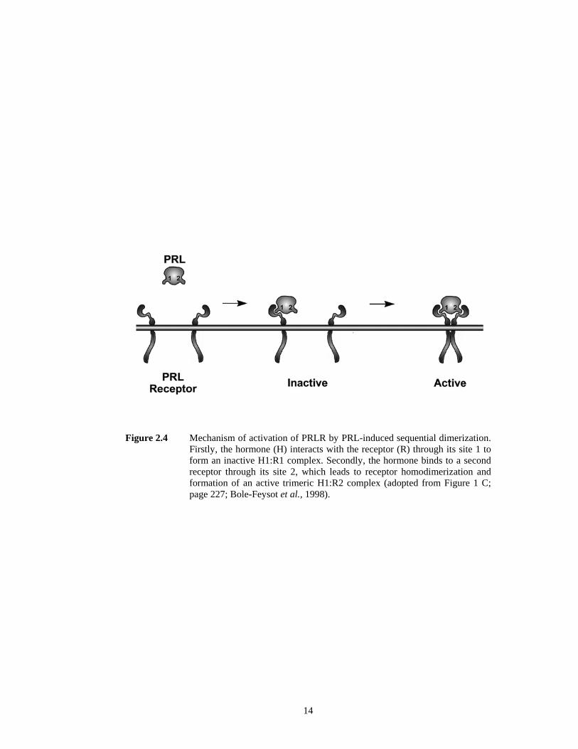
14
Figure 2.4 Mechanism of activation of PRLR by PRL-induced sequential dimerization. Firstly, the hormone (H) interacts with the receptor (R) through its site 1 to form an inactive H1:R1 complex. Secondly, the hormone binds to a second receptor through its site 2, which leads to receptor homodimerization and formation of an active trimeric H1:R2 complex (adopted from Figure 1 C; page 227; Bole-Feysot et al., 1998).

15
for further action. Secondly, the hormone binds to a second yet identical molecule of the
receptor through its binding site 2 to form an active 1:2 trimeric complex which composed of
one hormone molecule and one receptor homodimer (Goffin et al., 1998; Bole-Feysot et al.,
1998; Freeman et al., 2000; Manzon, 2002).
Dimerization of PRLR induces signal transduction pathway to initiate a cellular
response in various target cells. PRLR itself doesn’t possess any intrinsic enzymatic activity
therefore tyrosine phosphorylation and activation of the JAK kinase followed by
phosphorylation of the receptor are essential to start the signal transduction mechanisms (Rui
et al., 1992; Taga & Kishimoto, 1993; Ihle, 1994; Kishimoto et al., 1994; Ihle & Kerr, 1995;
Bole-Feysot et al., 1998). Besides JAK, another major signaling pathway involves the
phosphorylation of cytoplasmic Stat proteins, which themselves dimerize and translocate to
the nucleus and bind to specific promoter elements on PRL-responsive genes (DaSilva et al.,
1994; Bole-Feysot et al., 1998).
Engineered hormone analogues with binding site 2 blocked sterically are unable to
induce receptor homodimerization and receptors remain inactive. However, these analogues
are still able to bind to binding site 1 of the receptor and hence act as antagonists to the wild
type hormones (Goffin et al., 1998). Studies on mammalian (Gertler et al., 1996) and trout
(Sandowski et al., 2000; Le Rouzic et al., 2001) PRLRs have revealed that the 1:2 complex
is transient and rapidly dissociates to an inactive 1:1 complex. This is because an increase in
the stability of the 1:2 complex may not guarantee a greater physiological response, whereas
a rapid dissociation of the active trimeric complex back to the inactive form would give way
to further functional interactions (Gertler et al., 1996).
To support this argument, study on GH analogues showed that even if there was an
increase in the affinity to the GH receptor, the analogues failed to enhance the signal
transduction or cell proliferation (Pearce et al., 1999). Another study suggested that there
may be a “minimal time” for homodimering of the receptor in order to activate the signal
transduction pathways (Helman et al., 2001).

16
2.3 Biological effects of Prolactin (PRL)
The significance of PRL studies lies in its broad area of biological effects in
vertebrates. PRL was initially identified as a hormone from anterior pituitary which can
stimulate the development of mammary glands (Riddle et al., 1933). Continuously, various
other functions from this multifaceted hormone have been discovered and they have been
reviewed and grouped into five broad areas including reproduction, osmoregulation, growth,
integument and synergism with steroids (Nicoll & Bern, 1972; Nicoll, 1974 in Bole-Feysot
et al., 1998). The number of effects of PRL kept on increasing and was then categorized into
six categories; water and electrolyte balance, growth and development, endocrinology and
metabolism, brain and behaviour, reproduction, and lastly immunoregulation and protection
(Bole-Feysot et al., 1998).
All the actions mentioned in the cited references covered a wide range of animals
including lower species and some of them may not directly be related to fish or mammals.
This suggests that some actions of PRL may have been lost with evolution or may only be
seen in higher animals during certain stages of development (Bole-Feysot et al., 1998).
However, the present study is primarily on discus fish (Symphysodon aequifasciata)
regarding its natural parenting behaviour and mucus production as a nourishment for young,
hence other PRL effects will be described in Section 2.3.1 in general following the categories
done by Bole-Feysot et al. (1998).
2.3.1 PRL effects in general
2.3.1.1 Water and electrolyte balance
From all the studies that have been done, PRL plays a key role in regulating fluid
balance through the gill and kidney of many fish and has been referred to as a fresh water-
adapting hormone (Sandra et al., 1995, 2000; Le Rouzic et al., 2001; Prunet et al., 2000; Tse
et al., 2000; Higashimoto et al., 2001; Santos et al., 2001; Manzon, 2002; Lee et al., 2006a;
San Martín et al., 2007). Water and electrolyte balance is of particular importance for fish as
they are exposed constantly to media with varying osmotic degrees. Therefore, PRL plays

17
pivotal role in the homeostatic control of body fluid balance in freshwater and seawater fish
(Bole-Feysot et al., 1998; Freeman et al., 2000; Manzon, 2002).
Seawater fish lose quite an amount of water to the hyperosmotic environment. In
order to replenish the lost, seawater fish reabsorb the water from intestine and produce only a
minimal volume of urine (Loretz, 2001). In freshwater, fish body fluids are hypertonic to the
external environment. Freshwater fish are capable to reduce the gill permeability to retain the
ions and salts while actively secrete excessive intake of water (Nicoll, 1974 in Bole-Feysot et
al., 1998). For fish that migrate between fresh and seawater habitat, osmoregulation of water
and electrolyte balance is highly essential (Bole-Feysot et al., 1998). Besides, there are also
suggestions that PRL may protect the hatchlings of rainbow trout from failure in freshwater
adaptation (Ensor, 1978).
2.3.1.2 Growth and development
There are a great amount of studies reporting that PRL stimulates growth and
development. For some mammals and fish species, studies failed to prove that there is a
direct effect of PRL on body growth but in birds PRL-treated males showed successful
increase of body weight (Silverin, 1980). Since PRL and GH share high homology in amino
acid and structural similarity, both of these hormones are suggested to share many biological
functions in many vertebrates especially in the context of growth. In amphibians and reptiles,
PRL reverses metamorphic changes but promotes skin molting (Tata et al., 1991; Bres &
Nicoll, 1993; Bole-Feysot et al., 1998; Hasunuma et al., 2004). Antimetamorphic effect also
occurs in fish species fundamentally in Japanese flounder (De Jesus et al., 1994). Regarding
other reported studies on developmental processes, PRL also plays an important role in
maturation of the lung and surfactant production, differentiation of preadipocytes, maturation
of germ cells and hypothalamic dopamine development (Bole-Feysot et al., 1998). PRL is
also found to affect angiogenesis (Corbacho et al., 2002).

18
2.3.1.3 Endocrinology and metabolism
In monkey, studies showed that PRL has specific effect on ATPase activity in
different regions of the brain therefore modulating energy metabolism (Kumaran et al.,
1989). PRL also has proven to regulate lipid and carbohydrate metabolism in mammals
(Machida et al., 1990). In an endocrinology context, PRL was shown to have direct effect on
pancreatic function and insulin secretion (Nielsen, 1982; Sorenson et al., 1987). Studies
revealed that PRLR expression can be up- and down-regulated by GH and PRL depending
on the concentration. Due to the wide spectrum of biological effects of PRL and the
existence of multiple forms of PRLR, there is always more than one responsive mechanism
designated for this multifaceted hormone. In order to respond efficiently to numerous target
tissues, PRL is capable of acting promptly by increasing the number of specific PRLRs
themselves (Kelly et al., 1991; Matsuda & Mori, 1997; Leclerc et al., 2007).
2.3.1.4 Reproduction
PRL has long been related to luteotropic and luteolytic actions and has been labelled
as luteal hormone. In general, PRL enhances progesterone secretion by luteal cells. Results
have strongly proven that PRL is involved directly in modulating the physiological states of
oestrus, pregnancy, and lactation seeing the fact that it is an important factor in regulating the
formation and destruction of the corpus luteum (Matsuyama et al., 1990; Cecim et al., 1995,
Bole-Feysot et al., 1998; Freeman et al., 2000). In uterus, PRL increases the concentration of
progesterone receptors and hence all uterine actions associated with progesterone are
enhanced (Daniel et al., 1984; Chilton & Daniel, 1987). For male species, PRL has been
proposed to stimulate steroidogenesis and androgen production when synergized with LH
(Binart et al., 2003). PRL also increases total lipids and the conversion of spermatogenesis in
Sertoli cells (Nag et al., 1981; Guillaumot & Benahmed, 1999). However, PRL has been
reported that it causes a decrease in the gonads and the reproductive tract tissue weights in
both male and female ring doves (Buntin & Tesch, 1985).

19
2.3.1.5 Immunoregulation and protection
Injection of PRL into hypophysectomized rats causes an increase in the weight of the
spleen and thymus and promotes the thymus development (Carreño et al., 2004). In
lymphocytes, PRL is known to increase hormonal and cellular immunity, to reverse anemia,
leukopenia and thrombocytopenia that were induced by hypophysectomy. PRL is also found
to enhance antibody formation, including IgG and IgM antibodies (Bole-Feysot et al., 1998;
Freeman et al., 2000). In fish, PRL regulates melanogenesis (Sage, 1970) and maintains
specific immune functions through prevention of immunosuppression by cortisol (Yada et al.,
2004). To date, no genetic diseases associated with a mutation of the gene encoding PRL or
the PRLR have been identified in humans or animals (Kelly et al., 1991). However, PRL has
been associated with a number of different forms of cancer and tumor growth (Barker et al.,
1992; Wennbo & Törnell, 2000). PRL is also shown to increase and to affect a number of
autoimmune circumstances (Bole-Feysot et al., 1998).
2.3.2 Parental behaviour
Besides the well known effects of PRL in stimulating the production of milk proteins
and osmoregulation, there is a wealth of evidence to support the idea that PRL is involved in
parental behaviour of fish, birds, and mammals (Bole-Feysot et al., 1998; Schradin &
Anzenberger, 1999; Freeman et al., 2000). In behavioural studies, parental care is a good
example that would seem to benefit the species. It promotes the survival and well-being of
the next generation at a cost to the resources of the current generation in order to ensure the
continuity of the species (Freeman et al., 2000; Gross, 2005). This link between PRL and
parental care added another label for PRL as the “hormone of parenthood” (Schradin &
Anzenberger, 1999). Although PRL plays an important role in parental care, it is not the only
factor which induces caregiving behaviour for the young. This complex physiological
process normally involves a number of factors to ensure its proper function especially in
higher vertebrates. Hence, steroid hormones including estradiol and progesterone are
important for the proper functions of PRL (Schradin & Anzenberger, 1999).

20
2.3.2.1 Parental care in mammals
In almost all mammals, maternal care is long well known and females play vital role
in parental care including the two families of monotremes, the platypus (Ornithorhynchidae)
and the spiny anteater (Tachyglossidae) which bear egg (Bole-Feysot et al., 1998; Freeman
et al., 2000; Reynolds et al., 2002). PRL and PL are suggested to induce maternal care
because there is a significant increase in the circulating concentration of these hormones
during and after the pregnancy (Bridges et al., 1990, 1997). PRL remains the sole factor in
milk production in the mammary glands even though the stimulation and regulation of milk
production may vary among vertebrates (Bole-Feysot et al., 1998; Neville et al., 2002).
In terms of paternal care, males are suggested to contribute care to the young.
However, this is not always the case for all the mammals. Only a small portion in mammals
is socially monogamous. However, monogamy isn’t always equivalent to parental care
(Schradin & Anzenberger, 1999; Freeman et al., 2000). Fathers showed a significant
increase in plasma PRL in California mouse (Peromyscus californicus) and exhibited
caregiving to the young (Gubernick et al., 1993). Among carnivores, wolves showed
biparental care where both parents participate in caregiving by regurgitation of food, licking
and defending the pups. For higher vertebrates, not only mothers, fathers also showed
significant higher level of plasma PRL if compared with nonfathers in primates and human
(Schradin & Anzenberger, 1999). Even though there is a connection between PRL and
parental behaviour, in higher animals, the concentration of PRL may not regulate the
parental behaviour of the animal (Schradin & Anzenberger, 1999). Across the mammalian
taxa, only 9% of males help to provide care for the young including feeding, guarding,
grooming, carrying, defending, teaching and play with the young offspring (Reynolds et al.,
2002).
2.3.2.2 Parental care in birds
All birds showed biparental care behaviour, which is to say, both parents participate
in taking care of their offspring (Reynolds et al., 2002). Studies showed that avian PRL

21
involves in incubation and brooding processes, including nesting behaviour and nest
attendance (Hector & Goldsmith, 1985; March et al., 1994). Even though PRL is essential in
incubation in doves, but other factors like gonadal hormones must also be taken into
consideration (Janik & Buntin, 1985).
PRL secretion increased before hatching, especially during incubation period and it
controls the parental behaviour (Buntin et al., 1996) and elevation of plasma PRL has been
observed before incubation period in domestic chicken (Hall et al., 1986). This lends further
support regarding the involvement of PRL in pre-hatching phase. Extensive research has
been done mainly on ring dove regarding PRL and parental care. Results showed that
injection of PRL made both parents show extensive care towards the young (Schradin &
Anzenberger, 1999).
In birds, PRL induces proliferation and thickening of mucosal epithelial lining of
pigeons and doves crop sac directly and indirectly, but not in every bird (Anderson et al.,
1984). This crop sac is connected to the stomach and serves as a food storage organ. PRL
acts as a mitogen on the epithelium and sloughs it off into the crop in order to produce a
substance to nourish their young, called crop milk, similar function of mammary secretion
(Anderson et al., 1984; Horseman & Buntin, 1995; Buntin et al., 1996). Crop milk contains
clumps of epithelial cells from the crop sac mucosal lining wall and is fed to the young by
regurgitation. PRL is proven to stimulate regurgitation feeding in birds (Horseman & Buntin,
1995; Buntin et al., 1991).
Regulation of PRL release in birds is controlled by a PRL-releasing factor, PRF, of
the hypothalamus. Electrochemical (EC) stimulation of the preoptic-anterior hypothalamic
region showed an increase in the weight of the crop sac of the pigeon. And studies showed
that PRL strongly stimulates feeding activity and body weight gain in ring dove (Kanematsu,
1980). Other than stimulates crop sac proliferation, PRL synergizes with ovarian steroids to
maintain the brood patch in many bird species (Bole-Feysot et al., 1998).

22
2.3.2.3 Parental Care in fish
Various studies have shown that PRL induced parental fanning to provide a constant
supply of fresh water to the eggs in several teleosts (Slijkhuis et al., 1984; De Ruiter et al.,
1986; Páll et al., 2004). In stickleback, PRL cells of the anterior pituitary were found to be
more active during the later parental phase than during the initial sexual phase. Results
suggested that this upsurge of PRL induces fanning but reduces courtship behaviour in
stickleback (De Ruiter et al., 1986; Páll et al., 2004).
Besides, PRL induced nest building (Kindler et al., 1991) and also stimulated mucus
production on body surface as a supplementary nutrient to feed the young. PRL treatment to
discus fish enhanced the production of mucus-secreting cells, which serves as nutrient for the
fries. In many fish, mucus secretion is responsive to PRL. It is further proven that the
production of a substance for the survival of the young is not a feature restricted only to
mammals (Ogawa, 1970; Blüm & Fiedler, 1965).
PRL was also shown to calm fish and to depress feeding response in cichlid. Results
suggested that this behaviour can prevent the eggs to be eaten up by the parents. Further
studies on other hormones including progesterone and several adenophypophysial hormones
showed that PRL remained the sole factor in promoting parental behaviour of cichlids (Blüm
& Fiedler, 1965). In paradise fish (Macropodus opercularis), males exhibited better parental
behaviour than the females. However, studies showed that PRL alone could not increase
bubble nests building, ie. bubble nests were formed by mixing air bubbles with mucus during
egg laying process. Furthermore, androgen is found to increase nest building with the
presence of PRL (Machemer & Fiedler, 1965 in Bole-Feysot et al., 1998). PRL is also
involved in migration of some teleosts from seawater to fresh water (Bole-Feysot et al.,
1998).
2.4 Discus as an animal model
Discus fish (Symphysodon aequifasciata) is a well-known and popular freshwater
ornamental fish produced in several countries in South East Asia. These living jewels are

23
widely bred throughout Asian countries such as Malaysia, Singapore, Thailand and
Indonesia with several breeding techniques developed to boost the production. From just a
few original forms of wild discus, there is a variety of strains exists due to active
interbreeding among wild forms and also among new varieties (Koh et al., 1999).
Discus fish was classified in the family of Cichlidae and Symphysodon discus was
the first discus fish found and described by Johann Jacob Heckel at the Amazon River basin
in 1840 while Jacques Pellegrin described the second species, the green discus,
Symphysodon aequifasciatus in 1904. Subsequently, there were five subspecies documented
in the later years, which are the S. discus discus, S. discus willischwartzi, S. aequifasciatus
aequifasciatus, S. aequifasciatus axelrodi and S. aequifasciatus haraldi. Unfortunately, the
identification and classification of new varieties were getting tougher since there was no
genetic background recorded for the new varieties, like newborns that drifted from the
lineage of their ancestors. Nowadays, discus was classified based on the colour and the
physical characteristic, either from the wild-type or the domestic-type (Leibel, 1996).
Despite all nomenclature issues, more and more varieties of discus were produced
and progressively exported to developed countries such as Japan, United Kingdom and the
United States. Malaysia is one of the top discus fish contributors in the world with successful
discus fish breeders and active discus breeding in the country. Today, discus fish are priced
from RM30 to several thousands per fish according to the strains and sizes.
Discus fish (Symphysodon aequifasciata) belongs to Cichlidae family. Like other
cichlids, all Symphysodon species are laterally compressed and round in shape like a disc.
This is how the common name, “discus” is derived. The sides of the fish body are patterned
generally. The height and length of the grown fish are both about 20–25 cm (8–10 in). This
fish demonstrates unique parental-care behaviour towards newly hatched fries where parental
fish feed their newborns with epidermal-secreted mucus (Chong et al., 2005). There are other
members in this family that feed their newborns with secreted mucus such as tilapia. The
male parental-fish tilapia “incubates” free swim larvae in the mouth where newborns were
fed with mucus secreted from the mouth (Kishida and Specker, 2000).

24
Parental discus fish will look after the eggs until they hatch and are able to free
swim. During the eggs guarding period, the male and female parental fish will take turn to
fan the eggs (Leibel, 1996). This study demonstrated that one very important contribution
from parental-care behaviour in fish is indirect transferring of antimicrobial substances from
epidermal mucus to fertilized eggs during egg-guarding session. Parental fish will normally
move hatched embryos to other places in the aquarium which is safer and well covered in
order to minimize distractions. Parental-care behaviour will continue until the larvae free
swim and detach from the yolk sac. When larvae free swim, they will feed on the mucus
secreted on the epidermis of the parental fish. Figure 2.5 shows discus fish and their unique
parental-care behaviour.
There are more than 1300 species in the family Cichlidae and they are widespread in
Africa and Central and South America. All species provide parental care. The diversity of
forms of parental care is unusually large for a single family of animals. A review found
evidence that 73 genera provide biparental care, and 108 genera have female care (Goodwin
et al., 1998). Cichlid fish provide the best opportunity for the study of parental care in
teleosts because they show diverse forms of parental care from substrate guarding to delayed
and immediate mouthbrooding. There are also variations in which sex provides care
(biparental, female-only and male-only care) (Blumer, 1982). Moreover, extensive
ecological, behavioural and phylogenetic information is available for this family, which has
led to considerable speculation about the studies of the uniqueness of fish parental care.