clinical and epidemiological studies in … · clinical and epidemiological studies in ......
TRANSCRIPT

From Department of Medicine, Division of Hematology,
Karolinska University Hospital and Karolinska Institutet,
Stockholm, Sweden
CLINICAL AND EPIDEMIOLOGICAL STUDIES IN
MYELOPROLIFERATIVE NEOPLASMS
Malin Hultcrantz
Stockholm 2013

Printed by Reproprint AB, Stockholm 2013.
All previously published papers were reproduced with permission from the publisher.
Published by Karolinska Institutet.
© Malin Hultcrantz, 2013
ISBN 978-91-7549-139-4
Printed by
2013
Gårdsvägen 4, 169 70 Solna

ABSTRACT
Myeloproliferative neoplasms (MPN), including polycythemia vera (PV), essential thrombocythemia (ET), and
primary myelofibrosis (PMF), are clonal hematopoietic disorders characterized by excessive terminally
differentiated myeloid cells. MPNs can progress to secondary myelofibrosis or acute myeloid leukemia
(AML)/myelodysplastic syndromes (MDS). Although progress has been made in the understanding of the
pathogenesis and management of MPNs, there are still unresolved issues regarding prognosis, causes of
death, and risk factors for leukemic transformation. In patients with hematological malignancies, the risk of
suicide and suicide attempts is largely unknown.
We conducted a population-based study to establish patterns of survival in 9,384 MPN patients
identified from the Swedish Cancer Registry between 1973 and 2008. Relative survival ratios were computed
as measures of patient survival. Relative survival was significantly lower in all MPN subtypes compared to
expected survival in the general population, reflected in10-year relative survival ratios of 0.64 (95%
confidence interval (CI); 0.62-0.67) in PV, 0.68 (0.64-0.71) in ET, and 0.21 (0.18-0.25) in PMF,
respectively. Excess mortality was observed in patients of all MPN subtypes during all four calendar periods
(p <0.001). Nevertheless, survival improved significantly over time (p <0.001); however, the improvement
was less pronounced after the year 2000 and was confined to patients with PV and ET. In conclusion, our
findings underline the assertion that all MPNs should be considered serious diseases that reduce life
expectancy and highlight the need to improve treatment strategies for these patients.
Through the Swedish Cancer Registry and our national MPN cohort we identified 9,563 MPN patients
diagnosed between 1973 and 2005 and their 37,643 matched controls to assess excess mortality and causes
of death. Cumulative incidence functions, calculated using a flexible parametric model, were used to estimate
10-year probabilities of death with 95% CIs for six categories of causes of death. The 10-year probability of
dying from infections in male MPN patients aged 70-79 years at diagnosis were 4.5% (matched controls;
2.3%), from hematological malignancy 13.7% (0.2%), from cardiovascular disease 16.8% (15.0%), from
cerebrovascular disease 5.5% (5.1%), from solid tumor 9.7% (11.5%), and from other disorders 24.9%
(14.9%). The excess mortality in MPN patients declined due to a decrease in deaths from hematological
malignancies during the first calendar period (1973-1982), infections, and in younger MPN patients, from
cardiovascular disease. The overall improvement in 10-year mortality, observed in both patients and matched
controls over time, was mainly explained by declines in cardiovascular death. In conclusion, the improved
survival over time is multifactorial and can only partly be attributed to improved management of the
underlying hematological malignancy.
We conducted a nested case-control study to assess the role of MPN treatment and subsequent
AML/MDS risk. From a nationwide MPN cohort (n=11,039; diagnosed 1958-2005), we identified 162
patients (cases) with transformation (153 and nine with subsequent AML and MDS diagnosis, respectively)
and their 242 matched controls (MPN patients without AML/MDS transformation). Using logistic
regression, odds ratios (ORs) were calculated as measures of AML/MDS risk. Forty-one (25%) of the 162
MPN patients with AML/MDS transformation were never exposed to alkylating agents, radioactive
phosphorous (P32
), or hydroxyurea (HU). The ORs for cases receiving 1 to 499 g, 500 to 999 g, more than 1,000 g of HU were 1.5 (95% CI; 0.6-2.4), 1.4 (0.6-3.4), and 1.3 (0.5-3.3), respectively, for AML/MDS
development (not significant). Patients with MPNs who received P32
doses greater than 1,000 MBq and more than 1 g of alkylating agents had a 4.6-fold (2.1-9.8; p<0.002) and 3.4-fold (1.1-10.6; p<0.015) increased risk of AML/MDS, respectively. Thus, the risk of AML/MDS development after MPN diagnosis was not associated with HU treatment at any dosage. The fact that only 32% of patients with AML/MDS transformation received doses found here to be leukemogenic indicates a major role for non-treatment-
related factors.
To define incidence and risk factors for suicide and suicide attempts in patients with hematological
malignancies, we conducted a population-based study in 47,220 patients with hematological malignancies
and their 235,868 matched controls. Using Cox regression, the hazard ratios (HRs) for suicide and suicide
attempts (combined end-point) in patients with hematological malignancies was 1.9 (95% CI; 1.5-2.3)
compared to matched controls during the first three years after diagnosis. When more than three years had
elapsed, there was no excess risk of suicide/suicide attempts (HR 1.1; 0.9-1.4). Patients with multiple
myeloma carried the highest risk, HR 3.4 (2.3-5.0), and a pre-existing psychiatric disorder was strongly
associated with an increased risk of suicide and suicide attempts (HR 23.3; 16.6-32.6). Although suicides
contributed marginally to mortality in patients with hematological malignancies, awareness of risk factors for
suicide/suicide attempts can facilitate identification of high-risk patients and enable preventive interventions.

LIST OF PUBLICATIONS
This thesis is based on the following papers, which are referred to in the text by their Roman
numerals:
I. Hultcrantz M, Kristinsson SY, Andersson TM-L, Landgren O, Eloranta S,
Derolf ÅR, Dickman PW, Björkholm M. Patterns of survival among 9,384
patients with myeloproliferative neoplasms diagnosed in Sweden 1973-2008; a
population-based study. J Clin Oncol. 2012 Aug 20;30(24):2995-3001.
II. Hultcrantz M, Hinchliffe S, Kristinsson SY, Andersson TM-L, Derolf AR,
Samuelsson J, Landgren O, Dickman PW, Lambert PC, Björkholm M. Risk
and Cause of Death in 9,563 Patients Diagnosed with Myeloproliferative
Neoplasms in Sweden between 1973 and 2005. Manuscript 2013.
III. Björkholm M, Derolf ÅR, Hultcrantz M, Kristinsson SY, Ekstrand C, Goldin
LR, Andreasson B, Birgegård G, Linder O, Malm C, Markevärn B, Nilsson L,
Samuelsson J, Granath F, Landgren O. Treatment-Related Risk Factors for
Transformation to Acute Myeloid Leukemia and Myelodysplastic Syndromes
in Myeloproliferative Neoplasms. J Clin Oncol. 2011 Jun 10;29(17):2410-5.
IV. Hultcrantz M, Svensson T, Derolf ÅR, Kristinsson SY, Ekbom A, Granath F,
Björkholm M. Incidence and Risk Factors for Suicide and Attempted Suicide
Following a Diagnosis of Hematological Malignancy. Manuscript 2013.
TABLE OF CONTENTS
1 Introduction ...........................................................................................................................7
1.1 Myeloproliferative neoplasms (MPNs) ..........................................................................9
1.1.1 Polycythemia vera .................................................................................................9
1.1.2 Essential thrombocythemia .................................................................................10
1.1.3 Primary myelofibrosis .........................................................................................12
1.1.4 Myeloproliferative neoplasm unclassified..........................................................14
1.2 Molecular and cytogenetic background........................................................................14
1.3 Clinical course ...............................................................................................................15
1.3.1 Thrombosis and bleeding ....................................................................................16
1.3.2 Transformation to myelofibrosis and acute leukemia ........................................17
1.4 Treatment.......................................................................................................................17
1.4.1 Current Nordic guidelines ...................................................................................19
1.5 Quality of life and suicides in cancer patients ..............................................................19
1.5.1 Quality of life.......................................................................................................19
1.5.2 Suicide .................................................................................................................19
2 Aims.....................................................................................................................................21
3 Survival and causes of death in MPN patients (I-II) ..........................................................22
3.1 Methods, patients, and controls ....................................................................................22
3.1.1 Relative survival in MPN patients ......................................................................22
3.1.2 Causes of death in MPN patients and matched controls ....................................23
3.2 Results............................................................................................................................24
3.2.1 Relative survival in MPN patients ......................................................................24
3.2.2 Causes of death in MPN patients and matched controls ....................................26
3.3 Discussion......................................................................................................................28
4 Transformation to acute myeloid leukemia and myelodysplastic syndromes (III) ...........34
4.1 Methods, patients, and controls ....................................................................................34
4.2 Results and discussion...................................................................................................35
5 Suicide and suicide attempts in patients with hematological malignancies (IV) ..............39
5.1 Methods, patients, and controls ....................................................................................39
5.2 Results and discussion...................................................................................................40
6 Summary and conclusions...................................................................................................45
7 Acknowledgements .............................................................................................................46
8 References............................................................................................................................48

LIST OF ABBREVIATIONS
Alk Alkylating agents
AML Acute myeloid leukemia
BCR-ABL Breakpoint cluster region-Abelson fusion (gene)
BU Busulfan
CI Confidence interval
CLL Chronic lymphocytic leukemia
CML Chronic myeloid leukemia
DIPSS Dynamic international prognostic scoring system
EBMT European Group for Blood and Marrow Transplantation
EMRR Excess mortality rate ratio
ET Essential thrombocythemia
HL Hodgkin lymphoma
HR Hazard ratio
HU Hydroxyurea
ICD International Statistical Classification of Diseases
IL Interleukin
IPSET International prognostic scoring system for essential
thrombocythemia
IPSS International prognostic scoring system
JAK2 Janus Kinase 2
MBq Megabecquerel
MDS Myelodysplastic syndromes
MF Myelofibrosis
MM Multiple myeloma
MPL Gene coding for thrombopoietin receptor
MPN Myeloproliferative neoplasm
MPN-U Myeloproliferative neoplasm unclassifiable
NHL Non-Hodgkin lymphoma
OR Odds ratio
P32
Radioactive phosphorous
PET-MF Post essential thrombocythemia myelofibrosis
PMF Primary myelofibrosis
pPMF Prefibrotic primary myelofibrosis
PPV-MF Post polycythemia vera myelofibrosis
PV Polycythemia vera
PVSG Polycythemia Vera Study Group
RSR Relative survival ratio SCT
Stem cell transplantation
SIR Standardize incidence ratio
WBC White blood cell
WHO World Health Organization

7
1 INTRODUCTION
Myeloproliferative neoplasms (MPNs) are a group of clonal diseases characterized by excess
hematopoiesis affecting one, two, or three cell lineages. The MPNs consists of three major
subtypes: polycythemia vera (PV), essential thrombocythemia (ET), and primary myelofibrosis
(PMF). Although the entities PV, ET, and PMF, had been described earlier, the interrelatedness
of the MPNs was first proposed by Dr. William Dameshek in 1951 (Figure 1).1
He stated that these diagnoses were all characterized by excess bone marrow proliferation
“perhaps due to a hitherto undiscovered stimulus.” Due to the similarities he found and the
difficulties in distinguishing between PV, ET and PMF, he suggested that they should be
considered as “closely related” and called them “myeloproliferative disorders”.1,2
This was long
before the molecular background of the diseases was known.
In 2005, several research groups simultaneously discovered a mutation affecting the
pseudokinase region of the Janus Kinase 2 (JAK2).3-6
This mutation is found in all MPN
subtypes and due to the activating nature of the mutation, leads to constant stimulation of
myeloid proliferation as already suggested by Dameshek.2
Since then, a number of additional
disease-related mutations have been described.7
JAK2 mutation-status has been incorporated into
the classification systems and also, the nomenclature has been changed from myeloproliferative
disorders to myeloproliferative neoplasms to underline the neoplastic nature of these diseases.8
Figure 1. Dr. William Dameshek

8
MPNs are characterized by a relatively indolent course, which can be complicated by
thromboembolic events, progression to secondary myelofibrosis (MF), and transformation to
acute myeloid leukemia (AML) or myelodysplastic syndromes (MDS).8
Advances have been
made regarding the understanding of disease mechanisms and management of the MPNs. The
importance of phlebotomy to a hematocrit <0.45 in PV patients suggested by Thomas Pearson in
1978 and the discovery of a leukemogenic effect of several cytoreductive therapies led to
changes in the management of these diseases.9,10
There are however many unresolved clinical
issues both regarding disease pathophysiology and optimal treatment. There are still
uncertainties regarding survival among MPN patients due to the lack of large studies with long
follow-up time. In the hitherto studies, patients with PMF have consistently been reported to
have reduced life expectancy.11-15
Patients with PV have, in the majority of studies, been
observed to have moderately reduced survival.16-20
In contrast, according to most reports, but
not all, survival of patients with ET is not affected by the disease.15,17,21-25
In addition, causes of
death in MPN patients are not well described. There is a need for better prevention of disease
complications such as thromboembolism, progression to secondary MF and transformation to
AML/MDS. The potential leukemogenic effect of certain cytoreductive treatments, primarily
hydroxyurea (HU), is still a matter of debate.26
Even though great advances have been, the
molecular background of the MPNs is not completely known. How the MPN-associated genetic
mutations affect prognosis and if the recently introduced targeted treatments, such as JAK2
inhibitors, can affect disease progression remains to be elucidated.
MPNs and hematological malignancies can greatly affect the quality of life of patients. The
diseases, their complications and treatments as well as the psychological strain of a cancer
diagnosis can affect the emotional and physical well being of patients. Effects on quality of life
can range from minor complaints to fatigue, pain, severe depression with suicidal ideation and
even suicides. The incidence and risk factors for suicides in patients with hematological
malignancies are not fully known.

9
1.1 MYELOPROLIFERATIVE NEOPLASMS (MPNS)
1.1.1 Polycythemia vera
Polycythemia vera (PV) was first described in 1892 by Dr. Louis Henri Vaquez in a patient with
erythrocytosis and hepatomegaly.27
Later, Dr. William Osler described several similar patients
with a new clinical entity he called Vaquez’s disease.28
Both Vaquez and Osler recognized a
high incidence of stroke in these patients.
The incidence of PV is 1.5-2.0/100,000 persons/year.8,16,29
PV is characterized by an excess
erythropoiesis with elevated hemoglobin and hematocrit. The bone marrow is hypercellular and
dominated by erythropoiesis (Figure 2) but there is also a certain degree of panmyeloid
proliferation leading to elevated white blood cell (WBC) and platelets counts in many PV
patients.8
The JAK2 V617F mutation is found in >95% of patients with PV and an additional 3%
of PV patients are positive for mutations in exon12 of the JAK2 gene.3-6,30
The exon 12 mutation
results in a more isolated erythropoiesis without leukocytosis and thrombocytosis.31
In Sweden, PV was earlier diagnosed according to the Polycythemia Vera Study Group
(PVSG) diagnostic criteria32
and since 2001, patients are diagnosed according to the World
Health Organization (WHO) classification system (Table 1).33
The diagnostic criteria have been
fairly constant during the last decades with the addition of the JAK2 V617F mutation in the most
recent WHO-classification in 2008.8
Figure 2. Bone marrow of a patient with polycythemia vera showing dominance of erythropoiesis and
enlarged megakaryocytes.
Patients with PV have a high risk of thromboembolic events while progression to
secondary MF and transformation to AML/MDS are more rare events, see chapter 1.3. Other
signs and symptoms of PV can include headaches, dizziness, pruritus which often is aquagenic,
fatigue, splenomegaly, and microvascular disturbances such as erythromelalgia.34

10
Patients are defined as high risk if age is greater than 60 years or if there is a history of
previous thrombosis; cytoreductive treatment is recommended if any one of these risk factors are
present.18,35
A preliminary risk score in PV has been presented and a refined risk score is now
under construction.36
In addition to age >60 years and a history of thrombosis, this risk score
includes WBC counts >11 x109/L and cytogenetic abnormalities as risk factors for a worse
overall survival.36
In very early reports of untreated patients, median survival was around 18 months.10,28,37
With the introduction of modern treatments, life expectancy is now significantly longer but the
reported survival in PV varies in different studies. Patients with PV are in the majority of studies
observed to have a moderately reduced survival16,18-20,38
but survival has also been reported to be
similar to that of the general population.21
Table 1. Diagnostic criteria for PV according to the 2008 WHO classification system.
8
Major criteria
1. Hemoglobin > 185 g/L in men, >165 g/L in women, or hematocrit > 0.52 in men and > 0.48 in
women, or other evidence of increased red cell volume*
2. Presence of JAK2 V617F or other functionally similar mutation such as JAK2 exon 12 mutation
Minor criteria
1. Bone marrow biopsy showing hypercellularity for age with trilineage growth (panmyelosis) with
prominent erythroid, granulocytic, and megakaryocytic proliferation
2. Serum erythropoietin level below the reference range for normal
3. Endogenous erythroid colony formation in vitro
Diagnosis requires the presence of both major criteria and 1 minor criterion or the presence of the first
major criterion together with 2 minor criteria.
* Hemoglobin or hematocrit greater than 99th percentile of method-specific reference range for age, sex, altitude
of residence or hemoglobin greater than 170 g/L in men, 150 g/L in women if associated with a documented and
sustained increase of at least 20 g/L from an individual’s baseline value, or elevated red cell mass greater than
25% above mean normal predicted value.
1.1.2 Essential thrombocythemia
Essential thrombocythemia (ET) was the last of the classical MPN subtypes to be described, the
first report is from 1934 by Dr. Emil Epstein and Dr. Alfred Goedel.39
The incidence of ET is
similar to PV with 1.5-2.5 new cases per 100,000 inhabitants per year.15,29
ET is characterized by a normocellular bone marrow with an excess of large megakaryocytes
(Figure 3).8
A diagnosis of ET requires sustained thrombocytosis and bone marrow changes
consistent with ET, and in the absence of clonal markers, the exclusion of secondary
thrombocytosis.8
The clonal markers in clinical use in Sweden today are JAK2 V617F and MPL
mutations, positive in around 60%40,41
and 1-3%42,43
of patients, respectively. During recent years,
certain modifications of the diagnostic criteria have been made. Like PV, ET was initially
diagnosed with PVSG criteria,44
and since 2001, according to the WHO criteria (Table
2).33
In the WHO classification, presence of significant reticulin or any collagen fibrosis
excludes the diagnosis of ET.8
Other major changes are the differentiation of ET from

11
prefibrotic PMF (see chapter 1.1.3), the incorporation of JAK2 V617F mutation-status and the
lowering of the platelet threshold for ET from ≥ 600 x109/L to ≥ 450 x10
9/L.
8,45
Patients with ET are often asymptomatic, clinical signs and symptoms if present are fatigue,
headaches, and microvascular symptoms.8,46,47
There is a significant risk of arterial and venous
thrombosis but the risk of progression to secondary MF and transformation to AML/MDS is low
in patients with ET, see chapter 1.3.
ET patients with age >60 years, a history of thrombosis, or platelet counts >1,500 x109/L
have been considered as high risk patients and cytoreductive treatment is recommended if any
one of these risk factors are present.35,48
Recently, a prognostic scoring system, International
Prognostic Scoring System for ET (IPSET), was introduced which incorporates age >60 years,
history of thrombosis, and WBC count >11.0 x10⁹/L as risk factors.
40 IPSET predicts overall
survival but is not validated for predicting the risk of progression to post-ET myelofibrosis
(PET-MF) or AML since there were too few events during follow-up time.40
In addition, a
thrombosis-risk score called IPSET-thrombosis was developed including the risk factors age
>60 years, previous thrombosis, cardiovascular risk factors (hypertension, diabetes mellitus,
tobacco use), and presence of JAK2 V617F. Patients were divided into three different risk
groups low, intermediate, and high with annual thrombotic risks of 1.03%, 2.35%, and 3.56%,
respectively.47
Survival of patients with ET have in the majority of studies reported to be similar to that of
the general population.15,17,21,22,49
Most of these studies have however included a limited number
of patients and/or a limited follow-up time. However, in a study with follow-up longer than 10
years, observed survival was shorter compared to expected survival in the general population.25
Figure 3. Bone marrow of a patient with ET showing large mature megakaryocytes.
12
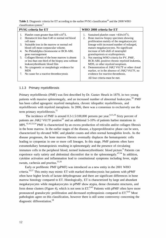
Table 2. Diagnostic criteria for ET according to the earlier PVSG classification44
and the 2008 WHO
classification system.8
PVSG criteria for ET WHO 2008 criteria for ET
1. Platelet count greater than 600 x109/L
2. Hematocrit less than 0.40 or normal red blood
cell mass
3. Stainable iron in the marrow or normal red
blood cell mean corpuscular volume.
4. No Philadelphia chromosome or BCR-ABL gene rearrangement
5. Collagen fibrosis of the bone marrow is absent
or less than one third of the biopsy area without
leukoerythroblastic blood film
6. No cytogenetic or morphologic evidence for MDS
7. No cause for a reactive thrombocytosis
1. Sustained platelet count >450x109/L
2. Bone marrow biopsy specimen showing
proliferation mainly of the megakaryocytic
lineage with increased number of enlarged,
mature megakaryocytes. No significant
increase of left-shift of neutrophil
granulopoiesis or erythropoiesis.
3. Not meeting WHO criteria for PV, PMF, BCR-ABL positive chronic myeloid leukemia,
MDS, or other myeloid neoplasm.
4. Demonstration of JAK2 V617F or other clonal
marker, or in the absence of JAK2 V617F, no
evidence for reactive thrombosis.
All four criteria must be met.
1.1.3 Primary myelofibrosis
Primary myelofibrosis (PMF) was first described by Dr. Gustav Heuck in 1879, in two young
patients with massive splenomegaly, and an increased number of abnormal leukocytes.50
PMF
has been called agnogenic myeloid metaplasia, chronic idiopathic myelofibrosis, and
myelofibrosis with myeloid metaplasia. In 2006, there was a consensus to exclusively use the
term primary myelofibrosis.51
The incidence of PMF is around 0.3-1.5/100,000 persons per year.8,15,29
Sixty percent of
patients are JAK2 V617F positive52
and an additional 5-10% of patients harbor mutations in
MPL.42,43,53,54
PMF is characterized by an excess production of reticulin and/or collagen fibrosis
in the bone marrow. In the earlier stages of the disease, a hyperproliferative phase can be seen,
characterized by elevated WBC and platelet counts and often normal hemoglobin levels. As the
disease progresses, the bone marrow fibrosis eventually displaces the hematopoietic cells
leading to cytopenias in one or more cell lineages. In this stage, PMF patients often have
extramedullary hematopoiesis resulting in splenomegaly and the presence of circulating
immature cells in the peripheral blood, termed leukeoerythroblastic blood picture.8
Patients can
experience early satiety and abdominal discomfort due to the splenomegaly.47,54
In addition,
cytokine activation and inflammation lead to constitutional symptoms including fever, night
sweats, cachexia and pruritus.55,56
Early or prefibrotic PMF (pPMF) was introduced as a new entity in the 2001 WHO
criteria.33,57
This entity may mimic ET with marked thrombocytosis but patients with pPMF
often have higher levels of lactate dehydrogenase and there are significant differences in bone
marrow histology compared to ET. Histologically, ET is characterized by large and abundant
megakaryocytes while megakaryocytes in pPMF show atypia, dense chromatin structures, and
form dense clusters (Figure 4), which is not seen in ET.8,45
Patients with pPMF often have more
pronounced granulocytic proliferation and decreased erythropoiesis compared to ET.8,45
Most
pathologists agree on this classification, however there is still some controversy concerning the
diagnostic differentiation.58

13
After the introduction of pPMF, a number of patients previously diagnosed as ET, for
example with thrombocytosis and a limited degree of bone marrow fibrosis, are now reclassified
as having pPMF.41,59,60
Patients with pPMF have a higher risk of progression to overt MF,
transformation to AML, and a worse overall survival compared to patients with ET.41
Patients
with pPMF also have a higher risk of major bleedings but the risk of thrombotic events appears
similar in the two entities.41,61
Three different risk scores for PMF patients have been developed during recent years; the
International Prognostic Scoring System (IPSS), the dynamic IPSS (DIPSS) and DIPSS-
plus.11,23,62
The IPSS assesses the prognosis at the time of PMF diagnosis while DIPSS and
DIPSS-plus can be used anytime during follow-up. Based on the number of clinical risk factors,
patients are divided into four risk groups; low, intermediate-1 intermediate-2, and high risk. The
IPSS and DIPSS incorporate five risk factors; age ≥60 years, haemoglobin <100 g/L,
constitutional symptoms, WBC count ≥25 x109/L, and ≥1% circulating blasts. In the DIPS-plus,
three additional risk factors have been included; cytogenetic abnormalities (+8, -7/7q-, I(17q),
inv(2), -5/5q-, 12p-, or 11q23 rearrangements), platelet count <100x109/L, and red blood cell
transfusion dependency.62
In addition to these risk scores, elevated levels of cytokines (interleukin
(IL)-8, IL-2R, IL-12, and IL-15) and free light chains have also been correlated with shortened
survival.56,63
PMF is the subtype associated with the worst survival, with overall survival reported to be
approximately five years,11-15
ranging from 27 to 135 months depending on IPSS score.11
Table 3. Diagnostic criteria for PMF according to the WHO 2008 criteria.
8
Major criteria
1. Presence of megakaryocyte proliferation and atypia*, usually accompanied by either reticulin
and/or collagen fibrosis, or, in the absence of significant reticulin fibrosis, the megakaryocyte
changes must be accompanied by an increased bone marrow cellularity characterized by
granulocytic proliferation and often decreased erythropoiesis (i.e., prefibrotic cellular-phase
disease)
2. Not meeting WHO criteria for PV, chronic myeloid leukemia, MDS, or other myeloid
neoplasm
3. Demonstration of JAK2 V617F or other clonal marker (e.g., MPL W515L/K), or in the absence
of a clonal marker, no evidence of bone marrow fibrosis due to underlying inflammatory or
other neoplastic diseases
Minor criteria
1. Leukoerythroblastosis
2. Increase in serum lactate dehydrogenase level
3. Anemia
4. Palpable splenomegaly
Diagnosis requires meeting all 3 major criteria and 2 minor criteria.
*Small to large megakaryocytes with an aberrant nuclear/cytoplasmic ratio and hyperchromatic, bulbous, or
irregularly folded nuclei and dense clustering.

14
Figure 4. Bone marrow of a patients with primary myelofibrosis showing large atypical megakaryocytes in
dense clusters.
1.1.4 Myeloproliferative neoplasm unclassified
Myeloproliferative neoplasm unclassified (MPN-U) was introduced in the Swedish Cancer
Register in 1993. This is not a strict entity but a group of patients with unclassified MPN who do
not fulfill the criteria for PV, ET, or PMF. These might be patients early in the disease course
but most often represent patients in later disease stages with fibrosis where the initial diagnosis
cannot be determined.8
1.2 MOLECULAR AND CYTOGENETIC BACKGROUND
The spectrum of genetic abnormalities found in the MPNs has increased over the last eight
years. As mentioned, in 2005 an activating mutation in the JAK2 gene was discovered by four
different research groups simultaneously.3-6
The JAK2 V617F is a point mutation in the JAK2
gene on chromosome 9 where phenylalanine is substituted for valine in the 617 position. The
mutation affects the pseudokinase region of the JAK2 which is situated in the intracellular part
of the erythropoietin receptor and leads to constant stimulation of hematopoiesis.3,4
More than
95% of PV patients are JAK2 V617F positive and around 50-60% of ET and PMF patients
harbor this mutation.8,40
In addition, mutations in the exon 12 region of the JAK2 gene have been
described; these are found in approximately 3% of PV patients and are not seen together with the
JAK2 V617F mutation.30
The allele burden of JAK2 V617F is highest in patients with PV and
lower in ET and PMF patients.64
Overall, the JAK2 V617F allele burden has not been correlated
to patient survival except in PMF where JAK2 V617F positivity with a low allele burden has
been associated with a reduced survival.64,65
A number of additional mutations have been described in MPN patients since the original
discovery of the JAK2 V617F mutation (Table 4). Many of these mutations affect the JAK-
STAT signaling pathway, some direct and some through epigenetic mechanisms.66,67
The

15
presence of some of these mutations has been associated with a worse prognosis, for example
ASXL1 and EZH2, but for the majority of mutations, the prognostic relevance is not known.67,68
Although JAK2 V617F is the most MPN-specific of these mutations, it can be found in a
number of other myeloid malignancies i.e. AML, MDS, MDS/MPN unclassifiable and
refractory anemia with ring sideroblasts and thrombocytosis.67,69
Low levels of JAK2 V617F has
also been observed in healthy blood donors.70
The genetic background of MPNs is complex and the disease is suggested to be comprised
of different competing clones, often with genetic differences.71
For example, in JAK2 V617F
positive MPN patients that transform to AML, the leukemic clone can be JAK2 V617F
negative.72
Table 4. Genes involved in MPN pathogenesis.67,73
JAK2 NRAS RUNX1 MPL KRAS TP53 LNK PRC2 IDH1/2 CBL ASXL1 SF3B1 SOCS1-3 EZH2 SRSF2 TET2 DNMT3A JARID2
The majority of MPN patients do not have cytogenetic abnormalities and cytogenetic
assessment is not part of routine work-up. Cytogenetic abnormalities are found in 33%, 11%,
and 7% of patients with PMF, PV and ET, respectively.74-76
The most common abnormalities are
del (20q), del (13q), +8, +9, and abnormalities in chromosomes 1, 5, and 7. Many of these are
seen in other hematological malignancies, most frequently in MDS, but abnormalities +9 and
13q are relatively specific to MPNs.77
Prognostic significance has been described for cytogenetic
abnormalities in PMF but not in PV or ET.62,74,76
As mentioned previously, in PV, a new
prognostic system is under construction where cytogenetic abnormalities are incorporated as one
of several prognostic factors.36
In addition to the above mentioned genetic changes, familial aggregation of MPNs has been
described with a 5-7 fold increased risk in first-degree relatives.78
In this study by Landgren et
al, no anticipation was seen; however Rumi et al at observed a lower median age in second-
generation compared to first-generation MPN patients in their family studies.79,80
The germline
mutations are currently unknown but a predisposing haplotype (46/1) in the JAK2 locus has been
described.81,82
1.3 CLINICAL COURSE
MPNs are associated with increased risks of thrombosis and bleeding. Disease progression can
occur; ET can phenotypically develop into PV49,83
and both PV and ET can progress to
secondary MF. PV, ET, and PMF can at any time transform to AML/MDS (Figure 5).

16
Figure 5. Disease course and complications associated with MPNs
1.3.1 Thrombosis and bleeding
Patients with MPNs have a high risk of both arterial and venous thromboembolic complications.
In PV, 11-39% of patients present with a major thrombosis and around 20% develop
thrombosis during follow-up.18,19,84,85
A high hematocrit is associated with an elevated risk of
major thrombosis and cardiovascular death, this finding was recently confirmed in a large
randomized controlled trial.9,86,87
In patients with ET, the frequency of thromboembolic events in different studies ranges from
10% to 30% at diagnosis and between 8% and 31% during follow-up.25,83,88,89
The risk of
arterial thrombosis is higher than the risk of venous thrombosis but patients of any subtype can
develop large liver and splanchnic vein thrombosis.85
In addition to age >60 years and history
of thrombosis, the presence of cardiovascular risk factors, a high WBC count, and JAK2
V617F positivity in ET have been observed as risk factors for thrombosis.40,88,90
Additional
underlying mechanisms and risk factors that have been suggested are activation of neutrophil
granulocytes and platelets, tissue-factor bearing microparticles, inflammation, endothelial
activation, and gender differences where young women have a higher risk of abdominal venous
thrombosis.91
No study has hitherto confirmed a correlation between high platelet counts and an
elevated risk of thrombosis.10,92
In both pPMF and overt PMF, the risk of thrombosis is similar to the risk observed in ET.
The annual risk of thrombosis was approximately 2% in a large study of PMF patients.41,52
JAK2
V617F positivity has also been associated with an elevated risk of thrombosis in PMF.52
The risk of major bleeding is elevated in MPN patients although bleeding complications are
not as common as thromboembolic complications.61
The cumulative incidence of bleeding in PV
and ET is around 5-6% and in pPMF, the cumulative incidence was 12% in a recent study.61,84
Risk factors for bleeding are history of previous bleeding event, thrombocytopenia, and the use
of aspirin, and vitamin K antagonists.61,93
A higher risk of bleeding has also been noted in
patients treated with a combination of aspirin and anagrelide.94
Platelet counts >1,500 x109
/L is
associated with a higher risk of bleeding, possibly due to acquired von Willebrand disease.95,96

17
In addition, patients with splenic vein thrombosis and/or splenomegaly are at risk of upper
gastrointestinal bleeding from gastric varices.91
1.3.2 Transformation to myelofibrosis and acute leukemia
In PV, the 15-year risk of progression to the so called spent phase or post-PV myelofibrosis
(PPV-MF) is 6-34%.38,97,98
In ET, the risk of progression to secondary MF is lower compared to
PV, the 15-year risk is around 4-9%.38,41
MPN patients who transform to AML/MDS have a dismal prognosis.99-102
In PV, the 10-
year risk of transformation to AML or MDS is 5-10% but the reported frequency ranges from
2% to 20%.20,103-106
The risk of transformation to AML/MDS increases with time after
diagnosis.103
The risk of leukemic transformation in ET is reported to be between 0.65% and
3.3% which is lower than in the other subtypes.40,49,107
PMF carries the highest risk of
transformation, around 20%11,54,101
Older age and elevated leukocyte counts have been proposed as risk factors for leukemic
transformation.49,103,108
In PMF, the DIPSS has apart from predicting survival also been shown
to predict transformation to AML.109
In addition, several mutations have been associated with
leukemic transformation, namely IDH1/2, IKZF1, TP53, NF1, RUNX1, NRAS, SRSF2, and
DNMT3A.7,110-113
JAK2 V617F positivity in MPN patients has not been correlated to an
increased risk of transformation to AML. As mentioned, patients with JAK2 V617F positive
disease at MPN diagnosis can transform to JAK2 V617F negative AML.72
AML secondary to
MPN is often characterized by a more complex karyotype compared to de novo AML indicative
of a worse prognosis.112
Treatment with alkylating agents and radioactive phosphorous (P32
) have been associated
with a higher risk of transformation.22,89,106,114
The potential leukemogenic effect of HU is still a
matter of debate.26
There are no conclusive randomized studies on treatment and AML
transformation in patients with MPNs due the late-appearing and rare events in a long-term
disease course, and reluctance to randomly assign patients to receive potentially leukemogenic
therapies.
1.4 TREATMENT
Patients with MPNs have been treated with different cytoreductive treatments and phlebotomy.
The goals of treatment have been to prevent disease complications, give symptom relief, and
palliation. There is currently no treatment that can prevent progression to myelofibrosis or
transformation to AML/MDS and only recently, studies have shown that specific treatments can
prolong survival in PMF patients.115
Apart from allogeneic stem cell transplantation there is no
known cure for MPN.
Phlebotomy for PV has been used since the disease was first described.28,116
In the 1970s the
importance of phlebotomy was emphasized by Pearson et al and the benefit of phlebotomy to a
hematocrit level <0.45 was recently stressed in a pivotal trial.9,87
Treatment of PV and the other MPNs have included skeletal radiation therapy (in 1917),
acetylphenylhydrazine (1918), potassium arsenite (1933), lead acetate (1942), nitrogen
mustard (1950), triethylene melamine (1952), pyrimethamine (1954), 6-mercaptopurine
(1962), uracil mustard (1964), chlorambucil (1965) and dapsone (1966). P32
was introduced in
1940, busulfan 1958, pipobroman 1962, hydroxyurea and melphalan in the 1970s and

18
interferon-α in the 1980s.116,117
The PVSG was founded in 1967 and conducted trials for optimization of treatment in PV
until 1997. One of their most important trials was PV01 in which a higher risk of transformation
to AML was observed in patients treated with chlorambucil and P32
compared to patients treated
with phlebotomy alone.106
Several studies have since then confirmed a leukemogenic effect and
an elevated risk of secondary cancer associated with alkylating agents.20,114
Alkylating agents,
primarily busulfan (BU), were previously one of the first line treatments in MPNs. However,
their use has decreased after the leukemogenic effects were recognized.10
HU is a non-alkylating agent which inhibits the enzyme ribonucleotide diphosphate
reductase thereby inhibiting DNA-synthesis and cell growth.118
The PVSG recommended HU as
first line therapy in PV since their prospective trials did not support a leukemogenic effect of
HU.10
Some investigators have reported a pronounced leukemogenic effect while others have
not.119-121
In the large European Collaboration on Low-dose Aspirin trial, 518 patients with PV were
randomized to low-dose aspirin or placebo. In the aspirin arm, there was a reduced risk of the
combined end point of nonfatal myocardial infarction, nonfatal stroke, pulmonary embolism,
major venous thrombosis, or death from cardiovascular causes (relative risk 0.40, p=0.03).
Since then, low-dose aspirin is standard treatment in PV and also many patients with ET and
PMF.122,123
In ET, Cortelazzo et al showed that patients treated with HU had a lower risk of thrombosis
compared to patients on placebo.124
In the British PT1-trial, HU was associated with a lower risk
of arterial thrombosis and progression to MF but a higher risk of venous thrombosis compared to
anagrelide in patients with high risk ET.94
Recently, the Anahydret study was presented where
HU was again compared to anagrelide but this time in high risk ET diagnosed according to the
WHO criteria. This study showed no difference in the risk of arterial or venous thrombosis or
bleeding.125
In phase II studies, treatment with interferon-α has been shown to induce molecular
remissions in PV and reduce bone marrow fibrosis in early stages of PMF.126,127
A lower than
expected rate of thrombosis has been observed during interferon-α treatment and interferon-α is
not considered to be leukemogenic.126
However, interferon-α is associated with considerable
side effects often leading to treatment discontinuation.126
The first two randomized controlled trials (Comfort I and II) on the effect of the JAK2
inhibitor ruxolitinib vs placebo and vs best available therapy in intermediate-2 and high risk
PMF were recently published.115,128
There was a decrease in spleen size and symptom burden in
the experimental arm of both studies and in Comfort I, a survival benefit was observed in the
ruxolitinib arm compared to patients on placebo.115
Therapies that are currently being tested primarily in PMF are additional JAK2 inhibitors,
immunomodulators, histone deacetylase inhibitors, mTOR inhibitors interfering with epigenetic
mechanisms and signaling pathways.20
Allogeneic stem cell transplantation (SCT), introduced during the 1970s and 80s, is the only
known cure for MPN but is associated with a substantial transplantation-related
mortality.129,130
Apart from the studies mentioned above, there has been few randomized controlled trials in
MPNs which explains why treatment recommendations are to a large extent based on expert
opinion. In 2011, a consensus committee consisting of European and American MPN experts
19
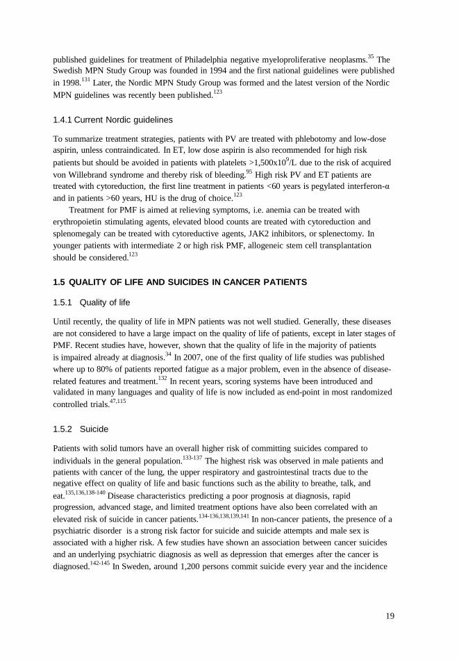
published guidelines for treatment of Philadelphia negative myeloproliferative neoplasms.35
The
Swedish MPN Study Group was founded in 1994 and the first national guidelines were published
in 1998.131
Later, the Nordic MPN Study Group was formed and the latest version of the Nordic
MPN guidelines was recently been published.123
1.4.1 Current Nordic guidelines
To summarize treatment strategies, patients with PV are treated with phlebotomy and low-dose
aspirin, unless contraindicated. In ET, low dose aspirin is also recommended for high risk
patients but should be avoided in patients with platelets >1,500x109/L due to the risk of acquired
von Willebrand syndrome and thereby risk of bleeding.95
High risk PV and ET patients are
treated with cytoreduction, the first line treatment in patients <60 years is pegylated interferon-α
and in patients >60 years, HU is the drug of choice.123
Treatment for PMF is aimed at relieving symptoms, i.e. anemia can be treated with
erythropoietin stimulating agents, elevated blood counts are treated with cytoreduction and
splenomegaly can be treated with cytoreductive agents, JAK2 inhibitors, or splenectomy. In
younger patients with intermediate 2 or high risk PMF, allogeneic stem cell transplantation
should be considered.123
1.5 QUALITY OF LIFE AND SUICIDES IN CANCER PATIENTS
1.5.1 Quality of life
Until recently, the quality of life in MPN patients was not well studied. Generally, these diseases
are not considered to have a large impact on the quality of life of patients, except in later stages of
PMF. Recent studies have, however, shown that the quality of life in the majority of patients
is impaired already at diagnosis.34
In 2007, one of the first quality of life studies was published
where up to 80% of patients reported fatigue as a major problem, even in the absence of disease-
related features and treatment.132
In recent years, scoring systems have been introduced and
validated in many languages and quality of life is now included as end-point in most randomized
controlled trials.47,115
1.5.2 Suicide
Patients with solid tumors have an overall higher risk of committing suicides compared to
individuals in the general population.133-137
The highest risk was observed in male patients and
patients with cancer of the lung, the upper respiratory and gastrointestinal tracts due to the
negative effect on quality of life and basic functions such as the ability to breathe, talk, and
eat.135,136,138-140
Disease characteristics predicting a poor prognosis at diagnosis, rapid
progression, advanced stage, and limited treatment options have also been correlated with an
elevated risk of suicide in cancer patients.134-136,138,139,141
In non-cancer patients, the presence of a
psychiatric disorder is a strong risk factor for suicide and suicide attempts and male sex is
associated with a higher risk. A few studies have shown an association between cancer suicides
and an underlying psychiatric diagnosis as well as depression that emerges after the cancer is
diagnosed.142-145
In Sweden, around 1,200 persons commit suicide every year and the incidence

20
has decreased steadily since the 1970s.146
In studies on time trends in cancer suicides declines
over time have been also observed.135,138,139,147
Suicide and suicide attempts have not been studied in detail in patients with hematological
malignancies. In some studies, hematological malignancies have been included in subgroup
analyses with diverging results.135,136,138,141
In Misono’s and Hem’s studies patients with any
hematological malignancy had a higher suicide risk while no risk increase was seen in a study
from California.136,148
Björkenstam et al showed a higher risk of suicides in patients with AML
while in patients with PV and MF, the risk of suicide was lower than that of the general
population.135
Males with multiple myeloma (MM) had an increased risk while males with
leukemia had risks lower than average in a study from the American Surveillance,
Epidemiology, and End Results data base.141

21
2 AIMS
The overall aim of these studies is to improve the management of patients with MPNs by
increasing our understanding of these disorders and more specifically to:
Define patterns of survival and causes of death in MPN patients and compare the findings to
those of the general population.
Elucidate the potential leukemogenic effect of different cytoreductive treatments with a special
focus on hydroxyurea.
Assess incidence and risk factors for suicide and suicide attempts in patients with hematological
malignancies including MPNs.

22
3 SURVIVAL AND CAUSES OF DEATH IN MPN PATIENTS (I-II)
3.1 METHODS, PATIENTS, AND CONTROLS
3.1.1 Relative survival in MPN patients
Information regarding patients diagnosed with a malignant disease in Sweden is by law reported
to the population-based nationwide Swedish Cancer Register which was established in
1958.149,150
It is mandatory for clinicians to prospectively report all incident cancers to the
register. From 1984, a double reporting system was introduced for MPNs (for both clinicians
and pathologists/cytologists) increasing the registries coverage. All dates and causes of death are
reported to the Cause of Death Register.151
The cross-linkage of registries is facilitated by the
unique national registration number which is given to all Swedish residents.152
All patients diagnosed with an MPN reported to the Swedish Cancer Register January 1st
1973 to December 31st
2008 were included. By cross-linkage to the Cause of Death Register,
information on date of deaths was obtained. Patients were followed until death, emigration or
end of follow-up (December 31st
2009), whichever occurred first.
Information on the number of allogeneic stem cell transplantations was obtained from the
European Group for Blood and Marrow Transplantation (EBMT) Registry which was founded
in 1974.
Relative survival ratios (RSRs) were computed as measures of patient survival.153,154
Relative survival is defined as the observed survival in the patient group (where all deaths are
considered events) divided by the expected survival of a comparable group from the general
population, which is assumed to be free from the cancer under study. RSR provides a measure of
total excess mortality associated with a diagnosis of MPN irrespective of whether the excess
mortality was directly or indirectly associated with the MPN. Expected survival was estimated
using the Ederer II155
method from the Swedish population life tables stratified by age, sex, and
calendar period.
As described in paper I, the 1-, 5-, 10-, 15- and 20-year RSRs with 95% confidence
intervals (CIs) were calculated for MPN patients during four calendar periods: 1973–1982, 1983-
1992, 1993–2000, and 2001-2008. In the most recent calendar period, 1-, 5- and (due to the
limited follow-up time) 8-year RSRs were calculated. Relative survival ratios were calculated
separately in patients with different MPN subtypes: PV, ET, PMF, and MPN-U and in addition,
RSRs were analyzed in patients diagnosed before versus after 1993 which was when the
category MPN-U was introduced. Separate RSRs were calculated for patients in five age
categories, <50, 50-59, 60-69, 70-79, and ≥80 years, and separately for men and women.
Excess mortality rate ratios (EMRRs) were calculated using Poisson regression in order to
estimate the effects of the factors described above while controlling for potential confounding
factors.154
All EMRRs were adjusted for age, sex and calendar period of diagnosis.

23
3.1.2 Causes of death in MPN patients and matched controls
We identified all patients diagnosed with an MPN reported to the Swedish Cancer Register from
1973 to 2005. In addition, we retrieved information on MPN patients through a national MPN
network (the Swedish Myeloproliferative Neoplasm Study Group) comprising all
hematology/oncology centers in Sweden, to include additional MPN patients.156
For each MPN patient, four controls matched by sex, year of birth, and county of residence
were selected randomly from the Swedish Register of Total Population. All controls had to be
alive and free of hematological malignancy at the time of MPN diagnosis for the corresponding
patients.
By linking the national registration number to the Causes of Death Register, data on cause
and date of death was collected from January 1st
1973 to December 31st
2007 (end of follow-up).
Cause of death was classified into six different categories: infection, solid tumor, hematological
malignancy, cardiovascular disease, cerebrovascular disease and other disorders. The category
hematological malignancy included patients that transformed to AML/MDS and non-
transformed MPN patients where no underlying cause of death other than the MPN was
specified. The category cardiovascular disease included deaths due to arterial thromboembolism
whereas deaths from venous thromboembolism, congestive heart disease, and cardiac
arrhythmias were included in other disorders.
A flexible parametric survival model was used to estimate the cause-specific mortality rates
for the six different categories of causes of death in MPN patients compared to matched controls
during the first 10 years after diagnosis.157,158
Results are presented as hazard ratios (HRs) with
95% confidence intervals (CIs). The main analysis considered all subtypes combined, and
results are presented for all MPN subtypes together, if not specified otherwise. Separate analyses
were performed for men and women and for five age groups, 18-49, 50-59, 60-69, 70-79 and
≥80 years. Calendar period of diagnosis was categorized into four groups, 1973-1982, 1983-
1992, 1993-2000, and 2001-2005. Likelihood ratio tests were used for model selection. The final
model included the variables patient status (MPN patient or matched control), sex, age group,
and period of diagnosis as well as an interaction term between patient status and age group. No
time-dependent effects were found to be significant so the proportional hazards assumption was
assumed to be reasonable for all six causes of death.
The different causes of death were treated as competing events and the probability of death
from each of the six causes was estimated as a function of time. This is known as the cumulative
incidence function and can be obtained through transformation of the cause-specific mortality
rates obtained from the flexible parametric model.158
An additional analysis was carried out to evaluate the probabilities of death for the subtypes
PV, ET, and PMF. As MPN-U was not introduced until 1993 and therefore does not cover all
four periods, it was not considered in the subtype analysis.

24
Rela
tive s
urv
iva
l ra
tio
3.2 RESULTS
3.2.1 Relative survival in MPN patients
A total of 9,384 patients with MPN were identified (PV n=4,389, ET n=2,559, PMF n=1,048
and MPN-U n=1,388). Patient survival was considerably lower in all subtypes of MPN
compared to expected survival in the general population. The RSRs for patients diagnosed
between 1993 and 2008 are shown in Figure 6. Compared to PV, patients with PMF and MPN-
U had higher overall excess mortality, EMRR in PMF was 4.38 (95% CI 3.90-4.91) and in
MPN-U 4.57 (3.87-5.41). Patients diagnosed with ET before 1993 had an inferior survival
compared to patients with PV. However, after 1993, the relationship was the opposite with 10-
year RSRs of 0.72 (0.67- 0.76) and 0.83 (0.79-0.88) for PV and ET, respectively. All MPN
subtypes were associated with a significantly increased excess mortality during all calendar
periods. During the most recent calendar period (2001-2008), the 8-year RSRs were 0.84 (0.77-
0.90), 0.91 (0.84-0.97), 0.48 (0.39-0.57), and 0.54 (0.47-0.60) in patients with PV, ET, PMF,
and MPN-U, respectively.
1.00
Survival by subtype, 1993-2008
0.80
0.60
0.40
0.20
0.00
PV ET
PMF MPN-U
0 1 2 3 4 5 6 7 8 9 10 11 12 13 14 15
Years since diagnosis
1-year RSR 5-year RSR 10-year RSR 15-year RSR
PV
ET
PMF
MPN-U
0.99 (0.98-
1.00)
1.00 (0.99-
1.01)
0.81 (0.76-
0.84)
0.87
(0.85-0.89)
0.91 (0.88-
0.94)
0.95 (0.92-
0.97)
0.42 (0.37-
0.48)
0.63
(0.59-0.66)
0.72 (0.67-
0.76)
0.83 (0.79-
0.88)
0.19 (0.12-
0.27)
0.49
(0.44-0.53)
0.56 (0.49-
0.63)
0.66 (0.59-
0.74)
0.13 (0.06-
0.23)
0.39
(0.32-0.47)
Figure 6. Cumulative relative survival among MPN patients diagnosed in Sweden between 1993 and 2008
stratified by subtype. 95% confidence intervals are shown within parenthesis.
25
Rela
tive s
urv
iva
l ra
tio
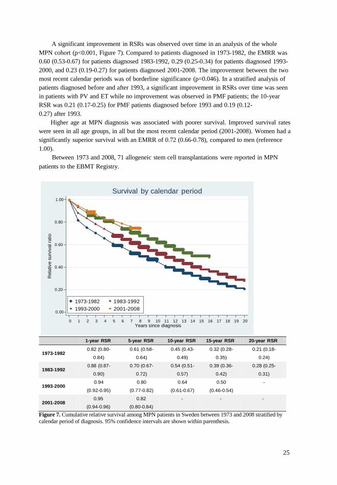
A significant improvement in RSRs was observed over time in an analysis of the whole
MPN cohort (p<0.001, Figure 7). Compared to patients diagnosed in 1973-1982, the EMRR was
0.60 (0.53-0.67) for patients diagnosed 1983-1992, 0.29 (0.25-0.34) for patients diagnosed 1993-
2000, and 0.23 (0.19-0.27) for patients diagnosed 2001-2008. The improvement between the two
most recent calendar periods was of borderline significance (p=0.046). In a stratified analysis of
patients diagnosed before and after 1993, a significant improvement in RSRs over time was seen
in patients with PV and ET while no improvement was observed in PMF patients; the 10-year
RSR was 0.21 (0.17-0.25) for PMF patients diagnosed before 1993 and 0.19 (0.12-
0.27) after 1993.
Higher age at MPN diagnosis was associated with poorer survival. Improved survival rates
were seen in all age groups, in all but the most recent calendar period (2001-2008). Women had a
significantly superior survival with an EMRR of 0.72 (0.66-0.78), compared to men (reference
1.00).
Between 1973 and 2008, 71 allogeneic stem cell transplantations were reported in MPN
patients to the EBMT Registry.
1.00
Survival by calendar period
0.80
0.60
0.40
0.20
0.00
1973-1982 1983-1992
1993-2000 2001-2008
0 1 2 3 4 5 6 7 8 9 10 11 12 13 14 15 16 17 18 19 20
Years since diagnosis
1-year RSR 5-year RSR 10-year RSR 15-year RSR 20-year RSR
1973-1982
1983-1992
1993-2000
0.82 (0.80-
0.84)
0.88 (0.87-
0.90)
0.94
(0.92-0.95)
0.61 (0.58-
0.64)
0.70 (0.67-
0.72)
0.80
(0.77-0.82)
0.45 (0.43-
0.49)
0.54 (0.51-
0.57)
0.64
(0.61-0.67)
0.32 (0.28-
0.35)
0.39 (0.36-
0.42)
0.50
(0.46-0.54)
0.21 (0.18-
0.24)
0.28 (0.25-
0.31)
-
2001-2008
0.95
(0.94-0.96)
0.82
(0.80-0.84)
- - -
Figure 7. Cumulative relative survival among MPN patients in Sweden between 1973 and 2008 stratified by
calendar period of diagnosis. 95% confidence intervals are shown within parenthesis.

26
3.2.2 Causes of death in MPN patients and matched controls
A total of 9,563 patients and 37,643 matched controls were included in the study. Mortality was
higher in MPN patients than in matched controls in all age groups, during all calendar periods,
and for all causes of death. In male MPN patients aged 70-79 at diagnosis, the HR of dying from
cardiovascular disease was 1.5 (95% CI 1.4-1.7), from cerebrovascular disease HR 1.5 (1.3-1.8),
from solid tumor HR 1.2 (0.99-1.3), and from other disorders HR 2.3 (2.1-2.6; Table 5). The risk
for MPN patients of dying from infection (HR 2.7; 2.4-3.1) and from hematological malignancy
(HR 92.8; 70.0-123.1) are shown for all ages combined due to low number of controls dying from
these causes.
Table 5. Hazard ratios and 95% confidence intervals (within parenthesis) of cause-specific deaths for MPN
patients compared to controls.
Patient age at diagnosis
(years)
18-49 50-59 60-69 70-79 ≥80
Infection 2.7
(2.4-3.1)
Solid tumor 2.5
(1.3-4.7)
1.3
(0.9-1.9)
1.2
(0.9-1.4)
1.2
(0.99-1.3)
1.0
(0.8-1.2)
Hematological malignancy 92.8
(70.0-123.1)
Cardiovascular disease 8.9
(4.0-19.8)
Cerebrovascular disease 8.8
(0.9-97.3)
Other disorders 5.2
(3.1-8.7)
2.2
(1.6-3.1)
4.7
(2.6-8.5)
4.3
(3.2-5.7)
1.8
(1.5-2.2)
2.8
(2.1-3.7)
3.7
(3.2-4.4)
1.5
(1.4-1.7)
1.5
(1.3-1.8)
2.3
(2.1-2.6)
1.6
(1.4-1.8)
1.4
(1.2-1.7)
1.8
(1.6-2.0)
In the cumulative incidence function analysis (competing risk model), male MPN patients
diagnosed between the ages 70 and 79 years during the calendar period 1993-2000 and their
matched controls were used as an example. These patients had an overall 10-year probability of
death of 75.0% compared to 49.0% in matched controls (Figure 8). The excess mortality in
MPN patients was mainly explained by death from infection, (MPN patients 4.5% vs. matched
controls 2.3%), hematological malignancy (13.7% vs. 0.2%), and other disorders (24.9% vs.
14.9%), all differences being statistically significant. There were no significant differences
regarding deaths due to cardiovascular disease (16.8% vs. 15.0%), cerebrovascular disease (5.5%
vs. 5.1%), or solid tumor (9.7% vs. 11.5%) between this group of male MPN patients and their
matched controls. In female patients (same age group and calendar period), the distribution of
causes of death was similar but the overall probability of death was lower; 61.0% for MPN
patients and 36.3% for matched controls 10 years after diagnosis (Figure 9). Figures 8 and 9
show 10-year probabilities with 95% confidence intervals for the six causes of death in males and
females, respectively. In the younger age groups, MPN patients had a higher probability of dying
from cardiovascular and cerebrovascular disease compared to matched controls (Figure
11).
The most common causes of death within the category other disorders were congestive
heart failure, pulmonary diseases, arrhythmias, and dementia. Venous thrombosis was a less
frequent cause of death in the category of other disorders.

27
Pe
rcen
tage D
ead
Males, Ages 70-79, Period 1993-2000
100
MPN Cases
100
Population Controls
80 80
60 60
40 40
20 20
0
0 2 4 6 8 10 0
0 2 4 6 8 10
Time Since Diagnosis (Years)
Infections Solid Tumors
Hematological Malignancy Cardiovascular Disease
Cerebrovascular Disease Other Causes
Patients Controls
Infection 4.5 (3.7-5.2) 2.3 (1.9-2.7)
Solid tumor 9.7 (8.4-11.0) 11.5 (10.5-12.4)
Hematological malignancy 13.7 (11.8-15.5) 0.2 (0.1-0.2)
Cardiovascular disease 16.8 (15.2-18.3) 15.0 (14.0-16.00)
Cerebrovascular disease 5.5 (4.6-6.4) 5.1 (4.5-5.7)
Other disorders 24.9 (23.0-26.8) 14.9 (14.0-15.9)
Total 75.0 49.0
Figure 8. Stacked cumulative probabilities of dying from the six different categories of causes of death in male MPN patients diagnosed during 1993-2000 aged 70-79 years at diagnosis. The 10-year probabilities (%)
with 95% confidence intervals (within parenthesis) of the different causes of death are given in the table.
In the analyses of MPN subtypes, patients with PMF had the highest total probability of
dying during the first 10-years after diagnosis. In PV and ET, the most common cause of death
was cardiovascular disease while patients with PMF had a higher probability of dying from
hematological malignancy. The pattern of causes of death due to infection, solid tumor,
cerebrovascular death and other disorders was similar in all MPN subtypes (Figure 10).
The excess mortality in MPN patients decreased over time primarily due to a decline in
deaths from hematological malignancy. This decline in 10-year mortality was observed during
the first calendar period (1973-1982), thereafter, the probability of dying from hematological
malignancy remained relatively stable during the three most recent calendar periods (Figures 11
and 12). Decreased 10-year mortality from infections and in the younger age groups, also from
cardiovascular diseases contributed to the reduction in excess mortality. The overall mortality
decreased over time in both patients and matched controls due to reduced probabilities of deaths
from cardiovascular disease (Figures 11 and 12).

28
Pe
rcen
tage D
ead
Females, Ages 70-79, Period 1993-2000
100
MPN Cases
100
Population Controls
80 80
60 60
40 40
20 20
0
0 2 4 6 8 10 0
0 2 4 6 8 10
Time Since Diagnosis (Years)
Infections Solid Tumors
Hematological Malignancy Cardiovascular Disease
Cerebrovascular Disease Other Causes
Patients Controls
Infection 3.4 (2.9-4.0) 1.6 (1.4-1.9)
Solid tumor 7.2 (6.2-8.2) 7.8 (7.2-8.5)
Hematological malignancy 10.4 (8.9-11.8) 0.1 (0.1-0.2)
Cardiovascular disease 12.2 (11.0-13.4) 10.0 (9.3-10.7)
Cerebrovascular disease 5.6 (4.7-6.5) 4.7 (4.2-5.2)
Other disorders 22.1 (20.4-23.8) 12.1 (11.3-12.9)
Total 61.0 36.3
Figure 9. Stacked cumulative probabilities of dying from the six different categories of causes of death in female MPN patients diagnosed during 1993-2000 aged 70-79 years at diagnosis. The 10-year probabilities
(%) with 95% confidence intervals (within parenthesis) of the different causes of death are given in the table.
3.3 DISCUSSION
In these two studies on survival and causes of death, patients with MPNs had an inferior relative
survival as well as a higher probability of death from any cause compared to the general
population. There was an excess mortality during all calendar periods and in all MPN subtypes,
including ET. The major causes of death in MPN patients were cardiovascular events and
hematological malignancy. Interestingly, the RSRs improved significantly over time. There was
a decrease in deaths due to cardiovascular disease in both patients and controls while the
decrease in excess mortality in MPN patients was mainly explained by reduced probability of
death due to hematological malignancies, infections, and in young patients, also cardiovascular
diseases.
Overall, PV was associated with an inferior relative survival similar to findings in earlier
reports.13,16,18-20,38
PV was the subtype with the highest proportion of cardiovascular deaths and
cerebrovascular deaths compared to the other subtypes. In previous studies, major causes of

29
death in PV were thromboembolic events and transformation to AML.18-20
Relative survival
improved significantly over time. Several factors may have contributed to the improved
survival observed in PV. Aspirin prophylaxis and stringent adherence to the hematocrit goals
for phlebotomy have been shown to be beneficial in preventing thromboembolic
complications.35,87,122
In addition, a more widespread blood count screening, introduced during
the second and third calendar periods,159
most likely led to an earlier establishment of MPN
diagnoses and thereby a better overall survival (lead time bias).153
ET was associated with an excess mortality throughout the study period. This finding
contrasts results from previous studies stating that survival is not affected by the
disease.15,21,22,24,49
Possible explanations for this discrepancy may be limited patient samples,
short follow-up, or patient selection in earlier studies. In a few studies with follow-up longer
than 10 years, an inferior survival in ET was also observed.24,25
In ET, the most common cause
of death was cardiovascular disease supporting reports on high risks of arterial thrombosis in
ET.88,94
RSRs improved significantly over time in ET, reflecting better disease management but
also improved diagnostics. Possible misclassification with inclusion of patients with early PMF
in the ET group may have contributed to the low RSR in ET before 1993. An accurate
diagnosis differentiating pPMF from ET is important not only for predicting overall survival
but also for assessing the risk of disease complications.41,59,60
The reported number of ET
patients in each calendar period increased over time, probably reflecting a better coverage of
the register rather than a true increase in incidence. A recent report from the Swedish Cancer
Register revealed a high coverage, >90%, of MPN diagnoses during recent years.123
There may
also have been a certain selection in reporting of only the severe ET cases during the first
calendar period, resulting in a better survival rate with increasing registration of patients with
less aggressive disease during the latter part of the study period. These factors may have
contributed to the low RSRs and higher probability of death from hematological malignancy in
ET patients during the earlier calendar periods of this study. Future studies will elucidate
whether or not ET classified according to WHO is associated with a reduced survival.
PMF was associated with the highest excess mortality, consistent with previous studies on
survival in PMF.11-15,23
In addition, we found no improvement in survival over time. In a later
detailed analysis stratified by age group, we observed an improvement over time in the younger
age groups,160
similar to the findings recently presented in an Italian study.12
The
most common cause of death in PMF patients was hematological malignancy reflecting the
higher risk of disease progression and transformation to AML/MDS compared with patients of
other subtypes.161
Additionally, these patients experience a high risk of thromboembolic and
hemorrhagic events, contributing to the excess mortality.41,52,61
Several novel therapies have
been investigated in PMF, such as thalidomide, lenalidomide, and JAK2 inhibitors, but only a
few patients during the study period were included in clinical trials of these therapies in
Sweden.128,162
Promising data on symptom relief, reduction of spleen size, and also improved
overall survival has been presented in patients treated with JAK2 inhibitors.115,128
Deaths due to cardiovascular events decreased over time in both patients and controls.
During the study period, there has been a general improvement in prevention and treatment of
cardiovascular disease. Reduced smoking, treatment of hyperlipidemia, and better acute
management of vascular events are factors contributing to the decreased mortality from vascular
disease.163,164

30
Perc
enta
ge D
ead
Males, Ages 70-79, Period 1993-2000
PV 100
ET 100
100
PMF
80 80 80
60 60 60
40 40 40
20 20 20
0
0 2 4 6 8 10 0
0 2 4 6 8 10 0
0 2 4 6 8 10
Time Since Diagnosis (Years)
Infections Solid Tumors
Hematological Malignancy Cardiovascular Disease
Cerebrovascular Disease Other Causes
PV ET PMF
Infection 4.5 (3.3-5.6) 2.6 (1.6-3.5) 10.2 (6.5-13.9)
Solid tumor 10.6 (8.7-12.6) 9.3 (6.8-11.8) 6.7 (3.8-9.6)
Hematological malignancy 7.6 (5.5-9.6) 12.7 (9.5-15.8) 20.2 (14.6-25.8)
Cardiovascular disease 18.8 (16.5-21.2) 16.2 (13.0-19.4) 12.4 (8.6-16.1)
Cerebrovascular disease 6.9 (5.4-8.4) 4.9 (3.1-6.7) 2.4 (0.9-3.8)
Other disorders 22.5 (19.9-25.2) 21.4 (18.0-24.8) 38.10 (31.9-44.3)
Total 70.8 67.0 89.9
Figure 10. Stacked cumulative probabilities of dying from the six different categories of causes of death in patients diagnosed during 1993-2000 aged 70-79 years at diagnosis for different MPN subtypes; a)
polycythemia vera, b) essential thrombocythemia, and c) primary myelofibrosis. The 10-year probabilities (%)
with 95% confidence intervals of the different causes of death are given in the table
Death from hematological malignancy was one of the main contributing factors to the excess
mortality in MPN patients compared to matched controls. The probability of dying from
hematological malignancy decreased between the first and second calendar period, which may in
part be explained by the decreasing use of leukemogenic treatments during the first calendar
period.10
Thereafter, the probability of dying from hematological malignancy remained stable
throughout the study period. Apart from the decreased use of P32
and alkylators, strategies for
cytoreductive treatment did not undergo any major changes during the three most recent
calendar periods.35,131,165
Allogeneic stem cell transplantation was introduced during the 1970s
but only a small number of MPN patients were eligible for this treatment during the study
period.

31
The risk of deaths from infectious diseases decreased over time reflecting improved
diagnostics and treatment of infectious complications. The probabilities of deaths due to solid
tumor were similar in MPN patients and in matched controls. The observed excess mortality due
to other disorders in MPN patients may, at least to a certain degree, reflect a higher frequency of
co-morbidities which may have contributed to the medical workup eventually leading to the
detection of the MPN.
In the cause of death study (II), we analyzed both HRs, showing relative risks, and the
cumulative incidence function, showing absolute risks in the presence of the competing risks.
Due to the competing causes of death, the results from the two measures differ slightly. The
cause-specific mortality rates of all of the six causes of death were higher in MPN patients than in
the matched controls. However, in absolute terms, when we take into account that patients are at
risk of dying from more than one cause, the proportion of deaths from each cause is not
consistently higher in MPN patients than in the controls. Also, by preventing patients from dying
of certain causes they will have a higher risk of dying from something else, hence the increases
of deaths from other causes.
There was a higher mortality in men compared to women. Similar patterns have been
observed in several hematological malignancies166-168
but the underlying causes for the better
outcome in women are unknown. There may be variations in tumor characteristics,
management, and/or life style factors; which need to be elucidated in future studies.
Older age at MPN diagnosis was associated with poorer survival. As survival is already
adjusted for the mortality rates in older patients, the higher mortality in older patients may reflect
more aggressive diseases and/or a higher rate of treatment related complications. Some authors
report a less optimal stringency to treatment guidelines and a less restrictive use of
leukemogenic therapies in older patients due to the expected shorter survival.103
Since life
expectancy is increasing in all age groups, the importance of keeping to treatment
recommendations also in older patients should be emphasized.
The highest relative risks of cardiovascular deaths were seen in the younger age groups,
partly since the low absolute number of deaths in the younger age groups will result in greater
relative change. Patients younger than 60 years are currently considered at low risk of
thrombosis and cytoreductive therapy is not recommended.35,123
This age group showed the
largest improvement over time however; further optimization of thromboprophylaxis in MPNs is
of great importance and better risk stratification measures to identify and treat high-risk patients
below the age of 60 years are needed.
Strengths of these studies are the population-based design, the large size and long follow-
up. Relative survival is a reliable measure of excess mortality, whether it is directly or directly
associated with the cancer under study, and estimates are independent of accurate classification
of cause of death. In the second study on causes of death, one of the limitations is the uncertainty
regarding classification in the Cause of Death Register. The reporting is user-dependent and the
rate of autopsies has decreased during the last decades.151
Another limitation in these studies is
the lack of detailed medical information such as clinical and laboratory data in the Swedish
Cancer Register, therefore, we were not able to confirm individual diagnoses. In addition, as
discussed, there were changes in the classification system during the study period which may
have influenced the comparison of survival and risk of complications in different MPN
subtypes over time.

32
Pe
rcen
tage d
ead 1
0 y
ea
rs a
fte
r d
iag
no
sis
P
erc
enta
ge d
ead 1
0 y
ea
rs a
fte
r d
iagn
osis
Males, Ages 50-59
Infections
15
Solid Tumours
15
Haematological Malignancy
15
10 10 10
5 5 5
0 0 0
1973-1982 1983-1992 1993-2000 2001-2005 1973-1982 1983-1992 1993-2000 2001-2005 1973-1982 1983-1992 1993-2000 2001-2005
Cardiovascular Disease
15
Cerebrovascular Disease 15 15
Other Disorders
10 10 10
5 5 5
0 0 0
1973-1982 1983-1992 1993-2000 2001-2005 1973-1982 1983-1992 1993-2000 2001-2005
Period of diagnosis
1973-1982 1983-1992 1993-2000 2001-2005
MPN Patients Population Controls
Figure 11. Probability (%) of dying from the six categories of causes of death in relation to calendar period of
diagnosis for male MPN patients aged 50-59 years at diagnoses and their matched controls.
Males, Ages 70-79
Infections 30
Solid Tumours
30
Haematological Malignancy
30
20 20 20
10 10 10
0 0 0
1973-1982 1983-1992 1993-2000 2001-2005 1973-1982 1983-1992 1993-2000 2001-2005 1973-1982 1983-1992 1993-2000 2001-2005
Cardiovascular Disease
30
Cerebrovascular Disease 30 30
Other Disorders
20 20 20
10 10 10
0 0 0
1973-1982 1983-1992 1993-2000 2001-2005 1973-1982 1983-1992 1993-2000 2001-2005
Period of diagnosis
1973-1982 1983-1992 1993-2000 2001-2005
MPN Patients Population Controls
Figure 12. Probability (%) of dying from the six categories of causes of death in relation to calendar period of
diagnosis for male MPN patients aged 70-79 years at diagnoses and their matched controls.

33
In summary, in this large population-based study, we found all MPN subtypes to have a
significantly reduced life expectancy compared to the general population, even in the most
recent calendar period. The excess mortality was mainly explained by death from the underlying
hematological malignancy, infections, and other disorders. Survival improved over time in all
age groups and in patients with PV and ET. Overall, there was no improvement in survival of
patients with PMF, and only a marginal improvement was observed between the two most
recent calendar periods in the whole MPN cohort. The excess mortality in MPN patients
decreased due to reduced probabilities of death from hematological malignancy and infections,
and in younger patients also cardiovascular disease. The decline in deaths from cardiovascular
disease over time in both MPN patients and controls most likely reflects a general improvement
in the prevention and treatment of cardiovascular diseases. Taken together, the reason for the
observed improvement in survival in MPN patients over time is multifactorial and can only
marginally be attributed to improvements in the disease-specific treatment of the underlying
MPN. The reduced life expectancy seen in this study in all MPN subtypes underlines the need
to optimize our current treatment strategies and highlights the need for effective disease-
modifying therapies to improve the outlook for MPN patients.

34
4 TRANSFORMATION TO ACUTE MYELOID LEUKEMIA AND MYELODYSPLASTIC SYNDROMES (III)
4.1 METHODS, PATIENTS, AND CONTROLS
All patients diagnosed with an MPN reported to the Swedish Cancer Register from 1958 to 2005
were identified. Additional cases not reported to cancer register were identified from a national
MPN network, the Swedish Myeloproliferative Neoplasm Study Group, which includes all
hematology/oncology centers in Sweden.
Information of a subsequent diagnosis of AML or MDS in patients within the MPN cohort
was obtained through the Swedish Cancer Register. Detailed information on treatment (type of
therapy, cumulative dose, duration of treatment) and laboratory variables at diagnosis,
including full blood count, bone marrow examination (at MPN diagnosis and at
transformation), and any other tumor preceding AML/MDS, was collected from medical
records of both cases and controls. Patients and controls were excluded if the diagnosis of
MPN or AML/MDS was incorrect, if there was lack of relevant medical information, or if they
had received chemotherapy or radiotherapy for a non-MPN malignancy.
The risk of AML was analyzed in the MPN patients identified through the Swedish
Cancer register. The absolute risks with respect to MPN subtype are presented by means of
Kaplan-Meier curves (Figure 13). The risk in relation to the expected risk in the population is
presented as standardized incidence ratio (SIR; i.e., ratio of observed to expected numbers of
AML cases). The expected number of cases was estimated by multiplying the age-, sex-, and
calendar year-specific person-years of follow-up for patients with MPNs who developed
AML, with the corresponding AML rates in the general population obtained from the Swedish
Cancer Register. Ninety-five percent CIs for the SIRs were determined based on the
assumption of Poisson-distributed number of observed cases. All patients were observed from
date of MPN diagnosis to date of death, emigration, diagnosis of AML, or end of follow-up,
whichever occurred first. In this set of patients, the risk of AML transformation in relation to
WBC count at diagnosis was calculated using χ2
test. Patients who developed MDS were not
included in these analyses because this diagnosis was not registered in the Swedish Cancer
Register until 1993.
We then performed a nested case-control study where the risk of transformation to
AML/MDS was analyzed in relation to exposure to MPN treatment. For each patient with MPN
and a subsequent AML/MDS diagnosis (i.e. cases), up to two patients with MPNs without
AML/MDS matched for MPN subtype, year of birth (±5 years), sex, and date of MPN
diagnosis (±1 year) were identified (i.e. controls). A control patient could be used for more than
one patient case but with adjusted follow-up time. In this analysis, 65 patient cases were
excluded because there were no matched controls available in the database.
Conditional logistic regression was used to analyze the risk of transformation in relation to
cumulative doses of HU, P32
, and alkylating agents. The matching factors were adjusted for age,
sex, calendar period, and MPN subtype. A subset analysis of patients with PV/ET and risk of
AML were performed. Results are presented as odds ratios (ORs) with 95% CIs. Analysis
of survival after AML in relation to MPN treatment was performed in all patients with relevant
clinical information.

35
4.2 RESULTS AND DISCUSSION
From the Swedish Cancer Register, we identified a total of 292 MPN patients who
transformed to AML (n=271) or MDS (n=21). Of these, 130 patients were excluded due to
reasons mentioned above. A total of 162 patients (153 with AML and 9 with MDS) were
included in the study of treatment exposure. Two hundred and forty-two control patients who
had not transformed to AML/MDS were identified. Thirty-four percent of patients were
diagnosed before the year 1980. The majority of MPN cases had PV (68%) followed by ET
(16%), PMF (9%) and MPN-U (7%).
The SIR for developing AML was 35.1 (95% CI 30.6-39.9) in MPN patients. The highest
risk of transformation was seen in PMF (SIR, 63.8; 42.7-91.6), followed by PV (SIR, 33.0;
27.8-38.9). ET was the subtype associated with the lowest risk (SIR, 24.7; 17.3-34.2). The
calculated SIRs are probably conservative estimates due to underreporting of AML/MDS
transformation to the cancer register. Overall, there was no significant difference in risk of
transformation to AML between men and women. A WBC count ≥9.0 x10⁹/L at MPN
diagnosis was observed in 62.0% of patient cases and 66.4% of controls, respectively. The
median WBC count of patient cases and controls with AML at diagnosis was 10.6 and 10.8
x109/L, respectively. Thus, earlier reports on the association between leukocytosis and
elevated risk of transformation to AML could not be confirmed.169
Figure 13. Risk of AML transformation in relation to time since MPN diagnosis according to subtype.
Analyses included all patients with MPNs with AML transformation who were identified from the Swedish
Cancer Register
There was no significant association between HU exposure and risk of transformation to
AML/MDS. The ORs for cases receiving 1 to 499 g, 500 to 999 g, and more than 1,000 g of
HU were 1.5 (95% CI, 0.6 to 2.4), 1.4 (0.6 to 3.4), and 1.3 (0.5 to 3.3), respectively, for
AML/MDS development (not significant). The portion of patients on HU alone was similar in
MPN cases and controls (21%; Table 6). The potential leukemogenic effect of HU has

36
remained a controversial issue for several years.26,103,170
Many of the earlier studies reporting a
leukemogenic effect of HU are retrospective and there may be a significant amount of patient
selection in these studies. There may also be confounding by indication171
i.e. patients may be
at higher risk of transformation not because of treatment-related factors but rather because of a
longer disease course and/or more aggressive disease biology, causing exposure to higher
doses and/or multiple drugs.172
We cannot rule out confounding by indication in our study,
however, there was no significant difference in cumulative dosages of HU in transformed
cases and controls (Table 7). Patients treated with HU during long time periods for benign
diseases such as sickle cell anemia have not been reported to carry an increased risk of
developing AML.173
Based on our findings, we cannot totally exclude a leukemogenic effect
of HU but if any, the potential risk is limited compared with the risk associated with the
disease itself.
Table 6. Risk of AML/MDS Transformation according to preceding treatment
Treatment No
Patient cases %
No
Controls %
None 41 25 78 32 Alk only 12 7 29 12 P
32 only 39 24 59 24
HU only 34 21 50 21 Alk + P
32 19 12 13 5 Alk + HU 5 3 4 2 HU + P
32 10 6 9 4 Alk + P
32 + HU 2 1 0 0
MPN cases who had received high doses of P32
or alkylators were at a higher risk of
transformation to AML/MDS. When the cumulative doses were analyzed, the ORs were 4.6
(2.1-9.8) in patients treated with P32
doses of ≥1000 MBq and 3.4 (1.1-10.6) in patients treated
with ≥1.0 g of alkylating agents (Table 7). Lower exposure to P32
and alkylating agents were
not associated with significantly increased risks of transformation. In an analysis restricted to
PV and ET only, results were essentially the same. Results were also unchanged when the
analysis was restricted to AML and MDS was excluded. Although based on small patient
numbers, this may indicate the existence of a threshold exposure of P32
and alkylators for
AML/MDS transformation in MPNs.
Time to AML transformation differed between the treatment groups. In patients with no
cytoreductive treatment and with HU only, 40% and 42% respectively, transformed more than
five years after diagnosis. In contrast, a majority of patients treated with alkylating agents
(76%), P32
(77%), or combinations of the two (91%) experienced transformation after more
than 5 years. This may corroborate the notion that HU is nonleukomogenic because the
majority of patients administered P32
and/or alkylators experienced transformation at a later
time point.
A higher risk of transformation was observed in MPN cases treated with two or more
types of cytoreductive treatments compared to patients receiving single-agent treatment at any
dose level (Table 8). This may reflect a true risk elevation associated with sequential therapy
but also, as mentioned, reflect a more aggressive underlying MPN contributing further to the
risk of leukemic transformation (confounding by indication).22,120,174

37
Importantly, 25% of transformed cases had never received cytoreductive therapy prior to
transformation, indicating that MPNs have an inherent propensity of leukemic transformation.
In addition, only 32% of patients with transformed disease were exposed to cumulative doses
of P32
and/or alkylating agents shown here to be leukemogenic.
Table 7. Risk of AML/MDS in all patients and controls with MPNs in relation to cumulative dose of
cytoreductive treatment. ORs are mutually adjusted for the other treatments. Figures in bold denote
significant differences.
Cumulative doses Patient cases
No. (%)
Controls No.
(%)
Adjusted OR 95% CI
HU (g) 0 111 (69) 179 (74) 1.0 Reference
1-499 24 (15) 29 (12) 1.5 0.6-2.4
500-999 14 (9) 15 (6) 1.4 0.6-3.4
≥1,000 13 (8) 19 (8) 1.3 0.5-3.3
P32
(MBq) 0 92 (57) 161 (67) 1.0 Reference
1-499 14 (9) 21 (9) 1.5 0.6-3.3
500-999 16 (10) 32 (13) 1.1 0.5-2.2
≥ 1,000 40 (25) 28 (12) 4.6 2.1-9.8
Alkylating 0 124 (77) 196 (81) 1.0 Reference
agents (g) 0.1-0.49 15 (9) 26 (11) 1.1 0.5-2.3
0.5-0.99 11 (7) 12 (5) 1.7 0.6-5.0
≥1.000 g 12 (7) 8 (3) 3.4 1.1-10.6
Survival in transformed patients was poor, median survival was three months and survival
was independent of previous therapy for the MPN (Figure 14). Patients with PV and PMF
tended to have worse survival after AML transformation compared to transformed ET
patients; however, the difference was not statistically significant. In the majority of earlier
studies on AML secondary to MPN, observed median survival has been 3-6 months.100,175
The
few long-term survivors in our cohort had all undergone allogeneic SCT, which has been
reported to be the only effective treatment of post-MPN AML.99
Table 8. Risk of AML/MDS transformation according to number of cytoreductive treatment types. Figures
in bold denote significant differences.
Treatment OR 95% CI None 1.0 Reference P
32 only 1.5 0.8-2.8
Alkylating agent only 0.9 0.4-2.1 HU only 1.2 0.6-2.4 Mixed treatment (two or three) 2.9 1.4-5.9
Strengths of this study are its large size and the population-based design. The use of a
nested case-control design based on information from nation-wide registries and medical
records minimizes the risk of recall bias. One limitation is the underreporting to the registries
both of MPN patients, particularly in the earlier years of this study, but most notably the
underreporting of transformed cases. In total, 2.6% of patients transformed to AML/MDS
which is low compared to other studies.54,103
The study was also limited by the relative lack of
matched controls in spite of the large cohort of MPN patients. We therefore did a sensitivity

38
Mixed
Alkylating
HydroxyU
None
P32
Fra
cti
on
alive
analysis with more relaxed matching criteria including 44 additional transformed AML/MDS
patients and the corresponding matched controls. The results were similar to the main analysis
which further corroborates the robustness of our data.
1
0.8
Mixed
Alkylating
HydroxyU
None
P32
0.6
0.4
0.2
0
0 0.5 1 1.5 2 2.5 3
Time since AML diagnosis (years)
Figure 14. AML survival in relation to previous MPN therapy
In conclusion, the MPNs have an inherent propensity to transform to AML/MDS
regardless of treatment as 25% of transformed patients had never been exposed to any
cytoreductive medication. Higher doses of P32
, alkylating agents, and sequential use of two or
more cytoreductive agents are associated with an elevated risk of leukemic transformation.
HU, even at high doses, was not associated with an increased risk of transformation to AML
or MDS. Thus, we conclude that the risk of transformation is to a large extent associated with
the disease itself and any potential leukemogenic effect of HU is of less importance.

39
5 SUICIDE AND SUICIDE ATTEMPTS IN PATIENTS WITH HEMATOLOGICAL MALIGNANCIES (IV)
5.1 METHODS, PATIENTS, AND CONTROLS
All patients with a hematological malignancy reported to the Swedish Cancer Register
between 1992 and 2006 were included in the study. For each patient, five controls matched for
age, sex, and county of residence were chosen randomly from the Swedish Register of Total
Population. All controls had to be alive and free of any preceding hematologic malignancy at
the time of diagnosis for the corresponding patient.
Since 1964, the National Inpatient Register holds information on all somatic and
psychiatric hospital discharges with a high level of coverage.176
From the Inpatient Register,
information on admissions due to suicide attempts and due to psychiatric disorder was
obtained. Pre-existing psychiatric disorder was defined as at least one admission with a
psychiatric diagnosis prior to the cancer diagnosis. All psychiatric diagnoses, including
substance abuse, were considered. Suicide and suicide attempts were defined as Intentional
self-harm X60-X84 from ICD10 and Suicide and self-inflicted injury E950-E959 from ICD9.
Events of undetermined intent Y10-Y34 (ICD10) and Injury Undetermined whether accidental
or purposely inflicted E980-E989 (ICD9) were also included because a substantial portion of
these deaths are considered to be suicides.134,177
Information of suicides, suicide attempts and pre-existing psychiatric disorder were
obtained by cross-linkage to the Cause of Death Register and the National Inpatient Register.
For patients with consummated suicides during the first three years following diagnosis,
detailed information on patient characteristics, stage and progression of the disease as well as
treatment was collected from patient medical records. The presence of substantial pain was
defined as pain complaints being noted in the medical record and/or treatment with continuous
pain medication.
Patients and controls were followed from the date of diagnosis or the corresponding time for
the controls, until death, emigration, or end of follow-up. Suicide attempts were assessed until
December 31st
2006 and suicides until December 31st
2005 due to delayed reporting to the Cause
of Death Register. Cox-regression was used to analyze the risk of suicide and suicide attempts
and results are presented as HRs with 95% CIs. In addition, incidence of suicide and suicide
attempts were analyzed in relation to pre-existing psychiatric disorder in both patients and
controls. Results are presented as events per thousand person-years of follow-up.
The HRs for suicide and suicide attempts were analyzed in relation to age, sex, and time
after diagnosis. Since the increased risk was confined to the first three years after diagnosis and
was of the same magnitude regarding both suicides and suicide attempts, the results represent
the combined end-point suicide/suicide attempts appearing during the first three years after
diagnosis unless not otherwise specified. Separate analyses were performed for the different
subtypes of malignancies: non-Hodgkin lymphoma (NHL), Hodgkin lymphoma (HL), multiple
myeloma (MM), acute leukemia (including both acute myeloid leukemia and acute
lymphoblastic leukemia), and chronic lympho- and myeloproliferative disorders. The latter
group consisted mainly of chronic lymphocytic leukemia (CLL), chronic myeloid leukemia

40
(CML), and myeloproliferative neoplasms (MPNs) including the MPN subtypes PV, ET, and
PMF.
5.2 RESULTS AND DISCUSSION
A total of 47,220 patients and 235,868 matched controls were identified. Fifty-five percent were
men and median age at diagnosis was 70 years (range 18-102 years). Among patients, there were
54 suicides and 158 suicide attempts, of which 36 and 100, respectively, occurred during the
first three years after diagnosis.
The risk of suicide and suicide attempt was twice as high in patients with hematological
malignancies compared to the matched controls, HR 1.9 (95% CI 1.5-2.3). The risk was elevated
during the first three years after diagnosis; there was no statistically significant excess risk when
more than three years had elapsed. An analysis only including consummated suicides showed
identical risks, HR 1.9 (1.3-2.8 p=0.0005) during the first three years after diagnosis, and HR 1.2
(0.8-1.8) after three or more years of follow-up (Table 9). Similar patterns, with highest risks
shortly after diagnosis, have been reported for patients with solid tumors and in studies where
hematological malignancies have been included in subgroup analyses.133-136,138,139,142,178
In a
recent Swedish study, the risk of suicide and cardiovascular death was significantly elevated
during the first year, even the first weeks, after a cancer diagnosis indicating that a cancer
diagnosis is associated with both emotional and physical stress.178
A higher risk of suicide has
also been associated with advanced disease stage, poor prognosis, and rapid progression.139
After more than three years following diagnosis, patients who are still alive may either be cured
or may have adapted emotionally to the cancer diagnosis.
The overall excess risk associated with hematological malignancy was not significantly
modified by age at diagnosis. The number of suicides/suicide attempts was higher among male
patients men but when compared to controls, the HRs were similar in men and women. In
general, men have a higher propensity of committing suicide but studies on gender differences in
cancer patients have shown great variations.137,139-141
There was a trend towards a lower risk in
patients diagnosed after 1999 (HR 1.6; 1.2-2.1) compared to patients diagnosed before 1999
(HR 2.2; 1.7-2.9) although this difference was not statistically significant (p=0.065; Table 10).
Similar results with decreasing risk of cancer suicides have been previously reported. 135,147
Table 9. Suicide and suicide attempts in relation to time after diagnosis in patients compared to matched
controls.
No of events HR 95% CI P-value
Suicides/suicide attempts ≤3 years after diagnosis 136 1.9 1.5-2.3 <0.0001
Suicides ≤3 years after diagnosis 36 1.9 1.3-2.8 0.0005
Suicides/suicide attempts >3 years after diagnosis 76 1.1 0.9-1.5 0.2855
MM was associated with the highest risk of suicide and suicide attempts (HR 3.4; 2.3-5.0)
during the first three years after diagnosis. MM is in most cases incurable and both the disease
itself, leading to physical impairment and painful bone lesions, and its treatment can greatly
impact the quality of life of patients.179
Treatment regimens, including chemotherapy,
autologous stem cell transplantation, irradiation, and novel agents are associated with side
effects such as neuropathy and fatigue.180
In addition, treatment with high doses of

41
corticosteroids, included in most treatment regimens, can induce depression and psychosis, both
known risk factors for suicide181,182
A high risk of suicide has been observed in patients with
solid tumors that greatly impair the quality of life and physical functioning.136
In a study from the
American Surveillance, Epidemiology, and End Results database, Kendal et al indicated an
elevated risk of suicide in male MM patients compared to women; this study did however not
include a control population.141
It is of great importance that disease-specific treatment as well as
the psychological care in MM patients is optimized.
There was a significant increase in risk of suicide and suicide attempts in patients with NHL
(HR 1.7; 1.2-2.3). In patients with HL, acute leukemia, and CLL/CML/MPN there was a
tendency towards an increased risk, however this was not statistically significant (Table 10).
Some investigators have reported that patients diagnosed with malignancies with a poor
prognosis, including acute leukemias, are at a higher risk of suicide.135,147
New treatment options
have emerged especially for patients with MM, NHL, and CML during the study period.183-186
Simultaneously, there was a trend towards a decrease in risk of suicide/suicide attempts in
patients diagnosed during the second calendar period. Many of the recently introduced
treatments have a favorable effect on the quality of life in these patients but their potential
impact on risk of suicide needs to be elucidated.
There was a multiplicative interaction in patients with a pre-existing psychiatric disorder and
a hematological malignancy (Table 10). Compared to controls without a pre-existing psychiatric
disorder, the following risks were seen during the first three years of follow-up: patients with a
pre-existing psychiatric disorder HR 23.3 (16.6-32.6), controls with a pre- existing psychiatric
disorder HR 10.8 (8.8-13.2), and patients without pre-existing psychiatric disorder HR 1.8 (1.4-
2.3; Table 10). It is well established that persons with psychiatric disorders are at an elevated risk
of committing suicide. However, to our knowledge, the interaction between hematological
malignancies and a pre-existing psychiatric disorder and risk of suicide/suicide attempts has not
been studied in detail before. The elevated relative risks were reflected also in higher incidences
of suicide/suicide attempts per 1,000 person years in patients and controls with pre-existing
psychiatric disorders (Figure 15). In accordance with the relative risks, there was no difference in
incidences between in patients and controls when three or more years had elapsed.
The most common psychiatric disorder in these patients was depression. Depression is often
underdiagnosed and undertreated in cancer patients187,188
and several authors have previously
shown that patients with cancer have an elevated risk of depression compared to the general
population.142,144,189,190
In addition, depression has been shown to correlate with more rapid cancer
progression.191
Hypotheses of a common immunological background of cancer and depression
have been suggested. Neurobiological and immunological changes including the serotonin
system, hyperactivity in the hypothalamo-pituitary-adrenal axis, and dysregulation of a number of
different cytokines have been shown to correlate with both depression and cancer and may be a
contributing underlying cause of both diseases.192-194
One of these cytokines was IL-6 which also
has been observed in high levels in MM patients and has been associated with MM
progression.195-197
In addition, IL-6 has been suggested as one of the cytokines associated with
disease-related fatigue seen in MPN.132
The potential immunological relationship needs to be
further clarified and if this association is shown to be truly causative, it may have great
implications for the treatment of depression in cancer patients as well as the cancer itself.

42
Table 10. Risk of suicide/suicide attempts in relation to calendar period of diagnosis, sex, age, type of
hematological malignancy, and history of psychiatric disorder during the first three years after diagnosis
Patients ≤ 3 years of follow-up
No of events HR 95% CI P-value
Calendar period of diagnosis
1992 – 1998 80 2.2 1.7-2.9
1999 – 2006 56 1.6 1.2-2.1 0.065
Sex
Male 72 1.7 1.3-2.2
Female 64 2.1 1.5-2.7 0.379
Age at diagnosis
≤69 63 1.9 1.5-2.5
≥70 73 1.8 1.3-2.4 0.762
Subtype
Non-Hodgkin lymphoma 46 1.7 1.2-2.3
Hodgkin lymphoma 11 1.8 0.9-3.6
Multiple myeloma 37 3.4 2.3-5.0
Acute leukemia (AML/ALL) 9 1.9 0.9-4.1
Other (CLL/CML/MPN) 33 1.5 0.99-2.1 0.001*
History of psychiatric disorder
Patients without a pre-existing psychiatric disorder
(n=45,178) 92 1.8 1.4-2.3
Patients with a pre-existing psychiatric disorder
(n=2,042) 44 23.3 16.7-32.6
Controls without a pre-existing psychiatric disorder
(n=224,841) 325 1.0 NA
Controls with a pre-existing psychiatric disorder
(n=11,027)
147 10.8 8.3-13.2
* Test of homogeneity. This significance is mainly contributed by a higher risk for multiple myeloma.
HR= hazard ratio, CI= confidence interval, AML= acute myeloid leukemia, ALL= acute lymphoblastic
leukemia. CLL= chronic lymphocytic leukemia, CML= chronic myeloid leukemia and MPN=
myeloproliferative neoplasms.
Thirty-six patients committed suicide during the first three years after diagnosis. Of these,
23 had NHL, nine had MM, two had AML, and two had PV. The majority of patients were in
remission or on active therapy aiming at remission. Twelve patients (33%) were on active
treatment which included corticosteroids in 10 patients. Fifteen patients were in remission with
no ongoing treatment and six patients were in a palliative phase. Substantial pain was noted in
the medical record of 14 patients. Sixteen patients (44%) had a pre-existing psychiatric disorder
and nine patients had attempted to commit suicide prior to the suicide. In these 36 patients,
depression was the most common psychiatric disorder followed by alcohol abuse. Thirteen
patients did not have a pre-existing psychiatric disorder and were not in a palliative phase, five
of these had MM. Only a minority of patients were in a palliative phase where the by others
termed “rational suicides”198
may be found, reflecting the wish to put an end to suffering and
pain from an advanced incurable cancer. The methods used for suicides were, in order of

43
Inci
den
ce p
er 1
,000
per
son-y
ears
frequency: hanging, intoxication (most often with sedatives), jumping/falling, drowning, use of
firearms, and deaths due to burns/fire.
The first step of suicide prevention is to identify patients at an increased risk, which in this
study indicated patients who were recently diagnosed, patients with MM, and a pre-existing
psychiatric disorder. Additional risk factors observed in previous studies are male sex, poor
social support, advanced disease, and emotional distress including a feeling of
hopelessness.199,200
There are reports stating that patients who committed suicide visited their
doctor shortly before the suicide, 25% within the week before, and that suicides are often
committed shortly after discharge from the hospital.145,177
Early identification of these patients is
important since effective treatment in multidisciplinary teams, including psychological
intervention and anti-depressive treatment, can lead to a better quality of life and reduce the risk
of suicide and suicide attempts.187,191,200-202
Strengths of this study are the population-based design including a large number of patients
under a period of 16 years. The study includes information from Swedish registries, which have a
high quality and a high level of coverage,150,176
as well as detailed information from individual
medical records. One limitation is the possibility of underreporting of suicides to the Cause of
Death Register. There may also be underestimation of suicide attempts and pre-existing
psychiatric disorders since this information was obtained from the National Inpatient Register
only including patients who had been admitted to hospital with these diagnoses. In addition,
psychiatric complaints emerging after the cancer diagnosis are not analyzed in this study. In a
preliminary report, we observed that patients with AML had a higher risk of suicide than MM
patients.203
In this report a different time period and statistical method were used. We therefore
find the results from the current study, where matched controls are used as comparison, more
reliable and accurate.
Years from diagnosis of hematological malignancy
Figure 15. Suicide and suicide attempts among patients and controls with and without a pre-existing
psychiatric disorder per year, starting at diagnosis shown as incidence per 1,000 person-years.

44
In summary, in this large population-based study we found patients with hematological
malignancies, especially MM, to have a higher risk of suicide and suicide attempts compared to
matched controls during the first three years following diagnosis. The risk elevation was strongly
associated with a pre-existing psychiatric disorder. Future studies are needed to elucidate the
underlying neuropsychiatric and immunological causes, which may be shared in depression and
cancer. Even though the absolute number of suicide/suicide attempts was small, awareness of
risk factors such as male sex, a MM diagnosis, short time since diagnosis, and a pre-existing
psychiatric disorder may facilitate identification of high-risk patients. Our results emphasize, in
addition to optimal treatment of the malignant disease, the need for psychological assessment and
early detection of cancer patients at high-risk of committing suicide in order to enable effective
prevention of suicide/suicide attempts.

45
6 SUMMARY AND CONCLUSIONS
Survival and cause of death studies (I-II)
Patients with MPNs have an inferior relative survival compared to the general population.
Cardiovascular disease was a major contributing factor for mortality in MPN patients. However,
the excess mortality mainly consisted of a high risk of dying from the underlying hematological
malignancy. Survival improved over time but there was still significant excess mortality during
the most recent calendar period under study (2001-2008).
Transformation to acute myeloid leukemia and myelodysplastic syndromes in MPN
patients (III)
There was a significantly increased risk of transformation to AML/MDS in patients treated with
high doses of alkylating agents and P32
, and in patients treated with two or more cytoreductive
regimens. There was no correlation between treatment with HU at any cumulative dose and risk
of transformation to AML or MDS. Twenty-five percent of cases with AML/MDS had never
been exposed to cytoreductive agents, indicating a major role for non-treatment-related factors.
Suicide and suicide attempts in patients with hematological malignancies (IV)
Patients with hematological malignancies carry a two-fold elevated risk of suicide and suicide
attempts. Risk factors are male sex, a diagnosis of MM, and a pre-existing psychiatric disorder.
Although suicides contributed marginally to mortality in patients with hematological
malignancies, awareness of risk factors can facilitate identification of high risk patients and
enable preventive interventions.

46
7 ACKNOWLEDGEMENTS
I would like to express my sincere gratitude to all the people that have helped me in completing
this thesis. In particular, I would like to thank:
My main supervisor Magnus Björkholm. Thank you for excellent supervision and for guiding me
from the first “square” project to writing this thesis. Thanks for sharing some of your enormous
knowledge regarding research and for helping me develop research skills and critical thinking.
Thank you for showing me the art of phrasing and rephrasing and for always seeing the details.
Also thank you for your patience when I wanted to try some of my own ideas. It’s an honor and
great fun to be a part of your research group.
Sigurdur Kristinsson for being the world’s best Icelandic co-supervisor. Thank you for sharing
your great skills in research, hematology, manuscript writing, and Black Death. Thank you for
all the stories and for the good laughs. Thank you for the career planning, the to-do lists, and for
always encouraging me. I have learnt a lot!
My co-supervisor Paul Dickman, thank you for guiding me through the world of advanced
biostatistics. Thank you for all the help in spite of the often very short notice. It’s very
instructive and a lot of fun to work with you and your skilled biostatisticians.
Thank you Per Ljungman and Viktoria Hjalmar for providing an optimal environment for
combining research and clinical work.
My mentor Jacob Odeberg. Thank you for encouraging me and looking out for me. Thank you
for the strategy discussions and the sailing trip after my half-time seminar.
Åsa Derolf for helping me through the first projects I did within the research group and for
always being there when needed.
Thank you Jan Sjöberg, Eva Johansson, Ebba Lindqvist, Dawei Xu, Jenny Dahlström, Sarianna
Cortes-Wiig, Petra Janeld, Charlotta Ekstrand, and everyone else in the research group and at the
hematology lab.
Thank you all co-authors for great collaborations on the different papers.
Therese Andersson and everyone else in the HEMEP group for all the help with the statistical
analyses. Thank you for a great collaboration, which I hope will continue, for all the stimulating
meetings and for all the cake!
Sally Hinchliffe, for co-writing the MPN causes of death paper with me. Thank you for the all
the interesting statistical discussions through various qualities of telephone/internet connections.
Thank you Tobias Svensson and Fredrik Granath at KEP for all your statistical help.

47
All colleagues at the Division of hematology, including Lotta Ohm, my clinical supervisor.
Thank you all for your support, for the willingness to share your knowledge and for the
stimulating discussions on everything.
Ola Landgren, thank you for sharing you epidemiological skills, for the career planning
discussions, and for all the encouraging e-mails.
Jan Samuelsson and the Nordic Myeloproliferative Study group. Thank you Jan for all the
support and for sharing your expertise in MPNs. Also, I would like to thank everyone within the
NMPN for finding patients for the MPN cohort patients.
Anna Porwit for the wonderful bone marrow pictures in this book.
I would like to thank the teachers and fellow students at Forskarskolan for the great education in
epidemiology and for all the stimulating discussions.
Thank you Ninni Petersen and Marinette Blücher for all the secretarial help. Thank you Ninni
for your tips, your unofficial guidance, and always being one step ahead.
A special thanks to Alexander Klingspor for painting the cover picture. It’s an honor to have an
original Klingspor decorating my book.
All my friends for all the fun and for all your support through the years.
My family and extended family, you are all fantastic people! My brothers for being such good
friends and for the great laughs. My parents for being fantastic role models and indirectly
showing me how fun research can be. Thank you for always supporting and encouraging me
through life (even though I decided to become a doctor).
This thesis was supported by grants from the Swedish Cancer Society, Karolinska Institutet
Foundations, the Stockholm Count Council, Shire pharmaceuticals, Adolf H Lundin Charitable
Foundation, and Blodcancerfonden.

48
8 REFERENCES
1. Dameshek W: Some speculations on the myeloproliferative syndromes. Blood 6:372-5,
1951
2. Levine RL, Gilliland DG: Myeloproliferative disorders. Blood 112:2190-8, 2008
3. Kralovics R, Passamonti F, Buser AS, et al: A gain-of-function mutation of JAK2 in
myeloproliferative disorders. N Engl J Med 352:1779-90, 2005
4. Levine RL, Wadleigh M, Cools J, et al: Activating mutation in the tyrosine kinase JAK2
in polycythemia vera, essential thrombocythemia, and myeloid metaplasia with
myelofibrosis. Cancer Cell 7:387-97, 2005
5. James C, Ugo V, Le Couedic JP, et al: A unique clonal JAK2 mutation leading to
constitutive signalling causes polycythaemia vera. Nature 434:1144-8, 2005
6. Baxter EJ, Scott LM, Campbell PJ, et al: Acquired mutation of the tyrosine kinase JAK2
in human myeloproliferative disorders. Lancet 365:1054-61, 2005
7. Milosevic JD, Kralovics R: Genetic and epigenetic alterations of myeloproliferative
disorders. Int J Hematol 97:183-97, 2013
8. Swerdlow SH, Jaffe ES, International Agency for Research on Cancer., et al: WHO
classification of tumours of haematopoietic and lymphoid tissues. Lyon, International
Agency for Research on Cancer, 2008
9. Pearson TC, Wetherley-Mein G: Vascular occlusive episodes and venous haematocrit in
primary proliferative polycythaemia. Lancet 2:1219-22, 1978
10. Berk PD, Goldberg JD, Donovan PB, et al: Therapeutic recommendations in
polycythemia vera based on Polycythemia Vera Study Group protocols. Semin Hematol
23:132-43, 1986
11. Cervantes F, Dupriez B, Pereira A, et al: New prognostic scoring system for primary
myelofibrosis based on a study of the International Working Group for Myelofibrosis
Research and Treatment. Blood 113:2895-901, 2009
12. Cervantes F, Dupriez B, Passamonti F, et al: Improving survival trends in primary
myelofibrosis: an international study. Journal of clinical oncology : official journal of the
American Society of Clinical Oncology 30:2981-7, 2012
13. Maynadie M, Girodon F, Manivet-Janoray I, et al: Twenty-five years of epidemiological
recording on myeloid malignancies: data from the specialized registry of hematologic
malignancies of Cote d'Or (Burgundy, France). Haematologica 96:55-61, 2011
14. Rollison DE, Howlader N, Smith MT, et al: Epidemiology of myelodysplastic
syndromes and chronic myeloproliferative disorders in the United States, 2001-2004,
using data from the NAACCR and SEER programs. Blood 112:45-52, 2008
15. Mesa RA, Silverstein MN, Jacobsen SJ, et al: Population-based incidence and survival
figures in essential thrombocythemia and agnogenic myeloid metaplasia: an Olmsted
County Study, 1976-1995. Am J Hematol 61:10-5, 1999
16. Ania BJ, Suman VJ, Sobell JL, et al: Trends in the incidence of polycythemia vera
among Olmsted County, Minnesota residents, 1935-1989. Am J Hematol 47:89-93, 1994
17. Passamonti F, Rumi E, Malabarba L, et al: Long-term follow-up of young patients with
essential thrombocythemia treated with pipobroman. Ann Hematol 83:495-7, 2004
18. Marchioli R, Finazzi G, Landolfi R, et al: Vascular and neoplastic risk in a large cohort
of patients with polycythemia vera. J Clin Oncol 23:2224-32, 2005

49
19. Gruppo Italiano Studio Policitemia: Polycythemia vera: the natural history of 1213
patients followed for 20 years. Ann Intern Med 123:656-64, 1995
20. Kiladjian JJ, Chevret S, Dosquet C, et al: Treatment of polycythemia vera with
hydroxyurea and pipobroman: final results of a randomized trial initiated in 1980. J Clin
Oncol 29:3907-13, 2011
21. Rozman C, Giralt M, Feliu E, et al: Life expectancy of patients with chronic
nonleukemic myeloproliferative disorders. Cancer 67:2658-63, 1991
22. Palandri F, Catani L, Testoni N, et al: Long-term follow-up of 386 consecutive patients
with essential thrombocythemia: safety of cytoreductive therapy. Am J Hematol 84:215-
20, 2009
23. Passamonti F, Cervantes F, Vannucchi AM, et al: A dynamic prognostic model to predict
survival in primary myelofibrosis: a study by the IWG-MRT (International Working
Group for Myeloproliferative Neoplasms Research and Treatment). Blood 115:1703-8,
2010
24. Bazzan M, Tamponi G, Schinco P, et al: Thrombosis-free survival and life expectancy in
187 consecutive patients with essential thrombocythemia. Ann Hematol 78:539-43, 1999
25. Wolanskyj AP, Schwager SM, McClure RF, et al: Essential thrombocythemia beyond
the first decade: life expectancy, long-term complication rates, and prognostic factors.
Mayo Clin Proc 81:159-66, 2006
26. Barbui T: The leukemia controversy in myeloproliferative disorders: is it a natural
progression of disease, a secondary sequela of therapy, or a combination of both? Semin
Hematol 41:15-7, 2004
27. Vaquez H: Sur une Forme Speciale De Cyanose S'Accompagnant D' Hyperglobulie
Excessive Et Peristante. C R Soc Biol 44:384-388, 1892
28. Osler W: Chronic cyanosis with polycythaemis and enlarged spleen: a new entity. Am J
Med Sci 126:187-192, 1903
29. Johansson P, Kutti J, Andreasson B, et al: Trends in the incidence of chronic
Philadelphia chromosome negative (Ph-) myeloproliferative disorders in the city of
Goteborg, Sweden, during 1983-99. J Intern Med 256:161-5, 2004
30. Scott LM, Tong W, Levine RL, et al: JAK2 exon 12 mutations in polycythemia vera and
idiopathic erythrocytosis. N Engl J Med 356:459-68, 2007
31. Scott LM: The JAK2 exon 12 mutations: a comprehensive review. Am J Hematol
86:668-76, 2011
32. Berlin NI: Diagnosis and classification of the polycythemias. Semin Hematol 12:339-51,
1975
33. Jaffe ES HN, Stein H, Vardiman JW, eds.: World Health Organization Classification of
Tumours: Pathology and Genetics of Tumours of Haematopoietic and Lymphoid
Tissues. 2001
34. Abelsson J, Andreasson B, Samuelsson J, et al: Patients with polycythemia vera have the
worst impairment of quality of life among patients with newly diagnosed
myeloproliferative neoplasms. Leuk Lymphoma, 2013. E-pub ahead of publication
35. Barbui T, Barosi G, Birgegard G, et al: Philadelphia-negative classical
myeloproliferative neoplasms: critical concepts and management recommendations from
European LeukemiaNet. J Clin Oncol 29:761-70, 2011
36. Tefferi A, Rumi E, Finazzi G, et al: Survival and Prognosis Among 1,263 Patients with
Polycythemia Vera: An International Study. Blood 118:127-128, 2011

50
37. Chievitz E, Thiede T: Complications and causes of death in polycythaemia vera. Acta
Med Scand 172:513-23, 1962
38. Passamonti F, Rumi E, Pungolino E, et al: Life expectancy and prognostic factors for
survival in patients with polycythemia vera and essential thrombocythemia. Am J Med
117:755-61, 2004
39. Epstein E, Goedel A: Hämorrhagische thrombocythämie bei vasculärer schrumpfmilz.
Virchows Arch f path Anat Physiol f klin Med 292:233-248, 1934
40. Passamonti F, Thiele J, Girodon F, et al: A prognostic model to predict survival in 867
World Health Organization-defined essential thrombocythemia at diagnosis: a study by
the International Working Group on Myelofibrosis Research and Treatment. Blood
120:1197-201, 2012
41. Barbui T, Thiele J, Passamonti F, et al: Survival and disease progression in essential
thrombocythemia are significantly influenced by accurate morphologic diagnosis: an
international study. J Clin Oncol 29:3179-84, 2011
42. Pardanani AD, Levine RL, Lasho T, et al: MPL515 mutations in myeloproliferative and
other myeloid disorders: a study of 1182 patients. Blood 108:3472-6, 2006
43. Beer PA, Campbell PJ, Scott LM, et al: MPL mutations in myeloproliferative disorders:
analysis of the PT-1 cohort. Blood 112:141-9, 2008
44. Murphy S, Peterson P, Iland H, et al: Experience of the Polycythemia Vera Study Group
with essential thrombocythemia: a final report on diagnostic criteria, survival, and
leukemic transition by treatment. Semin Hematol 34:29-39, 1997
45. Vardiman JW, Thiele J, Arber DA, et al: The 2008 revision of the World Health
Organization (WHO) classification of myeloid neoplasms and acute leukemia: rationale
and important changes. Blood 114:937-51, 2009
46. McIntyre KJ, Hoagland HC, Silverstein MN, et al: Essential thrombocythemia in young
adults. Mayo Clin Proc 66:149-54, 1991
47. Emanuel RM, Dueck AC, Geyer HL, et al: Myeloproliferative neoplasm (MPN)
symptom assessment form total symptom score: prospective international assessment of
an abbreviated symptom burden scoring system among patients with MPNs. J Clin
Oncol 30:4098-103, 2012
48. Barbui T, Finazzi G, Carobbio A, et al: Development and validation of an International
Prognostic Score of thrombosis in World Health Organization-essential
thrombocythemia (IPSET-thrombosis). Blood 120:5128-33; 2012
49. Passamonti F, Rumi E, Arcaini L, et al: Prognostic factors for thrombosis, myelofibrosis,
and leukemia in essential thrombocythemia: a study of 605 patients. Haematologica
93:1645-51, 2008
50. Heuck G: Zwei Falle von Leukamie mit eigenthumlichem Blut- resp.
Knochenmarksbefund (Two cases of leukemia with peculiar blood and bone marrow
findings, respectively). Arch Pathol Anat Physiol Virchows 78:475-496, 1879
51. Mesa RA, Verstovsek S, Cervantes F, et al: Primary myelofibrosis (PMF), post
polycythemia vera myelofibrosis (post-PV MF), post essential thrombocythemia
myelofibrosis (post-ET MF), blast phase PMF (PMF-BP): Consensus on terminology by
the international working group for myelofibrosis research and treatment (IWG-MRT).
Leuk Res 31:737-40, 2007
52. Barbui T, Carobbio A, Cervantes F, et al: Thrombosis in primary myelofibrosis:
incidence and risk factors. Blood 115:778-82, 2010

51
53. Pikman Y, Lee BH, Mercher T, et al: MPLW515L is a novel somatic activating mutation
in myelofibrosis with myeloid metaplasia. PLoS Med 3:e270, 2006
54. Tefferi A, Lasho TL, Jimma T, et al: One thousand patients with primary myelofibrosis:
the mayo clinic experience. Mayo Clin Proc 87:25-33, 2012
55. Verstovsek S, Kantarjian H, Mesa RA, et al: Safety and efficacy of INCB018424, a
JAK1 and JAK2 inhibitor, in myelofibrosis. N Engl J Med 363:1117-27, 2010
56. Tefferi A, Vaidya R, Caramazza D, et al: Circulating interleukin (IL)-8, IL-2R, IL-12,
and IL-15 levels are independently prognostic in primary myelofibrosis: a
comprehensive cytokine profiling study. J Clin Oncol 29:1356-63, 2011
57. Vardiman JW, Harris NL, Brunning RD: The World Health Organization (WHO)
classification of the myeloid neoplasms. Blood 100:2292-302, 2002
58. Wilkins BS, Erber WN, Bareford D, et al: Bone marrow pathology in essential
thrombocythemia: interobserver reliability and utility for identifying disease subtypes.
Blood 111:60-70, 2008
59. Ejerblad E, Kvasnicka HM, Thiele J, et al: Diagnosis according to World Health
Organization determines the long-term prognosis in patients with myeloproliferative
neoplasms treated with anagrelide: results of a prospective long-term follow-up.
Hematology 18:8-13, 2013
60. Thiele J, Kvasnicka HM: Chronic myeloproliferative disorders with thrombocythemia: a
comparative study of two classification systems (PVSG, WHO) on 839 patients. Ann
Hematol 82:148-52, 2003
61. Finazzi G, Carobbio A, Thiele J, et al: Incidence and risk factors for bleeding in 1104
patients with essential thrombocythemia or prefibrotic myelofibrosis diagnosed
according to the 2008 WHO criteria. Leukemia 26:716-9, 2012
62. Gangat N, Caramazza D, Vaidya R, et al: DIPSS plus: a refined Dynamic International
Prognostic Scoring System for primary myelofibrosis that incorporates prognostic
information from karyotype, platelet count, and transfusion status. J Clin Oncol 29:392-
7, 2011
63. Pardanani A, Lasho TL, Finke CM, et al: Polyclonal immunoglobulin free light chain
levels predict survival in myeloid neoplasms. J Clin Oncol 30:1087-94, 2012
64. Vannucchi AM, Antonioli E, Guglielmelli P, et al: Clinical correlates of JAK2V617F
presence or allele burden in myeloproliferative neoplasms: a critical reappraisal.
Leukemia 22:1299-307, 2008
65. Vannucchi AM, Pieri L, Guglielmelli P: JAK2 Allele Burden in the Myeloproliferative
Neoplasms: Effects on Phenotype, Prognosis and Change with Treatment. Ther Adv
Hematol 2:21-32, 2011
66. Santos FP, Verstovsek S: JAK2 inhibitors for myelofibrosis: why are they effective in
patients with and without JAK2V617F mutation? Anticancer Agents Med Chem
12:1098-109, 2012
67. Abdel-Wahab O, Pardanani A, Bernard OA, et al: Unraveling the genetic underpinnings
of myeloproliferative neoplasms and understanding their effect on disease course and
response to therapy: proceedings from the 6th International Post-ASH Symposium. Am J
Hematol 87:562-8, 2012
68. Lasho TL, Gangat N, Finke C, et al: Prognostic Interactions Between SRSF2, ASXL1,
and IDH Mutations in Primary Myelofibrosis and Determination of Added Value to
Cytogenetic Risk Stratification and DIPSS-Plus. Blood 120, 2012

52
69. Hellstrom-Lindberg E, Cazzola M: The role of JAK2 mutations in RARS and other
MDS. Hematology Am Soc Hematol Educ Program:52-9, 2008
70. Sidon P, El Housni H, Dessars B, et al: The JAK2V617F mutation is detectable at very
low level in peripheral blood of healthy donors. Leukemia 20:1622, 2006
71. Al-Kali A, Verstovsek S, Kantarjian H, et al: Competing cell clones in
myeloproliferative neoplasm. Blood 116:5074-5, 2010
72. Theocharides A, Boissinot M, Girodon F, et al: Leukemic blasts in transformed JAK2-
V617F-positive myeloproliferative disorders are frequently negative for the JAK2-
V617F mutation. Blood 110:375-9, 2007
73. Vainchenker W, Delhommeau F, Constantinescu SN, et al: New mutations and
pathogenesis of myeloproliferative neoplasms. Blood 118:1723-35, 2011
74. Gangat N, Tefferi A, Thanarajasingam G, et al: Cytogenetic abnormalities in essential
thrombocythemia: prevalence and prognostic significance. Eur J Haematol 83:17-21,
2009
75. Hussein K, Huang J, Lasho T, et al: Karyotype complements the International Prognostic
Scoring System for primary myelofibrosis. Eur J Haematol 82:255-9, 2009
76. Gangat N, Strand J, Lasho TL, et al: Cytogenetic studies at diagnosis in polycythemia
vera: clinical and JAK2V617F allele burden correlates. Eur J Haematol 80:197-200,
2008
77. Tefferi A: Myeloproliferative neoplasms 2012: the John M. Bennett 80th birthday
anniversary lecture. Leuk Res 36:1481-9, 2012
78. Landgren O, Goldin LR, Kristinsson SY, et al: Increased risks of polycythemia vera,
essential thrombocythemia, and myelofibrosis among 24,577 first-degree relatives of
11,039 patients with myeloproliferative neoplasms in Sweden. Blood 112:2199-204,
2008
79. Rumi E, Passamonti F, Picone C, et al: Disease anticipation in familial
myeloproliferative neoplasms. Blood 112:2587-8; author reply 2588-9, 2008
80. Rumi E, Passamonti F, Della Porta MG, et al: Familial chronic myeloproliferative
disorders: clinical phenotype and evidence of disease anticipation. J Clin Oncol 25:5630-
5, 2007
81. Jones AV, Chase A, Silver RT, et al: JAK2 haplotype is a major risk factor for the
development of myeloproliferative neoplasms. Nat Genet 41:446-9, 2009
82. Olcaydu D, Harutyunyan A, Jager R, et al: A common JAK2 haplotype confers
susceptibility to myeloproliferative neoplasms. Nat Genet 41:450-4, 2009
83. Campbell PJ, Scott LM, Buck G, et al: Definition of subtypes of essential
thrombocythaemia and relation to polycythaemia vera based on JAK2 V617F mutation
status: a prospective study. Lancet 366:1945-53, 2005
84. De Stefano V, Za T, Rossi E, et al: Recurrent thrombosis in patients with polycythemia
vera and essential thrombocythemia: incidence, risk factors, and effect of treatments.
Haematologica 93:372-80, 2008
85. Papadakis E, Hoffman R, Brenner B: Thrombohemorrhagic complications of
myeloproliferative disorders. Blood Rev 24:227-32, 2010
86. Crisa E, Venturino E, Passera R, et al: A retrospective study on 226 polycythemia vera
patients: impact of median hematocrit value on clinical outcomes and survival
improvement with anti-thrombotic prophylaxis and non-alkylating drugs. Ann Hematol
89:691-9, 2010

53
87. Marchioli R, Finazzi G, Specchia G, et al: Cardiovascular events and intensity of
treatment in polycythemia vera. N Engl J Med 368:22-33, 2013
88. Carobbio A, Finazzi G, Guerini V, et al: Leukocytosis is a risk factor for thrombosis in
essential thrombocythemia: interaction with treatment, standard risk factors, and Jak2
mutation status. Blood 109:2310-3, 2007
89. Chim CS, Kwong YL, Lie AK, et al: Long-term outcome of 231 patients with essential
thrombocythemia: prognostic factors for thrombosis, bleeding, myelofibrosis, and
leukemia. Arch Intern Med 165:2651-8, 2005
90. Landolfi R, Di Gennaro L, Barbui T, et al: Leukocytosis as a major thrombotic risk
factor in patients with polycythemia vera. Blood 109:2446-52, 2007
91. McMahon B, Stein BL: Thrombotic and bleeding complications in classical
myeloproliferative neoplasms. Semin Thromb Hemost 39:101-11, 2013
92. Di Nisio M, Barbui T, Di Gennaro L, et al: The haematocrit and platelet target in
polycythemia vera. Br J Haematol 136:249-59, 2007
93. De Stefano V, Za T, Rossi E, et al: Increased risk of recurrent thrombosis in patients with
essential thrombocythemia carrying the homozygous JAK2 V617F mutation. Ann
Hematol 89:141-6, 2010
94. Harrison CN, Campbell PJ, Buck G, et al: Hydroxyurea compared with anagrelide in
high-risk essential thrombocythemia. N Engl J Med 353:33-45, 2005
95. Michiels JJ, Berneman Z, Schroyens W, et al: The paradox of platelet activation and
impaired function: platelet-von Willebrand factor interactions, and the etiology of
thrombotic and hemorrhagic manifestations in essential thrombocythemia and
polycythemia vera. Semin Thromb Hemost 32:589-604, 2006
96. Campbell PJ, MacLean C, Beer PA, et al: Correlation of blood counts with vascular
complications in essential thrombocythemia: analysis of the prospective PT1 cohort.
Blood 120:1409-11, 2012
97. Alvarez-Larran A, Bellosillo B, Martinez-Aviles L, et al: Postpolycythaemic
myelofibrosis: frequency and risk factors for this complication in 116 patients. Br J
Haematol 146:504-9, 2009
98. Ellis JT, Peterson P, Geller SA, et al: Studies of the bone marrow in polycythemia vera
and the evolution of myelofibrosis and second hematologic malignancies. Semin
Hematol 23:144-55, 1986
99. Kennedy JA, Atenafu EG, Messner HA, et al: Treatment outcomes following leukemic
transformation in Philadelphia-negative myeloproliferative neoplasms. Blood 121:2725-
33, 2013
100. Abdulkarim K, Girodon F, Johansson P, et al: AML transformation in 56 patients with
Ph- MPD in two well defined populations. Eur J Haematol 82:106-11, 2009
101. Cervantes F, Tassies D, Salgado C, et al: Acute transformation in nonleukemic chronic
myeloproliferative disorders: actuarial probability and main characteristics in a series of
218 patients. Acta Haematol 85:124-7, 1991
102. Tam CS, Nussenzveig RM, Popat U, et al: The natural history and treatment outcome of
blast phase BCR-ABL- myeloproliferative neoplasms. Blood 112:1628-37, 2008
103. Finazzi G, Caruso V, Marchioli R, et al: Acute leukemia in polycythemia vera: an
analysis of 1638 patients enrolled in a prospective observational study. Blood 105:2664-
70, 2005

54
104. Najean Y, Rain JD: Treatment of polycythemia vera: use of 32P alone or in combination
with maintenance therapy using hydroxyurea in 461 patients greater than 65 years of age.
The French Polycythemia Study Group. Blood 89:2319-27, 1997
105. Passamonti F, Brusamolino E, Lazzarino M, et al: Efficacy of pipobroman in the
treatment of polycythemia vera: long-term results in 163 patients. Haematologica
85:1011-8, 2000
106. Berk PD, Goldberg JD, Silverstein MN, et al: Increased incidence of acute leukemia in
polycythemia vera associated with chlorambucil therapy. N Engl J Med 304:441-7, 1981
107. Gangat N, Wolanskyj AP, McClure RF, et al: Risk stratification for survival and
leukemic transformation in essential thrombocythemia: a single institutional study of 605
patients. Leukemia 21:270-6, 2007
108. Tefferi A, Pardanani A, Gangat N, et al: Leukemia risk models in primary myelofibrosis:
an International Working Group study. Leukemia 26:1439-41, 2012
109. Passamonti F, Cervantes F, Vannucchi AM, et al: Dynamic International Prognostic
Scoring System (DIPSS) predicts progression to acute myeloid leukemia in primary
myelofibrosis. Blood 116:2857-8, 2010
110. Abdel-Wahab O, Manshouri T, Patel J, et al: Genetic analysis of transforming events that
convert chronic myeloproliferative neoplasms to leukemias. Cancer Res 70:447-52, 2010
111. Zhang SJ, Rampal R, Manshouri T, et al: Genetic analysis of patients with leukemic
transformation of myeloproliferative neoplasms shows recurrent SRSF2 mutations that
are associated with adverse outcome. Blood 119:4480-5, 2012
112. Milosevic JD, Puda A, Malcovati L, et al: Clinical significance of genetic aberrations in
secondary acute myeloid leukemia. Am J Hematol 87:1010-6, 2012
113. Harutyunyan A, Klampfl T, Cazzola M, et al: p53 lesions in leukemic transformation. N
Engl J Med 364:488-90, 2011
114. Radaelli F, Onida F, Rossi FG, et al: Second malignancies in essential thrombocythemia
(ET): a retrospective analysis of 331 patients with long-term follow-up from a single
institution. Hematology 13:195-202, 2008
115. Verstovsek S, Mesa RA, Gotlib J, et al: A double-blind, placebo-controlled trial of
ruxolitinib for myelofibrosis. N Engl J Med 366:799-807, 2012
116. Tefferi A: The history of myeloproliferative disorders: before and after Dameshek.
Leukemia 22:3-13, 2008
117. Linkesch W, Gisslinger H, Ludwig H, et al: [Therapy with interferon (recombinant IFN-
alpha-2C) in myeloproliferative diseases with severe thrombocytoses]. Acta Med
Austriaca 12:123-7, 1985
118. Fruchtman SM, Mack K, Kaplan ME, et al: From efficacy to safety: a Polycythemia
Vera Study group report on hydroxyurea in patients with polycythemia vera. Semin
Hematol 34:17-23, 1997
119. Nielsen I, Hasselbalch HC: Acute leukemia and myelodysplasia in patients with a
Philadelphia chromosome negative chronic myeloproliferative disorder treated with
hydroxyurea alone or with hydroxyurea after busulphan. American journal of
hematology 74:26-31, 2003
120. Weinfeld A, Swolin B, Westin J: Acute leukaemia after hydroxyurea therapy in
polycythaemia vera and allied disorders: prospective study of efficacy and
leukaemogenicity with therapeutic implications. Eur J Haematol 52:134-9, 1994
121. Randi ML, Fabris F, Girolami A: Second malignancies in patients with essential
thrombocythaemia. Br J Haematol 116:923-4, 2002

55
122. Landolfi R, Marchioli R, Kutti J, et al: Efficacy and safety of low-dose aspirin in
polycythemia vera. N Engl J Med 350:114-24, 2004
123. Nordic Myeloproliferative Neoplasm Study Group: Nordic guidelines on the diagnosis
and treatment of patients with Myeloproliferative Neoplasms. Sweden, 2013
124. Cortelazzo S, Finazzi G, Ruggeri M, et al: Hydroxyurea for patients with essential
thrombocythemia and a high risk of thrombosis. N Engl J Med 332:1132-6, 1995
125. Gisslinger H, Gotic M, Holowiecki J, et al: Anagrelide compared with hydroxyurea in
WHO-classified essential thrombocythemia: the ANAHYDRET Study, a randomized
controlled trial. Blood 121:1720-8, 2013
126. Kiladjian JJ, Cassinat B, Chevret S, et al: Pegylated interferon-alfa-2a induces complete
hematologic and molecular responses with low toxicity in polycythemia vera. Blood
112:3065-72, 2008
127. Silver RT, Vandris K, Goldman JJ: Recombinant interferon-alpha may retard
progression of early primary myelofibrosis: a preliminary report. Blood 117:6669-72,
2011
128. Harrison C, Kiladjian JJ, Al-Ali HK, et al: JAK inhibition with ruxolitinib versus best
available therapy for myelofibrosis. N Engl J Med 366:787-98, 2012
129. Ballen KK, Shrestha S, Sobocinski KA, et al: Outcome of transplantation for
myelofibrosis. Biology of blood and marrow transplantation : journal of the American
Society for Blood and Marrow Transplantation 16:358-67, 2010
130. Abelsson J, Merup M, Birgegard G, et al: The outcome of allo-HSCT for 92 patients
with myelofibrosis in the Nordic countries. Bone Marrow Transplant 47:380-6, 2012
131. Birgegård G, Björkholm M, Kutti J, et al: Polycytemia vera, Essentiell trombocytemi och
Myelofibros. Diagnostik, behandling och uppföljning. Rekommendationer från en
nationell arbetsgrupp 1998. 1998
132. Mesa RA, Niblack J, Wadleigh M, et al: The burden of fatigue and quality of life in
myeloproliferative disorders (MPDs): an international Internet-based survey of 1179
MPD patients. Cancer 109:68-76, 2007
133. Allebeck P, Bolund C, Ringback G: Increased suicide rate in cancer patients. A cohort
study based on the Swedish Cancer-Environment Register. J Clin Epidemiol 42:611-6,
1989
134. Allebeck P, Bolund C: Suicides and suicide attempts in cancer patients. Psychol Med
21:979-84, 1991
135. Bjorkenstam C, Edberg A, Ayoubi S, et al: Are cancer patients at higher suicide risk than
the general population? Scand J Public Health 33:208-14, 2005
136. Misono S, Weiss NS, Fann JR, et al: Incidence of suicide in persons with cancer. J Clin
Oncol 26:4731-8, 2008
137. Fox BH, Stanek EJ, 3rd, Boyd SC, et al: Suicide rates among cancer patients in
Connecticut. J Chronic Dis 35:89-100, 1982
138. Hem E, Loge JH, Haldorsen T, et al: Suicide risk in cancer patients from 1960 to 1999. J
Clin Oncol 22:4209-16, 2004
139. Yousaf U, Christensen ML, Engholm G, et al: Suicides among Danish cancer patients
1971-1999. Br J Cancer 92:995-1000, 2005
140. Innos K, Rahu K, Rahu M, et al: Suicides among cancer patients in Estonia: a
population-based study. Eur J Cancer 39:2223-8, 2003
141. Kendal WS: Suicide and cancer: a gender-comparative study. Ann Oncol 18:381-7, 2007

56
142. Henriksson MM, Isometsa ET, Hietanen PS, et al: Mental disorders in cancer suicides. J
Affect Disord 36:11-20, 1995
143. Akechi T, Nakano T, Akizuki N, et al: Clinical factors associated with suicidality in
cancer patients. Jpn J Clin Oncol 32:506-11, 2002
144. Breitbart W, Rosenfeld B, Pessin H, et al: Depression, hopelessness, and desire for
hastened death in terminally ill patients with cancer. Jama 284:2907-11, 2000
145. Miller M, Mogun H, Azrael D, et al: Cancer and the risk of suicide in older Americans. J
Clin Oncol 26:4720-4, 2008
146. The National Board of Health and Welfare: Dödsorsaker 2006, in Epidemiology Cf (ed).
Sweden, Socialstyrelsen, 2008
147. Miccinesi G, Crocetti E, Benvenuti A, et al: Suicide mortality is decreasing among
cancer patients in Central Italy. Eur J Cancer 40:1053-7, 2004
148. Nasseri K, Mills PK, Mirshahidi HR, et al: Suicide in cancer patients in California, 1997-
2006. Arch Suicide Res 16:324-33, 2012
149. Cancer Incidence in Sweden 2010, Official Statistics of Sweden Stockholm, National
Board of Health and Welfare, Centre for Epidemiology, 2011
150. Turesson I, Linet MS, Bjorkholm M, et al: Ascertainment and diagnostic accuracy for
hematopoietic lymphoproliferative malignancies in Sweden 1964-2003. Int J Cancer
121:2260-6, 2007
151. Socialstyrelsen S: Dödsorsaksstatistik historik, produktionsmetoder och tillfo�rlitlighet,
in Socialstyrelsen (ed), 2010
152. Ludvigsson JF, Otterblad-Olausson P, Pettersson BU, et al: The Swedish personal
identity number: possibilities and pitfalls in healthcare and medical research. Eur J
Epidemiol 24:659-67, 2009
153. Dickman PW, Adami HO: Interpreting trends in cancer patient survival. J Intern Med
260:103-17, 2006
154. Dickman PW, Sloggett A, Hills M, et al: Regression models for relative survival. Stat
Med 23:51-64, 2004
155. Ederer F: Instructions to IBM 650 Programmers in processing Survival Computations
[Methodological note, No. 10]. National Cancer Institute Bethesda MD: End Results
Evaluation Section, 1959
156. Mattsson B, Wallgren A: Completeness of the Swedish Cancer Register. Non-notified
cancer cases recorded on death certificates in 1978. Acta Radiol Oncol 23:305-13, 1984
157. Royston P, Parmar MK: Flexible parametric proportional-hazards and proportional-odds
models for censored survival data, with application to prognostic modelling and
estimation of treatment effects. Statistics in medicine 21:2175-97, 2002
158. Hinchliffe SR, Lambert PC: Flexible parametric modelling of cause-specific hazards to
estimate cumulative incidence functions. BMC Med Res Methodol 13:13, 2013
159. Merk K, Mattsson B, Mattsson A, et al: The incidence of cancer among blood donors. Int
J Epidemiol 19:505-9, 1990
160. Hultcrantz M, Kristinsson SY, Andersson TM, et al: Improved survival in younger
patient with primary myelofibrosis over time, a study of 1,048 patients diagnosed with
primary myelofibrosis in Sweden 1973-2008. Presented at the European Hematology
Association 17th Conference Amsterdam, 2012-06-15 2012
57
161. Bjorkholm M, Derolf AR, Hultcrantz M, et al: Treatment-related risk factors for
transformation to acute myeloid leukemia and myelodysplastic syndromes in
myeloproliferative neoplasms. J Clin Oncol 29:2410-5, 2011
162. Merup M, Kutti J, Birgergard G, et al: Negligible clinical effects of thalidomide in
patients with myelofibrosis with myeloid metaplasia. Med Oncol 19:79-86, 2002
163. Stenestrand U, Lindback J, Wallentin L: Long-term outcome of primary percutaneous
coronary intervention vs prehospital and in-hospital thrombolysis for patients with ST-
elevation myocardial infarction. Jama 296:1749-56, 2006
164. Sverige. Socialstyrelsen: Hjärtsjukvård: öppna jämförelser och utvärdering 2009.
Stockholm, Socialstyrelsen, 2009
165. Barbui T, Barosi G, Grossi A, et al: Practice guidelines for the therapy of essential
thrombocythemia. A statement from the Italian Society of Hematology, the Italian
Society of Experimental Hematology and the Italian Group for Bone Marrow
Transplantation. Haematologica 89:215-32, 2004
166. Kristinsson SY, Landgren O, Dickman PW, et al: Patterns of survival in multiple
myeloma: a population-based study of patients diagnosed in Sweden from 1973 to 2003.
J Clin Oncol 25:1993-9, 2007
167. Derolf AR, Kristinsson SY, Andersson TM, et al: Improved patient survival for acute
myeloid leukemia: a population-based study of 9729 patients diagnosed in Sweden
between 1973 and 2005. Blood 113:3666-72, 2009
168. Bjorkholm M, Ohm L, Eloranta S, et al: Success story of targeted therapy in chronic
myeloid leukemia: a population-based study of patients diagnosed in Sweden from 1973
to 2008. J Clin Oncol 29:2514-20, 2011
169. Tefferi A, Gangat N, Wolanskyj AP, et al: 20+ yr without leukemic or fibrotic
transformation in essential thrombocythemia or polycythemia vera: predictors at
diagnosis. Eur J Haematol 80:386-90, 2008
170. McMullin MF: A review of the therapeutic agents used in the management of
polycythaemia vera. Hematol Oncol 25:58-65, 2007
171. Rothman KJ, Greenland S, Lash TL: Modern epidemiology (ed 3.). Philadelphia,
Lippincott Williams & Wilkins, 2008
172. Nielsen I, Hasselbalch HC: Acute leukemia and myelodysplasia in patients with a
Philadelphia chromosome negative chronic myeloproliferative disorder treated with
hydroxyurea alone or with hydroxyurea after busulphan. Am J Hematol 74:26-31, 2003
173. Maier-Redelsperger M, Labie D, Elion J: Long-term hydroxyurea treatment in young
sickle cell patients. Curr Opin Hematol 6:115-20, 1999
174. Finazzi G, Ruggeri M, Rodeghiero F, et al: Second malignancies in patients with
essential thrombocythaemia treated with busulphan and hydroxyurea: long-term follow-
up of a randomized clinical trial. Br J Haematol 110:577-83, 2000
175. Mesa RA, Li CY, Ketterling RP, et al: Leukemic transformation in myelofibrosis with
myeloid metaplasia: a single-institution experience with 91 cases. Blood 105:973-7,
2005
176. Ludvigsson JF, Andersson E, Ekbom A, et al: External review and validation of the
Swedish national inpatient register. BMC Public Health 11:450, 2011
177. Tanaka H, Tsukuma H, Masaoka T, et al: Suicide risk among cancer patients: experience
at one medical center in Japan, 1978-1994. Jpn J Cancer Res 90:812-7, 1999
178. Fang F, Fall K, Mittleman MA, et al: Suicide and cardiovascular death after a cancer
diagnosis. N Engl J Med 366:1310-8, 2012

58
179. Mols F, Oerlemans S, Vos AH, et al: Health-related quality of life and disease-specific
complaints among multiple myeloma patients up to 10 yr after diagnosis: results from a
population-based study using the PROFILES registry. Eur J Haematol 89:311-9, 2012
180. Eshaghian S, Berenson JR: Multiple myeloma: improved outcomes with new therapeutic
approaches. Curr Opin Support Palliat Care 6:330-6, 2012
181. Dubovsky AN, Arvikar S, Stern TA, et al: The neuropsychiatric complications of
glucocorticoid use: steroid psychosis revisited. Psychosomatics 53:103-15, 2012
182. Fardet L, Petersen I, Nazareth I: Suicidal behavior and severe neuropsychiatric disorders
following glucocorticoid therapy in primary care. Am J Psychiatry 169:491-7, 2012
183. Bjorkholm M, Ohm L, Eloranta S, et al: Success story of targeted therapy in chronic
myeloid leukemia: a population-based study of patients diagnosed in Sweden from 1973
to 2008. Journal of clinical oncology : official journal of the American Society of Clinical
Oncology 29:2514-20, 2011
184. Dimopoulos M, Spencer A, Attal M, et al: Lenalidomide plus dexamethasone for
relapsed or refractory multiple myeloma. The New England journal of medicine
357:2123-32, 2007
185. Richardson PG, Sonneveld P, Schuster MW, et al: Bortezomib or high-dose
dexamethasone for relapsed multiple myeloma. N Engl J Med 352:2487-98, 2005
186. Coiffier B, Lepage E, Briere J, et al: CHOP chemotherapy plus rituximab compared with
CHOP alone in elderly patients with diffuse large-B-cell lymphoma. N Engl J Med
346:235-42, 2002
187. Tiernan E, Casey P, O'Boyle C, et al: Relations between desire for early death,
depressive symptoms and antidepressant prescribing in terminally ill patients with
cancer. J R Soc Med 95:386-90, 2002
188. Bolund C: Suicide and Cancer: II. Medical and Care Factors in Suicides by Cancer
Patients in Sweden, 1973-1976. Journal of Psychosociacl Oncology 3:31-52, 1985
189. Akechi T, Okuyama T, Sugawara Y, et al: Suicidality in terminally ill Japanese patients
with cancer. Cancer 100:183-91, 2004
190. Walker J, Waters RA, Murray G, et al: Better off dead: suicidal thoughts in cancer
patients. J Clin Oncol 26:4725-30, 2008
191. Spiegel D, Giese-Davis J: Depression and cancer: mechanisms and disease progression.
Biol Psychiatry 54:269-82, 2003
192. Mann JJ, McBride PA, Brown RP, et al: Relationship between central and peripheral
serotonin indexes in depressed and suicidal psychiatric inpatients. Arch Gen Psychiatry
49:442-6, 1992
193. Coryell W, Schlesser M: The dexamethasone suppression test and suicide prediction.
Am J Psychiatry 158:748-53, 2001
194. Schiepers OJ, Wichers MC, Maes M: Cytokines and major depression. Prog
Neuropsychopharmacol Biol Psychiatry 29:201-17, 2005
195. Solary E, Guiguet M, Zeller V, et al: Radioimmunoassay for the measurement of serum
IL-6 and its correlation with tumour cell mass parameters in multiple myeloma. Am J
Hematol 39:163-71, 1992
196. Klein B, Zhang XG, Lu ZY, et al: Interleukin-6 in human multiple myeloma. Blood
85:863-72, 1995
197. O'Donovan A, Rush G, Hoatam G, et al: Suicidal ideation is associated with elevated
inflammation in patients with major depressive disorder. Depress Anxiety 30:307-14,
2013

59
198. Siegel K, Tuckel P: Rational suicide and the terminally ill cancer patient. Omega
(Westport) 15:263-9, 1984
199. Spoletini I, Gianni W, Caltagirone C, et al: Suicide and cancer: where do we go from
here? Crit Rev Oncol Hematol 78:206-19, 2011
200. Filiberti A, Ripamonti C: Suicide and suicidal thoughts in cancer patients. Tumori
88:193-9, 2002
201. Rehse B, Pukrop R: Effects of psychosocial interventions on quality of life in adult
cancer patients: meta analysis of 37 published controlled outcome studies. Patient Educ
Couns 50:179-86, 2003
202. Akechi T: Psychotherapy for depression among patients with advanced cancer. Jpn J
Clin Oncol 42:1113-9, 2012
203. Bjorkholm M, Hultcrantz M, Kristinsson S, et al: Suicide in multiple myeloma and acute
myeloid leukaemia. Ann Oncol 18:1122-3, 2007
