circulatory system. aka- cardiovascular system consists of _______, _________, and _______...
TRANSCRIPT
Circulatory System

• AKA- cardiovascular system• Consists of _______, _________, and _______• Function–1. Transports O2 and nutrients to
body cells2.
Takes CO2 and metabolic materials away from body cells
• Heart is formed when????

HEART• AKA– PUMP– Hollow, muscular organ– Size?– Located in the _____________ cavity• (which is between the lungs, behind sternum, and
above diaphragm)

3 layers of tissue that form the
Endocardium-smooth layer of cells-lines inside of heart
-allows for smooth flow of blood
Myocardium-muscular middle layer
Pericardium-double layered sac that covers the outside of the
heart

• Pericardial fluid (lubricant) fills the space between the 2 layers to prevent friction and damage as the beats/contracts (like with resp. pleura layers)
• Septum- a muscular wall that separates the heart into right and left side– Function is to prevent blood from moving
between the right and left side of the heart

• is divided into 4 chambers• • • Upper 2 chambers are called atria• Lower 2 chambers are called ventricles
Valves- keep blood flowing in the correct direction
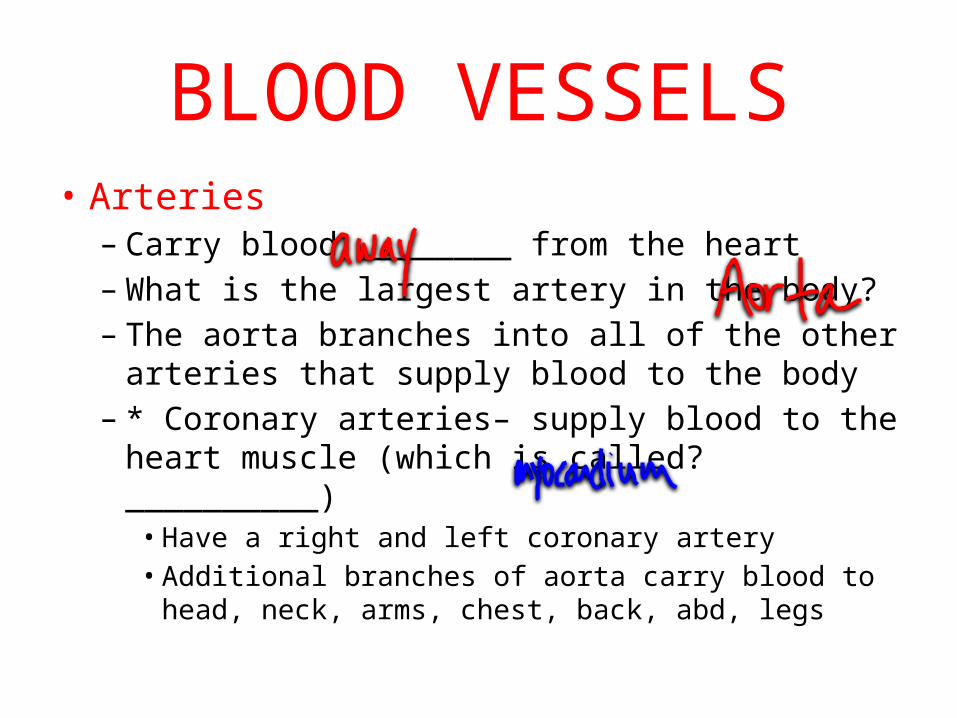
BLOOD VESSELS• Arteries– Carry blood ________ from the heart– What is the largest artery in the body?– The aorta branches into all of the other arteries
that supply blood to the body– * Coronary arteries– supply blood to the heart
muscle (which is called? __________)• Have a right and left coronary artery• Additional branches of aorta carry blood to head, neck,
arms, chest, back, abd, legs

• Arteries are more muscular/elastic than other blood vessels because they receive blood as it is pumped from the
• Smallest branches of arteries are called ______
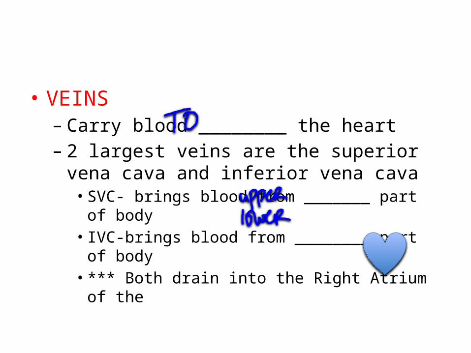
• VEINS – Carry blood ________ the heart– 2 largest veins are the superior vena cava and
inferior vena cava• SVC- brings blood from _______ part of body• IVC-brings blood from ________ part of body• *** Both drain into the Right Atrium of the

• Veins are thinner and have less muscle tissue than do arteries
• Have valves, which keep blood from flowing in a backward direction
• Smallest branches of veins are called _______

• Capillaries– Tiny vessels– Have thin walls- only contain one layer of cells
– Function- allow O2 and nutrients to pass through to cells and allow CO2 and metabolic products from the cells to enter the capillaries
– *** connect arterioles with venules****
BLOOD• Where does blood come from??? • Approx. 4-6 quarts of blood in average adult• Function– Transports O2 from lungs to body– Transports CO2 from the body cells to lungs– Transports nutrients from GI tract to body cells– Transports metabolic & waste products from the body cells
to organs for excretion– Transports heat produced by various body parts– Transports hormones produced by endocrine glands to the
body organs

Blood is made of the fluid called plasma & formed/solid elements called Blood Cells
• Plasma– 90% water with dissolved blood proteins (Ex-
fibrinogen and prothrombin– these are necessary for ___________); nutrients such as vitamins, carbs, proteins; electrolytes like K, Ca, Na ; gases like O2 and CO2; waste products; hormones; enzymes

Blood cells
3 kinds:Erythrocytes (RBC)
-live approx. _____ days, produced where??
- contains hemoglobin (which does what?)-normal count is 4.5-5.5 million per cm
Why are they RED???

• Leukocytes (WBC)– Live 3-9 days– Normal count 5,000-9,000 per cm of blood
– Function– to fight infection by engulfing, ingesting, and destroying pathogens by a process called _______________
– 5 types of leukocytes– neutrophils, eosinophils, basophils, monocytes, lymphocytes

• Thrombocytes (platelets)- Live about 5-9 days- Important for clotting process
** When a blood vessel is cut- thrombocytes collect at the site to form a “sticky plug”
**If larger vessel is cut– may have to have MD suture to close the opening to control __________


PATHWAY• HOW DOES BLOOD FLOW THROUGH
THE STRUCTURES OF THE

Cardiac Cycle (heartbeat)
• Right and Left sides of the work together in a cyclic manner even though they are separated by _____________
• Cycle consists of:– Diastole----– Systole ------

Conductive Pathway• So, what causes this cyclic contraction???____________ _________________
*A group of nerve cells in right atrium called the SA node (AKA pacemaker) sends out an electrical impulse which cause the atrial muscles to contract & push blood into ventricles

• * After impulse passes thru the atria, it reaches a group of nerve cells located between the atria & ventricles called the AV node (atrioventricular)
• *AV node sends the electrical impulse thru the Bundle of HIS, which divides into a right bundle branch & left bundle branch, which carries the impulse down thru the ventricles

* Bundle branches further into the Purkinje fibers (nerve fibers throughout the ventricles)
• Electrical impulses reach all the muscle tissue in the ventricles & the ventricles CONTRACT
• Pattern occurs approx. every 0.8 seconds• Movement of electrical impulse can be
recorded on _______________
Interference
• If something interferes with the normal conduction pattern of the arrhythmias occur
• What are arrhythmias??• Can be mild to life threatening• (Ex.- VF (ventricular fibrillation)- ventricles
contract at random without coordination– R/I ????

• Name 3 things used to dx arrhythmias
• What is a defibrillator?
• What is a pacemaker?– 2 types

Diseases/Conditions
• Anemia- not enough RBC’s, hemoglobin, or both (So, not enough O2 to the tissues)
• Sx: pallor (paleness), fatigue, dyspnea, rapid heart rate
• Types of anemia– 1. Iron deficiency- inadequate amount of iron to
form hemoglobin in RBC– Tx- take iron supplements, increase iron in diet

• 2. Aplastic anemia- injury or destruction of bone marrow which results in poor or no formation of RBC’s– Causes: chemo, radiation, viruses– Tx: determine cause and eliminate, blood
transfusions, severe cases- bone marrow transplant
– Can be fatal if unable to reverse damage

• 3. Pernicious anemia- RBC’s are abnormally large in size and inadequate in number
• Results from an inadequate absorption of Vit. B12 (Vit B12 and folic acid are required for development of mature RBC’s)
• Tx: B12 injections can control and correct

• 4. Sickle cell anemia– Chronic, inherited anemia– Erythrocytes (RBC) are crescent shaped and carry
less O2– Occurs almost exclusively with African Americans– Tx: Transfusions of packed cells, supportive
therapy during crisis

Aneurysm
• Ballooning out/ saclike formation on artery wall• Cause- disease, congenital defects, injuries• Sx: some cause pain/pressure, others no sx• Common sites- cerebral, aortal, abd.• If rupture---- hemorrhage----death• Tx: surgically removing damaged area of blood
vessel and replacing it with graft or another blood vessel

Arteriosclerosis
• Hardening or thickening of arterial walls which R/I loss of elasticity and contractility
• Commonly occurs because of aging• Can cause HTN and can lead to an aneurysm
or cerebral hemorrhage• Tx: lower BP through diet, meds

Atherosclerosis
• Fatty plaques (like cholesterol—type of lipid found in foods(eggs, meat, whole fat dairy)—produced by the liver—needed to make Vit. D, build cell walls, create bile salts that help you digest fat)
• Plaques narrow the arterial opening which reduces/eliminates blood flow
• These plaques can break loose and circulate through the bloodstream as an emboli
• Tx: low chol diet, cholesterol meds, no smoking reduce stress, exercise, angioplasty (remove plaques), bypass surgery if artery is completely blocked

Congestive Heart Failure (CHF)
• Heart muscles do not beat adequately to supply the blood to the body
• Sx: edema, dyspnea, weak but rapid pulse, pallor
• Tx: meds to slow and strengthen heart beat, diuretics, O2 therapy, low sodium diet

Coronary Artery Disease (CAD)
• Narrowing of the coronary arteries that supply blood to the heart
• Usually caused by atherosclerosis (build-up of fatty plaque inside blood vessels)
• CAD can lead to angina or MI• Tx: placing a stent inside the blocked vessels of
the heart

Embolus
• Foreign substance circulating in the bloodstream
• Can be air, blood clot, fat• Occurs when embolus enters artery too small
for passage--- R/I BLOCKAGE of blood vessel

Hemophilia
• Inherited disease• Lack a plasma protein required for clotting—
so, blood is unable to clot• Minor cut can lead to prolonged bleeding• Minor bump can lead to internal bleeding• Tx: transfusing whole blood or plasma,
administering missing protein factor

Hypertension (HTN)
• High blood pressure (BP 140/90)• Risk factors- family hx, obesity, stress,
smoking, aging, diet• NO CURE but can be controlled with meds,
limited stress, avoid tobacco, low sodium/low fat diet
• If not treated--- damage to heart, blood vessels, and kidneys
Leukemia
• Malignant disease of bone marrow or lymph tissue
• R/I high number of immature WBC’s• Sx- fever, pallor, swelling of lymph tissue,
fatigue, excessive bruising, joint pain• Tx: chemo, radiation, bone marrow transplant
Myocardial Infarction (MI)
• HEART ATTACK• Occurs when a blockage in the coronary arteries cuts off blood
supply to the heart• Affected heart tissue dies– known as an infarct• Sx: severe crushing pain that radiates to the arm, neck, and
jaw, pressure in the chest, perspiration, cold/clammy skin, dyspnea
• Tx:- If heart stops– CPR, can give “clot busting” meds (TPA) to open blood vessel—must given within first several hours, O2 therapy, vasodilators, pain meds, anticoagulants, BP control, diet changes no smoking, decrease stress, exercise, weight control

Phlebitis
• Inflammation of a vein (frequently in the leg)• Thrombophlebitis– a clot forms• Sx:- pain, edema, redness• Tx: anticoags, pain meds, elevation affected
area, support hose, surgery if needed to remove the clot

Varicose Veins
• Dilates, swollen veins that have lost elasticity and cause stasis (decreased blood flow)
• Frequently occurs in the legs, result from pregnancy, prolonged sitting/standing, heredity
• Tx: exercise, support hose, avoid prolonged sitting/standing, surgery to remove vein in severe cases
Stroke/CVA/TIA
• Can occur when a blood clot blocks the flow of blood in a vessel, or when a vessel bursts in the brain
• Sx: disoriented, difficulty with speech, loss of muscle control, paralysis (facial drooping), numbness, headache
• Tx: clot dissolving med (TPA), diet changes, make sure BP is controlled, Rehab- PT/OT