lab 12 – circulatory system circulatory system · circulatory system lab 12 – circulatory...
TRANSCRIPT
Circulatory SystemLab 12 – Circulatory System
IUSM – 2016
I. IntroductionII. Learning ObjectivesIII. KeywordsIV. Slides
A. Heart1. Layers
a. Epicardiumb. Myocardiumc. Endocardium
2. ValvesB. Vasculature
1. Grouped Structures2. Blood Vessels
a. Arteriali. Elastic arteriesii. Muscular arteriesiii. Arterioles
b. Capillariesc. Venous
i. Venulesii. Veins
3. Lymphatic VesselsV. Summary
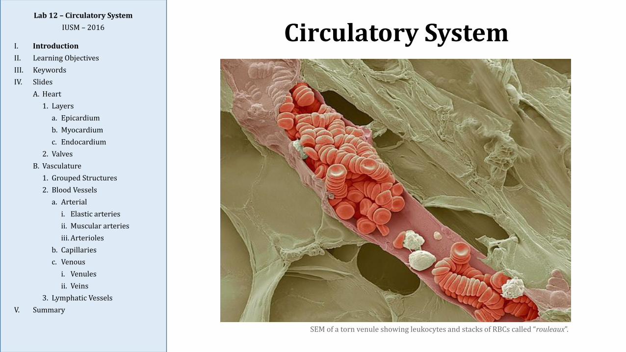
SEM of a torn venule showing leukocytes and stacks of RBCs called “rouleaux”.
Introduction
1. The circulatory system is composed of two major systems: the cardiovascular system and thelymphatic system.
a. The cardiovascular system consists of arteries (carry blood away from heart), arterioles,capillaries, venules, and veins (return blood to the heart).
b. The lymphatic system consists of lymph capillaries and lymph vessels that drain excessinterstitial fluid (lymph) from the tissues; after passing through at least one lymph node,the fluid is returned to the venous circulation via large lymph vessels (ducts) in the thorax.
2. Blood vessels larger than capillaries all share the same basic architecture consisting of threelayers of their walls; categories of vessels (e.g., elastic artery, muscular artery, vein) differ fromeach other based upon the size and composition of these layers:
a. The innermost tunica intima consists of endothelium and loose CT.
b. The middle tunica media consists primarily of smooth muscle fibers, or elastic fibers inthe large elastic arteries.
c. The outermost tunica adventitia consists of CT, and smooth muscle in larger veins.
Lab 12 – Circulatory SystemIUSM – 2016
I. IntroductionII. Learning ObjectivesIII. KeywordsIV. Slides
A. Heart1. Layers
a. Epicardiumb. Myocardiumc. Endocardium
2. ValvesB. Vasculature
1. Grouped Structures2. Blood Vessels
a. Arteriali. Elastic arteriesii. Muscular arteriesiii. Arterioles
b. Capillariesc. Venous
i. Venulesii. Veins
3. Lymphatic VesselsV. Summary
Learning Objectives
1. Explain how the circulatory system comprises the heart, blood vessels, andlymphatic vessels.
2. Identify the histological characteristics of the heart, including the epicardium,myocardium, and endocardium, and the conduction system.
3. Describe the histological features of arteries and veins as found within three layers:tunica intima, tunica media, and tunica adventitia.
4. Identify and distinguish between arteries, veins, capillaries, and lymph vessels(microvasculature vs. macrovasculature).
5. Describe the histological organization of lymphatic capillaries and vessels, theirrelationship to lymph nodes, and how they differ from blood vessels.
Lab 12 – Circulatory SystemIUSM – 2016
I. IntroductionII. Learning ObjectivesIII. KeywordsIV. Slides
A. Heart1. Layers
a. Epicardiumb. Myocardiumc. Endocardium
2. ValvesB. Vasculature
1. Grouped Structures2. Blood Vessels
a. Arteriali. Elastic arteriesii. Muscular arteriesiii. Arterioles
b. Capillariesc. Venous
i. Venulesii. Veins
3. Lymphatic VesselsV. Summary
Keywords
ArteriolesCapillariesElastic sheetsEndocardiumEndotheliumEpicardiumHeartHeart valveLarge muscular arteryLarge veinLymphatic vesselsMedium veinMesothelium
Muscular arteryMyocardiumPericytesPurkinje fibersSinusoidsSmall muscular arterySmall veinTunica adventitiaTunica intimaTunica mediaVasa vasorumVeinsVenule
Lab 12 – Circulatory SystemIUSM – 2016
I. IntroductionII. Learning ObjectivesIII. KeywordsIV. Slides
A. Heart1. Layers
a. Epicardiumb. Myocardiumc. Endocardium
2. ValvesB. Vasculature
1. Grouped Structures2. Blood Vessels
a. Arteriali. Elastic arteriesii. Muscular arteriesiii. Arterioles
b. Capillariesc. Venous
i. Venulesii. Veins
3. Lymphatic VesselsV. Summary
Slide 81 (NW): Cardiac Muscle, TrichromeLab 12 – Circulatory System
IUSM – 2016
I. IntroductionII. Learning ObjectivesIII. KeywordsIV. Slides
A. Heart1. Layers
a. Epicardiumb. Myocardiumc. Endocardium
2. ValvesB. Vasculature
1. Grouped Structures2. Blood Vessels
a. Arteriali. Elastic arteriesii. Muscular arteriesiii. Arterioles
b. Capillariesc. Venous
i. Venulesii. Veins
3. Lymphatic VesselsV. Summary
Slide Overview
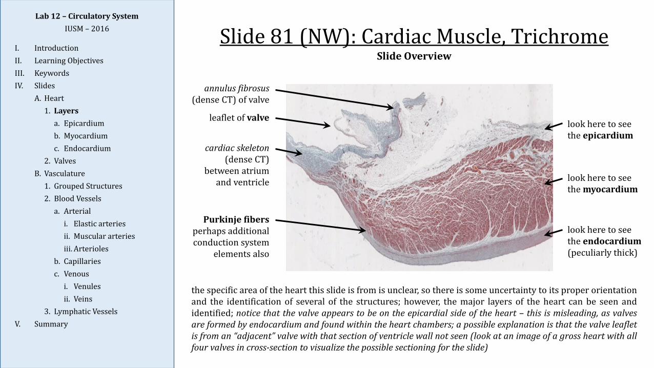
the specific area of the heart this slide is from is unclear, so there is some uncertainty to its proper orientationand the identification of several of the structures; however, the major layers of the heart can be seen andidentified; notice that the valve appears to be on the epicardial side of the heart – this is misleading, as valvesare formed by endocardium and found within the heart chambers; a possible explanation is that the valve leafletis from an “adjacent” valve with that section of ventricle wall not seen (look at an image of a gross heart with allfour valves in cross-section to visualize the possible sectioning for the slide)
look here to see the epicardium
look here to see the endocardium(peculiarly thick)
look here to see the myocardium
annulus fibrosus(dense CT) of valve
leaflet of valve
cardiac skeleton(dense CT)
between atrium and ventricle
Purkinje fibersperhaps additional conduction system
elements also
Slide 139: Heart, H&E
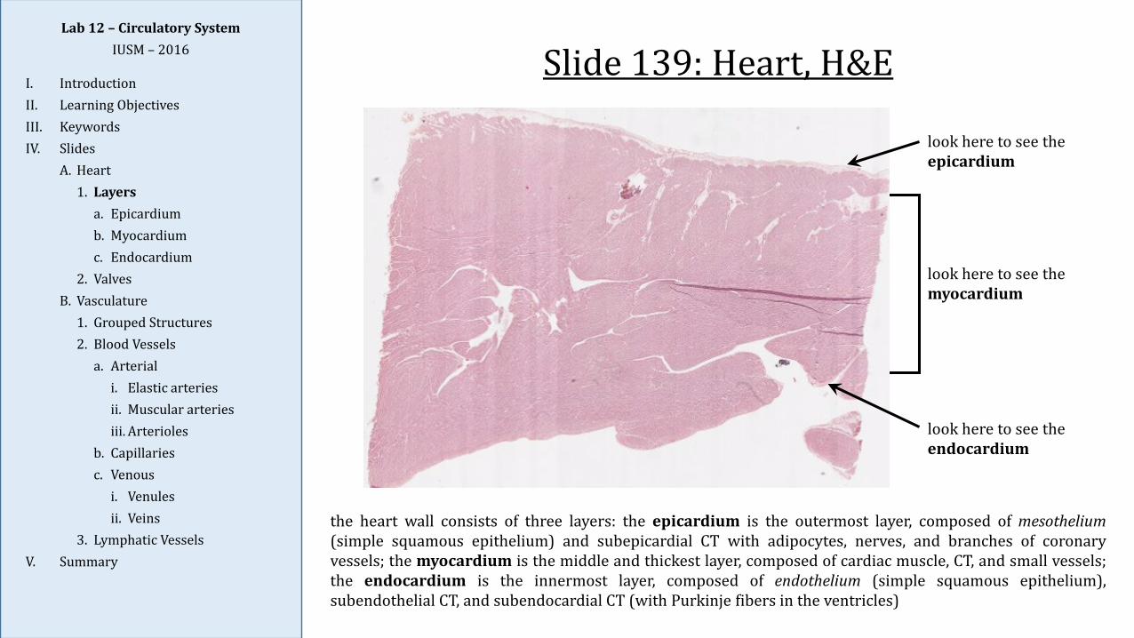
look here to see the epicardium
look here to see the endocardium
look here to see the myocardium
the heart wall consists of three layers: the epicardium is the outermost layer, composed of mesothelium(simple squamous epithelium) and subepicardial CT with adipocytes, nerves, and branches of coronaryvessels; the myocardium is the middle and thickest layer, composed of cardiac muscle, CT, and small vessels;the endocardium is the innermost layer, composed of endothelium (simple squamous epithelium),subendothelial CT, and subendocardial CT (with Purkinje fibers in the ventricles)
Lab 12 – Circulatory SystemIUSM – 2016
I. IntroductionII. Learning ObjectivesIII. KeywordsIV. Slides
A. Heart1. Layers
a. Epicardiumb. Myocardiumc. Endocardium
2. ValvesB. Vasculature
1. Grouped Structures2. Blood Vessels
a. Arteriali. Elastic arteriesii. Muscular arteriesiii. Arterioles
b. Capillariesc. Venous
i. Venulesii. Veins
3. Lymphatic VesselsV. Summary
Slide 48 (464): Cardiac Muscle, H&ELab 12 – Circulatory System
IUSM – 2016
I. IntroductionII. Learning ObjectivesIII. KeywordsIV. Slides
A. Heart1. Layers
a. Epicardiumb. Myocardiumc. Endocardium
2. ValvesB. Vasculature
1. Grouped Structures2. Blood Vessels
a. Arteriali. Elastic arteriesii. Muscular arteriesiii. Arterioles
b. Capillariesc. Venous
i. Venulesii. Veins
3. Lymphatic VesselsV. Summary
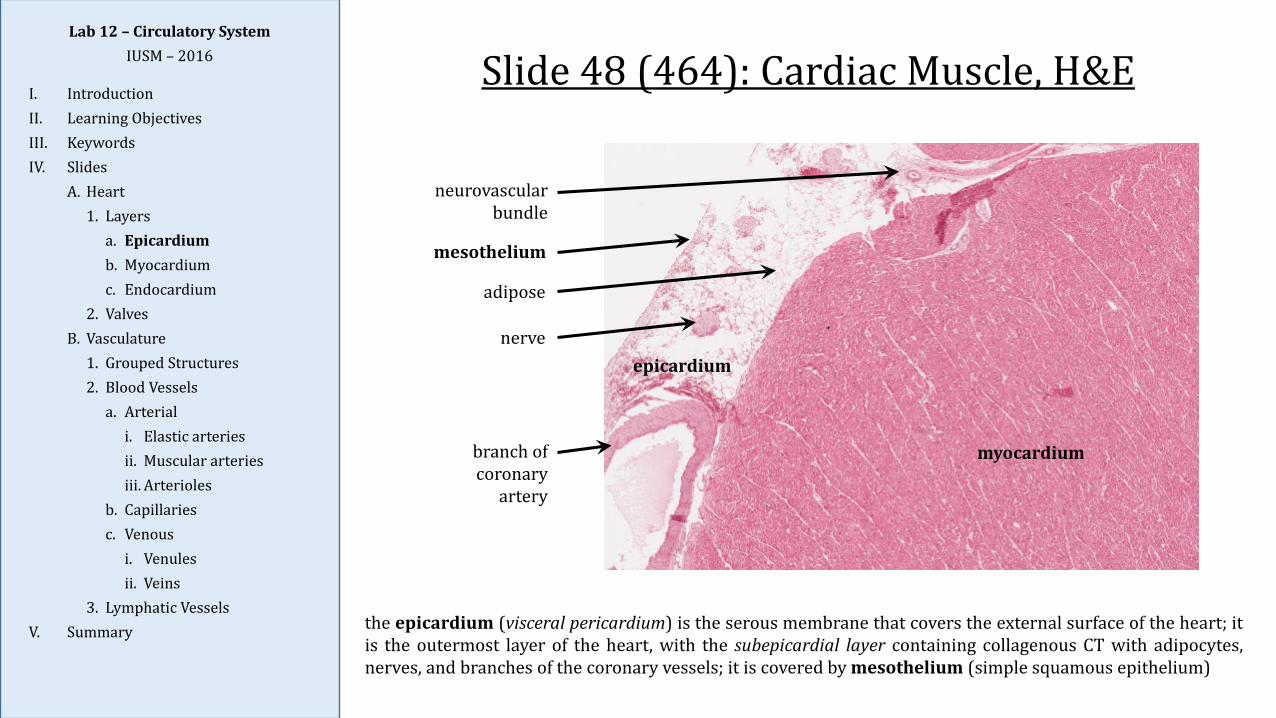
neurovascular bundle
mesothelium
adipose
nerve
branch of coronary
artery
myocardium
the epicardium (visceral pericardium) is the serous membrane that covers the external surface of the heart; itis the outermost layer of the heart, with the subepicardial layer containing collagenous CT with adipocytes,nerves, and branches of the coronary vessels; it is covered by mesothelium (simple squamous epithelium)
epicardium
Lab 12 – Circulatory SystemIUSM – 2016
I. IntroductionII. Learning ObjectivesIII. KeywordsIV. Slides
A. Heart1. Layers
a. Epicardiumb. Myocardiumc. Endocardium
2. ValvesB. Vasculature
1. Grouped Structures2. Blood Vessels
a. Arteriali. Elastic arteriesii. Muscular arteriesiii. Arterioles
b. Capillariesc. Venous
i. Venulesii. Veins
3. Lymphatic VesselsV. Summary
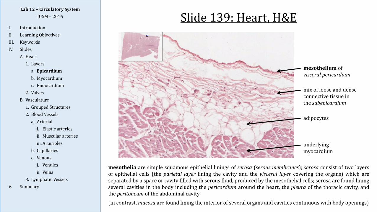
mesothelium of visceral pericardium
mix of loose and dense connective tissue in the subepicardium
adipocytes
underlying myocardium
mesothelia are simple squamous epithelial linings of serosa (serous membranes); serosa consist of two layersof epithelial cells (the parietal layer lining the cavity and the visceral layer covering the organs) which areseparated by a space or cavity filled with serous fluid, produced by the mesothelial cells; serosa are found liningseveral cavities in the body including the pericardium around the heart, the pleura of the thoracic cavity, andthe peritoneum of the abdominal cavity(in contrast, mucosa are found lining the interior of several organs and cavities continuous with body openings)
Slide 139: Heart, H&E
Lab 12 – Circulatory SystemIUSM – 2016
I. IntroductionII. Learning ObjectivesIII. KeywordsIV. Slides
A. Heart1. Layers
a. Epicardiumb. Myocardiumc. Endocardium
2. ValvesB. Vasculature
1. Grouped Structures2. Blood Vessels
a. Arteriali. Elastic arteriesii. Muscular arteriesiii. Arterioles
b. Capillariesc. Venous
i. Venulesii. Veins
3. Lymphatic VesselsV. Summary
Slide 139: Heart, H&E
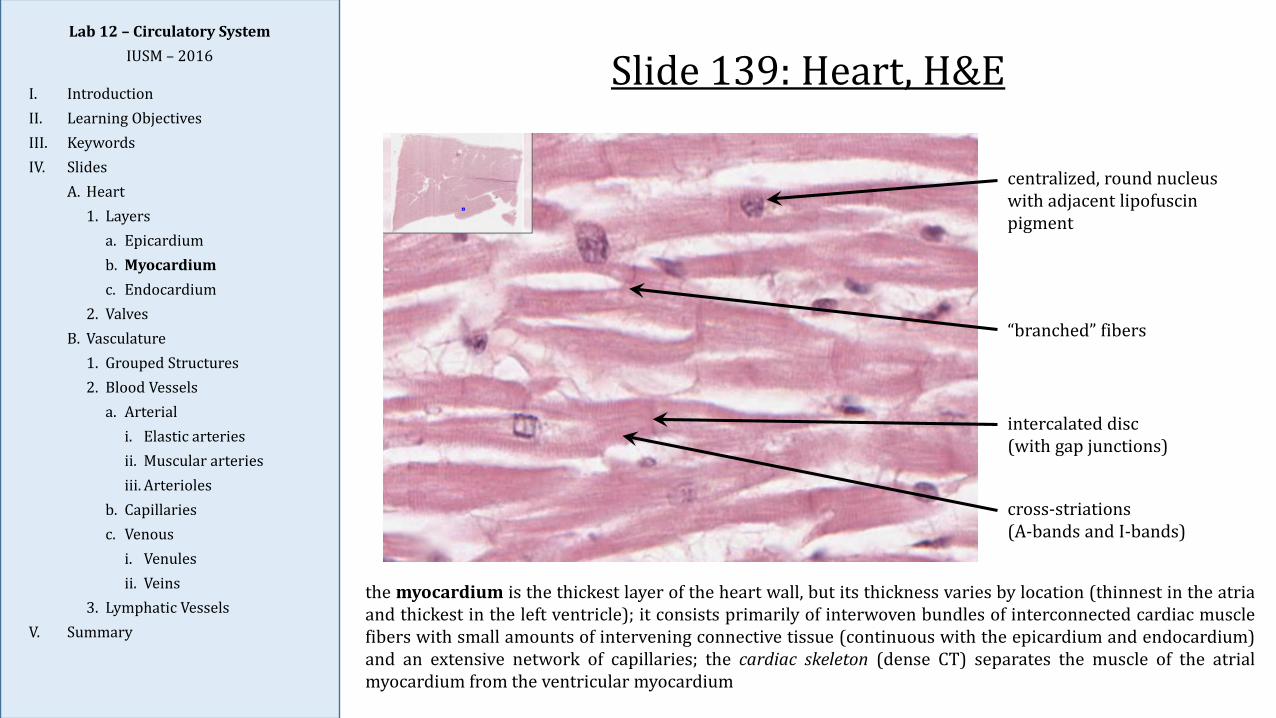
“branched” fibers
centralized, round nucleus with adjacent lipofuscin pigment
intercalated disc(with gap junctions)
cross-striations(A-bands and I-bands)
the myocardium is the thickest layer of the heart wall, but its thickness varies by location (thinnest in the atriaand thickest in the left ventricle); it consists primarily of interwoven bundles of interconnected cardiac musclefibers with small amounts of intervening connective tissue (continuous with the epicardium and endocardium)and an extensive network of capillaries; the cardiac skeleton (dense CT) separates the muscle of the atrialmyocardium from the ventricular myocardium
Lab 12 – Circulatory SystemIUSM – 2016
I. IntroductionII. Learning ObjectivesIII. KeywordsIV. Slides
A. Heart1. Layers
a. Epicardiumb. Myocardiumc. Endocardium
2. ValvesB. Vasculature
1. Grouped Structures2. Blood Vessels
a. Arteriali. Elastic arteriesii. Muscular arteriesiii. Arterioles
b. Capillariesc. Venous
i. Venulesii. Veins
3. Lymphatic VesselsV. Summary
Slide 139: Heart, H&E
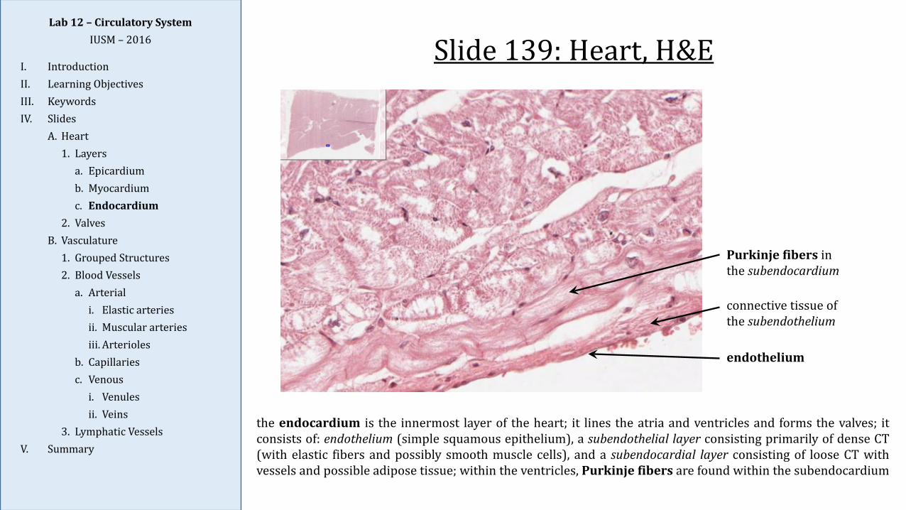
the endocardium is the innermost layer of the heart; it lines the atria and ventricles and forms the valves; itconsists of: endothelium (simple squamous epithelium), a subendothelial layer consisting primarily of dense CT(with elastic fibers and possibly smooth muscle cells), and a subendocardial layer consisting of loose CT withvessels and possible adipose tissue; within the ventricles, Purkinje fibers are found within the subendocardium
endothelium
Purkinje fibers in the subendocardium
connective tissue of the subendothelium
Lab 12 – Circulatory SystemIUSM – 2016
I. IntroductionII. Learning ObjectivesIII. KeywordsIV. Slides
A. Heart1. Layers
a. Epicardiumb. Myocardiumc. Endocardium
2. ValvesB. Vasculature
1. Grouped Structures2. Blood Vessels
a. Arteriali. Elastic arteriesii. Muscular arteriesiii. Arterioles
b. Capillariesc. Venous
i. Venulesii. Veins
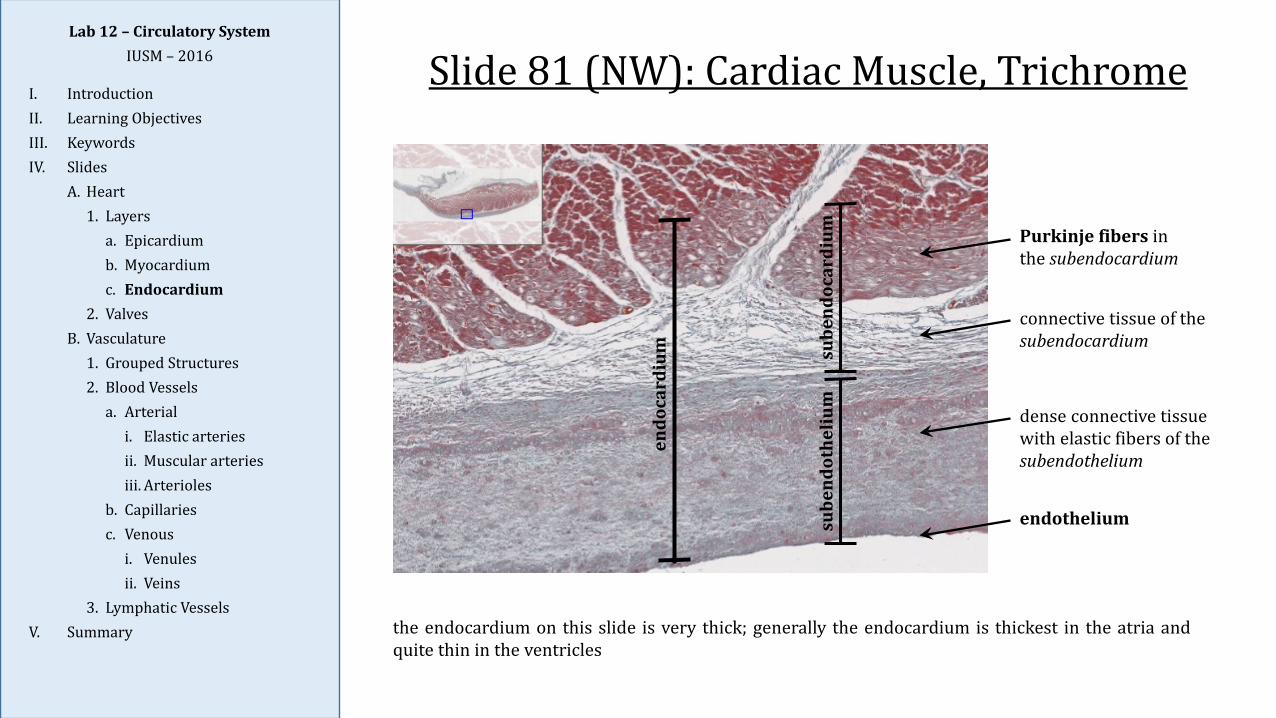
3. Lymphatic VesselsV. Summary the endocardium on this slide is very thick; generally the endocardium is thickest in the atria and
quite thin in the ventricles
Slide 81 (NW): Cardiac Muscle, Trichrome
Purkinje fibers in the subendocardium
connective tissue of the subendocardium
dense connective tissue with elastic fibers of the subendothelium
endothelium
endo
card
ium su
bend
ocar
dium
sube
ndot
heliu
m
Lab 12 – Circulatory SystemIUSM – 2016
I. IntroductionII. Learning ObjectivesIII. KeywordsIV. Slides
A. Heart1. Layers
a. Epicardiumb. Myocardiumc. Endocardium
2. ValvesB. Vasculature
1. Grouped Structures2. Blood Vessels
a. Arteriali. Elastic arteriesii. Muscular arteriesiii. Arterioles
b. Capillariesc. Venous
i. Venulesii. Veins
3. Lymphatic VesselsV. Summary
Slide 139: Heart, H&E
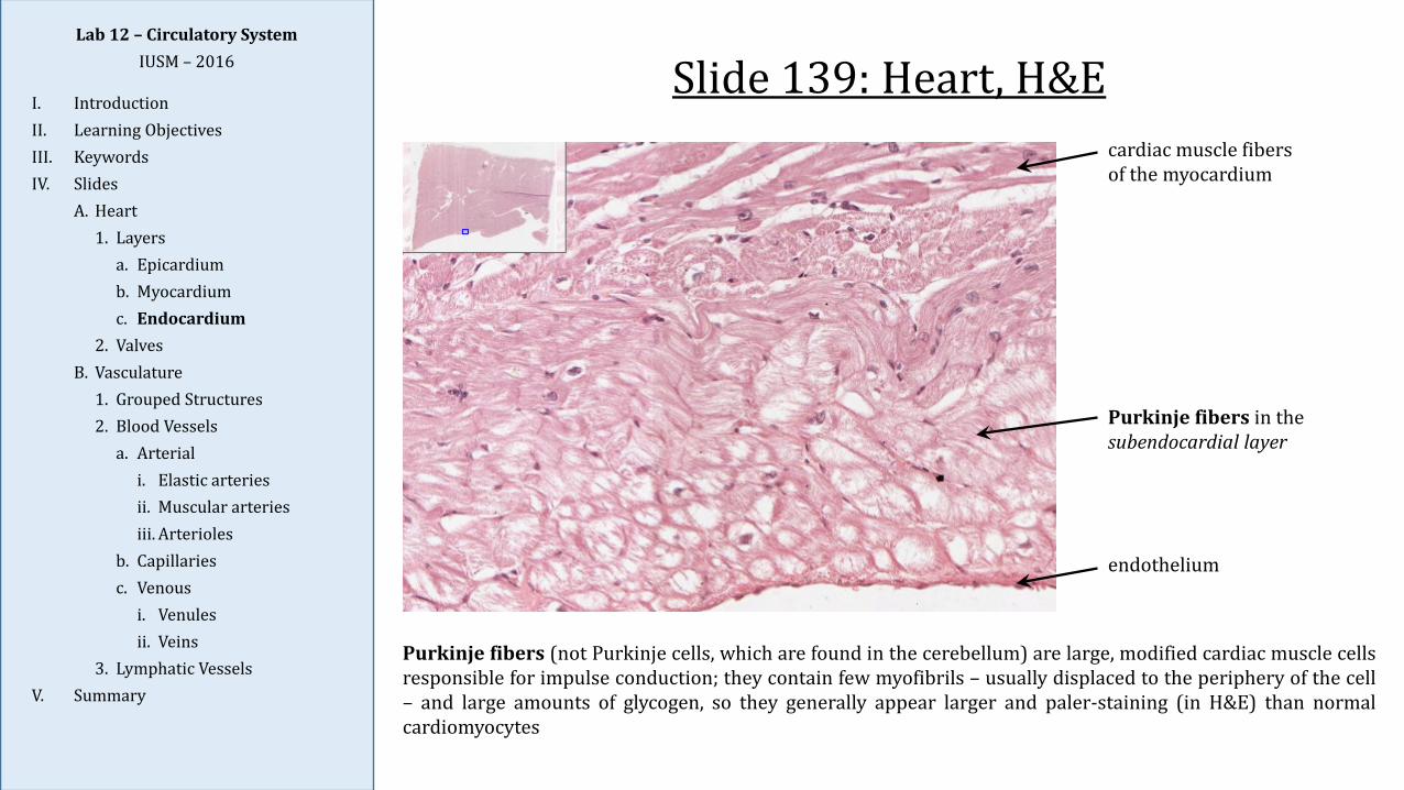
Purkinje fibers (not Purkinje cells, which are found in the cerebellum) are large, modified cardiac muscle cellsresponsible for impulse conduction; they contain few myofibrils – usually displaced to the periphery of the cell– and large amounts of glycogen, so they generally appear larger and paler-staining (in H&E) than normalcardiomyocytes
Purkinje fibers in the subendocardial layer
endothelium
cardiac muscle fibers of the myocardium
Lab 12 – Circulatory SystemIUSM – 2016
I. IntroductionII. Learning ObjectivesIII. KeywordsIV. Slides
A. Heart1. Layers
a. Epicardiumb. Myocardiumc. Endocardium
2. ValvesB. Vasculature
1. Grouped Structures2. Blood Vessels
a. Arteriali. Elastic arteriesii. Muscular arteriesiii. Arterioles
b. Capillariesc. Venous
i. Venulesii. Veins
3. Lymphatic VesselsV. Summary
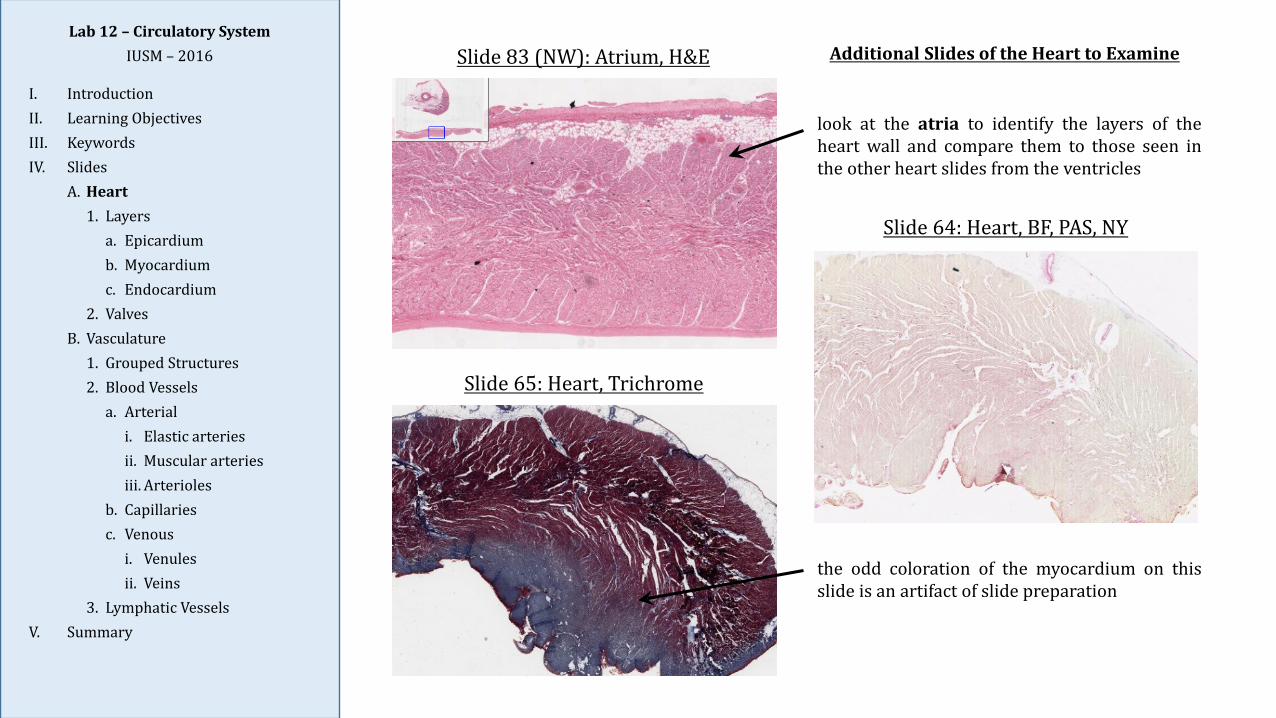
Slide 64: Heart, BF, PAS, NY
Slide 65: Heart, Trichrome
Slide 83 (NW): Atrium, H&E
look at the atria to identify the layers of theheart wall and compare them to those seen inthe other heart slides from the ventricles
the odd coloration of the myocardium on thisslide is an artifact of slide preparation
Additional Slides of the Heart to Examine
Lab 12 – Circulatory SystemIUSM – 2016
I. IntroductionII. Learning ObjectivesIII. KeywordsIV. Slides
A. Heart1. Layers
a. Epicardiumb. Myocardiumc. Endocardium
2. ValvesB. Vasculature
1. Grouped Structures2. Blood Vessels
a. Arteriali. Elastic arteriesii. Muscular arteriesiii. Arterioles
b. Capillariesc. Venous
i. Venulesii. Veins
3. Lymphatic VesselsV. Summary
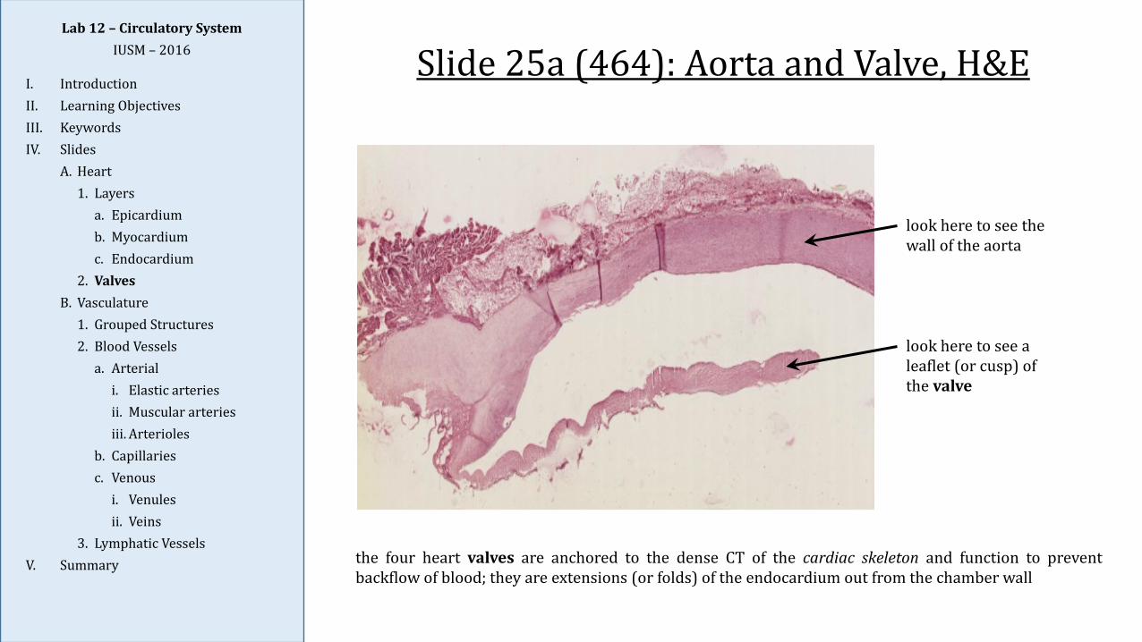
Slide 25a (464): Aorta and Valve, H&E
look here to see the wall of the aorta
look here to see a leaflet (or cusp) of the valve
the four heart valves are anchored to the dense CT of the cardiac skeleton and function to preventbackflow of blood; they are extensions (or folds) of the endocardium out from the chamber wall
Lab 12 – Circulatory SystemIUSM – 2016
I. IntroductionII. Learning ObjectivesIII. KeywordsIV. Slides
A. Heart1. Layers
a. Epicardiumb. Myocardiumc. Endocardium
2. ValvesB. Vasculature
1. Grouped Structures2. Blood Vessels
a. Arteriali. Elastic arteriesii. Muscular arteriesiii. Arterioles
b. Capillariesc. Venous
i. Venulesii. Veins
3. Lymphatic VesselsV. Summary
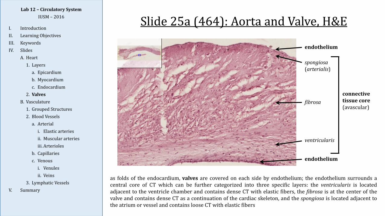
Slide 25a (464): Aorta and Valve, H&E
as folds of the endocardium, valves are covered on each side by endothelium; the endothelium surrounds acentral core of CT which can be further categorized into three specific layers: the ventricularis is locatedadjacent to the ventricle chamber and contains dense CT with elastic fibers, the fibrosa is at the center of thevalve and contains dense CT as a continuation of the cardiac skeleton, and the spongiosa is located adjacent tothe atrium or vessel and contains loose CT with elastic fibers
endothelium
endothelium
connective tissue core(avascular)
ventricularis
fibrosa
spongiosa(arterialis)
Lab 12 – Circulatory SystemIUSM – 2016
I. IntroductionII. Learning ObjectivesIII. KeywordsIV. Slides
A. Heart1. Layers
a. Epicardiumb. Myocardiumc. Endocardium
2. ValvesB. Vasculature
1. Grouped Structures2. Blood Vessels
a. Arteriali. Elastic arteriesii. Muscular arteriesiii. Arterioles
b. Capillariesc. Venous
i. Venulesii. Veins
3. Lymphatic VesselsV. Summary
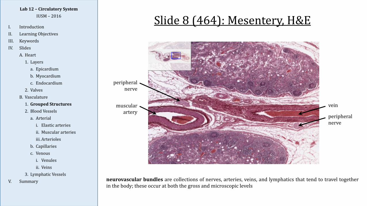
peripheral nerve
muscular artery
vein
peripheral nerve
Slide 8 (464): Mesentery, H&E
neurovascular bundles are collections of nerves, arteries, veins, and lymphatics that tend to travel togetherin the body; these occur at both the gross and microscopic levels
Lab 12 – Circulatory SystemIUSM – 2016
I. IntroductionII. Learning ObjectivesIII. KeywordsIV. Slides
A. Heart1. Layers
a. Epicardiumb. Myocardiumc. Endocardium
2. ValvesB. Vasculature
1. Grouped Structures2. Blood Vessels
a. Arteriali. Elastic arteriesii. Muscular arteriesiii. Arterioles
b. Capillariesc. Venous
i. Venulesii. Veins
3. Lymphatic VesselsV. Summary
Slide 115: Aorta, H&E
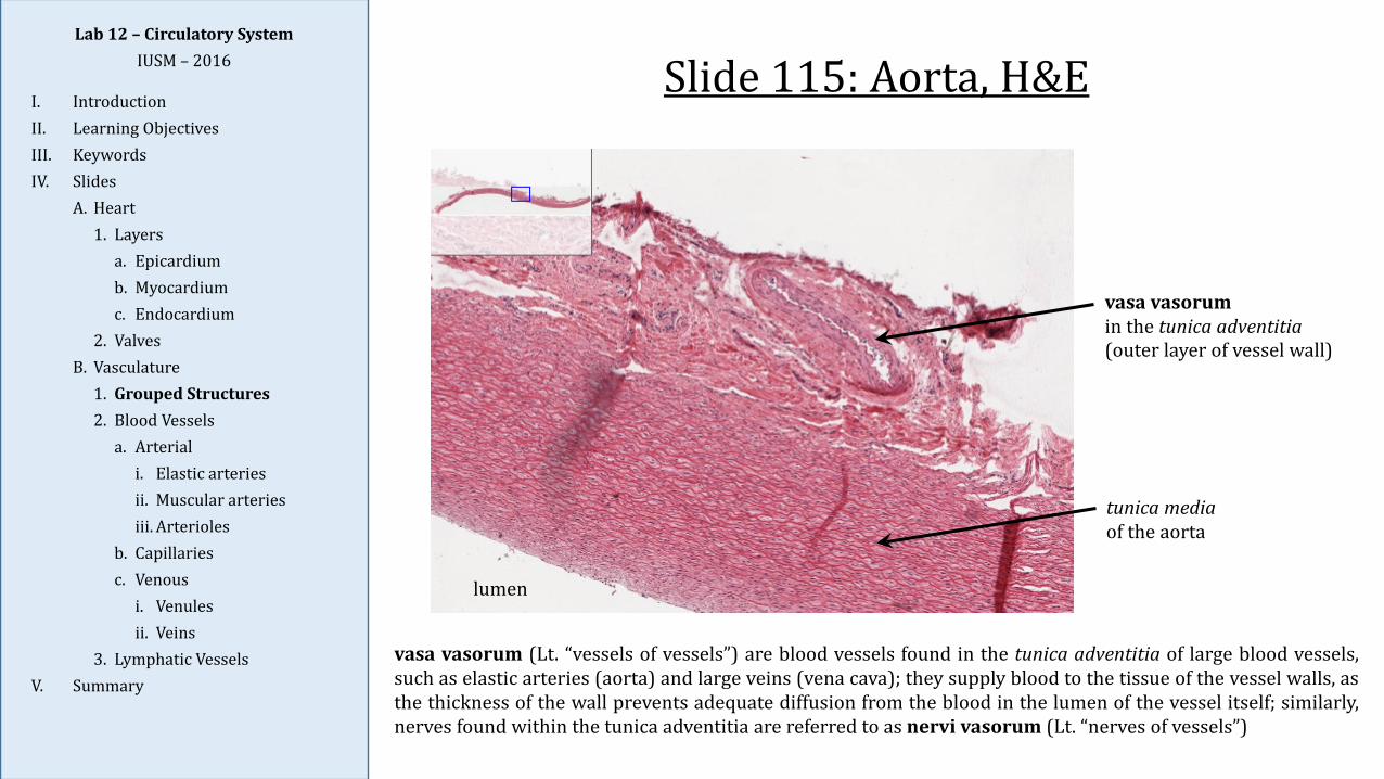
vasa vasorum (Lt. “vessels of vessels”) are blood vessels found in the tunica adventitia of large blood vessels,such as elastic arteries (aorta) and large veins (vena cava); they supply blood to the tissue of the vessel walls, asthe thickness of the wall prevents adequate diffusion from the blood in the lumen of the vessel itself; similarly,nerves found within the tunica adventitia are referred to as nervi vasorum (Lt. “nerves of vessels”)
vasa vasorumin the tunica adventitia(outer layer of vessel wall)
lumen
tunica mediaof the aorta
Lab 12 – Circulatory SystemIUSM – 2016
I. IntroductionII. Learning ObjectivesIII. KeywordsIV. Slides
A. Heart1. Layers
a. Epicardiumb. Myocardiumc. Endocardium
2. ValvesB. Vasculature
1. Grouped Structures2. Blood Vessels
a. Arteriali. Elastic arteriesii. Muscular arteriesiii. Arterioles
b. Capillariesc. Venous
i. Venulesii. Veins
3. Lymphatic VesselsV. Summary
Slide 8 (464): Mesentery, H&E
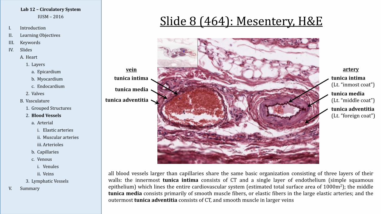
all blood vessels larger than capillaries share the same basic organization consisting of three layers of theirwalls: the innermost tunica intima consists of CT and a single layer of endothelium (simple squamousepithelium) which lines the entire cardiovascular system (estimated total surface area of 1000m2); the middletunica media consists primarily of smooth muscle fibers, or elastic fibers in the large elastic arteries; and theoutermost tunica adventitia consists of CT, and smooth muscle in larger veins
tunica intima(Lt. “inmost coat”)tunica media(Lt. “middle coat”)tunica adventitia(Lt. “foreign coat”)
tunica intima
tunica media
tunica adventitia
vein artery
Lab 12 – Circulatory SystemIUSM – 2016
I. IntroductionII. Learning ObjectivesIII. KeywordsIV. Slides
A. Heart1. Layers
a. Epicardiumb. Myocardiumc. Endocardium
2. ValvesB. Vasculature
1. Grouped Structures2. Blood Vessels
a. Arteriali. Elastic arteriesii. Muscular arteriesiii. Arterioles
b. Capillariesc. Venous
i. Venulesii. Veins
3. Lymphatic VesselsV. Summary
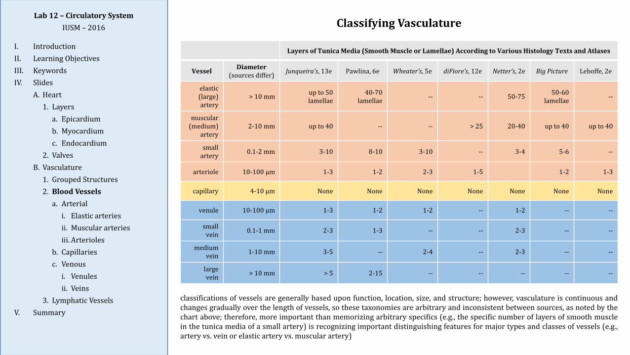
Layers of Tunica Media (Smooth Muscle or Lamellae) According to Various Histology Texts and Atlases
Vessel Diameter(sources differ) Junqueira’s, 13e Pawlina, 6e Wheater’s, 5e diFiore’s, 12e Netter’s, 2e Big Picture Leboffe, 2e
elastic (large) artery
> 10 mm up to 50 lamellae
40-70 lamellae -- -- 50-75 50-60
lamellae --
muscular (medium)
artery2-10 mm up to 40 -- -- > 25 20-40 up to 40 up to 40
small artery 0.1-2 mm 3-10 8-10 3-10 -- 3-4 5-6 --
arteriole 10-100 μm 1-3 1-2 2-3 1-5 1-2 1-3
capillary 4-10 μm None None None None None None None
venule 10-100 μm 1-3 1-2 1-2 -- 1-2 -- --
small vein 0.1-1 mm 2-3 1-3 -- -- 2-3 -- --
medium vein 1-10 mm 3-5 -- 2-4 -- 2-3 -- --
largevein > 10 mm > 5 2-15 -- -- -- -- --
Classifying Vasculature
classifications of vessels are generally based upon function, location, size, and structure; however, vasculature is continuous andchanges gradually over the length of vessels, so these taxonomies are arbitrary and inconsistent between sources, as noted by thechart above; therefore, more important than memorizing arbitrary specifics (e.g., the specific number of layers of smooth musclein the tunica media of a small artery) is recognizing important distinguishing features for major types and classes of vessels (e.g.,artery vs. vein or elastic artery vs. muscular artery)
Lab 12 – Circulatory SystemIUSM – 2016
I. IntroductionII. Learning ObjectivesIII. KeywordsIV. Slides
A. Heart1. Layers
a. Epicardiumb. Myocardiumc. Endocardium
2. ValvesB. Vasculature
1. Grouped Structures2. Blood Vessels
a. Arteriali. Elastic arteriesii. Muscular arteriesiii. Arterioles
b. Capillariesc. Venous
i. Venulesii. Veins
3. Lymphatic VesselsV. Summary
Slide 115: Aorta, H&E
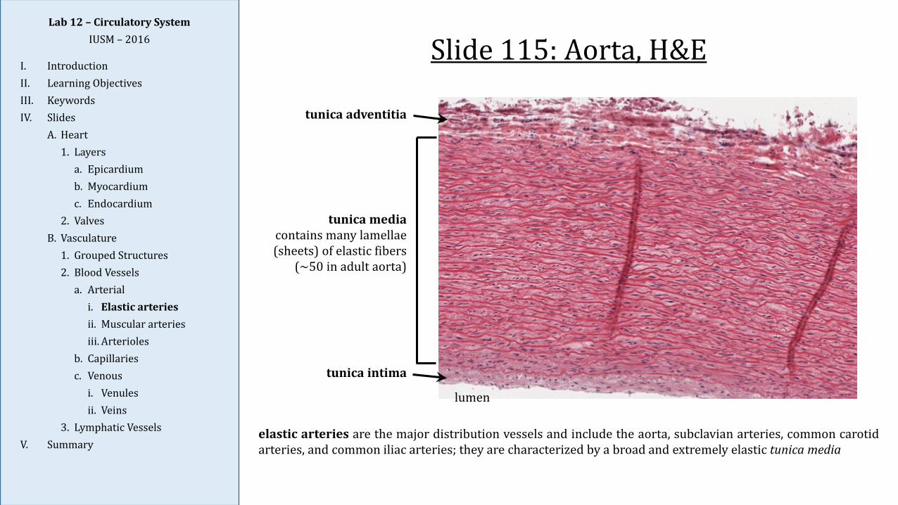
elastic arteries are the major distribution vessels and include the aorta, subclavian arteries, common carotidarteries, and common iliac arteries; they are characterized by a broad and extremely elastic tunica media
tunica intima
tunica mediacontains many lamellae (sheets) of elastic fibers
(~50 in adult aorta)
tunica adventitia
lumen
Lab 12 – Circulatory SystemIUSM – 2016
I. IntroductionII. Learning ObjectivesIII. KeywordsIV. Slides
A. Heart1. Layers
a. Epicardiumb. Myocardiumc. Endocardium
2. ValvesB. Vasculature
1. Grouped Structures2. Blood Vessels
a. Arteriali. Elastic arteriesii. Muscular arteriesiii. Arterioles
b. Capillariesc. Venous
i. Venulesii. Veins
3. Lymphatic VesselsV. Summary
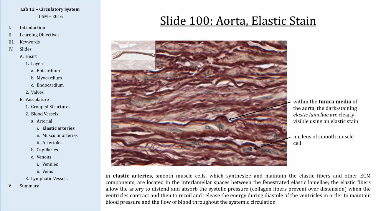
Slide 100: Aorta, Elastic Stain
within the tunica media of the aorta, the dark-staining elastic lamellae are clearly visible using an elastic stain
nucleus of smooth muscle cell
in elastic arteries, smooth muscle cells, which synthesize and maintain the elastic fibers and other ECMcomponents, are located in the interlamellar spaces between the fenestrated elastic lamellae; the elastic fibersallow the artery to distend and absorb the systolic pressure (collagen fibers prevent over distension) when theventricles contract and then to recoil and release the energy during diastole of the ventricles in order to maintainblood pressure and the flow of blood throughout the systemic circulation
Lab 12 – Circulatory SystemIUSM – 2016
I. IntroductionII. Learning ObjectivesIII. KeywordsIV. Slides
A. Heart1. Layers
a. Epicardiumb. Myocardiumc. Endocardium
2. ValvesB. Vasculature
1. Grouped Structures2. Blood Vessels
a. Arteriali. Elastic arteriesii. Muscular arteriesiii. Arterioles
b. Capillariesc. Venous
i. Venulesii. Veins
3. Lymphatic VesselsV. Summary
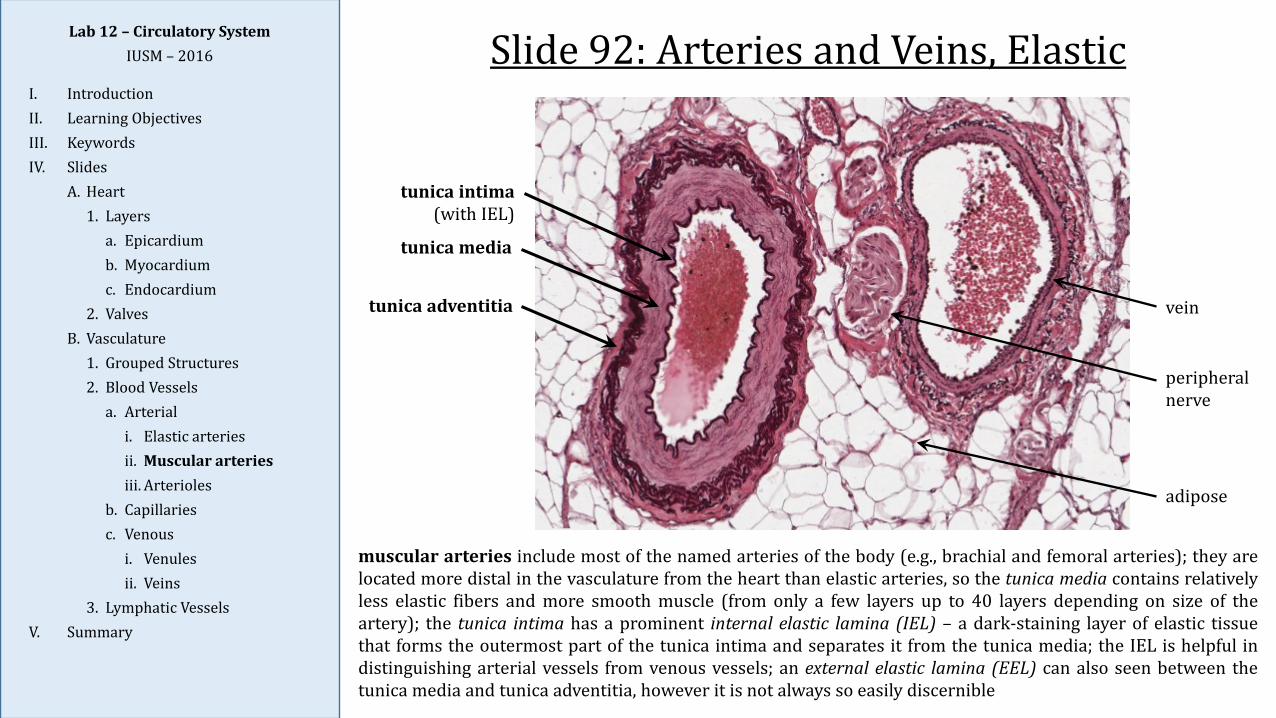
Slide 92: Arteries and Veins, Elastic
vein
peripheral nerve
tunica intima(with IEL)
tunica media
tunica adventitia
adipose
muscular arteries include most of the named arteries of the body (e.g., brachial and femoral arteries); they arelocated more distal in the vasculature from the heart than elastic arteries, so the tunica media contains relativelyless elastic fibers and more smooth muscle (from only a few layers up to 40 layers depending on size of theartery); the tunica intima has a prominent internal elastic lamina (IEL) – a dark-staining layer of elastic tissuethat forms the outermost part of the tunica intima and separates it from the tunica media; the IEL is helpful indistinguishing arterial vessels from venous vessels; an external elastic lamina (EEL) can also seen between thetunica media and tunica adventitia, however it is not always so easily discernible
Lab 12 – Circulatory SystemIUSM – 2016
I. IntroductionII. Learning ObjectivesIII. KeywordsIV. Slides
A. Heart1. Layers
a. Epicardiumb. Myocardiumc. Endocardium
2. ValvesB. Vasculature
1. Grouped Structures2. Blood Vessels
a. Arteriali. Elastic arteriesii. Muscular arteriesiii. Arterioles
b. Capillariesc. Venous
i. Venulesii. Veins
3. Lymphatic VesselsV. Summary
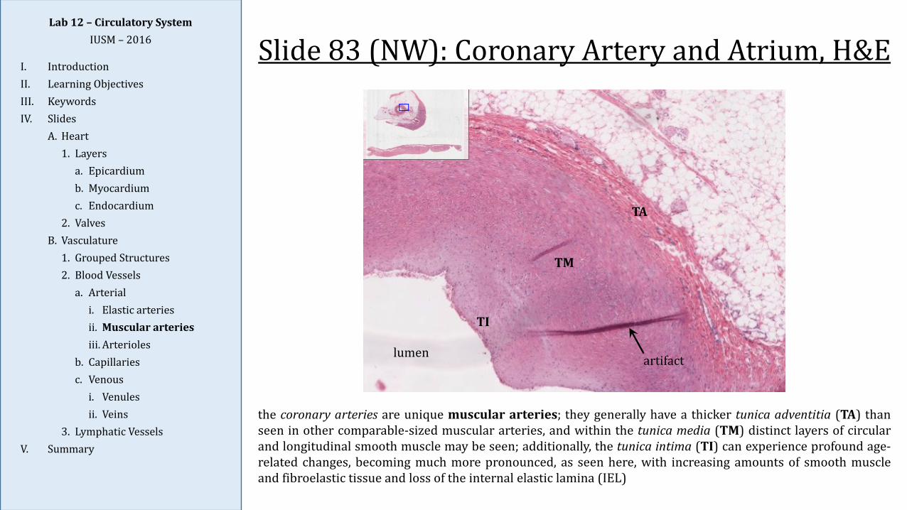
Slide 83 (NW): Coronary Artery and Atrium, H&E
the coronary arteries are unique muscular arteries; they generally have a thicker tunica adventitia (TA) thanseen in other comparable-sized muscular arteries, and within the tunica media (TM) distinct layers of circularand longitudinal smooth muscle may be seen; additionally, the tunica intima (TI) can experience profound age-related changes, becoming much more pronounced, as seen here, with increasing amounts of smooth muscleand fibroelastic tissue and loss of the internal elastic lamina (IEL)
lumen
TI
TM
TA
artifact
Lab 12 – Circulatory SystemIUSM – 2016
I. IntroductionII. Learning ObjectivesIII. KeywordsIV. Slides
A. Heart1. Layers
a. Epicardiumb. Myocardiumc. Endocardium
2. ValvesB. Vasculature
1. Grouped Structures2. Blood Vessels
a. Arteriali. Elastic arteriesii. Muscular arteriesiii. Arterioles
b. Capillariesc. Venous
i. Venulesii. Veins
3. Lymphatic VesselsV. Summary
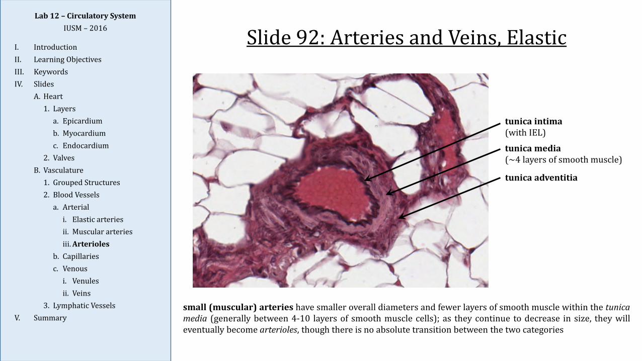
Slide 92: Arteries and Veins, Elastic
tunica intima(with IEL)
tunica media(~4 layers of smooth muscle)
tunica adventitia
small (muscular) arteries have smaller overall diameters and fewer layers of smooth muscle within the tunicamedia (generally between 4-10 layers of smooth muscle cells); as they continue to decrease in size, they willeventually become arterioles, though there is no absolute transition between the two categories
Lab 12 – Circulatory SystemIUSM – 2016
I. IntroductionII. Learning ObjectivesIII. KeywordsIV. Slides
A. Heart1. Layers
a. Epicardiumb. Myocardiumc. Endocardium
2. ValvesB. Vasculature
1. Grouped Structures2. Blood Vessels
a. Arteriali. Elastic arteriesii. Muscular arteriesiii. Arterioles
b. Capillariesc. Venous
i. Venulesii. Veins
3. Lymphatic VesselsV. Summary
Slide 92: Arteries and Veins, Elastic
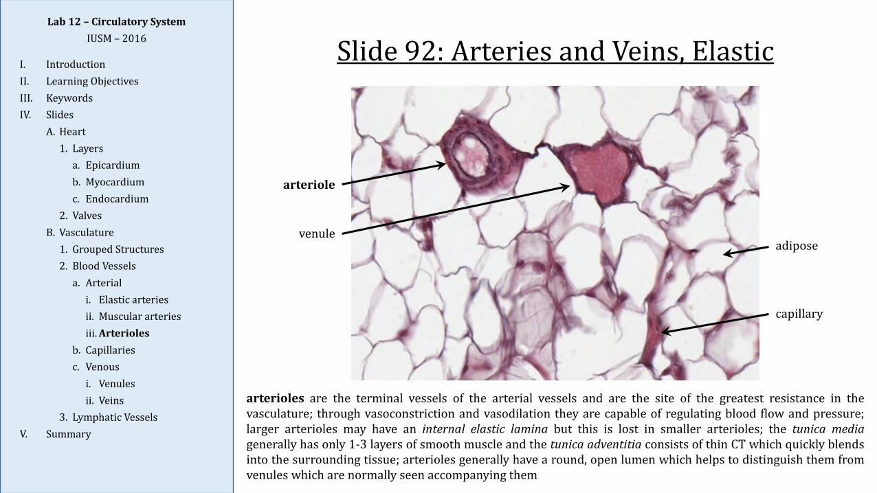
arterioles are the terminal vessels of the arterial vessels and are the site of the greatest resistance in thevasculature; through vasoconstriction and vasodilation they are capable of regulating blood flow and pressure;larger arterioles may have an internal elastic lamina but this is lost in smaller arterioles; the tunica mediagenerally has only 1-3 layers of smooth muscle and the tunica adventitia consists of thin CT which quickly blendsinto the surrounding tissue; arterioles generally have a round, open lumen which helps to distinguish them fromvenules which are normally seen accompanying them
arteriole
venule
capillary
adipose
Lab 12 – Circulatory SystemIUSM – 2016
I. IntroductionII. Learning ObjectivesIII. KeywordsIV. Slides
A. Heart1. Layers
a. Epicardiumb. Myocardiumc. Endocardium
2. ValvesB. Vasculature
1. Grouped Structures2. Blood Vessels
a. Arteriali. Elastic arteriesii. Muscular arteriesiii. Arterioles
b. Capillariesc. Venous
i. Venulesii. Veins
3. Lymphatic VesselsV. Summary
Slide 8: Tongue, Trichrome
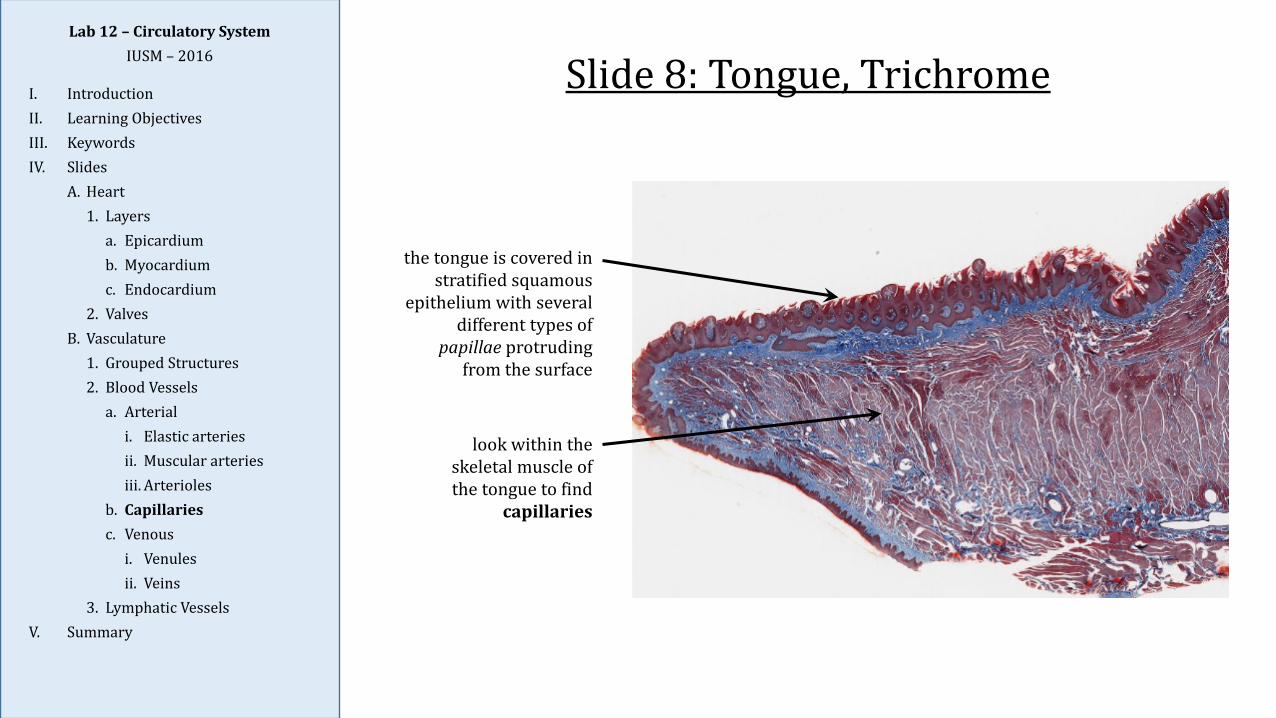
look within the skeletal muscle of the tongue to find
capillaries
the tongue is covered in stratified squamous
epithelium with several different types of
papillae protruding from the surface
Lab 12 – Circulatory SystemIUSM – 2016
I. IntroductionII. Learning ObjectivesIII. KeywordsIV. Slides
A. Heart1. Layers
a. Epicardiumb. Myocardiumc. Endocardium
2. ValvesB. Vasculature
1. Grouped Structures2. Blood Vessels
a. Arteriali. Elastic arteriesii. Muscular arteriesiii. Arterioles
b. Capillariesc. Venous
i. Venulesii. Veins
3. Lymphatic VesselsV. Summary
Slide 8: Tongue, Trichrome
capillary
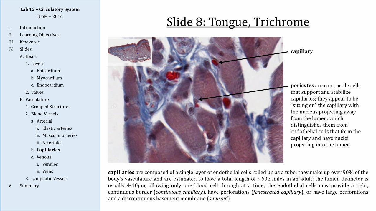
pericytes are contractile cells that support and stabilize capillaries; they appear to be “sitting on” the capillary with the nucleus projecting away from the lumen, which distinguishes them from endothelial cells that form the capillary and have nuclei projecting into the lumen
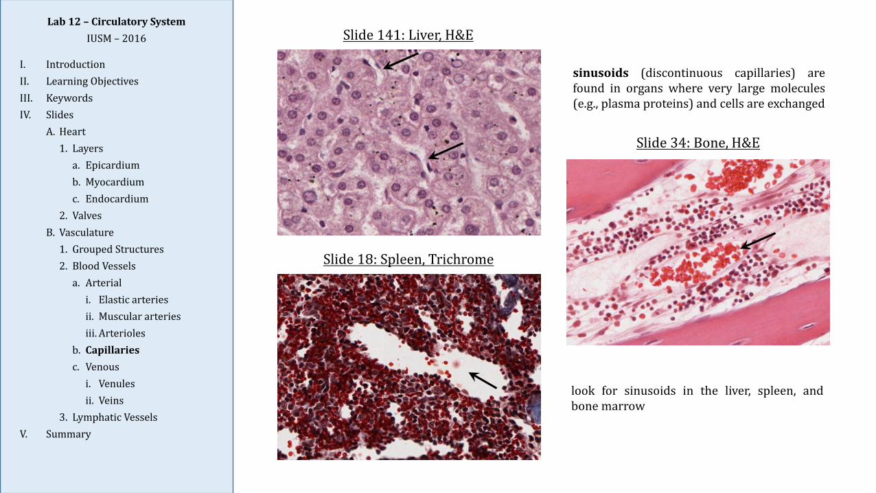
capillaries are composed of a single layer of endothelial cells rolled up as a tube; they make up over 90% of thebody’s vasculature and are estimated to have a total length of ~60k miles in an adult; the lumen diameter isusually 4-10µm, allowing only one blood cell through at a time; the endothelial cells may provide a tight,continuous border (continuous capillary), have perforations (fenestrated capillary), or have large perforationsand a discontinuous basement membrane (sinusoid)
Lab 12 – Circulatory SystemIUSM – 2016
I. IntroductionII. Learning ObjectivesIII. KeywordsIV. Slides
A. Heart1. Layers
a. Epicardiumb. Myocardiumc. Endocardium
2. ValvesB. Vasculature
1. Grouped Structures2. Blood Vessels
a. Arteriali. Elastic arteriesii. Muscular arteriesiii. Arterioles
b. Capillariesc. Venous
i. Venulesii. Veins
3. Lymphatic VesselsV. Summary
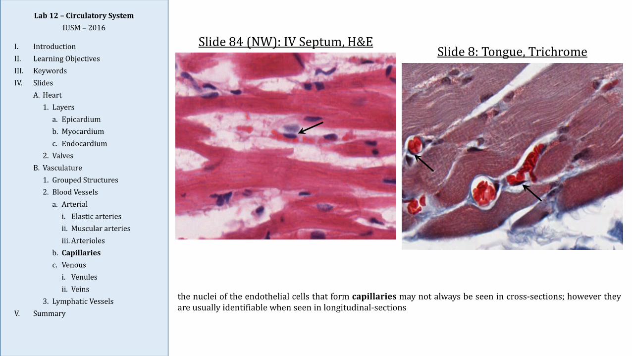
the nuclei of the endothelial cells that form capillaries may not always be seen in cross-sections; however theyare usually identifiable when seen in longitudinal-sections
Slide 84 (NW): IV Septum, H&ESlide 8: Tongue, Trichrome
Lab 12 – Circulatory SystemIUSM – 2016
I. IntroductionII. Learning ObjectivesIII. KeywordsIV. Slides
A. Heart1. Layers
a. Epicardiumb. Myocardiumc. Endocardium
2. ValvesB. Vasculature
1. Grouped Structures2. Blood Vessels
a. Arteriali. Elastic arteriesii. Muscular arteriesiii. Arterioles
b. Capillariesc. Venous
i. Venulesii. Veins
3. Lymphatic VesselsV. Summary
Slide 34: Bone, H&E
Slide 18: Spleen, Trichrome
Slide 141: Liver, H&E
sinusoids (discontinuous capillaries) arefound in organs where very large molecules(e.g., plasma proteins) and cells are exchanged
look for sinusoids in the liver, spleen, andbone marrow
Lab 12 – Circulatory SystemIUSM – 2016
I. IntroductionII. Learning ObjectivesIII. KeywordsIV. Slides
A. Heart1. Layers
a. Epicardiumb. Myocardiumc. Endocardium
2. ValvesB. Vasculature
1. Grouped Structures2. Blood Vessels
a. Arteriali. Elastic arteriesii. Muscular arteriesiii. Arterioles
b. Capillariesc. Venous
i. Venulesii. Veins
3. Lymphatic VesselsV. Summary
Slide 8 (464): Mesentery, H&E
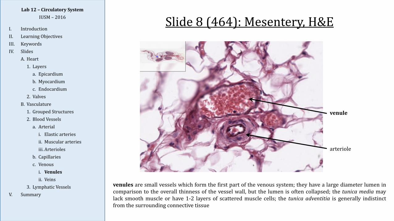
venules are small vessels which form the first part of the venous system; they have a large diameter lumen incomparison to the overall thinness of the vessel wall, but the lumen is often collapsed; the tunica media maylack smooth muscle or have 1-2 layers of scattered muscle cells; the tunica adventitia is generally indistinctfrom the surrounding connective tissue
venule
arteriole
Lab 12 – Circulatory SystemIUSM – 2016
I. IntroductionII. Learning ObjectivesIII. KeywordsIV. Slides
A. Heart1. Layers
a. Epicardiumb. Myocardiumc. Endocardium
2. ValvesB. Vasculature
1. Grouped Structures2. Blood Vessels
a. Arteriali. Elastic arteriesii. Muscular arteriesiii. Arterioles
b. Capillariesc. Venous
i. Venulesii. Veins
3. Lymphatic VesselsV. Summary
Slide 28a (464): Lymph Node, H&E
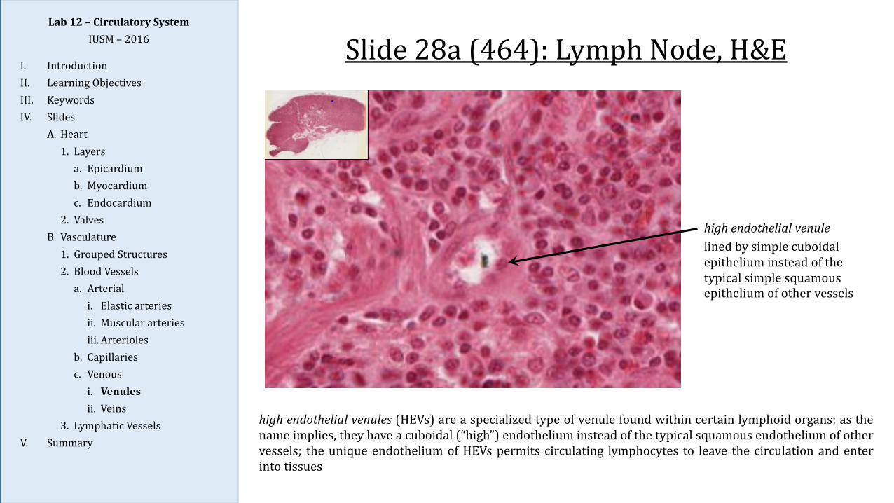
high endothelial venules (HEVs) are a specialized type of venule found within certain lymphoid organs; as thename implies, they have a cuboidal (“high”) endothelium instead of the typical squamous endothelium of othervessels; the unique endothelium of HEVs permits circulating lymphocytes to leave the circulation and enterinto tissues
high endothelial venulelined by simple cuboidal epithelium instead of the typical simple squamous epithelium of other vessels
Lab 12 – Circulatory SystemIUSM – 2016
I. IntroductionII. Learning ObjectivesIII. KeywordsIV. Slides
A. Heart1. Layers
a. Epicardiumb. Myocardiumc. Endocardium
2. ValvesB. Vasculature
1. Grouped Structures2. Blood Vessels
a. Arteriali. Elastic arteriesii. Muscular arteriesiii. Arterioles
b. Capillariesc. Venous
i. Venulesii. Veins
3. Lymphatic VesselsV. Summary
Slide 92: Arteries and Veins, Elastic
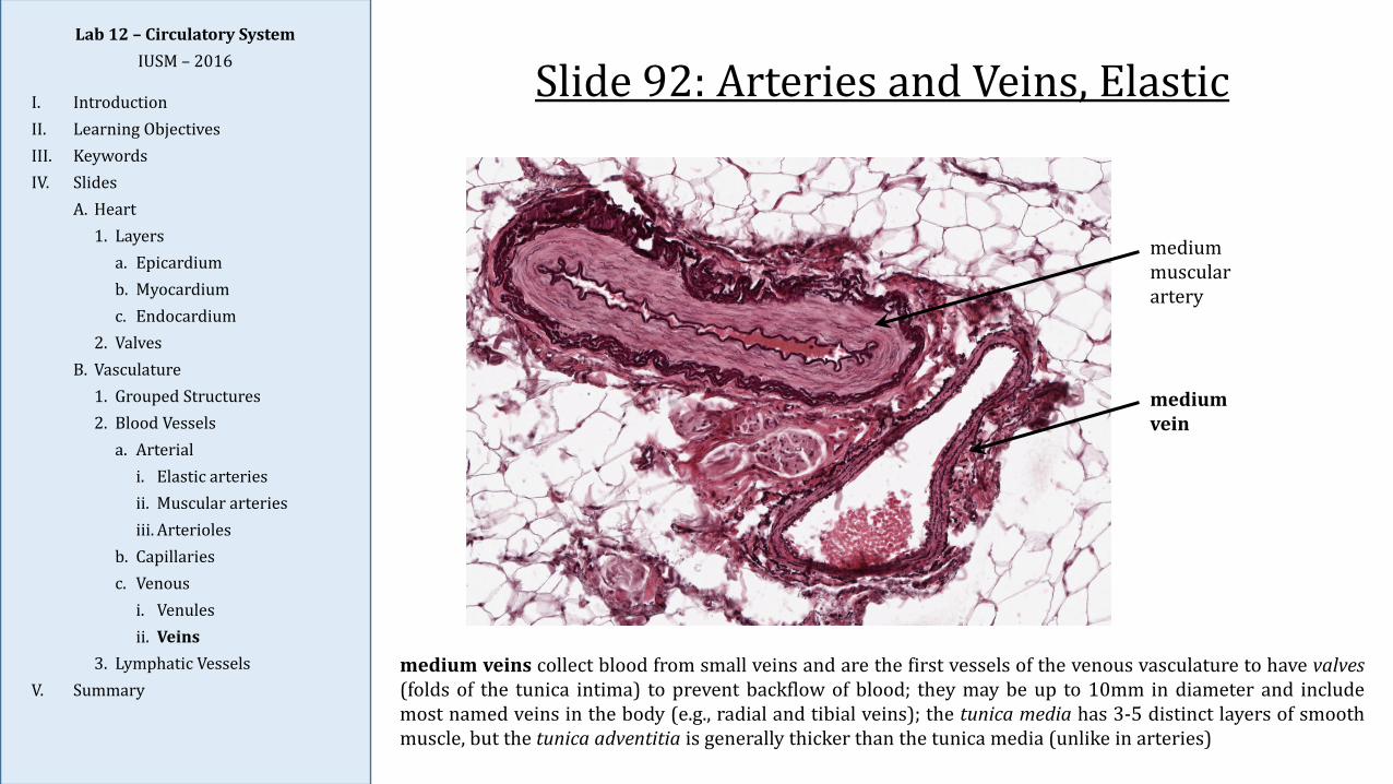
medium veins collect blood from small veins and are the first vessels of the venous vasculature to have valves(folds of the tunica intima) to prevent backflow of blood; they may be up to 10mm in diameter and includemost named veins in the body (e.g., radial and tibial veins); the tunica media has 3-5 distinct layers of smoothmuscle, but the tunica adventitia is generally thicker than the tunica media (unlike in arteries)
medium muscularartery
medium vein
Lab 12 – Circulatory SystemIUSM – 2016
I. IntroductionII. Learning ObjectivesIII. KeywordsIV. Slides
A. Heart1. Layers
a. Epicardiumb. Myocardiumc. Endocardium
2. ValvesB. Vasculature
1. Grouped Structures2. Blood Vessels
a. Arteriali. Elastic arteriesii. Muscular arteriesiii. Arterioles
b. Capillariesc. Venous
i. Venulesii. Veins
3. Lymphatic VesselsV. Summary
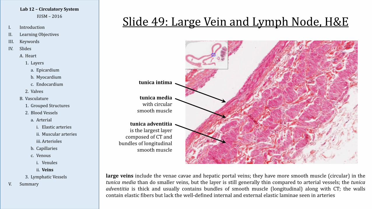
Slide 49: Large Vein and Lymph Node, H&E
large veins include the venae cavae and hepatic portal veins; they have more smooth muscle (circular) in thetunica media than do smaller veins, but the layer is still generally thin compared to arterial vessels; the tunicaadventitia is thick and usually contains bundles of smooth muscle (longitudinal) along with CT; the wallscontain elastic fibers but lack the well-defined internal and external elastic laminae seen in arteries
tunica intima
tunica mediawith circular
smooth muscle
tunica adventitiais the largest layer
composed of CT and bundles of longitudinal
smooth muscle
Lab 12 – Circulatory SystemIUSM – 2016
I. IntroductionII. Learning ObjectivesIII. KeywordsIV. Slides
A. Heart1. Layers
a. Epicardiumb. Myocardiumc. Endocardium
2. ValvesB. Vasculature
1. Grouped Structures2. Blood Vessels
a. Arteriali. Elastic arteriesii. Muscular arteriesiii. Arterioles
b. Capillariesc. Venous
i. Venulesii. Veins
3. Lymphatic VesselsV. Summary
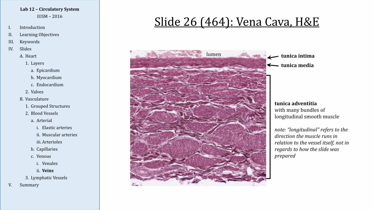
Slide 26 (464): Vena Cava, H&E
tunica intima
tunica media
tunica adventitiawith many bundles of longitudinal smooth muscle
note: “longitudinal” refers to the direction the muscle runs in relation to the vessel itself, not in regards to how the slide was prepared
lumen
Lab 12 – Circulatory SystemIUSM – 2016
I. IntroductionII. Learning ObjectivesIII. KeywordsIV. Slides
A. Heart1. Layers
a. Epicardiumb. Myocardiumc. Endocardium
2. ValvesB. Vasculature
1. Grouped Structures2. Blood Vessels
a. Arteriali. Elastic arteriesii. Muscular arteriesiii. Arterioles
b. Capillariesc. Venous
i. Venulesii. Veins
3. Lymphatic VesselsV. Summary
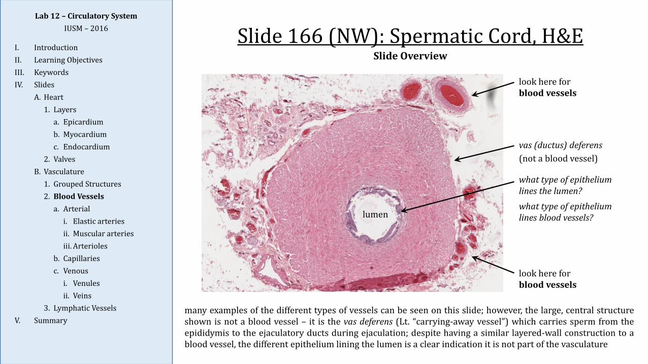
Slide 166 (NW): Spermatic Cord, H&E
many examples of the different types of vessels can be seen on this slide; however, the large, central structureshown is not a blood vessel – it is the vas deferens (Lt. “carrying-away vessel”) which carries sperm from theepididymis to the ejaculatory ducts during ejaculation; despite having a similar layered-wall construction to ablood vessel, the different epithelium lining the lumen is a clear indication it is not part of the vasculature
Slide Overview
what type of epithelium lines the lumen? what type of epithelium lines blood vessels?lumen
vas (ductus) deferens(not a blood vessel)
look here for blood vessels
look here for blood vessels
Lab 12 – Circulatory SystemIUSM – 2016
I. IntroductionII. Learning ObjectivesIII. KeywordsIV. Slides
A. Heart1. Layers
a. Epicardiumb. Myocardiumc. Endocardium
2. ValvesB. Vasculature
1. Grouped Structures2. Blood Vessels
a. Arteriali. Elastic arteriesii. Muscular arteriesiii. Arterioles
b. Capillariesc. Venous
i. Venulesii. Veins
3. Lymphatic VesselsV. Summary
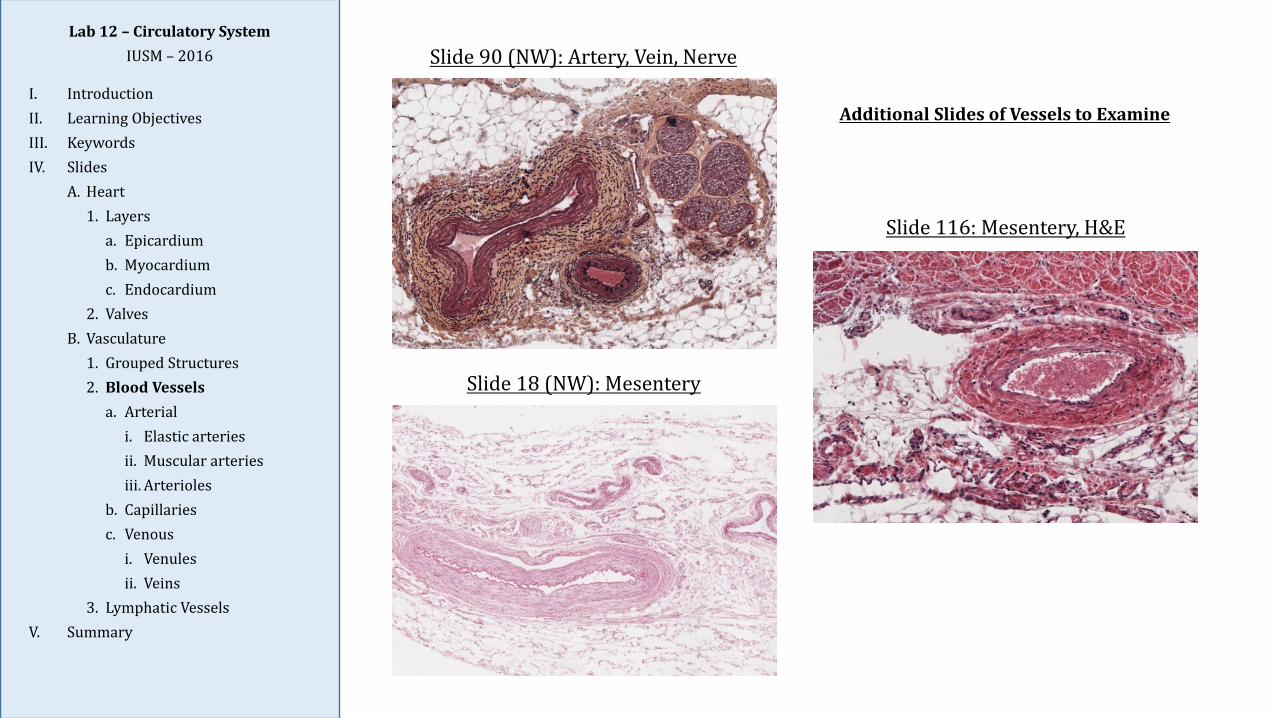
Slide 116: Mesentery, H&E
Slide 18 (NW): Mesentery
Slide 90 (NW): Artery, Vein, Nerve
Additional Slides of Vessels to Examine
Lab 12 – Circulatory SystemIUSM – 2016
I. IntroductionII. Learning ObjectivesIII. KeywordsIV. Slides
A. Heart1. Layers
a. Epicardiumb. Myocardiumc. Endocardium
2. ValvesB. Vasculature
1. Grouped Structures2. Blood Vessels
a. Arteriali. Elastic arteriesii. Muscular arteriesiii. Arterioles
b. Capillariesc. Venous
i. Venulesii. Veins
3. Lymphatic VesselsV. Summary
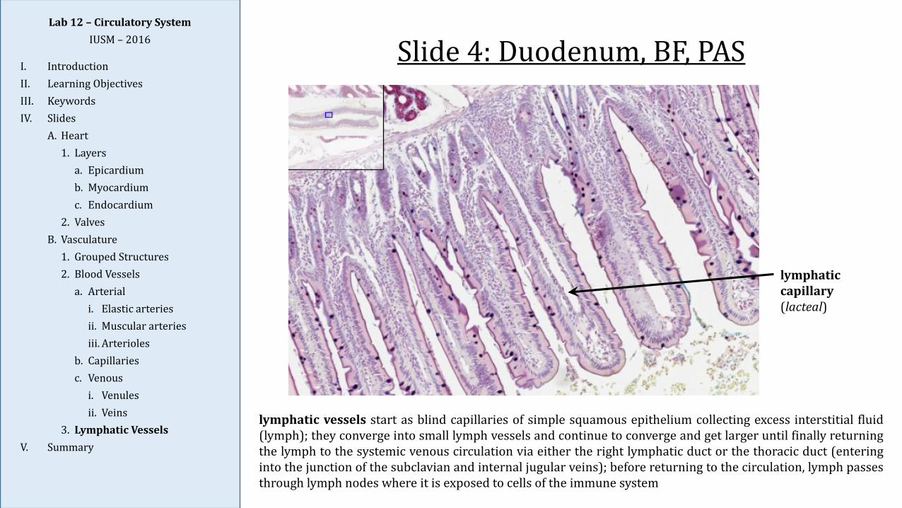
Slide 4: Duodenum, BF, PAS
lymphatic capillary(lacteal)
lymphatic vessels start as blind capillaries of simple squamous epithelium collecting excess interstitial fluid(lymph); they converge into small lymph vessels and continue to converge and get larger until finally returningthe lymph to the systemic venous circulation via either the right lymphatic duct or the thoracic duct (enteringinto the junction of the subclavian and internal jugular veins); before returning to the circulation, lymph passesthrough lymph nodes where it is exposed to cells of the immune system
Lab 12 – Circulatory SystemIUSM – 2016
I. IntroductionII. Learning ObjectivesIII. KeywordsIV. Slides
A. Heart1. Layers
a. Epicardiumb. Myocardiumc. Endocardium
2. ValvesB. Vasculature
1. Grouped Structures2. Blood Vessels
a. Arteriali. Elastic arteriesii. Muscular arteriesiii. Arterioles
b. Capillariesc. Venous
i. Venulesii. Veins
3. Lymphatic VesselsV. Summary
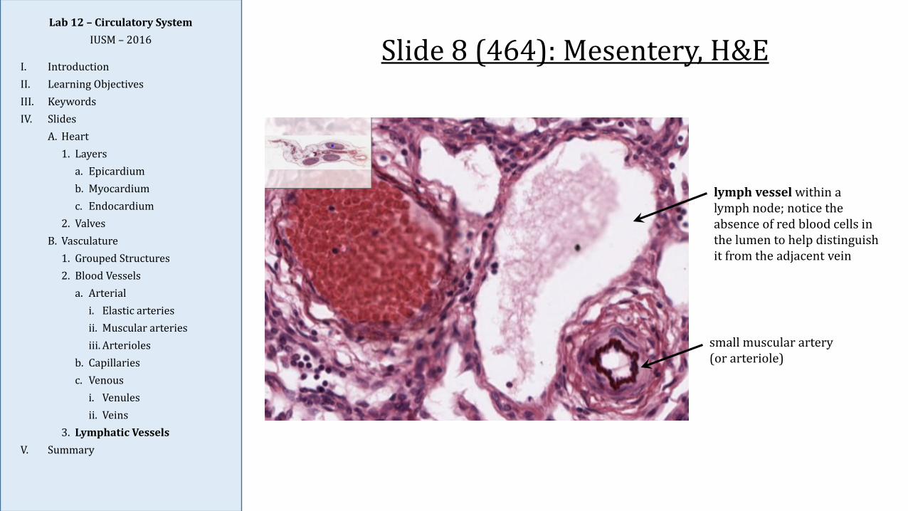
Slide 8 (464): Mesentery, H&E
lymph vessel within a lymph node; notice the absence of red blood cells in the lumen to help distinguish it from the adjacent vein
small muscular artery(or arteriole)
Lab 12 – Circulatory SystemIUSM – 2016
I. IntroductionII. Learning ObjectivesIII. KeywordsIV. Slides
A. Heart1. Layers
a. Epicardiumb. Myocardiumc. Endocardium
2. ValvesB. Vasculature
1. Grouped Structures2. Blood Vessels
a. Arteriali. Elastic arteriesii. Muscular arteriesiii. Arterioles
b. Capillariesc. Venous
i. Venulesii. Veins
3. Lymphatic VesselsV. Summary
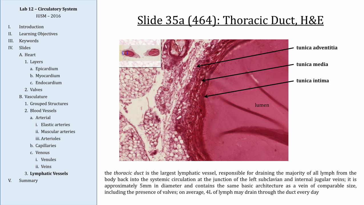
Slide 35a (464): Thoracic Duct, H&E
lumen
tunica adventitia
tunica media
tunica intima
the thoracic duct is the largest lymphatic vessel, responsible for draining the majority of all lymph from thebody back into the systemic circulation at the junction of the left subclavian and internal jugular veins; it isapproximately 5mm in diameter and contains the same basic architecture as a vein of comparable size,including the presence of valves; on average, 4L of lymph may drain through the duct every day
Lab 12 – Circulatory SystemIUSM – 2016
I. IntroductionII. Learning ObjectivesIII. KeywordsIV. Slides
A. Heart1. Layers
a. Epicardiumb. Myocardiumc. Endocardium
2. ValvesB. Vasculature
1. Grouped Structures2. Blood Vessels
a. Arteriali. Elastic arteriesii. Muscular arteriesiii. Arterioles
b. Capillariesc. Venous
i. Venulesii. Veins
3. Lymphatic VesselsV. Summary
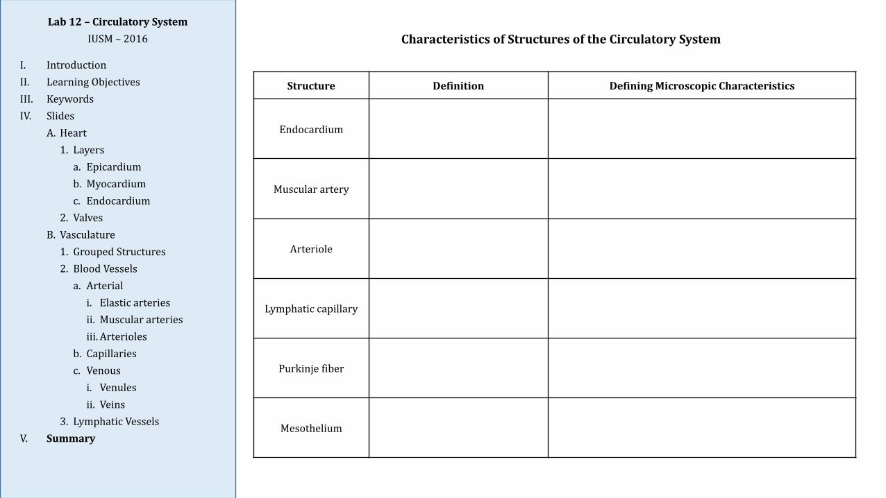
Structure Definition Defining Microscopic Characteristics
Endocardium
Muscular artery
Arteriole
Lymphatic capillary
Purkinje fiber
Mesothelium
Characteristics of Structures of the Circulatory System