circulatory diseases and treatments practicum i 2011-2012
TRANSCRIPT
Circulatory Diseases and Treatments
Practicum I2011-2012
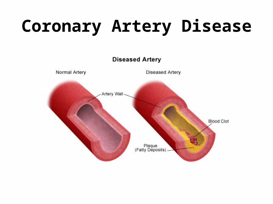
Coronary Artery Disease

Coronary Arteries
Supply the heart muscle (myocardium)
with oxygenated
blood.
Coronary Artery Disease
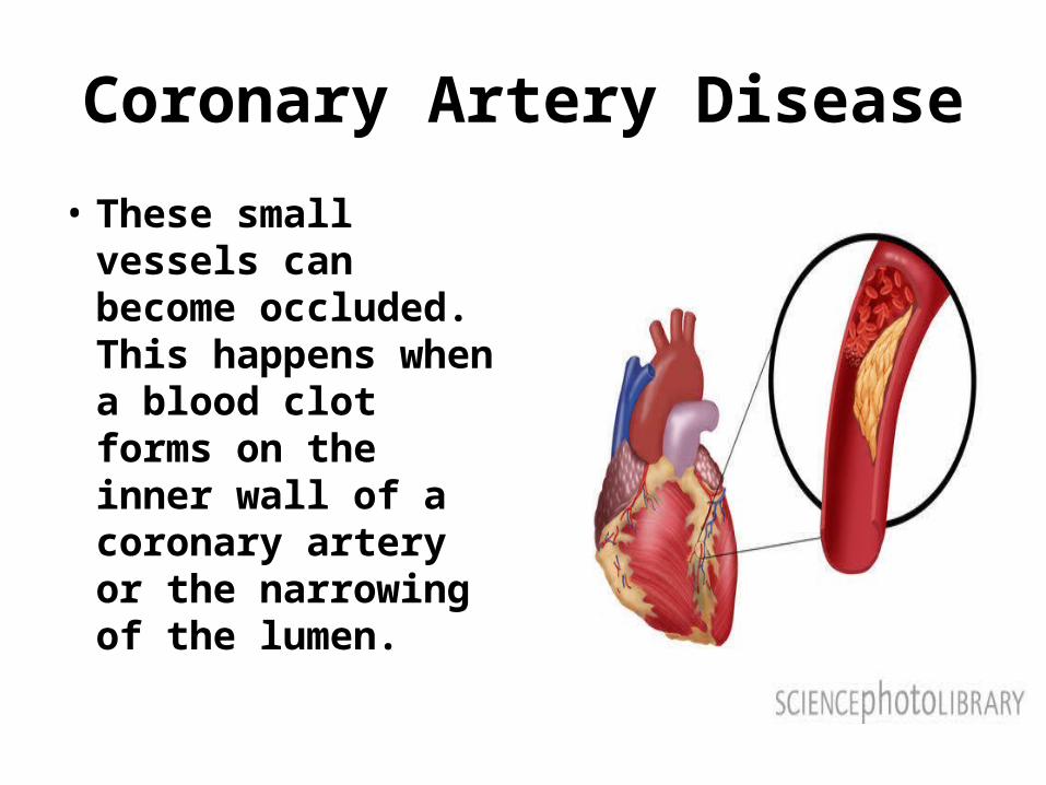
• These small vessels can become occluded. This happens when a blood clot forms on the inner wall of a coronary artery or the narrowing of the lumen.
Coronary Artery Disease
• Result of atherosclerosis.• The fatty plaque first causes plugging of
the artery. • Next, the roughened lining may rupture or
cause abnormal clotting of the blood leading to a thrombotic occlusion.


Coronary Artery Disease
• Blood flow is then decreased (ischemia) or stopped completely.
• Lack of oxygen leads to tissue death (necrosis) of the myocardium. The area of necrosis is called an infarct. (MI=mycardial infarction=heart attack)

Coronary Artery Disease
• Arteriosclerosis is hardening of the arteries. Artery walls become thick and loose their elasticity.
• Can be a result of aging and chronic hypertension.

Coronary Artery Disease
• High Blood Pressure• High Cholesterol/Obesity• Smoking• Diabetes• Genetics has a role• Stress• Aging

Signs and Symptoms
• Angina• Shortness Of Breath
(SOB)• Heart attack (crushing
pain, diaphoresis, nausea, pallor, light headedness.
• Fatigue• Lack of energy
Diagnosis
• History (medical past, symptoms, risk factors). Also current medications, vital sign recordings, and previous surgeries.
• Physical exam (listen for murmurs, abnormal pulse, skin color and temp, swelling.)
• Vital signs (BP, HR, RR, Temp)
Diagnosis
• EKG/ECG• Chest x-ray• Angiography/Cath lab• Stress test• Echo (EF%)• Normal EF is 50-70%• Average is 58%• Labs: Cardiac Enzymes,
Lipid panels, BNP.


Acute Coronary Syndromes (ACSs)
• Conditions caused by myocardial ischemia. (Temporary oxygen insufficiency). MI is when the myocardium is suddenly completely deprived of oxygen.
• Some heart muscle cells die and are replaced by scar tissue.
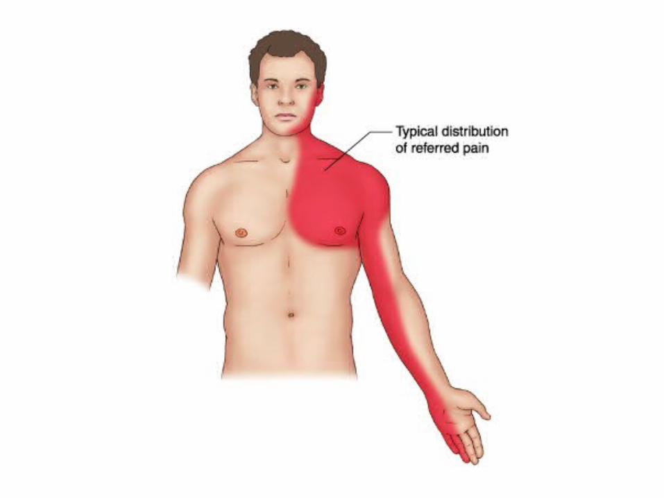
• Symptoms include angina (chest pain) which may radiate to the neck, jaw, and left arm.
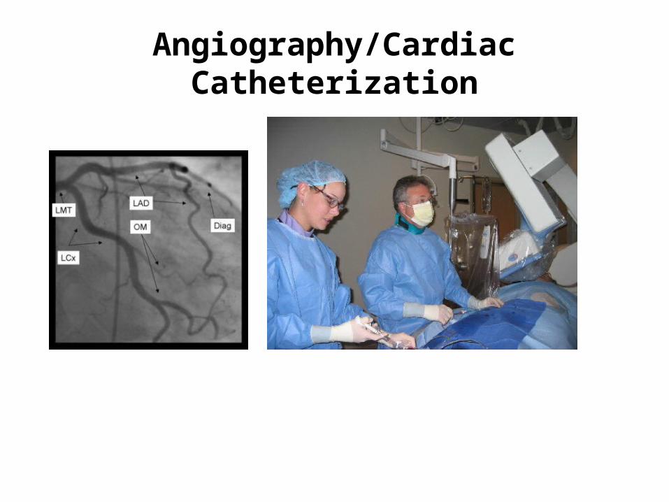
Angiography/Cardiac Catheterization
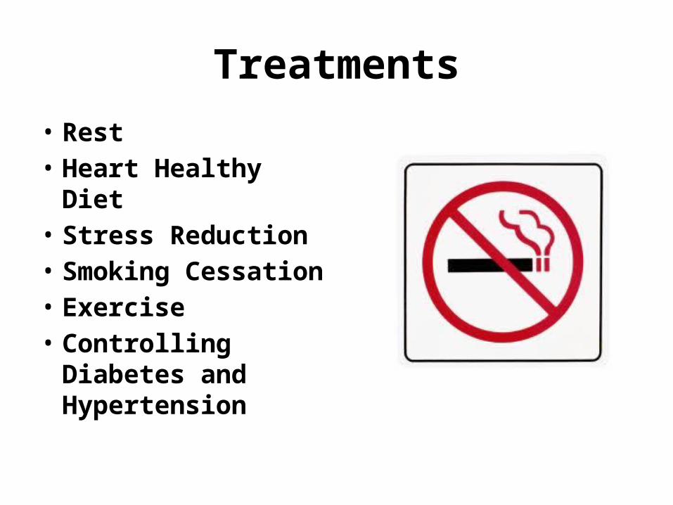
Treatments
• Rest• Heart Healthy Diet• Stress Reduction• Smoking Cessation• Exercise• Controlling Diabetes
and Hypertension

Statins
• Cholesterol-lowering drugs. They block the enzyme in the liver responsible for making cholesterol.
• Simvastatin (Zocor)• Atorvastatin (Lipitor)• Rosuvastatin (Crestor)
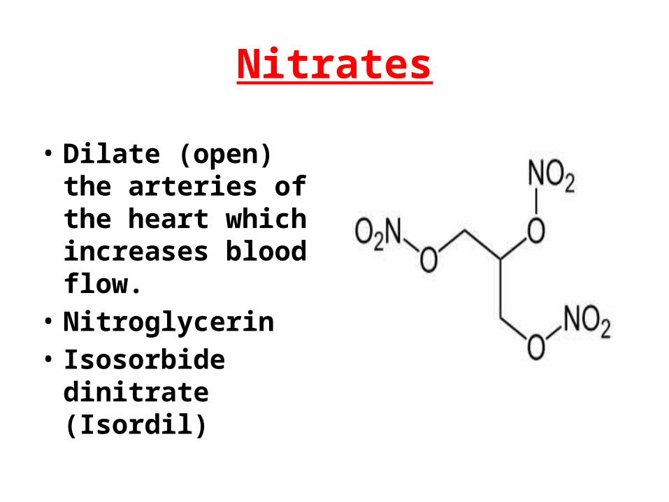
Nitrates
• Dilate (open) the arteries of the heart which increases blood flow.
• Nitroglycerin• Isosorbide dinitrate
(Isordil)

Thrombolytics
• Thrombolytics are clot busting drugs.• tPA (alteplase)• Urokinase• Streptokinase
• These must be used in first few hours of an MI.
Aspirin and Plavix
• Aspirin is given to help prevent clot formation. It doesn’t break up clots.
• If a patient is intolerant of aspirin (ASA) clopidogrel (Plavix) is prescribed.
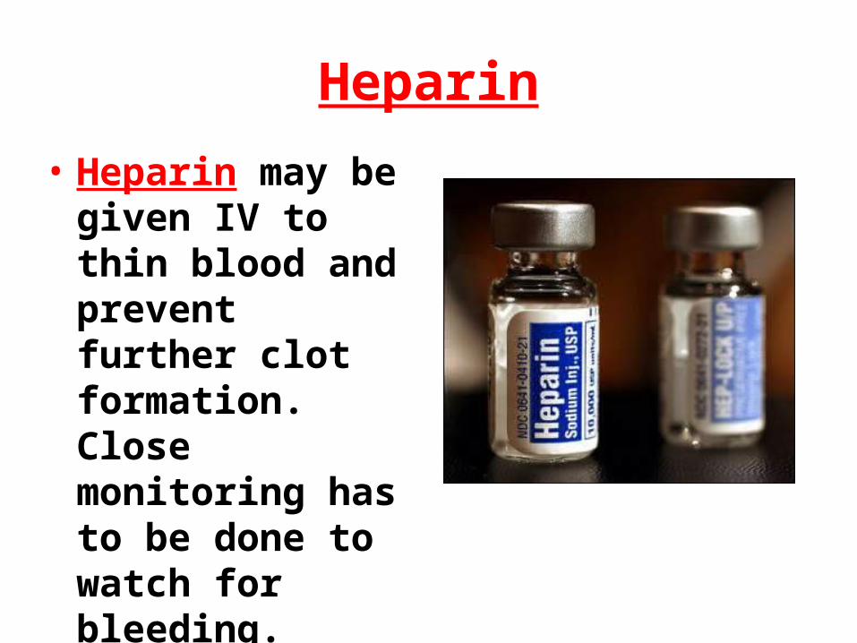
Heparin
• Heparin may be given IV to thin blood and prevent further clot formation. Close monitoring has to be done to watch for bleeding.
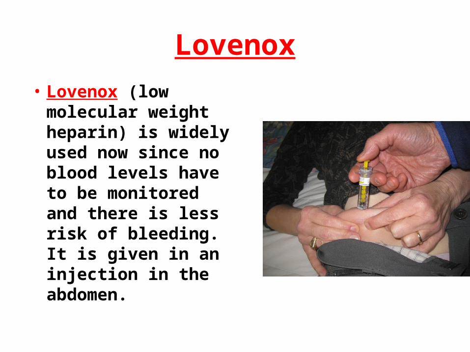
Lovenox
• Lovenox (low molecular weight heparin) is widely used now since no blood levels have to be monitored and there is less risk of bleeding. It is given in an injection in the abdomen.

ACE inhibitors (angiotension-converting enzyme)
• Angiotension in a hormone produced by the liver. It causes vasoconstriction which increases blood pressure.
• These medications decrease production of angiotension converting enzyme.
• Captopril• Lisinopril

Beta Blockers
• Improve the heart's ability to relax, decrease the production of harmful substances produced by the body in response to heart failure, and slow the heart rate. Over time, beta-blockers improve the heart's pumping ability.
• Block beta-adrenergic receptors, preventing adrenaline (epinephrine) from stimulating these receptors.
• Metoprolol• Propranolol• Carvedilol

Calcium Channel Blockers
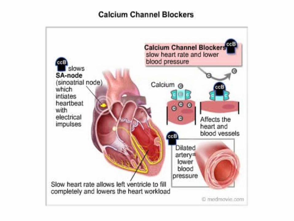
• Blocks the entry of calcium into the muscle cells of the heart and the arteries. It is the entry of calcium into these cells that causes the heart to contract and arteries to narrow. By blocking the entry of calcium, calcium channel blocker decrease the contraction of the heart and dilate (widen) the arteries.
Calcium Channel Blockers
By dilating the arteries, CCBs reduce the pressure in the arteries. This makes it easier for the heart to pump blood, and, as a result, the heart needs less oxygen. By reducing the heart's need for oxygen, CCBs prevent or relieve angina
• Nicardipine (Cardene)• Amlodipine (Norvasc)• Nifedipine (Procardia)• Diltiazem (Cardizem)

Treatments
• Percutaneous Coronary Intervention (PCI).
• Types of (Balloon Angioplasty, Stents)
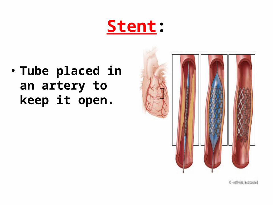
Stent:
• Tube placed in an artery to keep it open.

Coronary Artery Bypass Graft
• Surgical procedure in which one or more blocked coronary arteries are bypassed by a blood vessel graft to restore normal blood flow to the heart. These grafts usually come from the patient’s own arteries and veins located in the chest (thoracic), leg (saphenous) or arm (radial). The graft goes around the blocked artery (or arteries) to create new pathways for blood to flow to the heart.
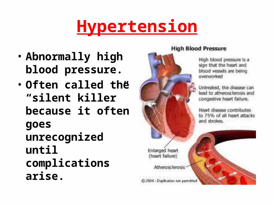
Hypertension
• Abnormally high blood pressure.
• Often called the “silent killer” because it often goes unrecognized until complications arise.
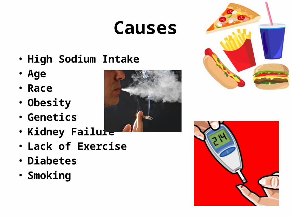
Causes
• High Sodium Intake• Age• Race• Obesity• Genetics• Kidney Failure• Lack of Exercise• Diabetes • Smoking

Signs and Symptoms
• Headache• Dizziness• Nausea• Blurred vision• Chest pain• Shortness of breath• Flushing• Nose bleeds
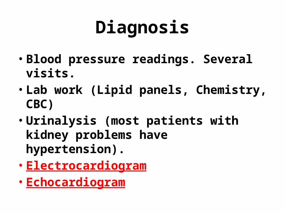
Diagnosis
• Blood pressure readings. Several visits.• Lab work (Lipid panels, Chemistry, CBC)• Urinalysis (most patients with kidney
problems have hypertension).• Electrocardiogram• Echocardiogram
Treatment
• Lifestyle changes: low sodium, low fat diet; smoking cessation, exercise, stress reduction, weight loss.
• Medications: Diuretics (water pills), Beta blockers, ACE inhibitors, Calcium channel blockers.
• Treat underlying problem.
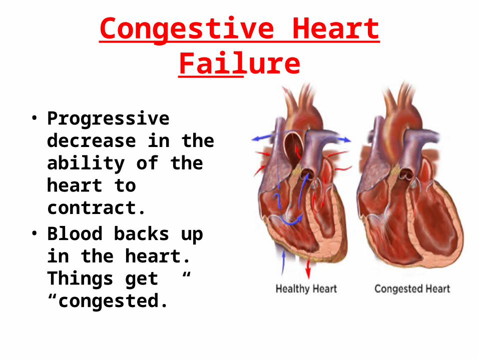
Congestive Heart Failure
• Progressive decrease in the ability of the heart to contract.
• Blood backs up in the heart. Things get “congested.”
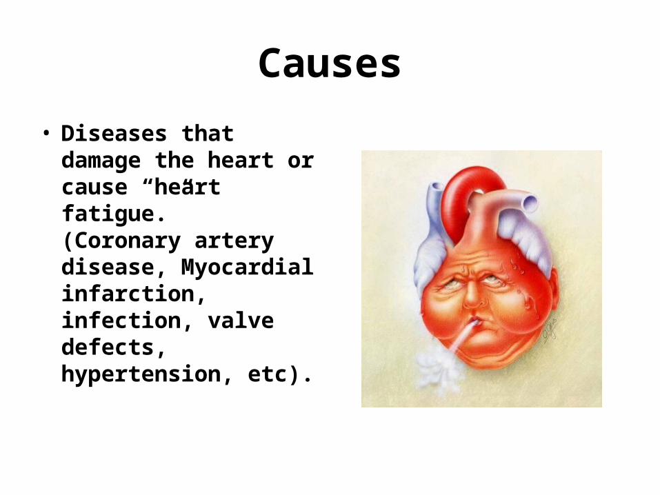
Causes
• Diseases that damage the heart or cause “heart fatigue.” (Coronary artery disease, Myocardial infarction, infection, valve defects, hypertension, etc).
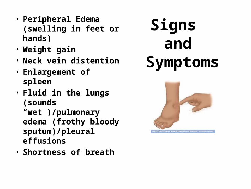
Signs and
Symptoms
• Peripheral Edema (swelling in feet or hands)
• Weight gain• Neck vein distention• Enlargement of spleen• Fluid in the lungs
(sounds “wet”)/pulmonary edema (frothy bloody sputum)/pleural effusions
• Shortness of breath

Diagnosis
• Assessment• Chest X-Ray• BNP – Lab test• Electrocardiogram• Echocardiogram
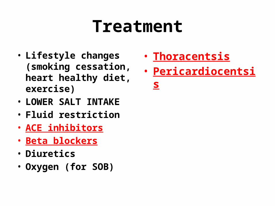
Treatment
• Lifestyle changes (smoking cessation, heart healthy diet, exercise)
• LOWER SALT INTAKE• Fluid restriction• ACE inhibitors• Beta blockers• Diuretics• Oxygen (for SOB)
• Thoracentsis• Pericardiocentsis
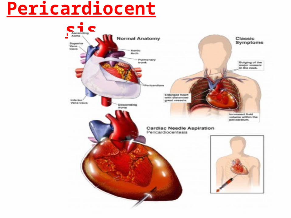
Pericardiocentsis

Thoracentsis
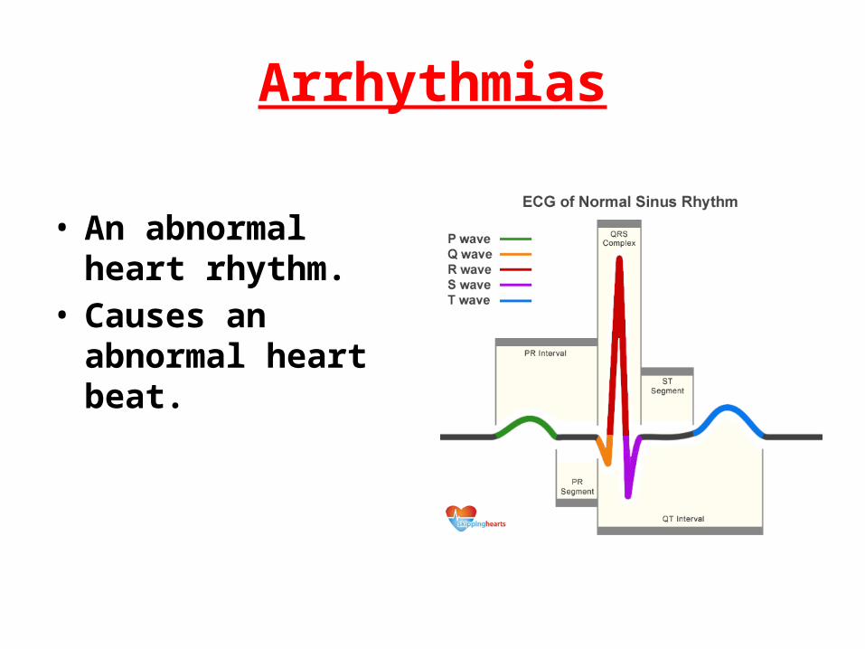
Arrhythmias
• An abnormal heart rhythm.
• Causes an abnormal heart beat.

Causes
• Myocardial Infarction
• Electrolyte imbalance
• Drugs• Electrical Shock• Congenital• Idiopathic

Signs and Symptoms
• Palpitations• Dizziness• Irregular Pulse• Shortness of breath• Weakness• Fatigue• Chest pain

Diagnosis
• ECG• Lab work
(chemistry sets)• Stress Tests• Holter monitor

Greater than 100bpm= Tachycardia

Less than 60bpm = Bradycardia
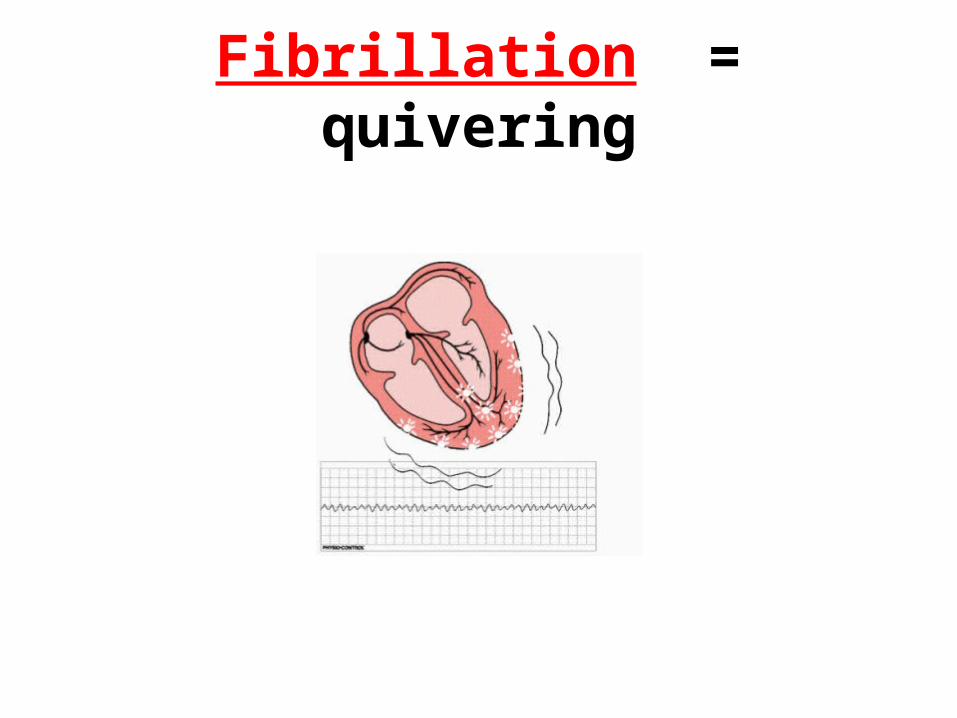
Fibrillation = quivering
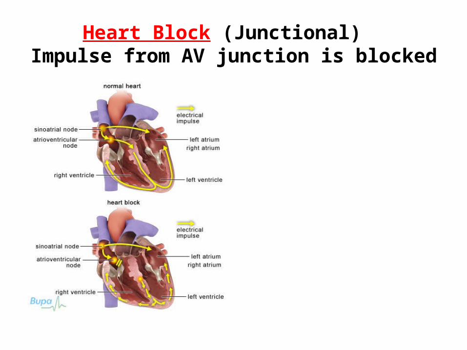
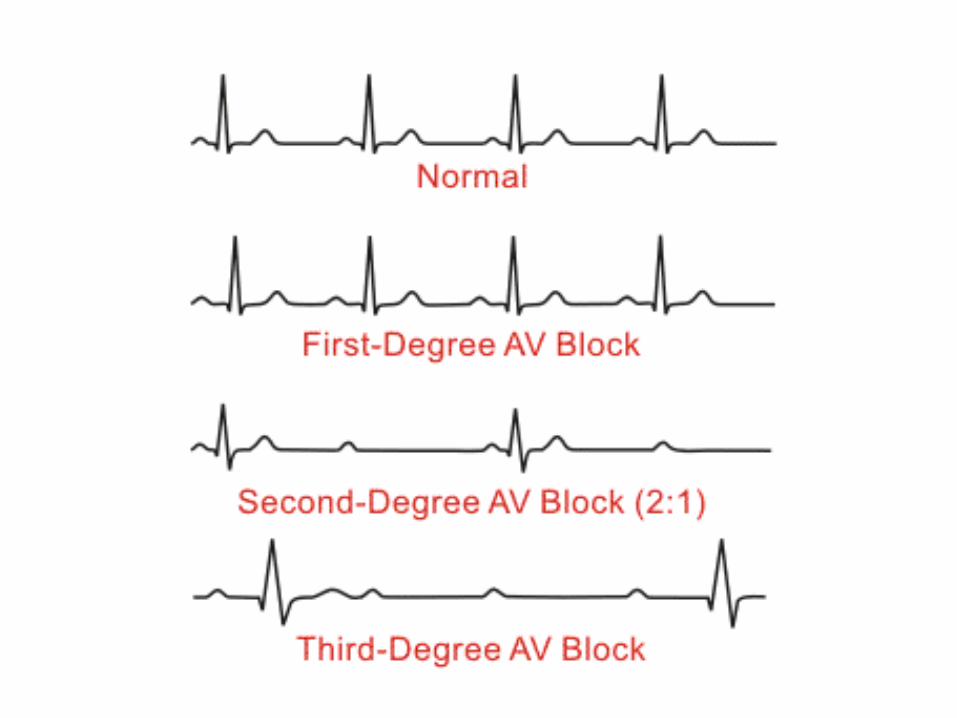
Heart Block (Junctional) Impulse from AV junction is blocked
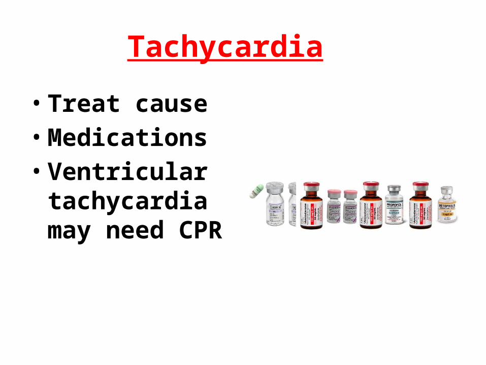
Tachycardia
• Treat cause• Medications• Ventricular
tachycardia may need CPR

Bradycardia
• Medications• Epinephrine• Pacing (Implanted,
transcutaneous, and transvenous)

Fibrillation
• Cardioversion• Defibrillation• Medication• Treat underlying
cause

AICD (Automated Implantable Cardiac Defibrillation)

Cardioversion

Congenital Heart DiseaseFetal Circulation


Fetal Heart Development
Congenital Heart Defects
• Structural problems with the heart, which are present at birth. They result when a mishap occurs during heart development soon after conception
Septal Defects
• Most common congenital heart defects.• An opening in the septum that separates the right
and the left side. This allows for mixing of oxygenated and deoxygenated blood. This can stress the heart as it compensates for lower oxygen levels.
• Left side of heart has greater pressures than right side. If there is a defect blood is generally shunted from left (oxygenated) to right (deoxygenated).

Cyanosis
• If the pressure becomes greater in the right side of the heart, blood may be shunted from right to left causing cyanosis (blue color in the tissues).
• Deoxygenated blood is now being pumped out to the body.
Tetralogy of Fallot
• The classic form of tetralogy includes four related defects of the heart and its major blood vessels:
• Ventricular septal defect (hole between the right and left ventricles)
• Narrowing of the pulmonary outflow tract (the valve and artery that connect the heart with the lungs)
• Overriding aorta (the artery that carries oxygen-rich blood to the body) that is shifted over the right ventricle and ventricular septal defect, instead of coming out only from the left ventricle
• A thickened muscular wall of the right ventricle (right ventricular hypertrophy)


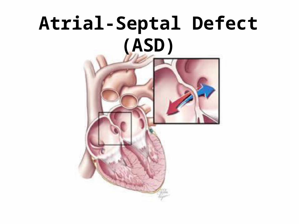
Atrial-Septal Defect (ASD)
• In fetal circulation, there is normally an opening between the two atria (the upper chambers of the heart) to allow blood to bypass the lungs. This opening usually closes around the time the baby is born.
Atrial-Septal Defect (ASD)
• If the ASD is persistent, blood continues to flow from the left to the right atria. This is called a shunt. If too much blood moves to the right side of the heart, pressures in the lungs build up. The shunt can be reversed so that blood flows from right to left. Small atrial septal defects often cause very few problems and may be found much later in life. Many problems can occur if the shunt is large, however. In advanced and severe cases with large shunts the increased pressure on the right side of the heart would result in reversal of blood flow (now from right to left). This usually results in significant shortness of breath.
Atrial-Septal Defect (ASD)
Ventricular-Septal Defect (VSD)
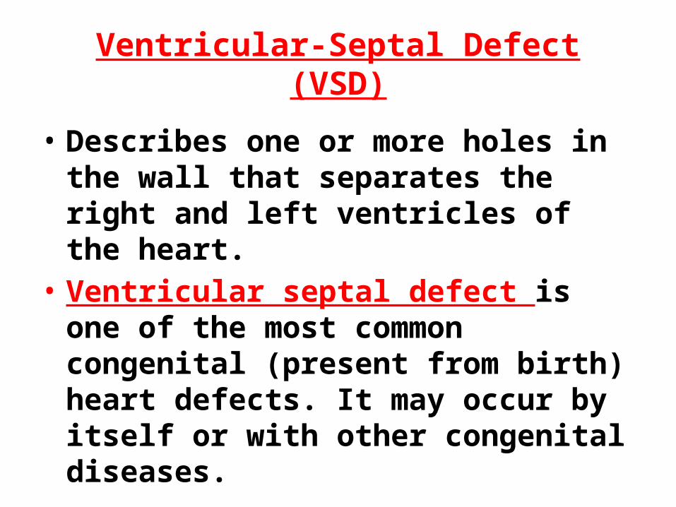
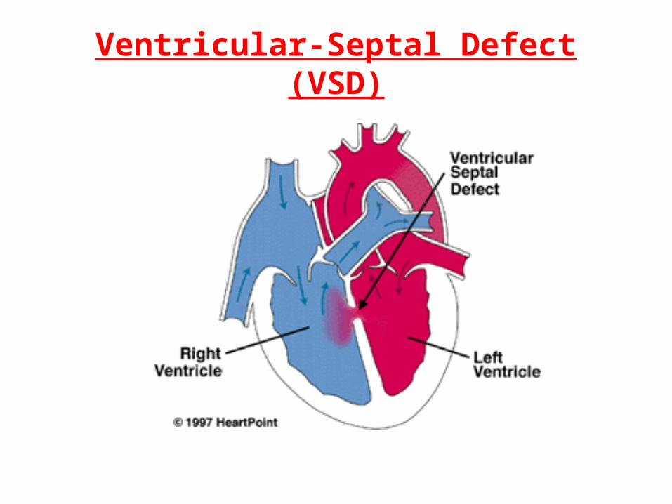
• Describes one or more holes in the wall that separates the right and left ventricles of the heart.
• Ventricular septal defect is one of the most common congenital (present from birth) heart defects. It may occur by itself or with other congenital diseases.
Ventricular-Septal Defect (VSD)
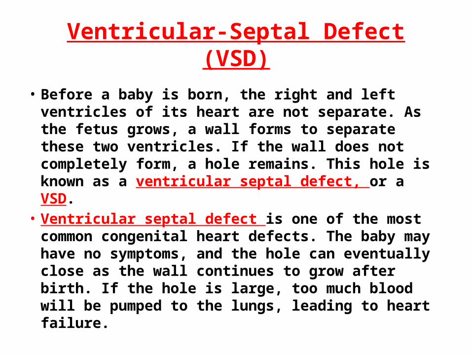
• Before a baby is born, the right and left ventricles of its heart are not separate. As the fetus grows, a wall forms to separate these two ventricles. If the wall does not completely form, a hole remains. This hole is known as a ventricular septal defect, or a VSD.
• Ventricular septal defect is one of the most common congenital heart defects. The baby may have no symptoms, and the hole can eventually close as the wall continues to grow after birth. If the hole is large, too much blood will be pumped to the lungs, leading to heart failure.
Ventricular-Septal Defect (VSD)

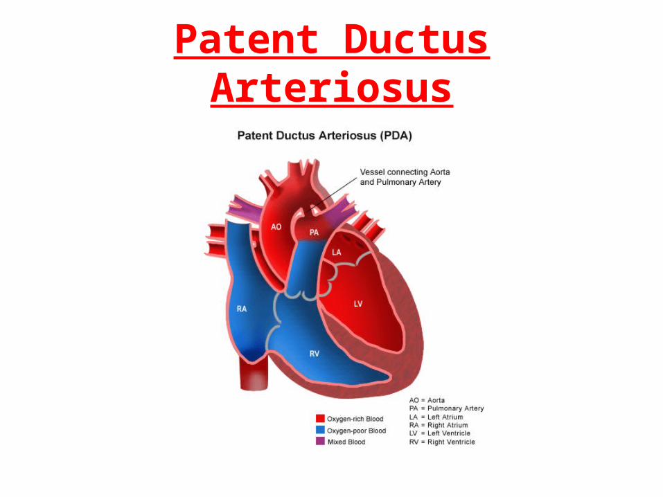
Patent Ductus Arteriosus (PDA)
• A condition in which the ductus arteriosus does not close.
• The ductus arteriosus is a blood vessel that allows blood to go around the baby's lungs before birth. Soon after the infant is born and the lungs fill with air, the ductus arteriosus is no longer needed. It usually closes in a couple of days after birth.
• PDA leads to abnormal blood flow between the aorta and pulmonary artery, two major blood vessels that carry blood from the heart.
Patent Ductus Arteriosus

Transposition of the Arteries
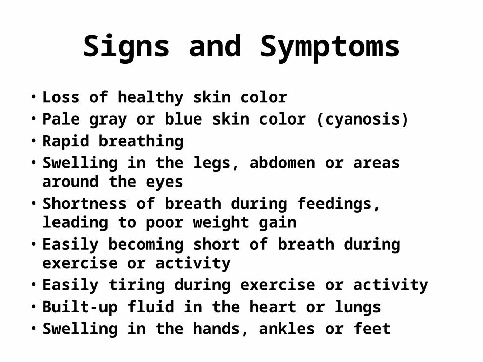
Signs and Symptoms
• Loss of healthy skin color• Pale gray or blue skin color (cyanosis)• Rapid breathing• Swelling in the legs, abdomen or areas around the eyes• Shortness of breath during feedings, leading to poor
weight gain• Easily becoming short of breath during exercise or
activity• Easily tiring during exercise or activity• Built-up fluid in the heart or lungs• Swelling in the hands, ankles or feet

Diagnosis
• Echocardiogram• Chest X-ray• Heart catheterization• Pulse Oximeter • Electrocardiogram

Treatment
• Heart Catheterization• Surgery• Cardiac Transplant• Medications to reduce heart workload

Aneurysms
• A weakening in the wall of a blood vessel.
• It will cause local dilation.
• Most common in the abdominal aorta and brain.
• The danger in aneurysms is they may increase in size and could rupture.
Aortic Dissection
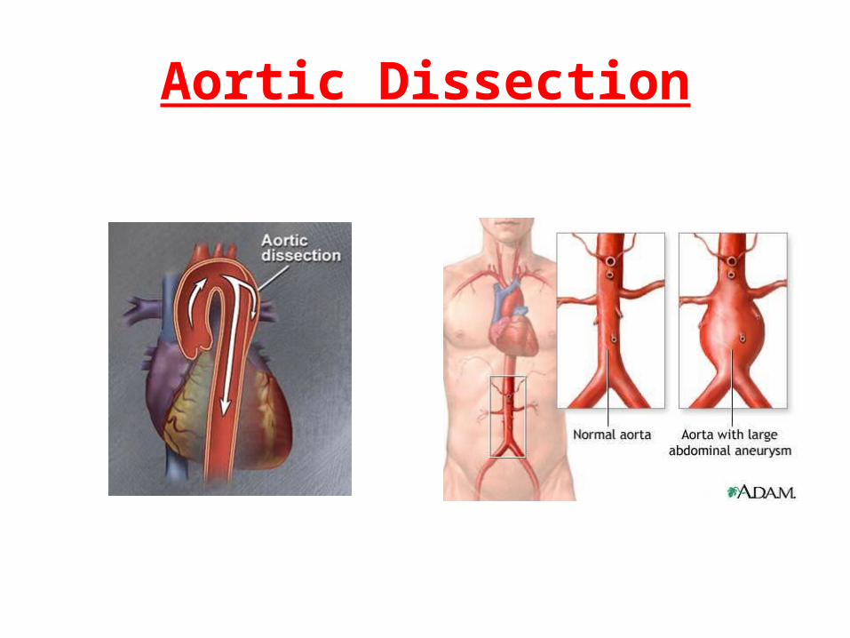
Aortic Dissection
• Aortic dissection is a potentially life-threatening condition in which there is bleeding into and along the wall of the aorta, the major artery carrying blood out of the heart.
Aortic Dissection
• When it leaves the heart, the aorta first moves up through the chest toward the head (the ascending aorta). It then bends or arches, and finally moves down through the chest and abdomen (the descending aorta).
• Aortic dissection most often occurs because of a tear or damage to the inner wall of the aorta.
• This usually occurs in the thoracic (chest) portion of the artery, but may also occur in the abdominal portion.

Causes
• The force of blood pushing against the walls of an artery combined with damage or injury to the artery’s walls can cause an aneurysm.
• Examples: Aging, smoking, hypertension.

Signs and Symptoms
• Typically none.• If in the brain, may exhibit stroke like
symptoms.
Diagnosis
• X-ray• Ultrasound• CT• MRI• Angiography

Treatment
• Surgical repair• Medications to lower blood pressure and
relax vessels (most common are beta blockers and calcium channel blockers)

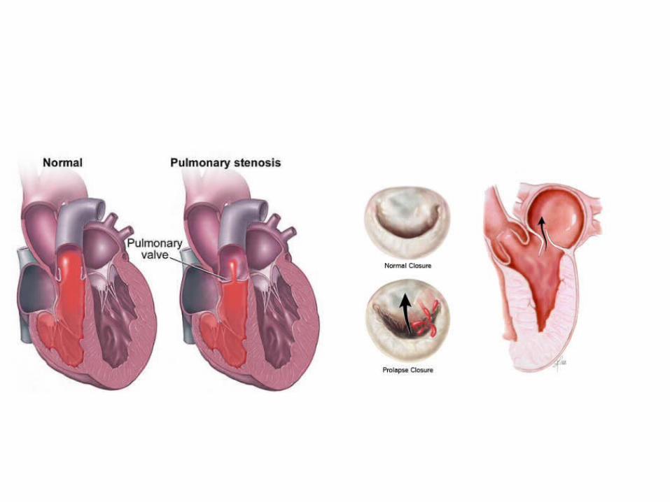
Valve Disorders
• Valves maintain unidirectional (one way) flow of blood through the heart. If it isn’t working correctly, blood may flow the wrong way.
• Valve disorders include stenosis (narrowing) and prolapsed (insufficiency).

Causes
• Congenital• Vegetations caused by Rheumatic Fever or
Endocarditis
Signs and Symptoms
• SOB• Weakness• Palpitations• Edema• Weight gain• Chest pain

Diagnosis
• Murmur on auscultation• Echocardiogram• Heart Catheterization• MRI

Treatment
• If severe enough, valvular replacement
• Heart medications
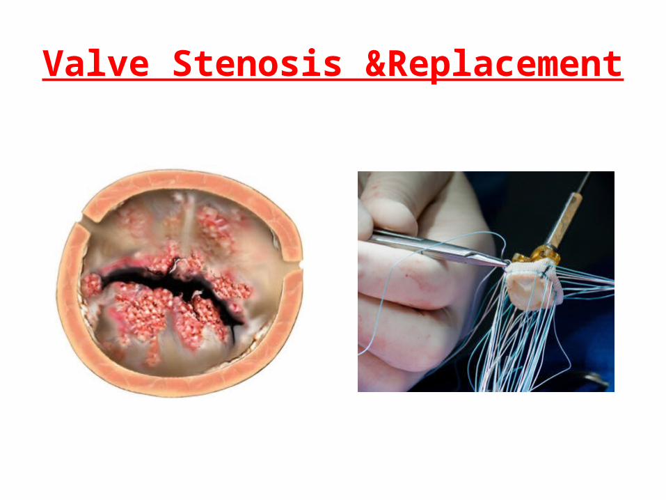
Valve Stenosis &Replacement

Valve Replacement

Carditis
• Inflammation of the heart
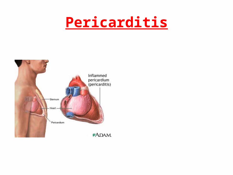
Pericarditis
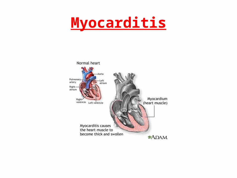
Myocarditis
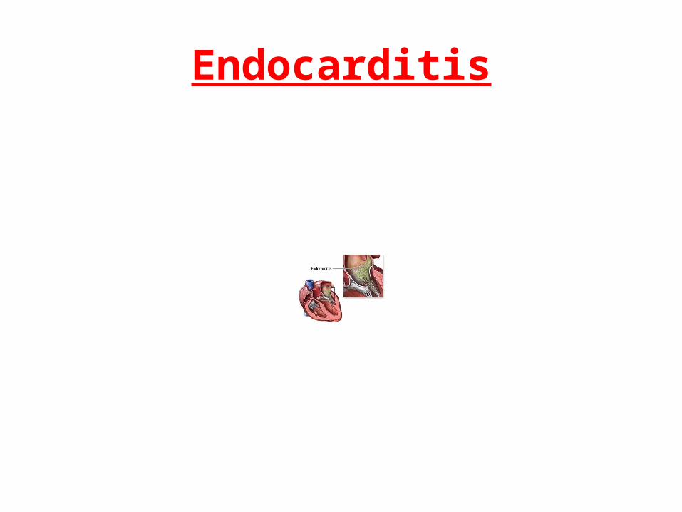
Endocarditis

Causes of Carditis
• Infections (viral and bacterial)• Lupus
Signs and Symptoms
• Chest pain• SOB (shortness of breath)• Fatigue• Edema
Diagnosis
• History• ECG• CXR• Echo
Treatment
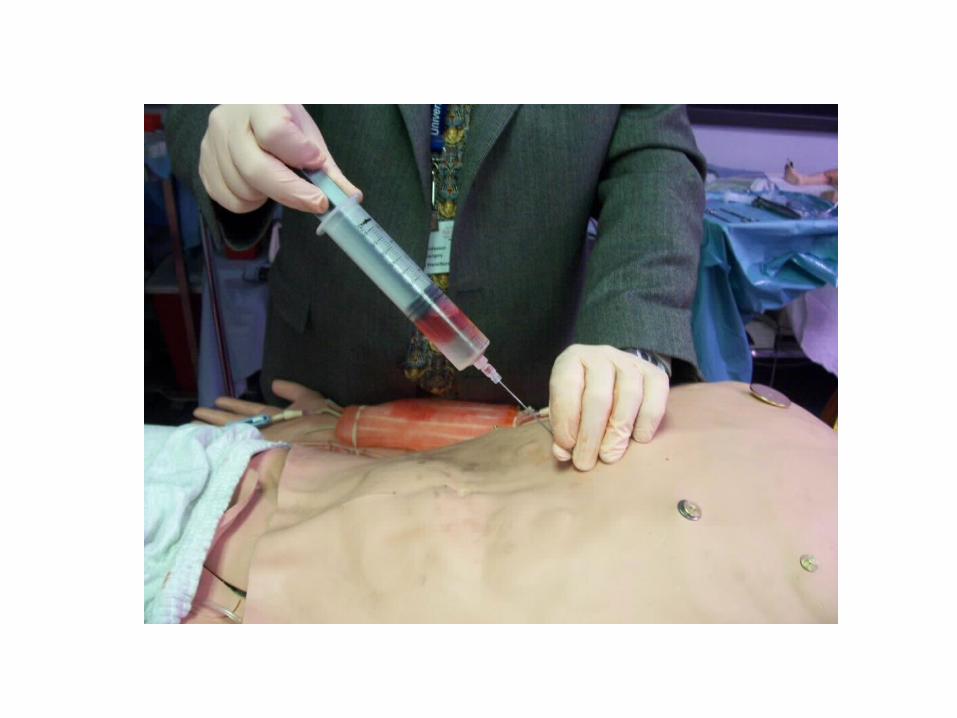
• Antibiotics• Centsis (for effusions)

Cardiac Needle Aspiration
Cardiogenic Shock
• Inadequate pumping of blood due to low blood pressure.
• “Global hypoperfusion”• Decreased pumping ability of the
heart.
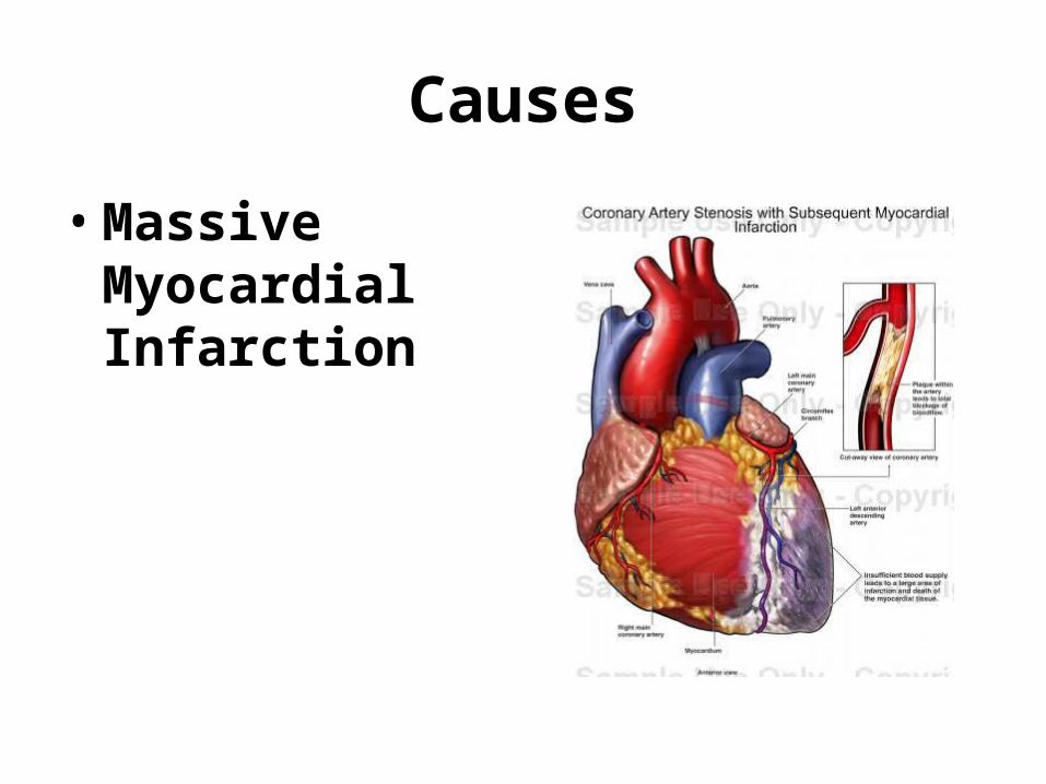
Causes
• Massive Myocardial Infarction
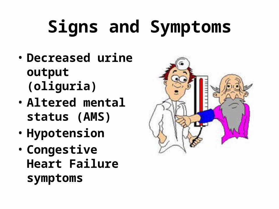
Signs and Symptoms
• Decreased urine output (oliguria)
• Altered mental status (AMS)
• Hypotension• Congestive Heart
Failure symptoms
Diagnosis
• Assessment• Echo• Labs: CBC, CE,
Lactate• ECG
• Serial lactate measurements are useful markers of hypoperfusion and are also used as indicators of prognosis. Elevated lactate values in a patient with signs of hypoperfusion indicate a poor prognosis; rising lactate values during resuscitation portend a very high mortality rate.
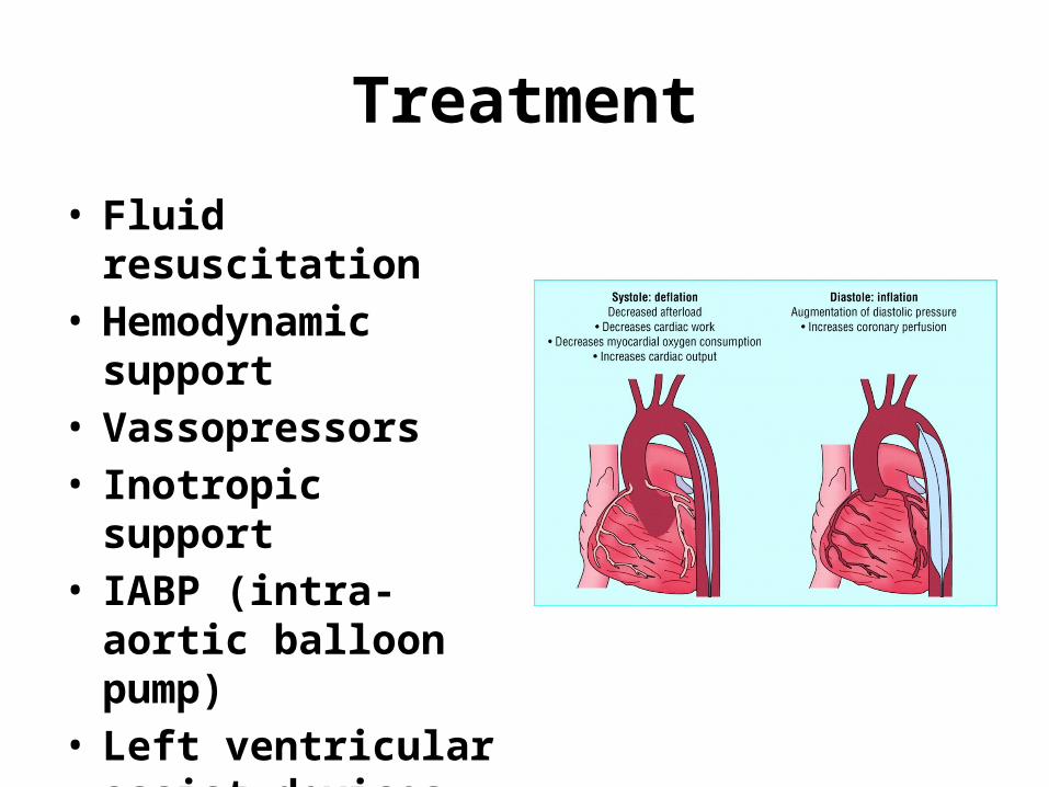
Treatment
• Fluid resuscitation• Hemodynamic support• Vassopressors• Inotropic support• IABP (intra-aortic
balloon pump)• Left ventricular assist
devices

LVAD (left ventricular assist device)
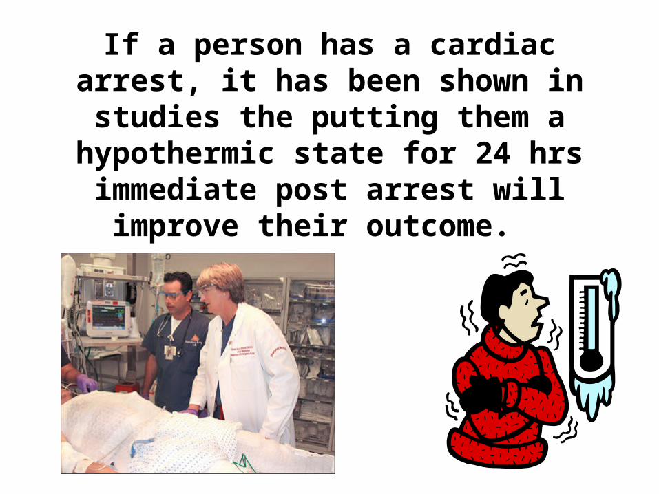
If a person has a cardiac arrest, it has been shown in studies the putting
them a hypothermic state for 24 hrs immediate post arrest will improve
their outcome.
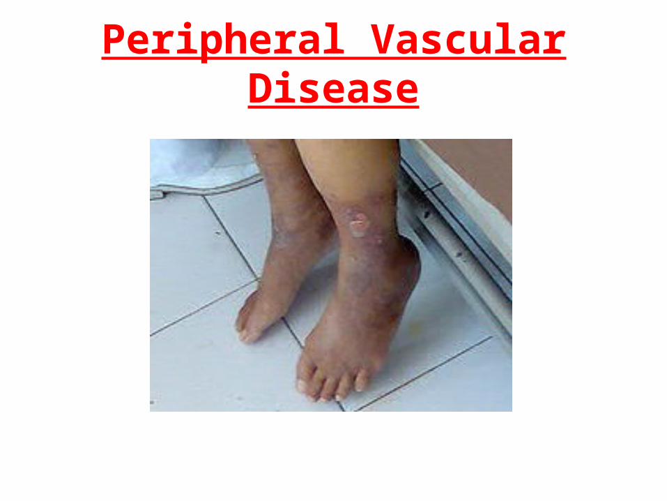
Peripheral Vascular Disease
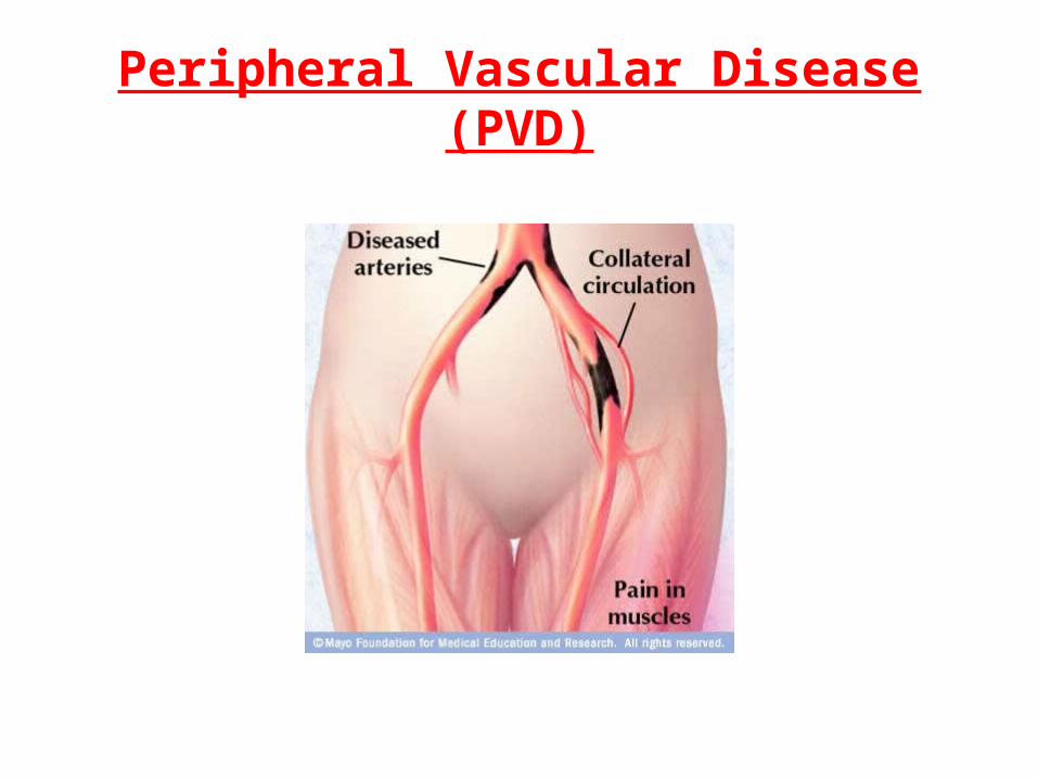
Peripheral Vascular Disease (PVD)
Peripheral Vascular Disease
• Narrowed arteries reduce blood flow to your limbs.
• When you develop peripheral artery disease (PAD), your extremities — usually your legs — don't receive enough blood flow to keep up with demand.

Peripheral Vascular Disease Thrombosis
• Formation of blood clots on blood vessel walls. Caused by slow blood flow. Because blood flows more slowly in veins, it is more common to develop a clot there.
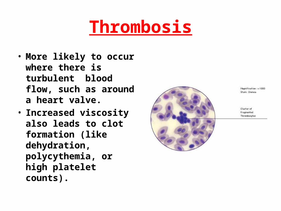
Thrombosis
• More likely to occur where there is turbulent blood flow, such as around a heart valve.
• Increased viscosity also leads to clot formation (like dehydration, polycythemia, or high platelet counts).

Embolus
• If a thrombus breaks free, it becomes an embolus.
• An embolus typically will lodge in the coronary arteries, lung, or brain vessels.
Causes
• High cholesterol• Diabetes• Heart disease (coronary
artery disease)• Hypertension• Kidney disease
involving hemodialysis
• Smoking• Polycythemia• Dehydration• Stasis• High Platelet Counts• Arrhythmias
Signs and Symptoms
• Claudication• Numbness• Coolness in extremity• Pain • Bruit• Weak or absent pulse• Slow wound healing• Clubbing• Decreased blood pressure in the extremity

Diagnosis
• Physical Exam• Lab Tests: CBC, Lipid Panels• Angiography of the arteries in the legs • Blood pressure measured in the arms and
legs for comparison • Doppler Ultrasound• CT• MRI
Treatment
• Balance exercise and rest.
• Smoking cessation• Reduce weight• Heart health diet• Aspirin or Plavix
• Statin drugs• Pain medication• Angioplasty• Stents• Bypass• Amputations

Phlebitis
• Inflammation of a vein. (usually deep veins of the leg)
• Caused by infection, injury, poor circulation, and obesity.

Femoral - Popliteal (Bypass)
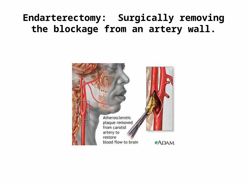
Endarterectomy: Surgically removing the blockage from an artery wall.

Thrombophlebitits
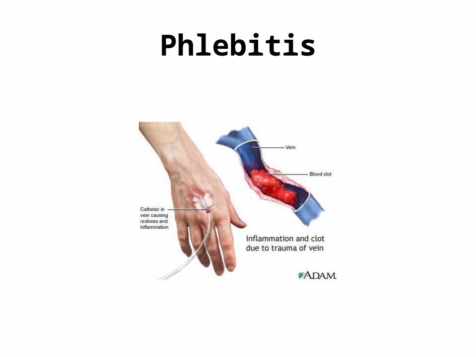
Phlebitis
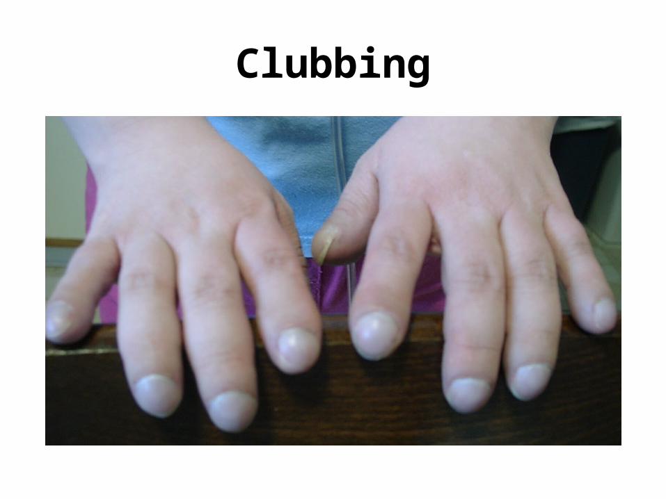
Clubbing

Gangrene