chromatin rearrangements in the prnd-prnb bidirectional promoter
TRANSCRIPT
EUKARYOTIC CELL, Feb. 2004, p. 144–156 Vol. 3, No. 11535-9778/04/$08.00�0 DOI: 10.1128/EC.3.1.144–156.2004Copyright © 2004, American Society for Microbiology. All Rights Reserved.
Chromatin Rearrangements in the prnD-prnB Bidirectional Promoter:Dependence on Transcription Factors
Irene García,1†* Ramon Gonzalez,1‡ Dennis Gomez,1§ and Claudio Scazzocchio1,2
Institut de Genetique et Microbiologie1 and Institut Universitaire de France,2 Universite Paris-Sud, UMR8621,91405 Orsay Cedex, France
Received 20 November 2003/Accepted 24 November 2003
The prnD-prnB intergenic region regulates the divergent transcription of the genes encoding proline oxidaseand the major proline transporter. Eight nucleosomes are positioned in this region. Upon induction, thepositioning of these nucleosomes is lost. This process depends on the specific transcriptional activator PrnAbut not on the general GATA factor AreA. Induction of prnB but not prnD can be elicited by amino acidstarvation. A specific nucleosomal pattern in the prnB proximal region is associated with this process. Underconditions of induction by proline, metabolite repression depends on the presence of both repressing carbon(glucose) and nitrogen (ammonium) sources. Under these repressing conditions, partial nucleosomal posi-tioning is observed. This depends on the CreA repressor’s binding to two specific cis-acting sites. Threeconditions (induction by the defective PrnA80 protein, induction by amino acid starvation, and induction in thepresence of an activated CreA) result in similar low transcriptional activation. Each results in a differentnucleosome pattern, which argues strongly for a specific effect of each signal on nucleosome positioning.Experiments with trichostatin A suggest that both default nucleosome positioning and partial positioningunder induced-repressed conditions depend on deacetylated histones.
In simple eukaryotes, some genes are transcribed diver-gently from a common bidirectional promoter. Well-studiedexamples are the GAL1-GAL10 promoter of Saccharomycescerevisiœ (reviewed in references 10 and 35) and the niiA-niaDpromoter of Aspergillus nidulans (37, 41, 52). Here we analyzedthe nucleosome rearrangements of the bidirectional prnD-prnBintergenic region. This is a 1.7-kb region located between thegene coding for proline oxidase (prnD) and the one coding forthe major, specific proline transporter (prnB) (29, 50). Thesegenes are located in the prn gene cluster in the right arm ofchromosome VII (Fig. 1). The regulation of prnD and prnBinvolves a multiplicity of metabolic signals. The pathway-spe-cific transcription factor PrnA is essential for proline inductionof both genes (11, 21, 22, 46). The prnD-prnB intergenic regionis a genuine bidirectional promoter, as mutations in the twoPrnA binding sites present in this region affect the transcrip-tion of both prnD and prnB (21; I. García, D. Gomez, and C.Scazzocchio, unpublished results).
Transcription of prnB but not of prnD can also be induced byamino acid starvation. This effect depends on the integrity of acanonical GCN4 binding site in the proximity of the prnBTATA box (56).
Repression of both prnB and prnD occurs only when both
carbon (glucose) and nitrogen-repressing (ammonium)sources are present simultaneously (2, 5, 6, 21, 22). Repressionacts directly on prnB expression, while repression of prnD isindirect and results from inducer exclusion (5, 16, 22). Repres-sion necessitates both the activation of the negative regulatorCreA and the inactivation of the GATA factor AreA. A modelto account for this pattern of repression has been publishedpreviously (23). Figure 1 shows the prnD-prnB intergenic re-gion with the cis-acting sites that have been shown to be phys-iologically relevant.
In this article we show that eight nucleosomes are positionedin the prnD-prnB region. Upon induction, these nucleosomesare no longer positioned, while a single nucleosome is partiallypositioned at a new location. In conditions of simultaneouscarbon and nitrogen metabolite repression in the presence ofinducer, a partial repositioning of nucleosomes occurs. Weanalyze in detail the role of the three transcription factorsinvolved in prnD-prnB regulation, PrnA, CreA, and AreA, inthis process.
MATERIALS AND METHODS
Strains. A pabaA1 strain was used as the wild type. creA loss-of-functionstrains were creAd1 pabaA1 (48) and creAd25 pabaA1 (4). The areA loss-of-function strain was areA600 biA1 sB43. areA600 is an early chain termination nullmutation (1, 30). The prnA loss-of-function mutations analyzed in this work arelisted in Table 1. Strains used were prnA404 pabaA1, prnA15 cnxJ1 pabaA1 fwA1,prnA80 pabaA1, prnA407 pabaA1 fwA, and prnA442 pabaA1 alcR125 fwA1. alcR125is an alcR loss of function (43). For definitions of the standard geneticmarkers, see the Glasgow Stock List at http://www.gla.ac.uk/Acad/IBLS/molgen/aspergillus/strintro.html.
Sequences of new prnA alleles. The sequence changes of a number of prnAalleles were determined (Table 1). The approximate position in the gene inrelation to deletion mutations was known (11, 46). Thus, the appropriate se-quence was amplified by PCR with specific primers and the newly introducedchanges were checked by sequencing (automatic sequencing; MWG Biotech,Ebersberg, Germany).
* Corresponding author. Mailing address: Instituto de BioquímicaVegetal y Fotosíntesis (CSIC), Centro de Investigaciones Científicasde la Isla de la Cartuja, Avda. Americo Vespucio s/n, 41092 Seville,Spain. Phone: 34 954489516. Fax: 34 954460065. E-mail: [email protected].
† Present address: Instituto de Bioquímica Vegetal y Fotosíntesis(CSIC), Centro de Investigaciones Científicas de la Isla de la Cartuja,41092 Seville, Spain.
‡ Present address: Instituto de Fermentaciones Industriales (CSIC),C/ Juan de la Cierva 3, 28006 Madrid, Spain.
§ Present address: Laboratoire de Physiologie Humaine, Faculte dePharmacie-Medecine, 51100 Reims, France.
144
on March 26, 2018 by guest
http://ec.asm.org/
Dow
nloaded from

Growth conditions. A total of 106 spores of each strain per ml were inoculatedat 37°C into liquid minimal medium with the appropriate supplements plus 0.1%fructose as the carbon source and 5 mM urea as the nitrogen source, except forthe areA600 strain. Mycelia were grown for 8 h at 37°C and then incubated for2 additional hours at 37°C or repressed with glucose (1%) and ammonium (20mM ammonium-L[�]-tartrate), or induced with 20 mM L-proline, or inducedwith 20 mM L-proline and simultaneously either carbon repressed (1% glucose),nitrogen repressed (20 mM ammonium-L[�]-tartrate), or carbon and nitrogenrepressed and incubated 2 h at 37°C. The areA600 strain was grown at 37°C inliquid minimal medium with the appropriate supplements plus 1% glucose and5 mM ammonium-L(�)-tartrate for 7 h at 37°C, and then cultures were filteredand shifted to minimal medium containing the appropriate supplements andneutral carbon and nitrogen sources (5 mM urea and 0.1% fructose) without orwith 20 mM proline and incubated for 2 additional hours at 37°C. An areA�
strain was grown in parallel in the same culture conditions. Mycelia were har-vested by filtration through sterile Blutex tissue, washed with sterile distilledwater, and frozen in liquid nitrogen.
RNA preparation and Northern blots. Total RNA was isolated with the RNAPlus Extraction Solution (Biogen) following the manufacturer’s instructions.RNA electrophoresis and Northern blot hybridizations were carried out as de-scribed previously (22, 23). prnB, prnD, and acnA probes were prepared asdescribed by Gomez et al. (22).
Nucleosome positioning. Micrococcal nuclease I digestions were performed bythe method adapted by Gonzalez and Scazzocchio (24). Micrococcal nucleasewas used at concentrations ranging between 0.5 and 2.5 U/g of mycelium. DNAwas digested with an appropriate restriction enzyme: PstI (SC1 hybridization) orHindIII (SC2 hybridization). Probe SC1 is the 389-bp PstI-AccI fragment of theprnD-prnB intergenic region (24). Probe SC2 is the 332-bp EcoRI-HindIII frag-ment of the prnD-prnB intergenic region (23).
For the restriction enzyme protection assay, a method adapted from Gregoryet al. (25) was used (M. Mathieu, personal communication). Probe HP is the
836-bp HindIII-PstI fragment of bAN926, which contains a 907-bp fragment ofthe prnD open reading frame, the prnD–prnB intergenic region, and 1,512 bp ofthe prnB open reading frame (23). At least three independent experiments werecarried out for each mutant and/or condition with identical results. In every case,induction and repression were checked by Northern blots made in parallel withthe same mycelia.
In vivo footprinting. Binding of PrnA to sites PrnA-2 and PrnA-3 was detectedby in vivo footprinting as described by Gomez et al. (23) following the techniqueof Wolschek et al. (59).
RESULTS
Chromatin rearrangements in the prnD-prnB bidirectionalpromoter. Throughout this article we compare three growthconditions: noninduced, absence of proline in a medium whichcontains nonrepressing carbon and nitrogen sources (underthese conditions expression of prnD and prnB is minimal andvirtually undetectable in Northern blots); induced, the samebut in the presence of proline; and induced-repressed, whereproline is added together with repressing nitrogen and carbonsources. The simultaneous presence of these repressing me-tabolites strongly diminishes transcription, but it does not abol-ish it (22, 51). While the exact experimental conditions aregiven in Materials and Methods, it is important to keep in mindthe conceptual differences between the three conditions. Whenother conditions are used, these are defined in the text.
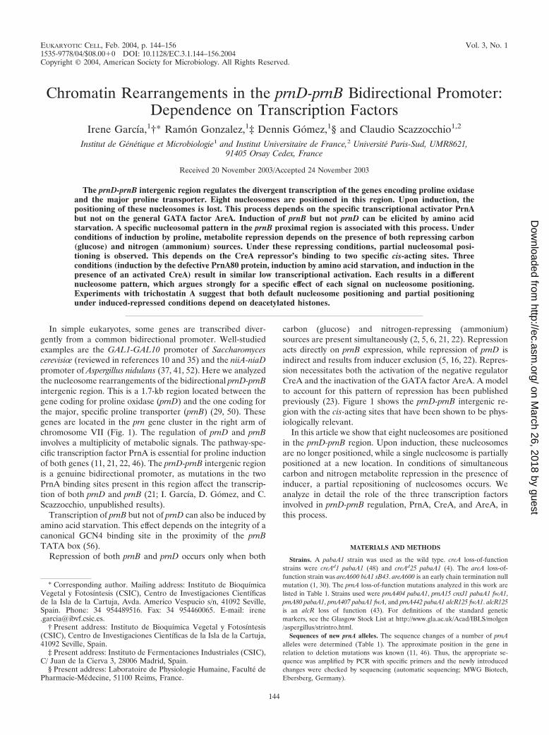
Under noninduced conditions, eight nucleosomes are posi-tioned in the prnD-prnB promoter. This pattern is identical,under noninducing, nonrepressing (noninduced, definedabove, Fig. 2), or repressing conditions (glucose and ammo-nium in the absence of proline, not shown). Upon induction(under nonrepressing conditions), nucleosome positioning islost (Fig. 2). Between positions 904 and 1205, the pattern ofmicrococcal nuclease I cuts is different from that of the nakedDNA and indicates that a nucleosome is partially positionedbetween these boundaries. The nuclease cut at position 1055 ismuch weaker under induced than noninduced conditions,which indicates that this nucleosome is positioned differentlyfrom either nucleosome �1 or �2 under noninducing condi-tions. The pattern of micrococcal nuclease I digestion does notallow us to conclude whether a new nucleosome is positionedbetween the cutting sites at nucleotides 510 and 725. However,the presence of a SacI restriction site in position 685 allowed usto investigate the positioning of this putative nucleosome. No
FIG. 1. prnD-prnB intergenic region. prnD encodes proline oxidase, and prnB encodes the specific proline transporter (20, 28, 29, 50). TheCreA-binding sites 3.1 and 3.2 (grey lozenges) are essential for prnB and indirectly for prnD repression (51, 15, 16). The AreA-binding sites 13 and14 (grey ovals) are necessary to set the maximal level of transcription of prnB and to integrate carbon and nitrogen metabolite repression of thisgene (24). High-affinity PrnA binding sites 2 and 3 are shown by white triangles; their occupancy upon induction by PrnA in vivo has beendemonstrated (23). A putative binding site for a GCN4-like factor is shown as a thick arrow (56) A black triangle indicates the prnB TATA box(24). The positions of the transcriptional start points (�1) and of the ATG of prnD and prnB are shown (20, 50; S. Demais and C. Scazzocchio,unpublished results). Relevant restriction sites mentioned in the text are also shown.
TABLE 1. Sequence changes of the prnA mutations used inthis work
Mutation Nucleotide sequencechangea
Amino acid sequencechangea Reference
prnA15 T(2209)C L(621)P This workprnA80 G(2319)C A(658)P This workprnA407 C(2101)A S585Y 11prnA442 G(2744)T AMB(819)Y�18-residue
extensionThis work
prnA469 Insertion of G(1911)and deletion ofC(1987)
Substitution of residues522–547
11
prnA404 Deletion of 67–1510 Deletion of residues23–398
11, 12
a Numbering of nucleotides and amino acids is that of Cazelle et al. (11).
VOL. 3, 2004 CHROMATIN STRUCTURE IN prnD-prnB PROMOTER 145
on March 26, 2018 by guest
http://ec.asm.org/
Dow
nloaded from
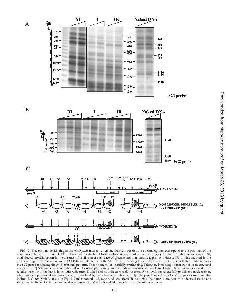
FIG. 2. Nucleosome positioning in the prnD-prnB intergenic region. Numbers besides the autoradiograms correspond to the positions of themain cuts relative to the prnD ATG. These were calculated from molecular size markers run in every gel. Three conditions are shown. NI,noninduced, mycelia grown in the absence of proline in the absence of glucose and ammonium; I, proline-induced; IR, proline-induced in thepresence of glucose and ammonium. (A) Pattern obtained with the SC1 probe (revealing the prnD proximal pattern). (B) Pattern obtained withthe SC2 probe (revealing the prnB proximal pattern). These patterns are partially overlapping. Triangles, increasing concentration of micrococcalnuclease I. (C) Schematic representation of nucleosome positioning. Arrows indicate micrococcal nuclease I cuts. Their thickness indicates therelative intensity of the bands in the autoradiogram. Dashed arrows indicate weakly cut sites. White ovals represent fully positioned nucleosomes,while partially positioned nucleosomes are shown by diagonally hatched ovals (see text). The positions and lengths of the probes used are alsoindicated. Other symbols are as in Fig. 1. Under noninduced, repressed conditions (R, see text), the nucleosome pattern is identical to the oneshown in the figure for the noninduced conditions. See Materials and Methods for exact growth conditions.
146
on March 26, 2018 by guest
http://ec.asm.org/
Dow
nloaded from

protection of this restriction site is seen, and thus inductiondoes not result in the positioning of an additional nucleosome(not shown) between these boundaries.
Addition of either glucose or ammonium, which singly donot result in significant repression (22, 23), does not affect thedestabilization of nucleosome positioning seen in induced cul-tures; the pattern is identical to that found under inducedconditions in the absence of any repressing metabolite (resultsnot shown). When both glucose and ammonium are added toan induced culture, nucleosome positioning is seen for nucleo-somes �2 to �4. The pattern is, except for nucleosome �1,which is fully positioned, one of partial positioning (see above),while the prnB proximal nucleosomes, �3 and �4, are notpositioned at all. By partial positioning we mean that the pat-tern observed is what would be generated by superimposingthe fully positioned and the fully nonpositioned patterns. Thepossible significance of this finding will be discussed below.
Induction results in nucleosome delocalization, not in com-plete nucleosome loss. It is possible to differentiate betweennucleosome delocalization and nucleosome loss if, rather thanrevealing micrococcal nuclease I cuts by indirect terminal la-beling, one hybridizes a chromatin micrococcal nuclease I di-gest with a probe covering the whole region that is beinganalyzed. When this is done, the typical digestion laddershould be seen whether nucleosomes are positioned or not(J. L. Barra, personal communication).
The prnD-prnB intergenic region shows a typical nucleosomerepeat under all induction and repression conditions, with alength of �160 bp, the length previously reported for Aspergil-lus nidulans (24, 36, 42). This is shown for the prnB-proximalregion in Fig. 3. Note that under induction conditions thebands became fuzzy and that this effect it more pronounced thelonger the polynucleosome is. This is exactly what is to beexpected if in a given segment of DNA nucleosomes is presentbut not translationally positioned, the distribution of sizes ob-tained by digestion being broader the larger the number ofnucleosomes. Bands are fuzzy in induced conditions, clear innoninduced conditions, and intermediate under induced-re-pressed conditions. The latter finding supports the data ob-tained (Fig. 2) with indirect terminal labeling, which corre-sponds to a pattern of partial positioning for the induced-repressed conditions (see Discussion). Other blots, hybridizedwith suitable probes, show the same pattern for the whole
intergenic region (not shown). As this experiment does notdistinguish individual nucleosomes, it cannot be excluded thatsome nucleosomes may be lost and some delocalized, nor canit be excluded that the population of nuclei be heterogeneousvis a vis these two possibilities.
PrnA is necessary for nucleosome delocalization upon in-duction. Figure 4 shows that in a deletion of prnA, all nucleo-somes remain completely positioned upon induction. A num-ber of prnA mutants outside the DNA binding domain havebeen characterized (11, 12, 46; this article). We investigatedwhether any of these mutants, unable to activate prnD andprnB transcription, as assessed by Northern blots, maintain theability to delocalize nucleosomes. The sequence changes ofthese mutations are shown in Table 1. All mutations tested,with the exception of prnA80, are unable to elicit transcriptionand nucleosome delocalization (not shown). The binding ofsome PrnA mutant proteins to high-affinity sites 2 and 3 (23)was also investigated by in vivo methylation protection. Theresults are shown in Fig. 5. All mutations tested, with theexception of prnA80, resulted in inability to bind both PrnAsites 2 and 3. prnA80 does not affect the binding to either site2 or 3. The prnA80 mutant was classified as cryosensitive ingrowth tests (46); it equally affects transcription of prnD at37°C and at 25°C, while showing a cryosensitive phenotype forprnB transcription (Fig. 6A). At the level of chromatin, aprnA80 strain behaves exactly like a prnA� strain: upon induc-tion all nucleosomes are delocalized and a nucleosome is newly(and partially) positioned between nucleotides 904 and 1205(Fig. 6B). The significance of these results will be discussedbelow.
Induction of prnB transcription by amino acid starvationresults in loss of positioning of prnB proximal nucleosomes.The transcription of prnB (but not of prnD) can be elicitedindependently from proline induction by amino acid starva-tion, possibly mediated by a GCN4-like factor (56). This acti-vation is lower, but noticeable, in a strain with prnA deleted(56). We used 3-amino-1,2,4-triazole, a competitive inhibitorof the histidine biosynthetic enzyme His3p, to induce aminoacid starvation (as in reference 56). Under these conditions,the positioning of the prnB proximal nucleosomes �3 and �4is lost (Fig. 7). All other nucleosomes remained positioned asthey are under noninduced conditions. This chromatin rear-rangement is identical in prnA� and prnA404 strains.
AreA is not necessary for chromatin remodeling upon in-duction. The GATA factor AreA is necessary to achieve themaximal levels of transcription of prnB and prnD (22). Weinvestigated here the role of AreA in nucleosome positioningupon induction. Under both inducing and noninducing condi-tions, the nucleosomal pattern is the same in areA� andareA600 strains (Fig. 8).
CreA is essential for nucleosome positioning upon repres-sion but is not involved in the establishment of the defaultchromatin structure. The creA loss-of-function mutant creAd1,which bears a point mutation in the DNA binding domain (48),results in complete derepression of prnB and prnD (Fig. 9A).Figure 9B shows that the nucleosome positioning associatedwith repression depends strictly on CreA. The pattern of nu-cleosome positioning in creA mutant strains is the same underinduced nonrepressed and induced-repressed conditions andidentical to the wild-type pattern obtained under induced con-
FIG. 3. Presence of nucleosomes in the prnD-prnB promoter, inde-pendent of positioning. We show here only the prnB proximal region(bp 998 to 1834 from the prnD ATG) NI, I, and IR are as in Fig. 2.
VOL. 3, 2004 CHROMATIN STRUCTURE IN prnD-prnB PROMOTER 147
on March 26, 2018 by guest
http://ec.asm.org/
Dow
nloaded from

ditions. creAd25, a mutation in the carboxy terminus of theprotein which results specifically in derepression of prnB andprnD but not of alcR and alcA (4; B. Cubero, M. Mathieu, B.Felenbok, and C. Scazzocchio, unpublished results) has the
same effect on nucleosomal positioning as the more extremecreAd1 mutation (not shown). Figure 9B also shows the chro-matin pattern obtained under noninduced conditions in creA-derepressed mutants, which are identical to the one obtained
FIG. 4. Nucleosome positioning in a prnA deletion. For comparison, the micrococcal nuclease I protection pattern of a prnA� strain grownunder inducing conditions is also shown. Left, SC1 probe; right, SC2 probe. Asterisks indicate the positions of the relevant changes observed withprobe SC2. Symbols are as in Fig. 1 and 2. No transcription of either prnD or prnB is seen in this mutant (shown for prnB also in Fig. 7). Bottompanel, schematic representation of nucleosome positioning in a prnA404 mutant, which was the same under noninduced and induced conditions.
FIG. 5. In vivo footprints of a number of prnA mutants. Left panel, in vivo footprints of a prnA� and a prnA442 strain (prnD coding strandshown). Right panel, in vivo footprint pattern of a number of prnA missense mutants. The prnA404 deletion mutant is also included as a control.Footprints of a prnA� strain under both noninducing and inducing conditions are also shown. All mutant strains were grown under inducingconditions (prnD coding strand shown). The sequence corresponding to PrnA binding sites 2 and 3 is shown to the side of the autoradiograms. Theprotected G’s in PrnA binding sites 2 and 3 are indicated in bold in the sequence and by arrows pointing to the autoradiogram. Symbols are asin Fig. 2. Nucleotide and amino acid changes in each mutant are shown in Table 1.
148 GARCIA ET AL. EUKARYOT. CELL
on March 26, 2018 by guest
http://ec.asm.org/
Dow
nloaded from

FIG. 6. Transcription and nucleosomal rearrangements in a prnA80 mutant. (A) Northern blots of mycelia grown at 25 and 37°C. (B) Micro-coccal nuclease I digestion of the prnA80 induced mycelia at 25 and 37°C. For comparison, the prnA� strain grown at 25°C is shown. This is identicalto the pattern obtained for prnA� at 37°C (Fig. 2 and Fig. 4). (C) Schematic representation of nucleosome positioning of a prnA80 mutant grownunder inducing conditions at both 25 and 37°C. Asterisks indicate the positions of the relevant changes observed with probe SC2. Other symbolsare as in Fig. 2.
VOL. 3, 2004 CHROMATIN STRUCTURE IN prnD-prnB PROMOTER 149
on March 26, 2018 by guest
http://ec.asm.org/
Dow
nloaded from
in creA� strains grown in the same conditions. This demon-strates that CreA is not involved in default nucleosome posi-tioning.
We constructed a double creA prnA loss-of-function mutant
(prnA404 creAd1). Northern blots and chromatin analysis werecarried out, showing that the prnA404 deletion is completelyepistatic to a creAd1 mutation for both transcriptional activa-tion and nucleosome delocalization (not shown).
FIG. 7. Transcriptional activation and nucleosome positioning under conditions of amino acid starvation. (A) Northern blot. (B) Patternsobtained after micrococcal nuclease I treatment of mycelia grown in the presence of 3-amino-1,2,4-triazole (3-AT). For comparison, patterns ofa prnA� strain grown in noninduced and induced conditions are also shown. All other symbols are as in Fig. 2. Asterisks indicate the bands resultingfrom micrococcal nuclease I cuts and revealed by the SC2 probe that appear under conditions of both proline and 3-amino-1,2,4-triazole inductionand that show loss of positioning of nucleosomes �3 and �4. (C) Schematic representation of nucleosome positioning of both prnA� and prnA404strains grown in the presence of 3-amino-1,2,4-triazole.
150 GARCIA ET AL. EUKARYOT. CELL
on March 26, 2018 by guest
http://ec.asm.org/
Dow
nloaded from

Nucleosome positioning upon repression does not occur incis-derepressed mutants. Mutations in CreA-binding sites 3.1and 3.2 (prnd22 and prnd20, respectively) have shown thesesites to be essential for CreA-mediated repression (3, 5, 15,51). Figure 9C shows that the nucleosome positioning patternin a prnd22 mutant is the same under induced and induced-repressed conditions, indicating that the partial positioning ofnucleosomes �4 to �1 does not occur when this site is mu-tated. Experiments with prnd20 and the prnd22 prnd20 doublemutant gave identical results (not shown). Thus, mutation atthese sites results in the same chromatin pattern as mutationsin the CreA trans-acting factor.
Histone deacetylation is involved in default and CreA-pro-moted nucleosome positioning. creAd mutations suppress areAloss-of-function mutations for the utilization of proline in thepresence of a repressive carbon source (2, 8). This happensbecause AreA is only necessary for prnB transcription in thepresence of an active CreA-repressing protein (2, 23, 24). Wefound that the presence of trichostatin A, an inhibitor of his-tone deacetylation (61), results in a similar phenotypic sup-pression of an areA null mutation (Fig. 10A). For analogousreasons, creAd mutations also suppress areA loss-of-functionmutations for the utilization of acetamide and �-aminobenzoicacid as nitrogen sources in the presence of glucose. We thuschecked whether trichostatin A results in a similar phenotypicsuppression of an areA null mutation on the latter nitrogensources. We could not see any phenotypic suppression on ei-
ther acetamide or �-aminobenzoic as the nitrogen source at atrichostatin A concentration identical to that used in Fig. 10A.Higher concentrations were too toxic to be tested usefully (notshown).
We then investigated the effect of trichostatin A on bothtranscription and nucleosome positioning (Fig. 10B and Fig.11). The presence of trichostatin A results in an elevated basallevel of prnB but not prnD transcription. Proline affords opti-mal induction independently of the presence of the drug. Aspredicted by the partial suppression of an areA mutation bytrichostatin A, transcription of prnB and, to a lesser extent,prnD is partially derepressed when the drug is added to theculture medium. Trichostatin A results in specific changes inthe pattern of nucleosome positioning. In the presence of tri-chostatin A in noninduced conditions, positioning of nucleo-somes �1 to �4 is completely lost and nucleosomes �1, �3,and �4 are only partially positioned. It may be relevant thatnucleosome �4 occludes the prnB TATA box. This result isshown in Fig. 11.
The most striking differences, however, are found underinducing-repressing conditions. Trichostatin A treatment re-sults in total loss of nucleosome positioning; that is, the sameresult as obtained for a creA-derepressed mutant (Fig. 11,compare with Fig. 8). There is complete agreement betweenthe partial phenotypic suppression of areA600 by trichostatinA, the levels of transcription, and nucleosome positioning in
FIG. 8. Micrococcal nuclease I digestion pattern of an areA600 mutant. Noninduced and induced conditions are shown. Symbols are as in Fig.2. The growth conditions used to permit the growth of the areA600 strain are different from those used in other experiments (see Materials andMethods). Under the same growth conditions, the areA� strain behaves exactly as shown in Fig. 2 for noninduced and induced cultures (not shown).Asterisks indicate the positions of the relevant changes observed with probe SC2. Other symbols are as in Fig. 2.
VOL. 3, 2004 CHROMATIN STRUCTURE IN prnD-prnB PROMOTER 151
on March 26, 2018 by guest
http://ec.asm.org/
Dow
nloaded from
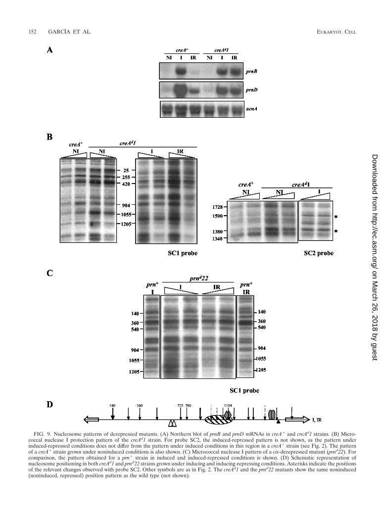
FIG. 9. Nucleosome patterns of derepressed mutants. (A) Northern blot of prnB and prnD mRNAs in creA� and creAd1 strains. (B) Micro-coccal nuclease I protection pattern of the creAd1 strain. For probe SC2, the induced-repressed pattern is not shown, as the pattern underinduced-repressed conditions does not differ from the pattern under induced conditions in this region in a creA� strain (see Fig. 2). The patternof a creA� strain grown under noninduced conditions is also shown. (C) Micrococcal nuclease I pattern of a cis-derepressed mutant (prnd22). Forcomparison, the pattern obtained for a prn� strain in induced and induced-repressed conditions is shown. (D) Schematic representation ofnucleosome positioning in both creAd1 and prnd22 strains grown under inducing and inducing-repressing conditions. Asterisks indicate the positionsof the relevant changes observed with probe SC2. Other symbols are as in Fig. 2. The creAd1 and the prnd22 mutants show the same noninduced(noninduced, repressed) position pattern as the wild type (not shown).
152 GARCIA ET AL. EUKARYOT. CELL
on March 26, 2018 by guest
http://ec.asm.org/
Dow
nloaded from

the prnD-prnB region. These results suggest that CreA-medi-ated repression acts via histone deacetylation.
DISCUSSION
Loss of nucleosome positioning in prnD-prnB depends onthe specific activator PrnA and is independent of the GATAfactor AreA. Transcriptional activation is often accompaniedby a loss of nucleosome positioning. Usually, specific transcrip-tion factors are necessary for this process (7, 53, 57), theniiA-niaD promoter of A. nidulans being an interesting excep-tion (37). In the prnD-prnB promoter, loss of nucleosome po-sitioning requires an active PrnA protein. Chromatin rear-rangements may be elicited directly or indirectly bytranscription factors or could be merely a passive result oftranscription. Of the eight nucleosomes in the prnD-prnB in-tergenic region, only nucleosomes �4 and �4 overlap theinitiation of transcription. The progress of RNA polymerasehas been shown to result in positive DNA supercoiling down-stream and negative DNA supercoiling upstream of its site ofaction (34). Positive supercoiling but not negative supercoilinghas been associated with nucleosome destabilization (32, 33).On the contrary, negative supercoiling has been associatedwith nucleosome stability (38). Thus, it is extremely unlikelythat the delocalization of nucleosomes �3 to �3 could be inany way related to topological alterations in the DNA resultingpassively from transcription.
In other experimental systems, mutations in the TATA boxhave been used to investigate the dependence of chromatinrearrangements on transcriptional activation (18, 47), but thisis not possible in this promoter, because a deletion of theputative prnB TATA box does not abolish transcription (22)and there is no obvious prnD TATA box. To discriminate thetranscriptional activation function of PrnA from its chromatinremodeling function, we have taken advantage of a number ofmutations available outside the DNA binding domain (Table1). All these mutants were tested for transcriptional activationand chromatin rearrangement activity. We failed to find amutant that had completely lost transcriptional activationwhile maintaining chromatin remodeling. Nevertheless, the re-sults with prnA80 strongly suggest that these functions areindeed separable. This mutation results in greatly impairedtranscriptional activation, but chromatin remodeling occursexactly as in a prnA� strain. The transcription of prnB andprnD in prnA80 mutants is as low as that found under induced-
repressed conditions in the wild type. Under the latter condi-tions, we see an intermediate pattern of nucleosome position-ing (see below). The fact that complete loss of nucleosomepositioning occurs in prnA80 strains in spite of the stronglydiminished transcription of prnB and prnD argues strongly fora specific effect of PrnA on nucleosomal delocalization and, bythe same token, for a specific role of CreA on nucleosomepositioning under inducing-repressing conditions (see below).
prnB transcription can also be induced by amino acid star-vation. While we have not shown by mutational analysis that aGCN4 homologue is directly involved in chromatin restructur-ing, this was previously shown for the transcriptional activationelicited by amino acid starvation (56). A mutation in a putativeGcn4p-like binding site abolishes this alternative inductionprocess but not PrnA-mediated proline induction. There is aclose homologue of GCN4 in A. nidulans (CpcA) (58), andthus it is very likely that this is the transcription factor involved(56). Gcn4p has been shown to be involved in destabilization ofnucleosome positioning in the HIS3 and PHO5 promoters (31,55). This protein interacts physically with coactivators such asGcn5p and other proteins of the SAGA and the SWI/SNFcomplexes (reference 54 and references therein).
The prnB transcription levels elicited by amino acid starva-tion are considerably higher than those found in a prnA80mutant induced by proline, where loss of nucleosome position-ing is complete. What is relevant here is that two differentinduction processes, mediated necessarily by different tran-scription factors, result in different chromatin rearrangementsand that these are not correlated with the levels of transcript.A similar uncoupling of transcriptional activation and nucleo-somal rearrangement has been observed in mutants resultingin derepression of the SUC2 promoter of Saccharomyces cer-evisiae (19).
The GATA factor AreA is totally irrelevant for nucleosomedelocalization upon induction in the prnD-prnB promoter. Thiscontrasts with its essential role in the niiA-niaD promoter (37).Ammonium repression on its own does not lead to nucleosomepositioning in the prnD-prnB promoter. As ammonium repres-sion prevents AreA function (30, 40) this is consistent with thefact that AreA is not necessary for nucleosome delocalizationupon induction.
Partial positioning of nucleosomes upon repression dependson the CreA repressor. Positioning of nucleosomes under non-induced conditions is independent from the presence of the
FIG. 10. Trichostatin A treatment. Left, growth tests showing partial suppression of an areA600 mutation. The medium contains proline as thesole nitrogen source plus glucose as the sole carbon source. The relevant genotypes of strains are indicated above the growth tests. Right, Northernblots showing the effect of trichostatin A on prnB and prnD expression. �TSA, no trichostatin A; �TSA, 3.3 �M trichostatin A.
VOL. 3, 2004 CHROMATIN STRUCTURE IN prnD-prnB PROMOTER 153
on March 26, 2018 by guest
http://ec.asm.org/
Dow
nloaded from

FIG. 11. Effect of trichostatin A on nucleosome positioning. (A) Micrococcal nuclease I patterns. In noninduced (NI) conditions, undertrichostatin A (TSA) treatment, nucleosomes �2 and �1 to �4 are partially positioned (probe SC1), and nucleosome �3 and �4 positioning islost (probe SC2). In induced-repressed (IR) conditions, positioning of nucleosomes �4 to �2 is lost after trichostatin A treatment, as revealed withprobe SC1. For probe SC2, the induced-repressed pattern is not shown, as in this region the pattern under induced-repressed conditions does notdiffer from the pattern under induced conditions (see Fig. 2). Under induced (I) conditions, the patterns obtained in the presence of trichostatinA are identical to those found in its absence (Fig. 2) and thus are not shown. Asterisks indicate the positions of the relevant changes observed withprobe SC2. (B) Schemes comparing nucleosome positioning under noninduced and induced-repressed conditions in the presence and absence oftrichostatin A. Symbols are as in Fig. 2. �TSA, no trichostatin A; �TSA, 3.3 �M trichostatin A. As trichostatin A is prepared in dimethyl sulfoxide,controls without trichostatin A were treated with equivalent amounts of this solvent.
154 GARCIA ET AL. EUKARYOT. CELL
on March 26, 2018 by guest
http://ec.asm.org/
Dow
nloaded from
CreA repressor. A specific pattern of nucleosome positioning,different from the default pattern and from the fully inducedpattern, is seen under inducing-repressing conditions (see be-low). Positioning upon simultaneous glucose and ammoniumrepression requires CreA. This has been shown by using mu-tations in both the CreA protein itself and in its cognate bind-ing sites in the prnD-prnB promoter. The comparison of theseresults with those obtained with the prnA80 mutant demon-strates that the partial positioning found under induced-re-pressed conditions is specific (see above).
We have shown by in vivo methylation protection experi-ments that under conditions of CreA-mediated repression,PrnA remains bound to the high-affinity sites 2 and 3 (Fig. 8 inreference 21). This implies that CreA acts by negating PrnAinteractions with the transcriptional and chromatin remodelingcomplexes rather than by preventing its binding to DNA.
Under conditions of CreA-mediated repression, positioningof nucleosomes �3 and �4 is completely lost, nucleosome 1 ispositioned, and nucleosomes �4 to �1 and �2 show a patternof partial positioning (see Fig. 2 and Results section). A similarpattern of partial positioning has been obtained for the PHO8promoter in S. cerevisae (9). This pattern can be due either toan “oscillation” in the state of each nucleosome or to a heter-ogeneity in the nuclear population, in which some nuclei showan “open” and others a “closed” chromatin pattern. We favorthe first alternative. Nuclear heterogeneity would imply thatthe intracellular concentration of CreA is limiting. It is unlikelyfor a protein that represses every single gene sensitive to car-bon catabolite repression to be present in limiting concentra-tions. Limiting concentrations of transcription factors lead tocodominance of loss-of-function mutations when tested in dip-loids with their wild-type allele (6, 14, 44, 45), while creAd (lossof function) mutations are clearly recessive (8).
In S. cerevisae, Mig1, the specific carbon catabolite repres-sor, acts by recruiting the Tup1-Ssn6 complex, and this in itsturn acts directly on chromatin structure, and specifically onH3 acetylation (17, 27; reviewed in references 49 and 60). Onecannot, however, extrapolate directly from S. cerervisae to A.nidulans. CreA shows similarity to Mig1 only in its DNA bind-ing domain. RcoA, the only clear Tup1 homologue present inthe A. nidulans genome, is not involved in carbon cataboliterepression of either the prn cluster or the alc regulon (26; I.García, M. Mathieu, B. Felenbok, and C. Scazzocchio, unpub-lished data). Its role will be analyzed in detail in anotherpublication.
Both default nucleosome positioning and positioning uponrepression are probably dependent on deacetylation. Tricho-statin A treatment results in loss of positioning of nucleosomes�1 to �4 and very mild transcriptional activation of prnB inthe absence of induction by proline. This implies that thedeacetylation of histones plays a role in the default positioningof at least some nucleosomes in the prnD-prnB promoter.Among the nucleosomes delocalized by trichostatin A treat-ment, we find nucleosome �4, the one that occludes the prnBTATA box. Previous work has shown that this element is notessential for prnB transcriptional activation, but that its dele-tion leads to halving the steady-state level of the prnB mRNA(22). A striking effect of trichostatin A is seen under conditionsof repression (induced-repressed). Here we see total loss ofnucleosome positioning and partial derepression. However,
the derepression observed is not nearly as drastic as that seenin a creAd mutation. This implies that while nucleosome repo-sitioning may be necessary for full repression, CreA can stillpartially repress on completely open chromatin.
The work presented here is an analysis of the chromatinstructure of a region subject to a multiplicity of transcriptionsignals in a simple eukaryote. The prnD-prnB promoter inte-grates four different signals, proline induction, amino acid star-vation, and nitrogen and carbon metabolite repression. Thislevel of complexity is higher than that found in some otherwell-studied promoters, such as GAL1-GAL10 of S. cerevisiaeand niiA-niaD of Aspergillus nidulans. We have been able todiscriminate between the roles of the different transcriptionfactors involved on nucleosome positioning. Transcription fac-tors act on chromatin structure by recruiting remodeling andacetylation complexes (see reference 39 for a review). Theavailability of the complete genomic sequence of A. nidulansand of new methods simplifying the procedures for gene inac-tivation (13; K.-H. Han, Z. Hamari, J.-H. Seo, C. Scazzocchio,and J.-H. Yu, unpublished results) will permit us to studysystematically the involvement of these complexes and theirinteraction with a multiplicity of transcription factors.
ACKNOWLEDGMENTS
We thank Jose L. Barra for suggesting the experiment used todiscriminate nucleosome delocalization from complete nucleosomeloss and Martine Mathieu and Beatrice Felenbok for communicatingtheir adaptation (25) before publication. We thank Rafael Fernandezfor help and discussion and Beatriz Cubero for critical reading of themanuscript.
I.G. was supported by postdoctoral fellowships from, successively,the Spanish Ministry of Education and the EU Marie Curie Pro-gramme. R.G. was the recipient of postdoctoral fellowships from,successively, the EMBO and the EU Marie Curie Programme. D.G.was supported by a predoctoral scholarship from the French Ministerede l’Education Nationale, de l’Enseignement Superieur et de la Re-cherche, and a postdoctoral fellowship from the Association pour laRecherche contre le Cancer. This work was supported by the Univer-site Paris-Sud, the CNRS, the IUF, and EU contract BIO4-CT96-0535.
REFERENCES
1. Al Taho, N., H. N. Sealy-Lewis, and C. Scazzocchio. 1984. Suppressiblealleles in a positive control gene in Aspergillus nidulans. Curr. Genet. 8:245–251.
2. Arst, H. N., Jr., and D. J. Cove. 1973. Nitrogen metabolite repression inAspergillus nidulans. Mol. Gen. Genet. 126:111–141.
3. Arst, H. N., Jr., and D. W. MacDonald. 1975. A gene cluster in Aspergillusnidulans with an internally located cis-acting regulatory region. Nature 254:26–31.
4. Arst, H. N., Jr., and C. R. Bailey. 1977. The regulation of carbon metabolismin Aspergillus nidulans, p. 131–146. In J. E. Smith and J. A. Pateman (ed.),Genetics and physiology of Aspergillus. Academic Press, London, England.
5. Arst, H. N., Jr., D. W. MacDonald, and S. A. Jones. 1980. Regulation ofproline transport in Aspergillus nidulans. J. Gen. Microbiol. 116:285–294.
6. Arst, H. N., Jr., and C. Scazzocchio. 1985. Formal genetics and molecularbiology of the control of gene expression in Aspergillus nidulans, p. 309–343.In J. W. Bennet and L. L. Lasure (ed.), Gene manipulations in fungi.Academic Press, Orlando, Fla.
7. Axelrod, J. D., M. S. Reagan, and J. Majors. 1993. GAL4 disrupts a repress-ing nucleosome during activation of GAL1 transcription in vivo. Genes Dev.7:857–869.
8. Bailey, C., and H. N. Arst, Jr. 1975. Carbon catabolite repression in Aspergil-lus nidulans. Eur. J. Biochem. 51:573–577.
9. Barbaric, S., K. D. Fascher, and W. Horz. 1992. Activation of the weaklyregulated PHO8 promoter in S. cerevisiae: chromatin transition and bindingsites for the positive regulatory protein PHO4. Nucleic Acids Res. 20:1031–1038.
10. Bash, R., and D. Lohr. 2001. Yeast chromatin structure and regulation ofGAL gene expression. Prog. Nucleic Acid Res. Mol. Biol. 65:197–259.
11. Cazelle, B., A. Pokorska, E. Hull, P. M. Green, G. Stanway, and C. Scaz-
VOL. 3, 2004 CHROMATIN STRUCTURE IN prnD-prnB PROMOTER 155
on March 26, 2018 by guest
http://ec.asm.org/
Dow
nloaded from
zocchio. 1998. Sequence, exon-intron organization, transcription and muta-tional analysis of prnA, the gene encoding the transcriptional activator of theprn gene cluster in Aspergillus nidulans. Mol. Microbiol. 28:355–370.
12. Cazelle, B., A. Pokorska, E. Hull, P. M. Green, G. Stanway, and C. Scaz-zocchio. 1999. Sequence, exon-intron organization, transcription and muta-tional analysis of prnA, the gene encoding the transcriptional activator of theprn gene cluster in Aspergillus nidulans (erratum). Mol. Microbiol. 31:1283.
13. Chaveroche M. K., J. M. Ghigo, and C. d’Enfert. 2000. A rapid method forefficient gene replacement in the filamentous fungus Aspergillus nidulans.Nucleic Acids Res. 28:E97.
14. Cove, D. J. 1966. The induction and repression of nitrate reductase in thefungus Aspergillus nidulans. Biochim. Biophys. Acta 113:51–56.
15. Cubero, B., and C. Scazzocchio. 1994. Two different adjacent and divergentzinc-finger binding sites are necessary for CREA-mediated carbon cataboliterepression in the proline gene cluster of Aspergillus nidulans. EMBO J.13:407–415.
16. Cubero, B., D. Gomez, and C. Scazzocchio. 2000. Metabolite repression andinducer exclusion in the proline utilization gene cluster of Aspergillus nidu-lans. J. Bacteriol. 182:233–235.
17. Edmonson, D. G., M. M. Smith, and S. Y. Roth. 1996. Repression domain ofthe yeast global repressor Tup1p interacts directly with histones H3 and H4.Genes Dev. 10:1247–1259.
18. Fascher, K. D., J. Schmitz, and W. Horz. 1993. Structural and functionalrequirements for the chromatin transition at the PHO5 promoter in Saccha-romyces cerevisiae upon PHO5 activation. J. Mol. Biol. 231:658–667.
19. Gavin, I. M., and R. T. Simpson. 1997. Interplay of yeast global transcrip-tional regulators Ssn6p-Tup1p and Swi-Snf and their effect on chromatinstructure. EMBO J. 16:6263–6271.
20. Gavrias, V. 1992. Etudes moleculaires sur la regulation et la structure dugroupe de genes prn chez Aspergillus nidulans. Ph.D. thesis. Universite Paris-Sud XI, Orsay, France.
21. Gomez, D., B. Cubero, G. Cecchetto, and C. Scazzocchio. 2002. PrnA, aZn2Cys6 activator with a unique DNA recognition mode, requires inducerfor in vivo binding. Mol. Microbiol. 44:585–597.
22. Gomez, D., I. García, C. Scazzocchio, and B. Cubero. 2004. Multiple GATAsites: protein binding and physiological relevance for the regulation of theproline transporter gene of Aspergillus nidulans. Mol. Microbiol. 50:277–289.
23. Gonzalez, R., V. Gavrias, D. Gomez, C. Scazzocchio, and B. Cubero. 1997.The integration of nitrogen and carbon catabolite repression in Aspergillusnidulans requires the GATA factor AreA and an additional positive-actingfactor, ADA. EMBO J. 16:2937–2944.
24. Gonzalez, R., and C. Scazzocchio. 1997. A rapid method for chromatinstructure analysis in the filamentous fungus Aspergillus nidulans. NucleicAcid Res. 25:3955–3956.
25. Gregory, P. D., A. Schmid, M. Zavari, M. Munsterkotter, and W. Horz. 1999.Chromatin remodelling at the PHO8 promoter requires SWI-SNF andSAGA at a step subsequent to activator binding. EMBO J. 18:6407–6414.
26. Hicks, J., R. A. Lockington, J. Strauss, D. Dieringer, C. P. Kubicek, J. Kelly,and N. Keller. 2001. RcoA has pleiotropic effects on Aspergillus nidulanscellular development. Mol. Microbiol. 39:1482–1493.
27. Huang, L., W. Zhang, and S. Y. Roth. 1997. Amino termini of histones H3and H4 are required for a1-�2 repression in yeast. Mol. Cell. Biol. 17:6555–6562.
28. Hull, E. P., P. M. Green, H. N. Arst, Jr., and C. Scazzocchio. 1989. Cloningand physical characterisation of the L-proline catabolism gene cluster ofAspergillus nidulans. Mol. Microbiol. 3:553–559.
29. Jones, S. A., H. N. Arst, Jr., and D. W. MacDonald. 1981. Gene roles in theprn cluster of Aspergillus nidulans. Curr. Genet. 27:150–158.
30. Kudla, B., M. X. Caddick, T. Langdon, N. M. Martinez-Rossi, C. F. Bennett,S. Sibley, R. W. Davies, and H. N. Arst, Jr. 1990. The regulatory gene areAmediating nitrogen metabolite repression in Aspergillus nidulans. Mutationsaffecting specificity of gene activation alter a loop residue of a putative zincfinger. EMBO J. 9:1355–1364.
31. Kim, Y., and D. J. Clark. 2002. SWI/SNF-dependent long-range remodelingof yeast HIS3 chromatin. Proc. Natl. Acad. Sci. USA 99:15381–15386.
32. Lee, M. S., and W. T. Garrard. 1991. Transcription-induced nucleosome‘splitting’: an underlying structure for DNase I sensitive chromatin. EMBOJ. 10:607–615.
33. Lee, M. S., and W. T. Garrard. 1991. Positive DNA supercoiling generates achromatin conformation characteristic of highly active genes. Proc. Natl.Acad. Sci. USA 88:9675–9679.
34. Liu, L. F., and J. C. Wang. 1987. Supercoiling of the DNA template duringtranscription. Proc. Natl. Acad. Sci. USA 84:7024–7027.
35. Lohr, D., P. Venkov, and J. Zlatanova. 1995. Transcriptional regulation inthe yeast GAL gene family: a complex genetic network. FASEB J. 9:777–787.
36. Morris, N. R. 1976. Nucleosome structure in Aspergillus nidulans. Cell 8:357–363.
37. Muro-Pastor, M. I., R. Gonzalez, J. Strauss, F. Narendja, and C. Scazzoc-chio. 1999. The GATA factor AreA is essential for chromatin remodelling inan eukaryotic bidirectional promoter. EMBO J. 18:1584–1597.
38. Patterton, H. G., and C. von Holt. 1993. Negative supercoiling and nucleo-some cores. I. The effect of negative supercoiling on the efficiency of nu-cleosome core formation in vitro. J. Mol. Biol. 229:623–636.
39. Peterson, C. L., and C. Logie. 2000. Recruitment of chromatin remodelingmachines. J. Cell. Biochem. 78:179–185.
40. Platt, A., T. Langdon, H. N. Arst, Jr., D. Kirk, D. Tollervey, J. M. Sanchez,and M. X. Caddick. 1996. Nitrogen metabolite signalling involves the Cterminus and the GATA domain of the Aspergillus transcription factorAREA and the 3� untranslated region of its mRNA. EMBO J. 15:2791–2801.
41. Punt, P. J., J. Strauss, R. Smit, J. R. Kinghorn, C. A. van den Hondel, andC. Scazzocchio. 1995. The intergenic region between the divergently tran-scribed niiA and niaD genes of Aspergillus nidulans contains multiple NirAbinding sites which act bidirectionally. Mol. Cell. Biol. 15:5688–5699.
42. Ramon, A., M. I. Muro-Pastor, C. Scazzocchio, and R. Gonzalez. 2000.Deletion of the unique gene encoding a typical histone H1 has no apparentphenotype in Aspergillus nidulans. Mol. Microbiol. 35:223–233.
43. Roberts, T., S. Martinelli, and C. Scazzocchio. 1979. Allele specific, geneunspecific suppressors in Aspergillus nidulans. Mol. Gen. Genet. 177:57–64.
44. Scazzocchio, C., N. Sdrin, and G. Ong. 1982. Positive regulation in a eu-karyote, a study of the uaY gene of Aspergillus nidulans. I. Characterizationof alleles, dominance and complementation studies, and a fine structure mapof the uaY-oxpA cluster. Genetics 100:185–208.
45. Scazzocchio, C. 1994. The proline utilisation pathway, history and beyond.Prog. Ind. Microbiol. 29:259–277.
46. Sharma, K. K., and H. N. Arst, Jr. 1985. The product of the regulatory geneof the proline catabolism gene cluster of Aspergillus nidulans is a positive-acting protein. Curr. Genet. 9:299–304.
47. Shen, C. H., B. P. Leblanc, J. A. Alfieri, and D. J. Clark. 2001. Remodelingof yeast CUP1 chromatin involves activator-dependent repositioning of nu-cleosomes over the entire gene and flanking sequences. Mol. Cell. Biol.21:534–547.
48. Shroff, R. A., R. A. Lockington, and J. M. Kelly. 1996. Analysis of mutationsin the creA gene involved in carbon catabolite repression in Aspergillusnidulans. Can. J. Microbiol. 42:950–959.
49. Smith, R. L., and A. D. Johnson. 2000. Turning genes off by Ssn6-Tup1: aconserved system of transcriptional repression in eukaryotes. Trends Bio-chem. Sci. 25:325–330.
50. Sophianopoulou, V., and C. Scazzocchio. 1989. The proline transport proteinof Aspergillus nidulans is very similar to amino acid transporters of Saccha-romyces cerevisiae. Mol. Microbiol. 3:705–714.
51. Sophianopoulou, V., T. Suarez, G. Diallinas, and C. Scazzocchio. 1993.Operator derepressed mutations in the proline utilisation gene cluster ofAspergillus nidulans. Mol. Gen. Genet. 236:209–213.
52. Strauss, J., M. I. Muro-Pastor, and C. Scazzocchio. 1998. The regulator ofnitrate assimilation in ascomycetes is a dimer which binds a nonrepeated,asymmetrical sequence. Mol. Cell. Biol. 18:1339–1348.
53. Svaren, J., and W. Horz. 1997. Transcription factors vs nucleosomes: regu-lation of the PHO5 promoter in yeast. Trends Biochem. Sci. 22:93–97.
54. Swanson, M. J., H. Qiu, L. Sumibcay, A. Krueger, S. J. Kim, K. Natarajan,S. Yoon, and A. G. Hinnebusch. 2003. A multiplicity of coactivators isrequired by Gcn4p at individual promoters in vivo. Mol. Cell. Biol. 23:2800–2820.
55. Syntichaki, P., I. Topalidou, and G. Thireos. 2000. The Gcn5 bromodomaincoordinates nucleosome remodelling. Nature 404:414–417.
56. Tazebay, U. H., V. Sophianopoulou, C. Scazzocchio, and G. Diallinas. 1997.The gene encoding the major proline transporter of Aspergillus nidulans isupregulated during conidiospore germination and in response to prolineinduction and amino acid starvation. Mol. Microbiol. 24:105–117.
57. Verdone, L., F. Cesari, C. L. Denism, E. Di Mauro, and M. Caserta. 1997.Factors affecting Saccharomyces cerevisiae ADH2 chromatin remodelling andtranscription. J. Biol. Chem. 272:30828–30834.
58. Wanke, C., S. Eckert, G. Albrecht, W. van Hartingsveldt, P. J. Punt, C. A.van den Hondel, and G. H. Braus. 1997. The Aspergillus niger GCN4 homo-logue, cpcA, is transcriptionally regulated and encodes an unusual leucinezipper. Mol. Microbiol. 23:23–33.
59. Wolschek, M. F., F. Narendja, J. Karlseder, P. Kubicek, C. Scazzocchio, andJ. Strauss. 1998. In situ detection of protein-DNA interactions in filamen-tous fungi by in vivo footprinting. Nucleic Acids Res. 26:3862–3864.
60. Wu, J., N. Suka, M. Carlson, and M. Grunstein. 2001. TUP1 utilizes histoneH3/H2B-specific HDA1 deacetylase to repress gene activity in yeast. Mol.Cell 7:117–126.
61. Yoshida, M., M. Kijima, M. Akita, and T. Beppu. 1990. Potent and specificinhibition of mammalian histone deacetylase both in vivo and in vitro bytrichostatin A. J. Biol. Chem. 265:17174–17179.
156 GARCIA ET AL. EUKARYOT. CELL
on March 26, 2018 by guest
http://ec.asm.org/
Dow
nloaded from