chapter ii ultrastructural analysis of swma, a protein
TRANSCRIPT
16
CHAPTER II
Ultrastructural analysis of SwmA, a protein required for non-
flagellar swimming motility
17
Abstract
The mechanism of swimming motility in marine cyanobacteria remains poorly
understood. Although the structural components used in swimming motility have not
been detected, presumably motility structures do exist. Electron microscopic
examination of motile cells, as well as comparisons between motile and non-motile
strains, should begin to identify the structures used for locomotion. Three-
dimensional tomographic electron micrographs verified the extracellular location of
SwmA, a protein required for swimming motility in this bacterium. Comparative
ultrastructural analysis of a motile strain and a non-motile mutant indicate possible
differences in cell surface characteristics between these strains. These results provide
a basis for further ultrastuctural investigations and incentive for continued study
presented in Chapter III.
Introduction
Although diverse techniques have been employed in attempts to identify
extracellular appendages on the surface of swimming Synechococcus cells (11, 12), to
date no approach has identified such a structure, leaving the cell surface itself as the
most likely thrust generating component. While the structures employed by
Synechococcus to swim through their liquid environment remain mysterious, one
protein that is required for their novel motility is known (1). SwmA is a glycosylated
protein of approximately 130 kDa that is present in all motile strains screened and
conspicuously absent in non-motile strains. Brief incubation of actively swimming
18
cells with proteinase K disrupts motility and whole cell extracts of treated cells show
SwmA to be largely degraded, suggesting that this protein is accessible to the protease
at or near the cell surface. Additionally, EDTA treatment, which strips the outer
membrane off of whole cells (5), efficiently extracts SwmA from cells. Preliminary
electron microscopic analysis of cells labeled with an antibody to SwmA indicated
that this protein is associated with the outer membrane and that SwmA may be
arranged on the cell surface with a periodic distribution (Brahamsha, unpublished). In
order to better understand the localization and distribution of this motility protein on
whole cells, additional experiments were undertaken. Ultrastructural analysis of cells
labeled with an antibody raised against SwmA using intermediate voltage TEM
combined with tomographic reconstruction was employed to investigate the three-
dimensional distribution of SwmA on whole cells.
In addition to tomographic analysis of wild-type cells, a comparison of motile
strain WH8102 and the non-motile, swmA mutant strain S1A1 was conducted. Strain
S1A1, which has an insertional inactivation of the swmA gene, continues to produces
all other cell surface proteins with the exception of SwmA itself (1). Thin sections of
both strains were analyzed by TEM to detect potential structural differences between
strains that could be correlated to swimming motility.
Materials and Methods
Bacterial strains and growth conditions. Synechococcus sp. strains
WH8102 (11) and its isogenic swmA mutant strain S1A1 (inactivated with the suicide
plasmid pBB1000 as previously described (3)) were both were grown in SN medium
19
(10) prepared with local seawater. 50-ml cultures were maintained in 125-ml flasks
under constant illumination (10 µE⋅m-2
⋅sec-1
) without shaking at 25°C. Cultures of
S1A1 contained 15 µg⋅ml-1
kanamycin to select for and maintain the insertion
inactivating swmA.
Immunolabeling for tomographic analysis. Cells were pre-fixed in 0.5%
EM grade glutaraldehyde for 1 hour at room temperature and then pelleted by
centrifugation at 7500 × g for 5 minutes. Following fixation, cells were washed three
times for 5 minutes each in PBS. Cells were then incubated for 1 hour in 1:50 dilution
of a rabbit polyclonal antibody to SwmA (Brahamsha, personal communication) in
PBS + 1% γ-globulins, followed by another three washes in PBS. Secondary
incubation with a 1:50 dilution of 10-nm gold conjugated goat anti-rabbit IgG (Ted
Pella Inc., Redding, CA) in PBS was carried out for 1 hour at room temperature
followed by 2 washes in PBS. Cells were post-fixed in 2% glutaraldehyde for 1 hour
at 4°C. Cells were then enrobed in 1.5% low melting point agarose (BRL,
Gaithersburg, MD), cut into 3mm cubes, and stained by soaking in a 2% solution of
osmium tetroxide (w/v) for 1.5 hours, either alone or with a subsequent staining in 2%
uranyl acetate for 30 minutes. Agar cubes were prepared as described by Rippka et
al.(6) with some modifications. Briefly, the cubes were dehydrated in a graded series
of ethanol and acetone washes. Following dehydration, the agar cubes were infiltrated
with acetone-Durcupan (Electron Microscopy Sciences, Hatfield, PA) resin mixtures
at room temperature (2:1 for 2 hours, 1:1 overnight, 1:2 for 2 hours) and then in pure
20
Durcupan EM resin (2×2hours). Finally the resin, containing the agar cubes, was
polymerized in a vacuum oven overnight at 60°C.
Chemical fixation for thin sectioning. Cells were fixed directly in SN
medium by the addition of 25% EM grade glutaraldehyde to a final concentration of
2% and incubated for 1 hour at 4°C. Cells were then enrobed in 1.5% low melting
point agar and prepared exactly as described for immunolabeled cells.
Electron microscopy and 3-D reconstruction. Sectioning was performed on
a Reichert-Jung ultramicrotome (Leica, Bannockburn, IL) using either a diamond
blade or freshly prepared glass blades. Thick sections of approximately 500 nm were
recorded using a JEOL 4000EX intermediate voltage electron microscope at an
acceleration voltage of 400kV. For each tomogram produced, single-axis tilt series of
images were recorded on film at angular increments of 2° from -60° to +60°. Images
were digitized and manipulated as described by Perkins et al.(4). The computer
software programs SUPRIM (8), FIDO (9), XVOXTRACE (S. Lamont, NCMIR),
ANALYZE (7), and SYNUVIEW (2) were used to generate and manipulate 3-D
images. Thin sections (approximately 80 nm as determined by a silver refractive color
to the sections) were visualized and recorded using a JEOL 100CX transmission
electron microscope at an acceleration voltage of 80kV.
Results
Intermediate voltage TEM allows for visualization of relatively thick sections
(~500 nm), which contain intact, or nearly intact, whole cells. Thick sections
containing anti-SwmA labeled cells demonstrate abundant labeling across the cell
21
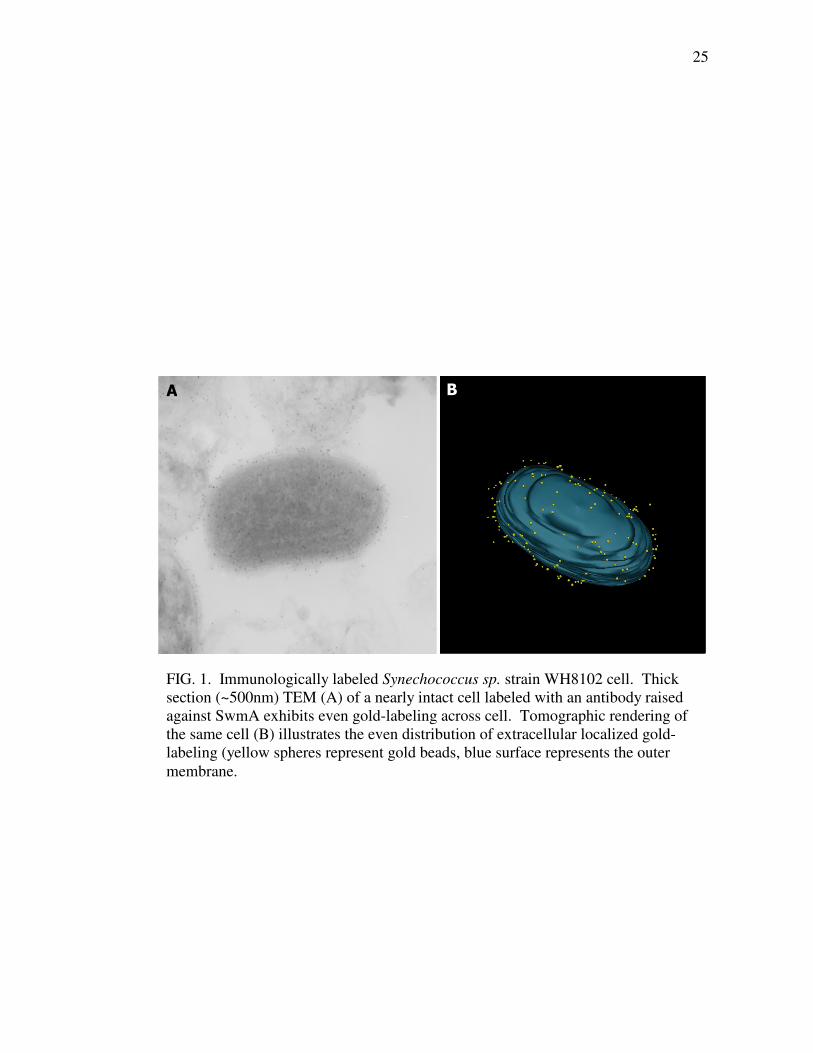
(Fig. 1A). While allowing for visualization of intact cells, interpretation of where the
labeling is positioned relative to the vertical axis of the image is difficult for these
projections through the entire cell. Tomographic reconstruction of the same cell
produces a three-dimensional image that more clearly displays the position of the gold
labeling. The tomogram shows that the anti-SwmA labeling is associated with the cell
surface (Fig. 1B). Labeling is located outside of the outer membrane (OM) yet still
closely associated with the OM. Furthermore, labeling is evenly spread across the
entire cell surface with no obvious pattern to its distribution apparent in the tomogram
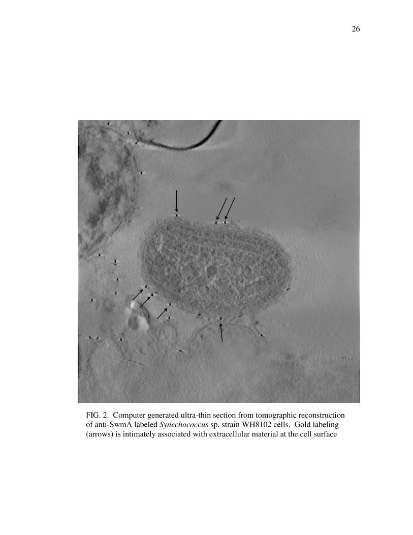
produced. Virtual ultra-thin sections produced by the 3-dimensional image processing
software XVOXTRACE also demonstrate the close association of gold particles and
the OM (Fig. 2). These virtual ultra-thin sections exhibit a diffusely staining layer of
irregular thickness external to the OM. Labeling of SwmA was always closely
associated with this extracellular material.
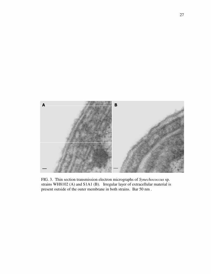
Thin sections of conventionally fixed cells also revealed diffusely stained
material on the surface of cells. Both the motile wild-type strain WH8102 and swmA
mutant strain S1A1 possess this cell-surface material (Fig. 3). Similar to the
observations from tomographic reconstruction, this diffuse staining material is
distributed around the entire cell surface of both strains in a layer of irregular
thickness. Although wild-type cells appear to possess more of the extracellular
material than do non-motile S1A1 cells, ultrastructural comparisons failed to detect
any clear, unambiguous differences between strains. Neither thin sections nor
tomographic reconstructions revealed any distinct structure present in one strain and
absent in the other.
22
Discussion
SwmA is associated with the cell envelope and is required for swimming
motility. Understanding the location and arrangement of SwmA may provide clues as
to the function of this protein. The immuno-labeling experiments presented here agree
with prior results and confirm the extracellular location of SwmA. Three-dimensional
tomographic reconstruction of a labeled Synechococcus cell provides a more detailed
picture of the surface localization of SwmA yet fails to reveal a conspicuous pattern to
its distribution. These results do however indicate the presence of some extracellular
material associated with SwmA labeling.
Chemically fixed cells failed to show an unambiguous difference between the
wild-type strain and a mutant lacking SwmA. Thin sections indicate that motile strain
WH8102 may possess more extracellular material than the non-motile swmA mutant,
but clear and reproducible differences were not observed. While these results did not
provide conclusive results, they do suggest possible structural differences between
stains, serve as a foundation for further studies, and provide a justification for
continued ultrastructural characterization, which is presented in the following chapter.
The following chapter contains additional analyses of electron microscopic
comparisons of wild-type strain WH8102 and swmA mutant strain S1A1. Those
results show that the chemical fixation techniques that have been employed here are
not sufficient for preserving the surface structures on these cells. Utilizing various
quick-freeze techniques, the outer-most layer of WH8102 cells is observed to be a
highly ordered S-layer, while S1A1 cells have some disordered fibrillar material
23
external to the OM. In retrospect, this largely explains the lack of a regular pattern to
the anti-SwmA labeling in the tomogram as the S-layer was not preserved by
glutaraldehyde fixation in these preparations. Perhaps with similar quick-freeze
fixation techniques, the regular crystalline lattice structure of the S-layer would be
observed in the distribution of the anti-SwmA gold label. Moreover, the fact that
chemical fixation does not preserve all of the cell surface structures, likely accounts
for the ambiguous differences in cell surface characteristics observed in comparisons
of WH8102 and S1A1 strains.
References
1. Brahamsha, B. 1996. An abundant cell-surface polypeptide is required for
swimming by the nonflagellated marine cyanobacterium Synechococcus. Proc.
Natl. Acad. Sci. USA. 93:6504-6509.
2. Hessler, D., S. J. Young, B. O. Carragher, M. Martone, S. Lamont, M. M.
Whittaker, R. A. Millgan, E. Masliah, J. E. Henshaw, and M. Ellisman.
1992. Programs for visualization in three-dimensional microscopy.
Neuroimage. 1:55-68.
3. McCarren, J., and B. Brahamsha. 2005. Transposon mutagenesis in a
marine Synechococcus strain: isolation of swimming motility mutants. J.
Bacteriol. 187:4457-4462.
4. Perkins, G., C. Renken, M. E. Martone, S. J. Young, M. Ellisman, and T.
Frey. 1997. Electron tomography of neuronal mitochondria: three-dimensional
structure and organization of cristae and membrane contacts. J. Struct. Biol.
119:260-272.
5. Resch, C., and J. Gibson. 1983. Isolation of the carotenoid-containing cell
wall of three unicellular cyanobacteria. J. Bacteriol. 55:345-350.
6. Rippka, R., J. B. Waterbury, and G. Cohen-Brazire. 1974. A
cyanobacterium which lacks thylakoids. Arch. Microbiol. 100:419-436.
24
7. Robb, R. A., and D. P. Hanson. 1991. A software system for interactive and
quantitative visualization of multidimensional biomedical images. Aust. Phys.
Eng. Sci. Med. 14:9-30.
8. Schroeter, J. P., and J.-P. Bretaudiere 1996. SUPRIM: easily modified
image processing software. J. Struct. Biol. 116:131-137.
9. Soto, G. E., S. J. Young, M. E. Martone, T. J. Deerinck, S. Lamont, B. O.
Carragher, K. Hama, and M. H. Ellisman. 1994. Serial section electron
tomography: A method for three-dimensional reconstruction of large
structures. NeuroImage. 1:230-243.
10. Waterbury, J. B., and J. M. Willey. 1988. Isolation and growth of marine
planktonic cyanobacteria. Meth. Enzymol. 167:100-105.
11. Waterbury, J. B., J. M. Willey, D. G. Franks, F. W. Valois, and S. W.
Watson. 1985. A cyanobacterium capable of swimming motility. Science.
230:74-76.
12. Willey, J. M. 1988. Characterization of swimming motility in a marine
cyanobacterium. Ph. D. Dissertation. Woods Hole Oceanographic Institution
and Massachusetts Institute of Technology, Cambridge, MA.
25
FIG. 1. Immunologically labeled Synechococcus sp. strain WH8102 cell. Thick
section (~500nm) TEM (A) of a nearly intact cell labeled with an antibody raised
against SwmA exhibits even gold-labeling across cell. Tomographic rendering of
the same cell (B) illustrates the even distribution of extracellular localized gold-
labeling (yellow spheres represent gold beads, blue surface represents the outer
membrane.
A B
26
FIG. 2. Computer generated ultra-thin section from tomographic reconstruction
of anti-SwmA labeled Synechococcus sp. strain WH8102 cells. Gold labeling
(arrows) is intimately associated with extracellular material at the cell surface
27
A B
FIG. 3. Thin section transmission electron micrographs of Synechococcus sp.
strains WH8102 (A) and S1A1 (B). Irregular layer of extracellular material is
present outside of the outer membrane in both strains. Bar 50 nm .