chapter 4: the musculoskeletal system - amazon s3s3.amazonaws.com/careertec/medical courses/medical...
TRANSCRIPT
Medical Terminology
Chapter 4: The Musculoskeletal System
CHAPTER OBJECTIVESThe chapter covers the musculoskeletal system, which includes bones, muscles and joints and the combining forms, terms and abbreviations used in building words that relate to it. Upon completion of this chapter, you should be able to:
• Name the parts of the musculoskeletal system and discuss their function
• Define combining forms used in building words that relate to the musculoskeletal system
• Identify the meaning of related abbreviations
• Name the common diagnoses, laboratory tests, and clinical procedures used in treating the musculoskeletal system
• Define the major pathological conditions, surgical terms, and pharmacological agents related to the musculoskeletal system
INTRODUCTION
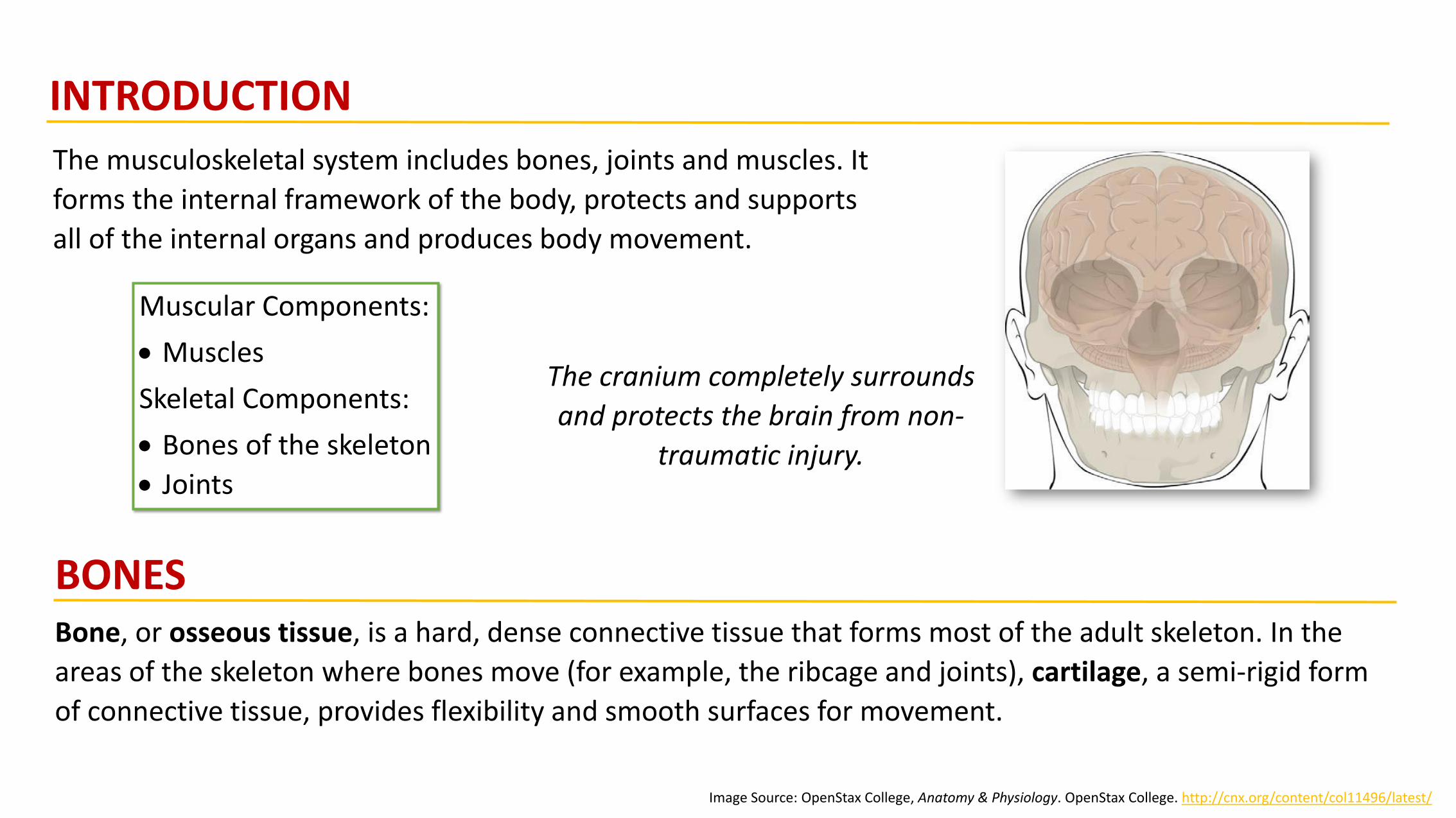
The musculoskeletal system includes bones, joints and muscles. It
forms the internal framework of the body, protects and supports
all of the internal organs and produces body movement.
Bone, or osseous tissue, is a hard, dense connective tissue that forms most of the adult skeleton. In the
areas of the skeleton where bones move (for example, the ribcage and joints), cartilage, a semi-rigid form
of connective tissue, provides flexibility and smooth surfaces for movement.
Muscular Components:
• Muscles
Skeletal Components:
• Bones of the skeleton
• Joints
The cranium completely surrounds
and protects the brain from non-
traumatic injury.
BONES
Image Source: OpenStax College, Anatomy & Physiology. OpenStax College. http://cnx.org/content/col11496/latest/
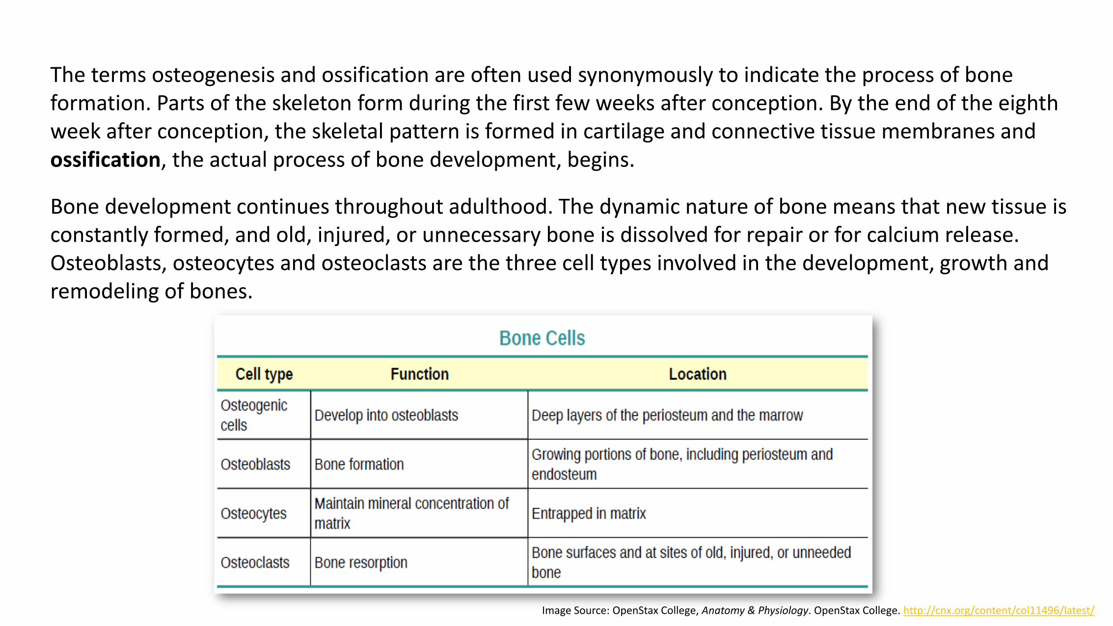
Bone development continues throughout adulthood. The dynamic nature of bone means that new tissue is constantly formed, and old, injured, or unnecessary bone is dissolved for repair or for calcium release. Osteoblasts, osteocytes and osteoclasts are the three cell types involved in the development, growth and remodeling of bones.
The terms osteogenesis and ossification are often used synonymously to indicate the process of bone formation. Parts of the skeleton form during the first few weeks after conception. By the end of the eighth week after conception, the skeletal pattern is formed in cartilage and connective tissue membranes and ossification, the actual process of bone development, begins.
Image Source: OpenStax College, Anatomy & Physiology. OpenStax College. http://cnx.org/content/col11496/latest/
Osteoblasts are bone-forming cells, osteocytes are mature bone cells and osteoclasts break down and reabsorb bone. Osteoclasts are continually breaking down old bone while osteoblasts are continually forming new bone. Even though bones stop growing in length in early adulthood, they can continue to increase in thickness or diameter throughout life in response to stress from increased muscle activity or to weight. The increase in diameter is called appositional growth. Osteoblasts in the periosteum form compact bone around the external bone surface. At the same time, osteoclasts in the endosteum break down bone on the internal bone surface, around the medullary cavity. These two processes together increase the diameter of the bone and, at the same time, keep the bone from becoming excessively heavy and bulky.
STRUCTURE OF BONE TISSUE:
Bone tissue (osseous tissue) differs greatly from other tissues in the body. Bone is hard and many of its
functions depend on that characteristic hardness. There are two types of bone tissue: compact and spongy.
Compact bone is dense so that it can withstand compressive forces, while spongy (cancellous) bone has
open spaces and supports shifts in weight distribution.
Link to Learning: Please read, Structure of Bone Tissue, to
learn more about the two types of bone tissue.
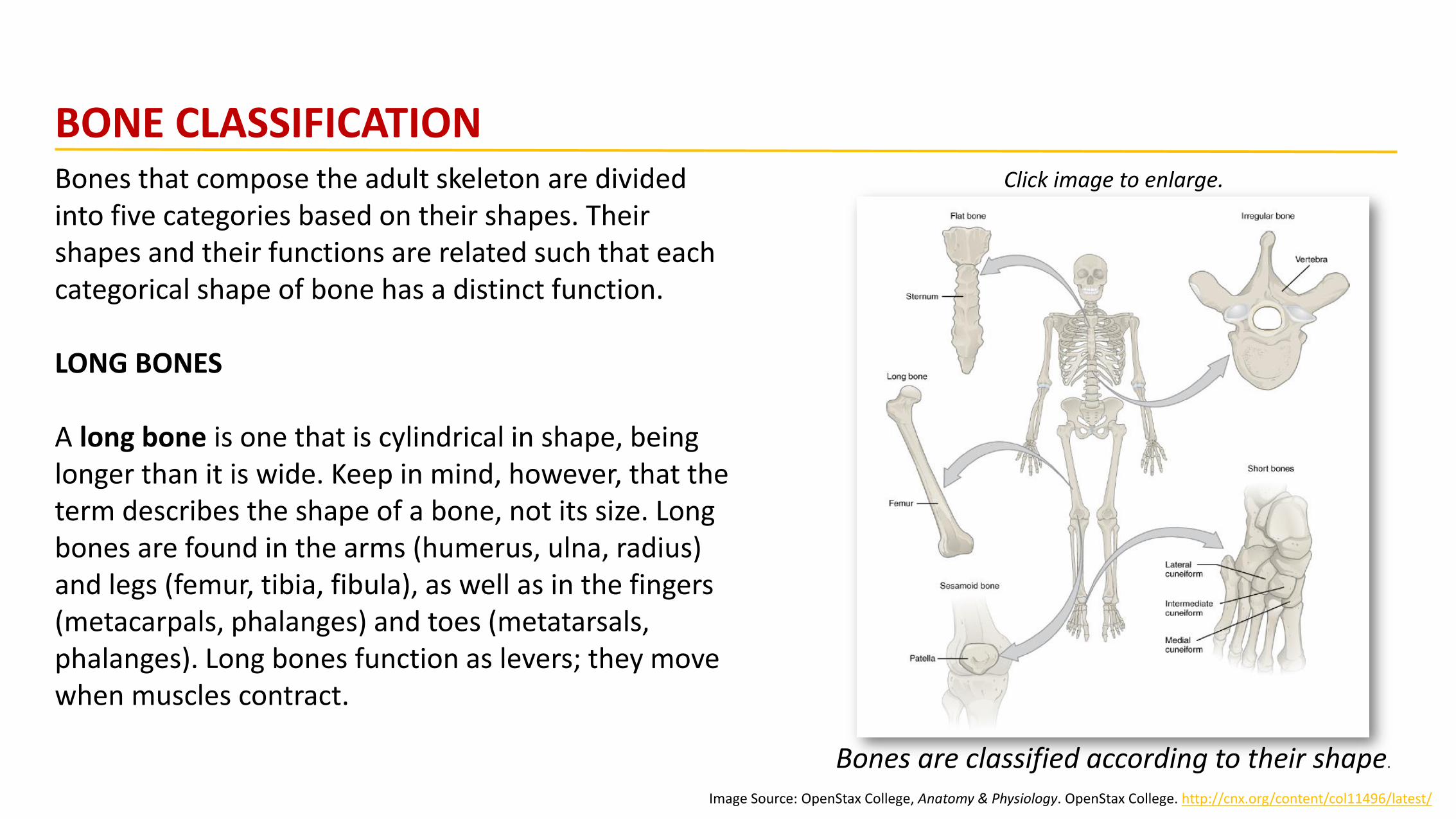
Bones that compose the adult skeleton are divided into five categories based on their shapes. Their shapes and their functions are related such that each categorical shape of bone has a distinct function.
LONG BONES
A long bone is one that is cylindrical in shape, being longer than it is wide. Keep in mind, however, that the term describes the shape of a bone, not its size. Long bones are found in the arms (humerus, ulna, radius) and legs (femur, tibia, fibula), as well as in the fingers (metacarpals, phalanges) and toes (metatarsals, phalanges). Long bones function as levers; they move when muscles contract.
Image Source: OpenStax College, Anatomy & Physiology. OpenStax College. http://cnx.org/content/col11496/latest/
BONE CLASSIFICATION
Bones are classified according to their shape.
Click image to enlarge.
SHORT BONES
A short bone is one that is cube-like in shape, being approximately equal in length, width, and thickness.
The only short bones in the human skeleton are in the carpals of the wrists and the tarsals of the ankles.
Short bones provide stability and support as well as some limited motion.
FLAT BONES
The term “flat bone” is somewhat of a misnomer because, although a flat bone is typically thin, it is also
often curved. Examples include the cranial (skull) bones, the scapulae (shoulder blades), the sternum
(breastbone), and the ribs. Flat bones serve as points of attachment for muscles and often protect internal
organs.
IRREGULAR BONES
An irregular bone is one that does not have any easily characterized shape and therefore does not fit any
other classification. These bones tend to have more complex shapes, like the vertebrae that support the
spinal cord and protect it from compressive forces. Many facial bones, particularly the ones containing
sinuses, are classified as irregular bones.
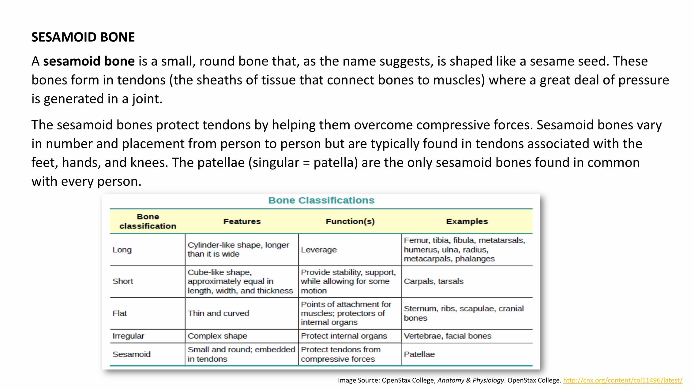
SESAMOID BONE
A sesamoid bone is a small, round bone that, as the name suggests, is shaped like a sesame seed. These
bones form in tendons (the sheaths of tissue that connect bones to muscles) where a great deal of pressure
is generated in a joint.
The sesamoid bones protect tendons by helping them overcome compressive forces. Sesamoid bones vary
in number and placement from person to person but are typically found in tendons associated with the
feet, hands, and knees. The patellae (singular = patella) are the only sesamoid bones found in common
with every person.
Image Source: OpenStax College, Anatomy & Physiology. OpenStax College. http://cnx.org/content/col11496/latest/
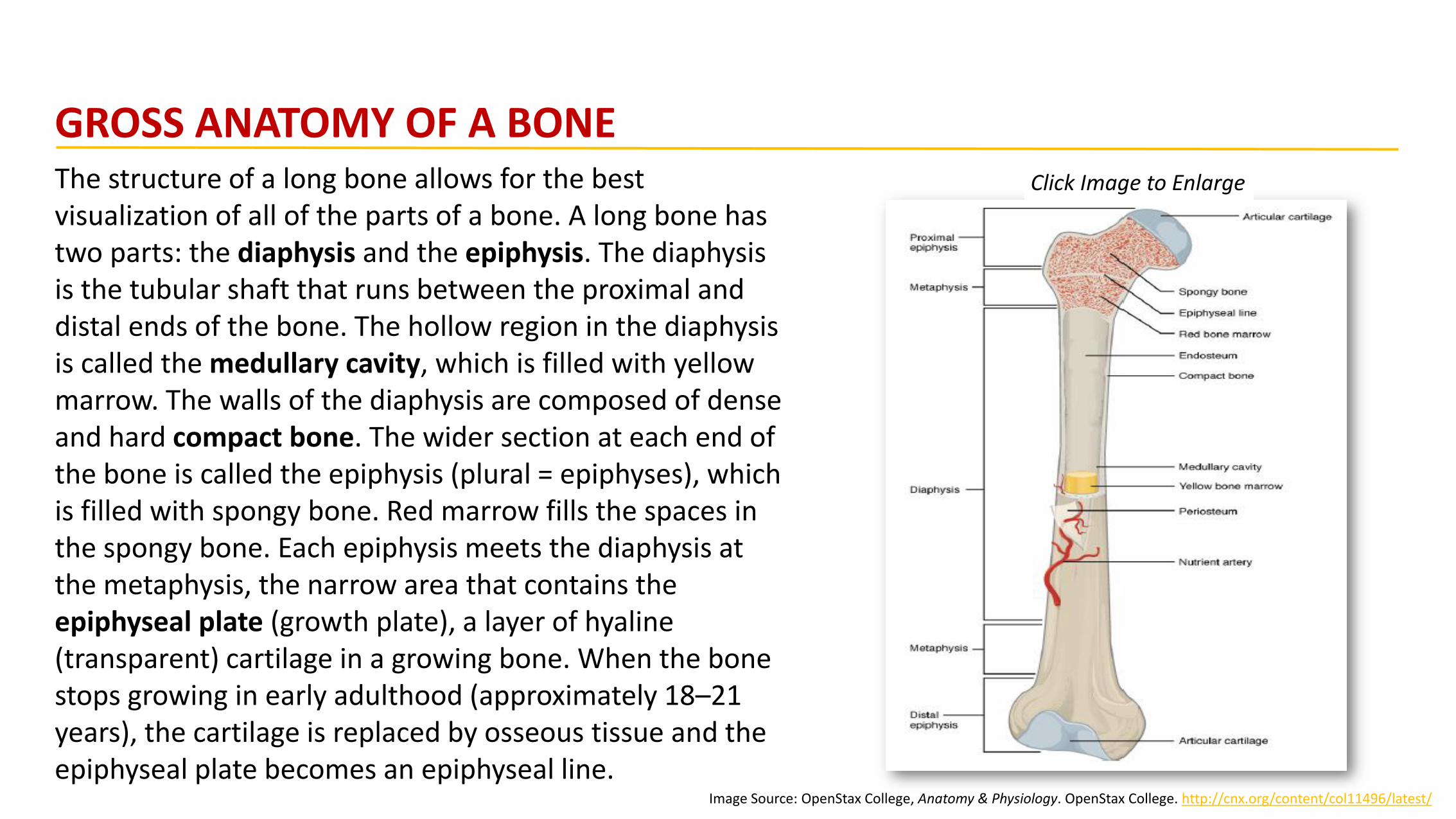
The structure of a long bone allows for the best visualization of all of the parts of a bone. A long bone has two parts: the diaphysis and the epiphysis. The diaphysis is the tubular shaft that runs between the proximal and distal ends of the bone. The hollow region in the diaphysis is called the medullary cavity, which is filled with yellow marrow. The walls of the diaphysis are composed of dense and hard compact bone. The wider section at each end of the bone is called the epiphysis (plural = epiphyses), which is filled with spongy bone. Red marrow fills the spaces in the spongy bone. Each epiphysis meets the diaphysis at the metaphysis, the narrow area that contains the epiphyseal plate (growth plate), a layer of hyaline (transparent) cartilage in a growing bone. When the bone stops growing in early adulthood (approximately 18–21 years), the cartilage is replaced by osseous tissue and the epiphyseal plate becomes an epiphyseal line.
Image Source: OpenStax College, Anatomy & Physiology. OpenStax College. http://cnx.org/content/col11496/latest/
GROSS ANATOMY OF A BONEClick Image to Enlarge

BONE MARKINGS
A projection is an area of a bone that projects above the surface of the bone. These are the attachment points for tendons and ligaments.
The surface features of bones depend on their function,
location, attachment of ligaments and tendons, or the
penetration of blood vessels and nerves.
The surface features of bones vary considerably,
depending on the function and location in the body.
There are three general classes of bone markings:
(1) articulations(2) projections(3) holes
As the name implies, an articulation is where two bone surfaces come together (articulus = “joint”).
Click Image to Enlarge
Image Source: OpenStax College, Anatomy & Physiology. OpenStax College. http://cnx.org/content/col11496/latest/
A hole is an opening or groove in the bone that allows blood vessels and nerves to enter the bone.
Bone also serves as a site for fat storage and blood cell production. The softer connective tissue that fills the interior of most bone is referred to as bone marrow. There are two types of bone marrow: yellow marrow and red marrow.
Image Source: OpenStax College, Anatomy & Physiology. OpenStax College. http://cnx.org/content/col11496/latest/
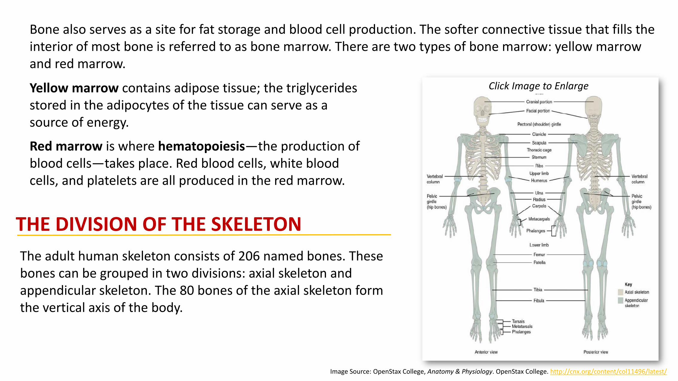
THE DIVISION OF THE SKELETON
The adult human skeleton consists of 206 named bones. These bones can be grouped in two divisions: axial skeleton and appendicular skeleton. The 80 bones of the axial skeleton form the vertical axis of the body.
Yellow marrow contains adipose tissue; the triglycerides stored in the adipocytes of the tissue can serve as a source of energy.
Red marrow is where hematopoiesis—the production of blood cells—takes place. Red blood cells, white blood cells, and platelets are all produced in the red marrow.
Click Image to Enlarge
The axial skeleton forms the vertical, central axis of the body and includes all bones of the head, neck, chest, and back. It serves to protect the brain, spinal cord, heart, and lungs. It also serves as the attachment site for muscles that move the head, neck, and back, and for muscles that act across the shoulder and hip joints to move their corresponding limbs.
The appendicular skeleton includes all bones of the upper and lower limbs, plus the bones that attach each limb to the axial skeleton. There are 126 bones in the appendicular skeleton of an adult.
Image Source: OpenStax College, Anatomy & Physiology. OpenStax College. http://cnx.org/content/col11496/latest/
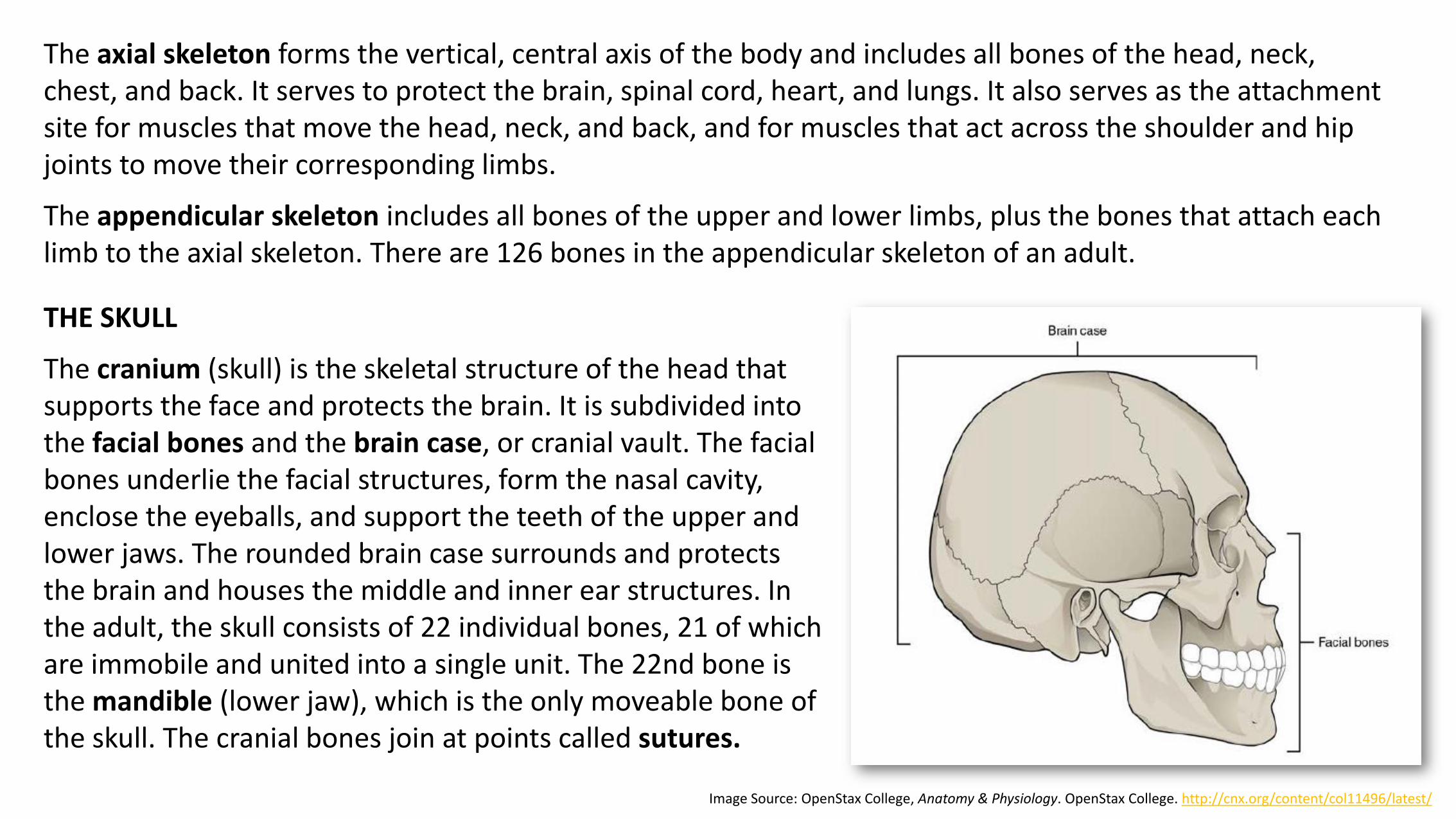
THE SKULL
The cranium (skull) is the skeletal structure of the head that supports the face and protects the brain. It is subdivided into the facial bones and the brain case, or cranial vault. The facial bones underlie the facial structures, form the nasal cavity, enclose the eyeballs, and support the teeth of the upper and lower jaws. The rounded brain case surrounds and protects the brain and houses the middle and inner ear structures. In the adult, the skull consists of 22 individual bones, 21 of which are immobile and united into a single unit. The 22nd bone is the mandible (lower jaw), which is the only moveable bone of the skull. The cranial bones join at points called sutures.
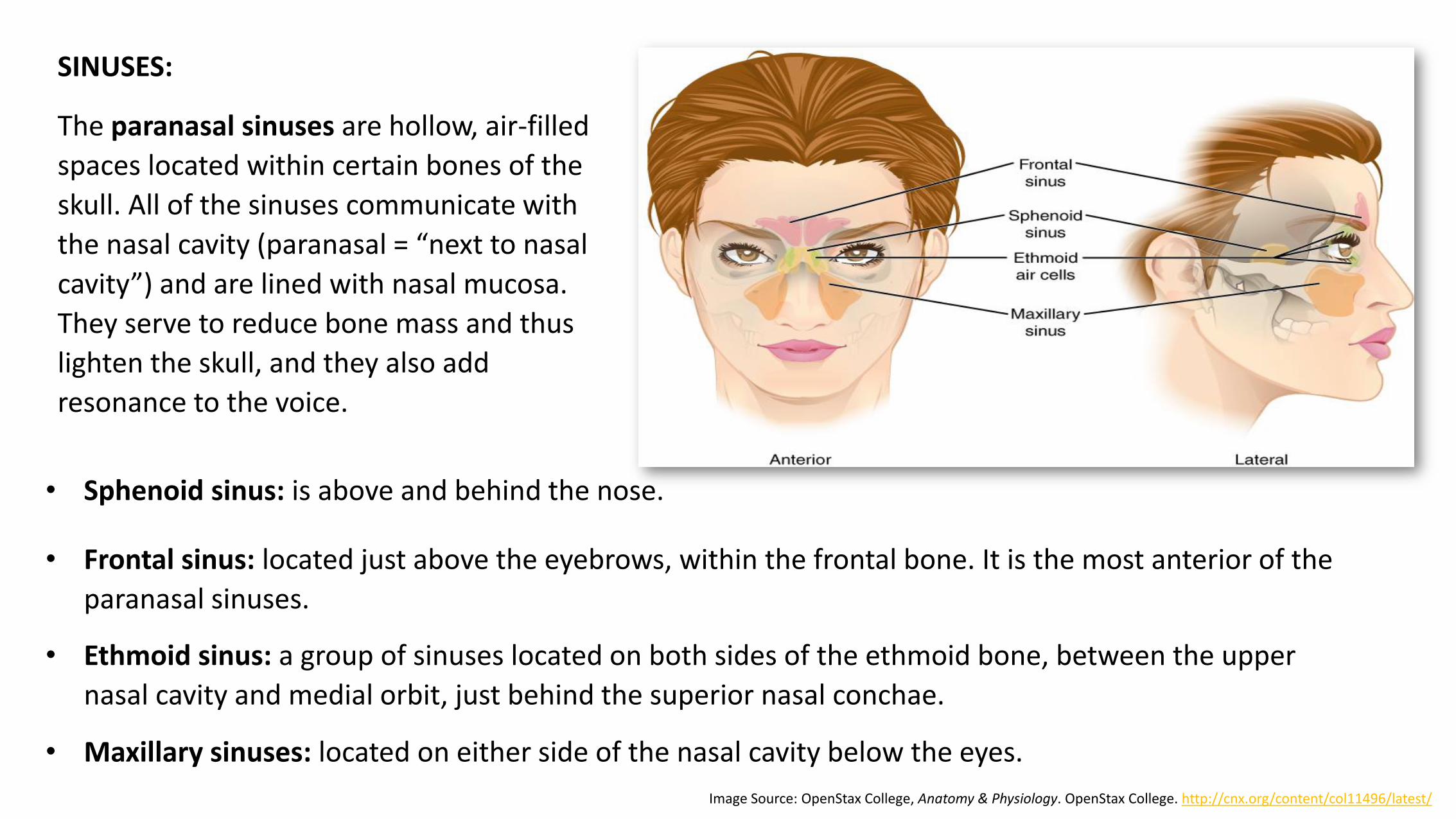
SINUSES:
The paranasal sinuses are hollow, air-filled
spaces located within certain bones of the
skull. All of the sinuses communicate with
the nasal cavity (paranasal = “next to nasal
cavity”) and are lined with nasal mucosa.
They serve to reduce bone mass and thus
lighten the skull, and they also add
resonance to the voice.
• Frontal sinus: located just above the eyebrows, within the frontal bone. It is the most anterior of the
paranasal sinuses.
Image Source: OpenStax College, Anatomy & Physiology. OpenStax College. http://cnx.org/content/col11496/latest/
• Sphenoid sinus: is above and behind the nose.
• Ethmoid sinus: a group of sinuses located on both sides of the ethmoid bone, between the upper
nasal cavity and medial orbit, just behind the superior nasal conchae.
• Maxillary sinuses: located on either side of the nasal cavity below the eyes.
For a more detailed overview of the bones in the skull, please watch the following video “Skull Bones” and then complete the Link to Learning material below. You will not be tested on this material in this course.
Link to Learning: Review these interactive tutorials of the human skull:
• Anterior View of the Skull Bones.
• Lateral View of the Skill Bones
• Posterior View of the Skull Bones
Then test your knowledge by completing this interactive quiz, WebAnatomy: Skull: Lateral View.

HYOID BONE
The hyoid bone is an independent bone that does not contact any other bone and thus is not part of the skull. It is a small U-shaped bone located in the upper neck near the level of the inferior mandible, with the tips of the “U” pointing posteriorly. The hyoid serves as the base for the tongue above, and is attached to the larynx below and the pharynx posteriorly. Movements of the hyoid are coordinated with movements of the tongue, larynx, and pharynx during swallowing and speaking.
Image Source: OpenStax College, Anatomy & Physiology. OpenStax College. http://cnx.org/content/col11496/latest/
THE VERTEBRAL COLUMN
The vertebral column is also known as the spinal column or spine. It consists of a sequence of vertebrae
(singular = vertebra), each of which is separated and united by an intervertebral disc. Together, the
vertebrae and intervertebral discs form the vertebral column. It is a flexible column that supports the
head, neck, and body and allows for their movements. It also protects the spinal cord, which passes down
the back through openings in the vertebrae.
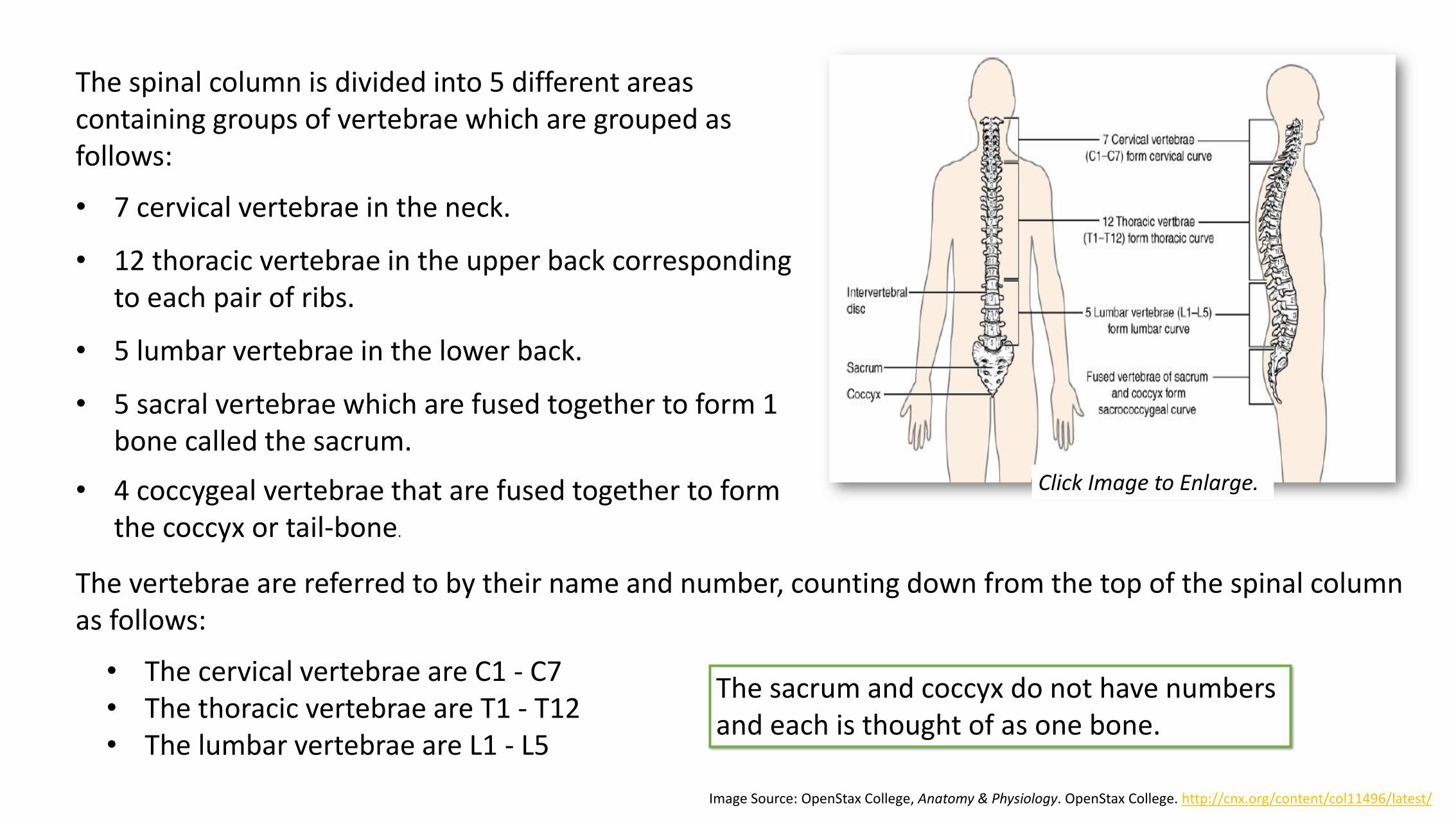
The spinal column is divided into 5 different areas containing groups of vertebrae which are grouped as follows:
• 7 cervical vertebrae in the neck.
• 12 thoracic vertebrae in the upper back corresponding to each pair of ribs.
• 5 lumbar vertebrae in the lower back.
• 5 sacral vertebrae which are fused together to form 1 bone called the sacrum.
Image Source: OpenStax College, Anatomy & Physiology. OpenStax College. http://cnx.org/content/col11496/latest/
The vertebrae are referred to by their name and number, counting down from the top of the spinal column as follows:
• The cervical vertebrae are C1 - C7• The thoracic vertebrae are T1 - T12• The lumbar vertebrae are L1 - L5
• 4 coccygeal vertebrae that are fused together to form the coccyx or tail-bone.
The sacrum and coccyx do not have numbers and each is thought of as one bone.
Click Image to Enlarge.

THE THORACIC CAGE
The thoracic cage (rib cage) forms the thorax (chest) portion of the body. It consists of the 12 pairs of ribs with their costal cartilages and the sternum. The ribs are anchored posteriorly to the 12 thoracic vertebrae (T1–T12). The thoracic cage protects the heart and lungs.
The sternum is the elongated bony structure that anchors the anterior thoracic cage. Each rib is a curved, flattened bone that contributes to the wall of the thorax. Ribs 1–7 are classified as true ribs (vertebrosternal ribs). The costal cartilage from each of these ribs attaches directly to the sternum. Ribs 8–12 are called false ribs (vertebrochondral ribs). The costal cartilages from these ribs do not attach directly to the sternum.
Image Source: OpenStax College, Anatomy & Physiology. OpenStax College. http://cnx.org/content/col11496/latest/
Link to Learning: Please watch: spineuniverse:
Spinal Anatomy Animation, for a review of the
anatomy of the spine.
Click image to enlarge.
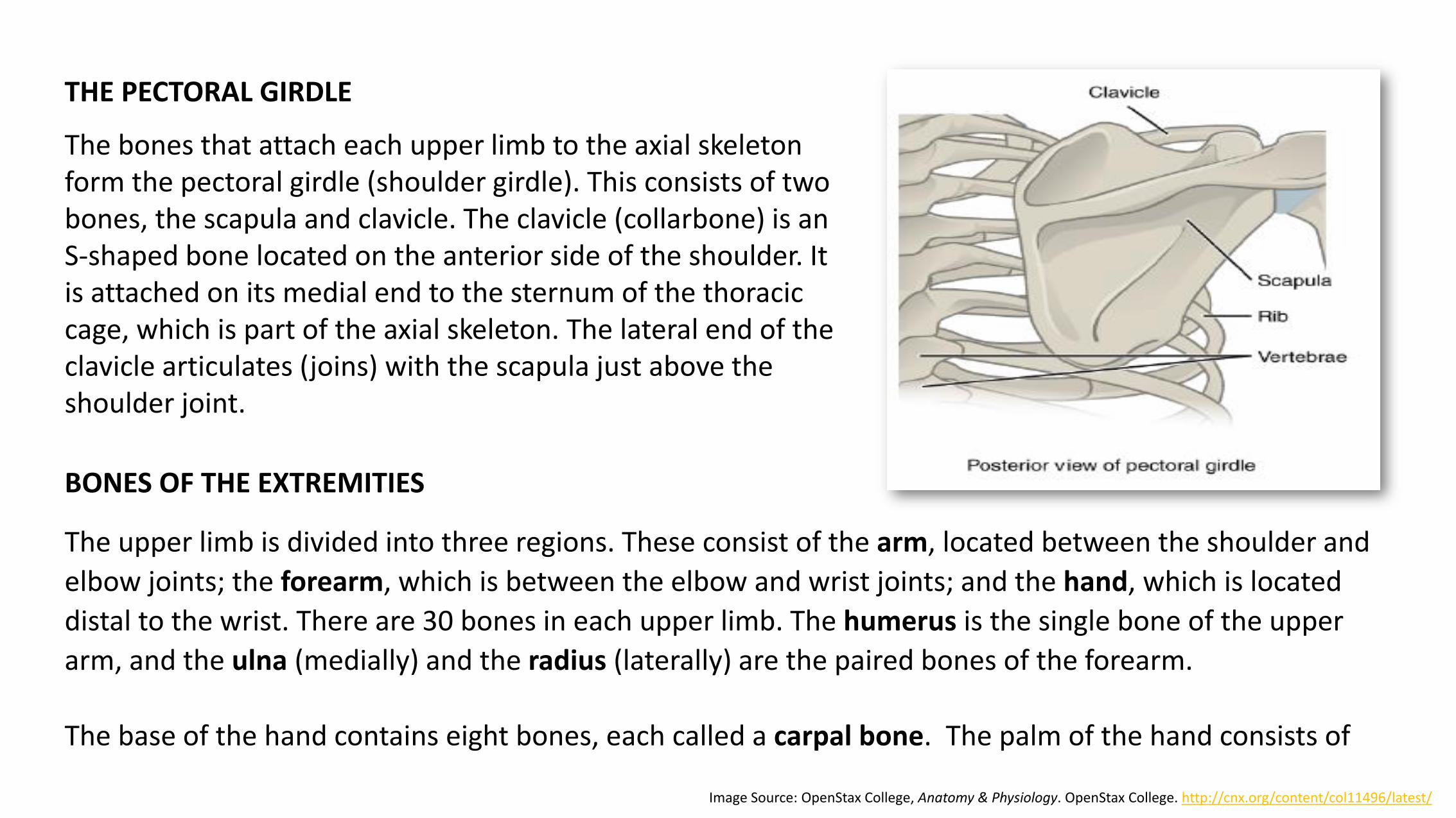
THE PECTORAL GIRDLE
The bones that attach each upper limb to the axial skeleton form the pectoral girdle (shoulder girdle). This consists of two bones, the scapula and clavicle. The clavicle (collarbone) is an S-shaped bone located on the anterior side of the shoulder. It is attached on its medial end to the sternum of the thoracic cage, which is part of the axial skeleton. The lateral end of the clavicle articulates (joins) with the scapula just above the shoulder joint.
Image Source: OpenStax College, Anatomy & Physiology. OpenStax College. http://cnx.org/content/col11496/latest/
BONES OF THE EXTREMITIES
The upper limb is divided into three regions. These consist of the arm, located between the shoulder and
elbow joints; the forearm, which is between the elbow and wrist joints; and the hand, which is located
distal to the wrist. There are 30 bones in each upper limb. The humerus is the single bone of the upper
arm, and the ulna (medially) and the radius (laterally) are the paired bones of the forearm.
The base of the hand contains eight bones, each called a carpal bone. The palm of the hand consists of

five bones, each called a metacarpal bone. The fingers and thumb contain a total of 14 bones, each of which is a phalanx bone of the hand. The distal end of the humerushas two articulation areas, which join the ulna and radius bones of the forearm to form the elbow joint.
Like the upper limb, the lower limb is divided into three regions. The thigh is that portion of the lower limb located between the hip joint and knee joint. The leg is specifically the region between the knee joint and the ankle joint. Distal to the ankle is the foot. The lower limb contains 30 bones. These are the femur, patella, tibia, fibula, tarsal bones, metatarsal bones, and phalanges. The femur is the single bone of the thigh and it is the longest bone in
Image Source: upload.wikimedia.org
the body. The patella is the kneecap and articulates with the distal femur. The tibia is the larger, weight-bearing bone located on the medial side of the leg, and the fibula is the thin bone of the lateral leg. The large expansion found on the medial side of the distal tibia is the medial malleolus (“little hammer”). This forms the large bony bump found on the medial side of the ankle region.
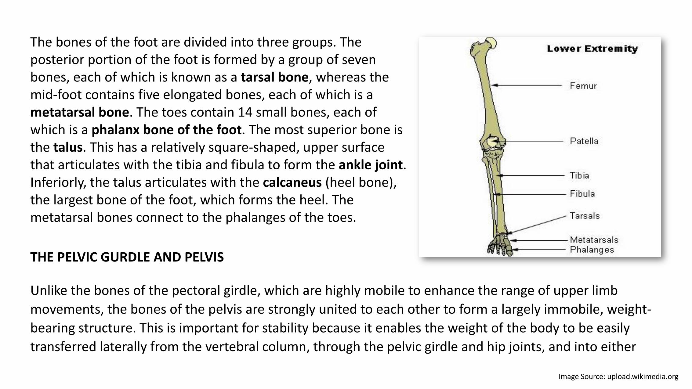
Image Source: upload.wikimedia.org
The bones of the foot are divided into three groups. The posterior portion of the foot is formed by a group of seven bones, each of which is known as a tarsal bone, whereas the mid-foot contains five elongated bones, each of which is a metatarsal bone. The toes contain 14 small bones, each of which is a phalanx bone of the foot. The most superior bone is the talus. This has a relatively square-shaped, upper surface that articulates with the tibia and fibula to form the ankle joint. Inferiorly, the talus articulates with the calcaneus (heel bone), the largest bone of the foot, which forms the heel. The metatarsal bones connect to the phalanges of the toes.
THE PELVIC GURDLE AND PELVIS
Unlike the bones of the pectoral girdle, which are highly mobile to enhance the range of upper limb
movements, the bones of the pelvis are strongly united to each other to form a largely immobile, weight-
bearing structure. This is important for stability because it enables the weight of the body to be easily
transferred laterally from the vertebral column, through the pelvic girdle and hip joints, and into either
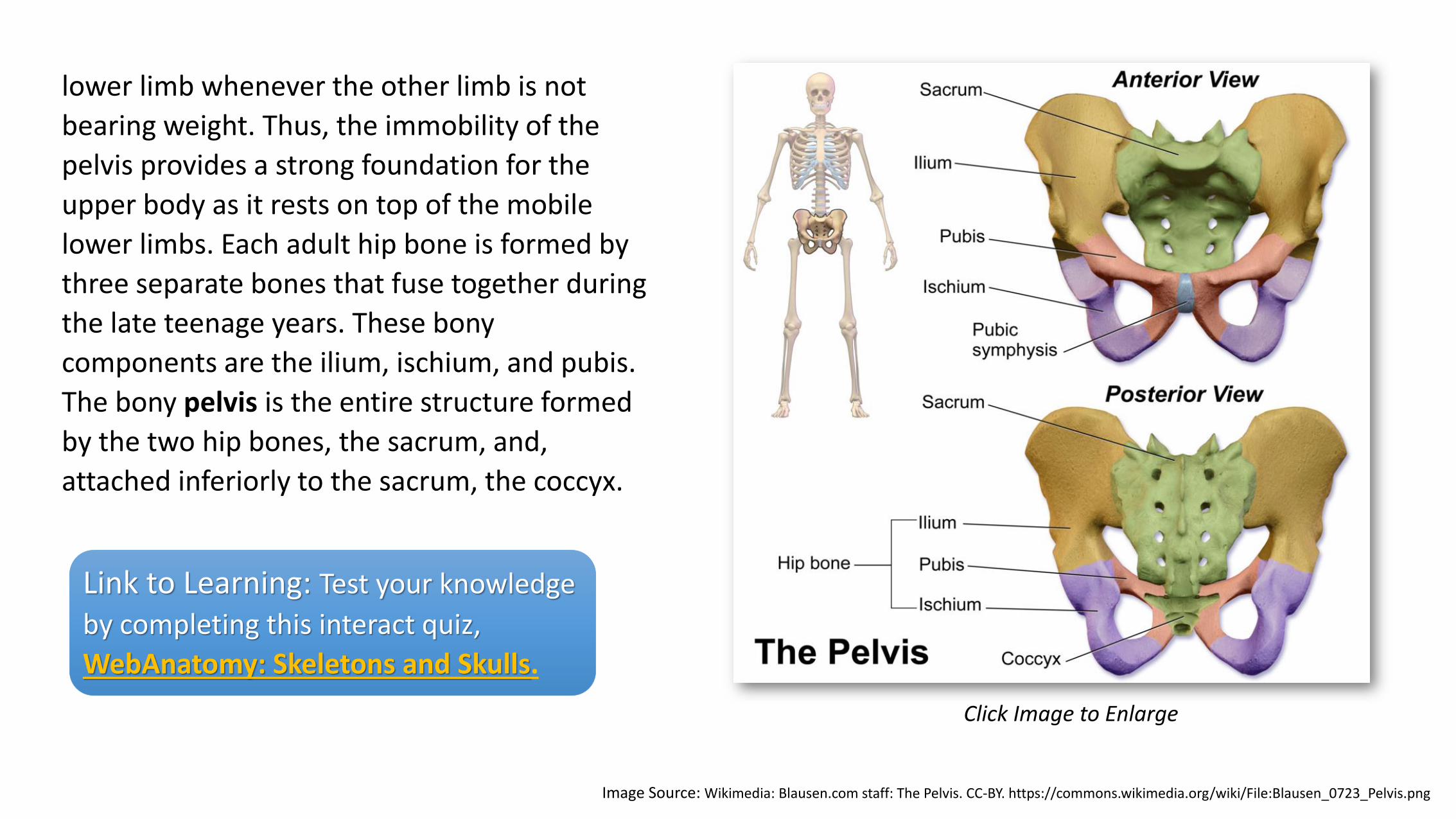
Image Source: Wikimedia: Blausen.com staff: The Pelvis. CC-BY. https://commons.wikimedia.org/wiki/File:Blausen_0723_Pelvis.png
lower limb whenever the other limb is not
bearing weight. Thus, the immobility of the
pelvis provides a strong foundation for the
upper body as it rests on top of the mobile
lower limbs. Each adult hip bone is formed by
three separate bones that fuse together during
the late teenage years. These bony
components are the ilium, ischium, and pubis.
The bony pelvis is the entire structure formed
by the two hip bones, the sacrum, and,
attached inferiorly to the sacrum, the coccyx.
Click Image to Enlarge
Link to Learning: Test your knowledge
by completing this interact quiz,
WebAnatomy: Skeletons and Skulls.
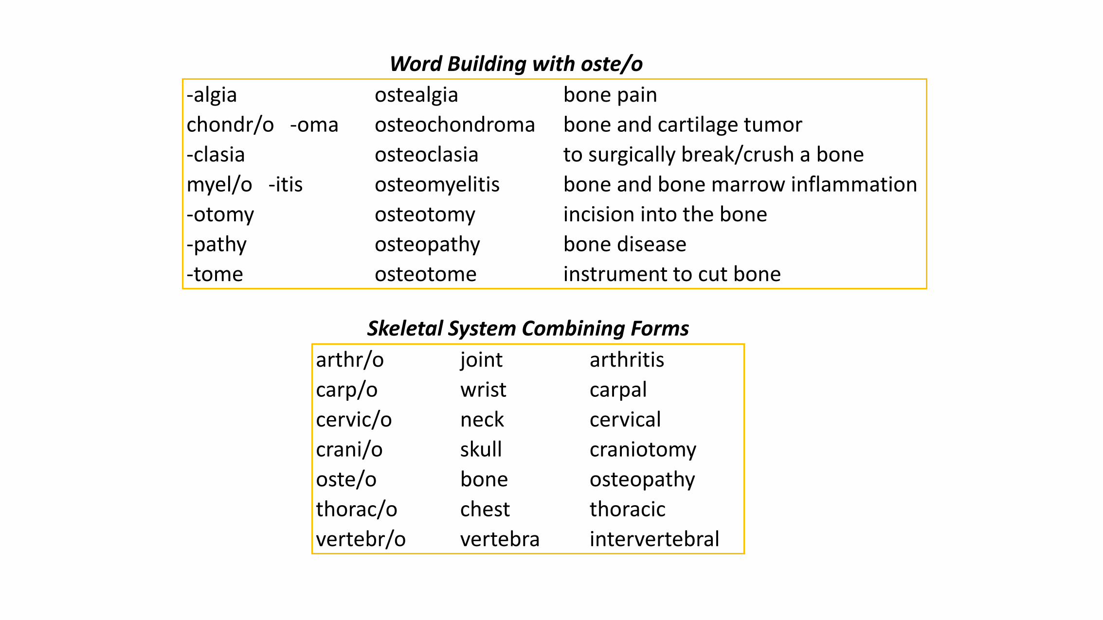
-algia ostealgia bone pain
chondr/o -oma osteochondroma bone and cartilage tumor
-clasia osteoclasia to surgically break/crush a bone
myel/o -itis osteomyelitis bone and bone marrow inflammation
-otomy osteotomy incision into the bone
-pathy osteopathy bone disease
-tome osteotome instrument to cut bone
Word Building with oste/o
Skeletal System Combining Forms
arthr/o joint arthritis
carp/o wrist carpal
cervic/o neck cervical
crani/o skull craniotomy
oste/o bone osteopathy
thorac/o chest thoracic
vertebr/o vertebra intervertebral

Adjective forms of bone names
iliac ilium tibial tibia
carpal carpus clavicular clavicle
cervical neck fibular fibula
costal rib lumbar low back
cranial cranium mandibular mandible
femoral femur patellar patella
humeral humerus scapular scapula
ischial ischium ulnar ulna
metacarpal metacarpus maxillary maxilla
metatarsal metatarsus coccygeal coccyx
radial radius phalangeal phalanges
sacral sacrum pelvic pelvis
sternal sternum pubic pubis
tarsal tarsus thoracic thorax
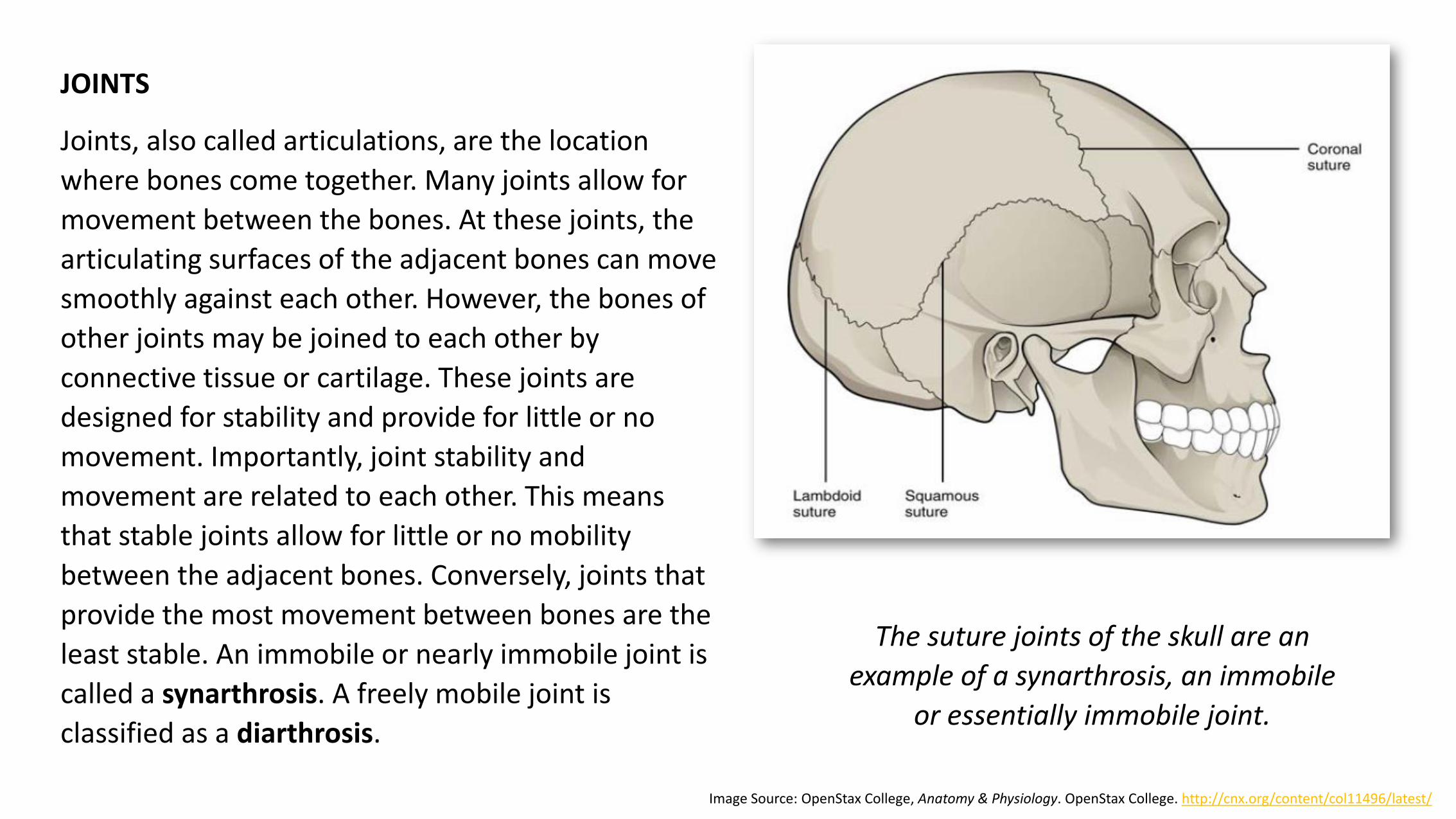
JOINTS
Joints, also called articulations, are the location
where bones come together. Many joints allow for
movement between the bones. At these joints, the
articulating surfaces of the adjacent bones can move
smoothly against each other. However, the bones of
other joints may be joined to each other by
connective tissue or cartilage. These joints are
designed for stability and provide for little or no
movement. Importantly, joint stability and
movement are related to each other. This means
that stable joints allow for little or no mobility
between the adjacent bones. Conversely, joints that
provide the most movement between bones are the
least stable. An immobile or nearly immobile joint is
called a synarthrosis. A freely mobile joint is
classified as a diarthrosis.
The suture joints of the skull are an
example of a synarthrosis, an immobile
or essentially immobile joint.
Image Source: OpenStax College, Anatomy & Physiology. OpenStax College. http://cnx.org/content/col11496/latest/
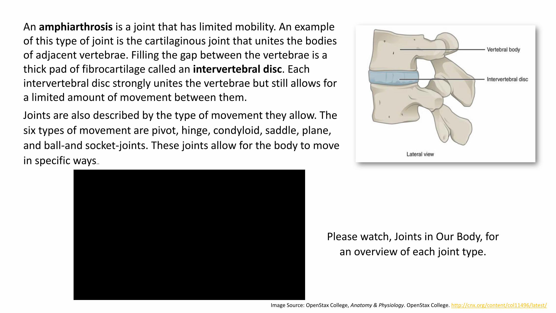
An amphiarthrosis is a joint that has limited mobility. An example of this type of joint is the cartilaginous joint that unites the bodies of adjacent vertebrae. Filling the gap between the vertebrae is a thick pad of fibrocartilage called an intervertebral disc. Each intervertebral disc strongly unites the vertebrae but still allows for a limited amount of movement between them.
Image Source: OpenStax College, Anatomy & Physiology. OpenStax College. http://cnx.org/content/col11496/latest/
Joints are also described by the type of movement they allow. The
six types of movement are pivot, hinge, condyloid, saddle, plane,
and ball-and socket-joints. These joints allow for the body to move
in specific ways..
Please watch, Joints in Our Body, for
an overview of each joint type.

Image Source: OpenStax College, Anatomy & Physiology. OpenStax College. http://cnx.org/content/col11496/latest/
Click Image to Enlarge.
Outside of their articulating surfaces, the
bones are connected together by
ligaments, which are strong bands of
fibrous connective tissue. These strengthen
and support the joint by anchoring the
bones together and preventing their
separation. Ligaments allow for normal
movements at a joint, but limit the range of
these motions, thus preventing excessive
or abnormal joint movements.
Synovial joints are characterized by the
presence of a joint cavity. They are covered
with a thin synovial membrane. The cells
of this membrane secrete synovial fluid
(synovia = “a thick fluid”), a thick, slimy
fluid that provides lubrication to further
reduce friction between the bones of the
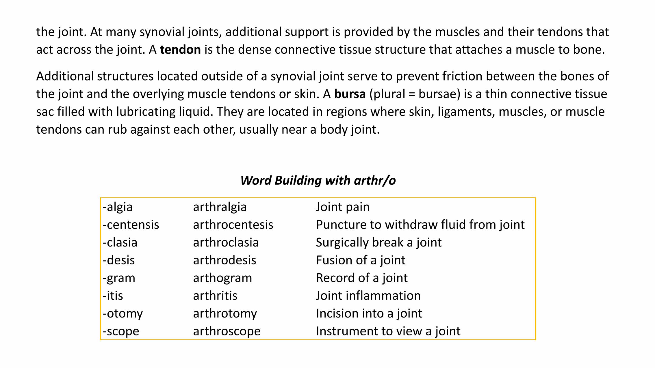
the joint. At many synovial joints, additional support is provided by the muscles and their tendons that
act across the joint. A tendon is the dense connective tissue structure that attaches a muscle to bone.
Additional structures located outside of a synovial joint serve to prevent friction between the bones of
the joint and the overlying muscle tendons or skin. A bursa (plural = bursae) is a thin connective tissue
sac filled with lubricating liquid. They are located in regions where skin, ligaments, muscles, or muscle
tendons can rub against each other, usually near a body joint.
-algia arthralgia Joint pain
-centensis arthrocentesis Puncture to withdraw fluid from joint
-clasia arthroclasia Surgically break a joint
-desis arthrodesis Fusion of a joint
-gram arthogram Record of a joint
-itis arthritis Joint inflammation
-otomy arthrotomy Incision into a joint
-scope arthroscope Instrument to view a joint
Word Building with arthr/o
Please watch, Synovial Joints, to see an animation of each movement.
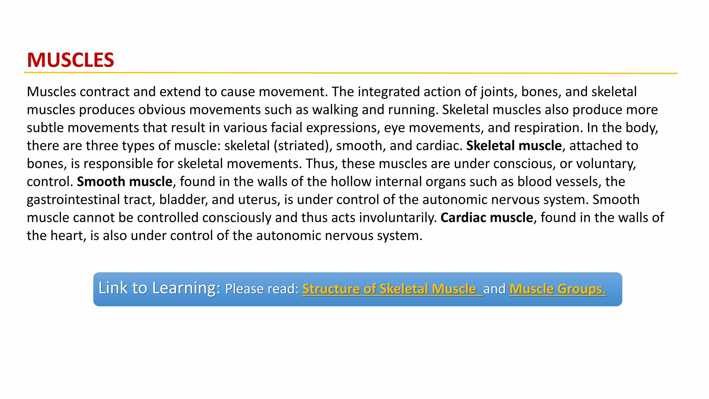
MUSCLES
Muscles contract and extend to cause movement. The integrated action of joints, bones, and skeletal muscles produces obvious movements such as walking and running. Skeletal muscles also produce more subtle movements that result in various facial expressions, eye movements, and respiration. In the body, there are three types of muscle: skeletal (striated), smooth, and cardiac. Skeletal muscle, attached to bones, is responsible for skeletal movements. Thus, these muscles are under conscious, or voluntary, control. Smooth muscle, found in the walls of the hollow internal organs such as blood vessels, the gastrointestinal tract, bladder, and uterus, is under control of the autonomic nervous system. Smooth muscle cannot be controlled consciously and thus acts involuntarily. Cardiac muscle, found in the walls of the heart, is also under control of the autonomic nervous system.
Link to Learning: Please read: Structure of Skeletal Muscle and Muscle Groups.
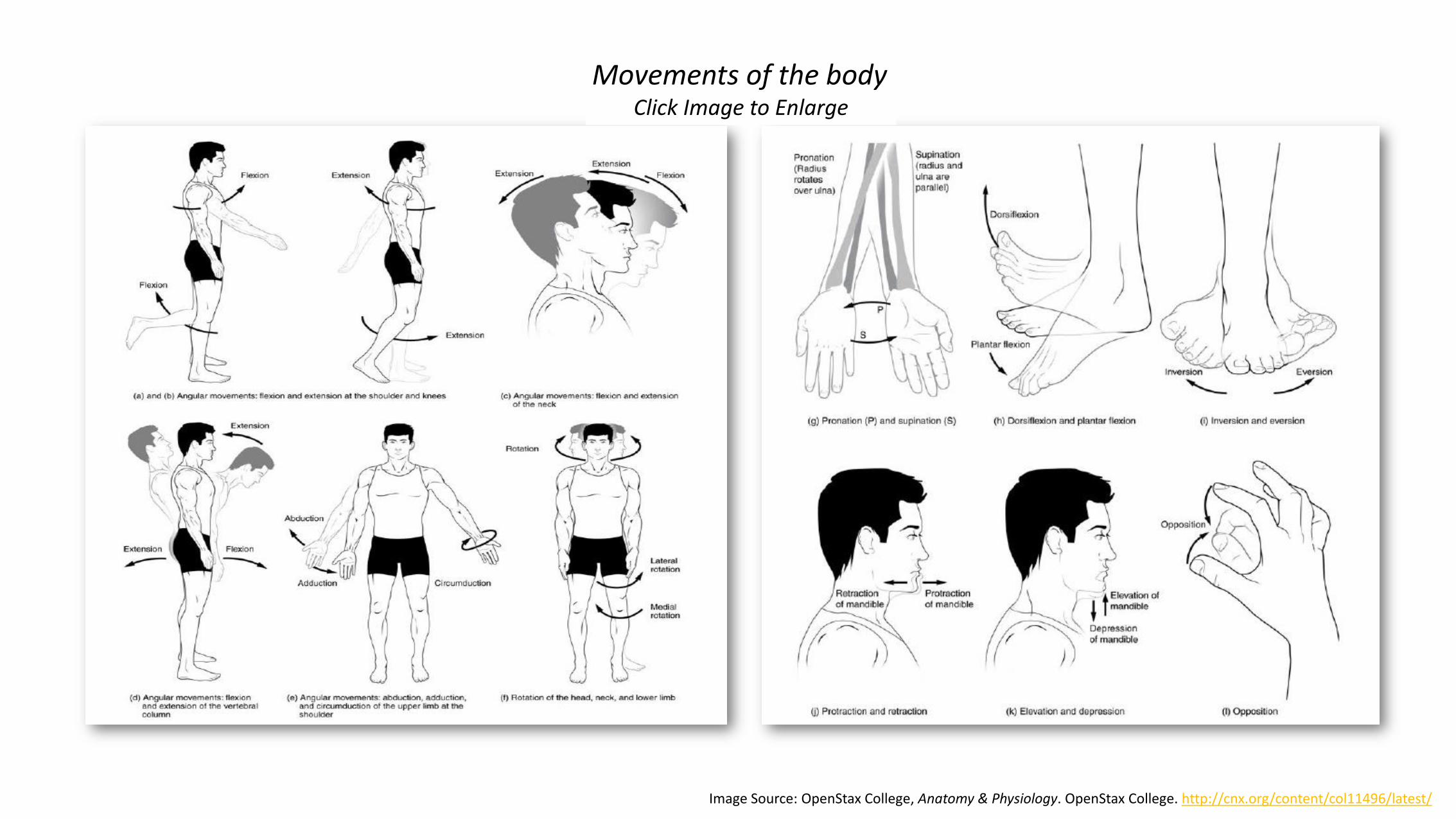
Image Source: OpenStax College, Anatomy & Physiology. OpenStax College. http://cnx.org/content/col11496/latest/
Movements of the bodyClick Image to Enlarge

Please watch “Anatomical Terms of Movement” to see an animation of the different types of body movements.
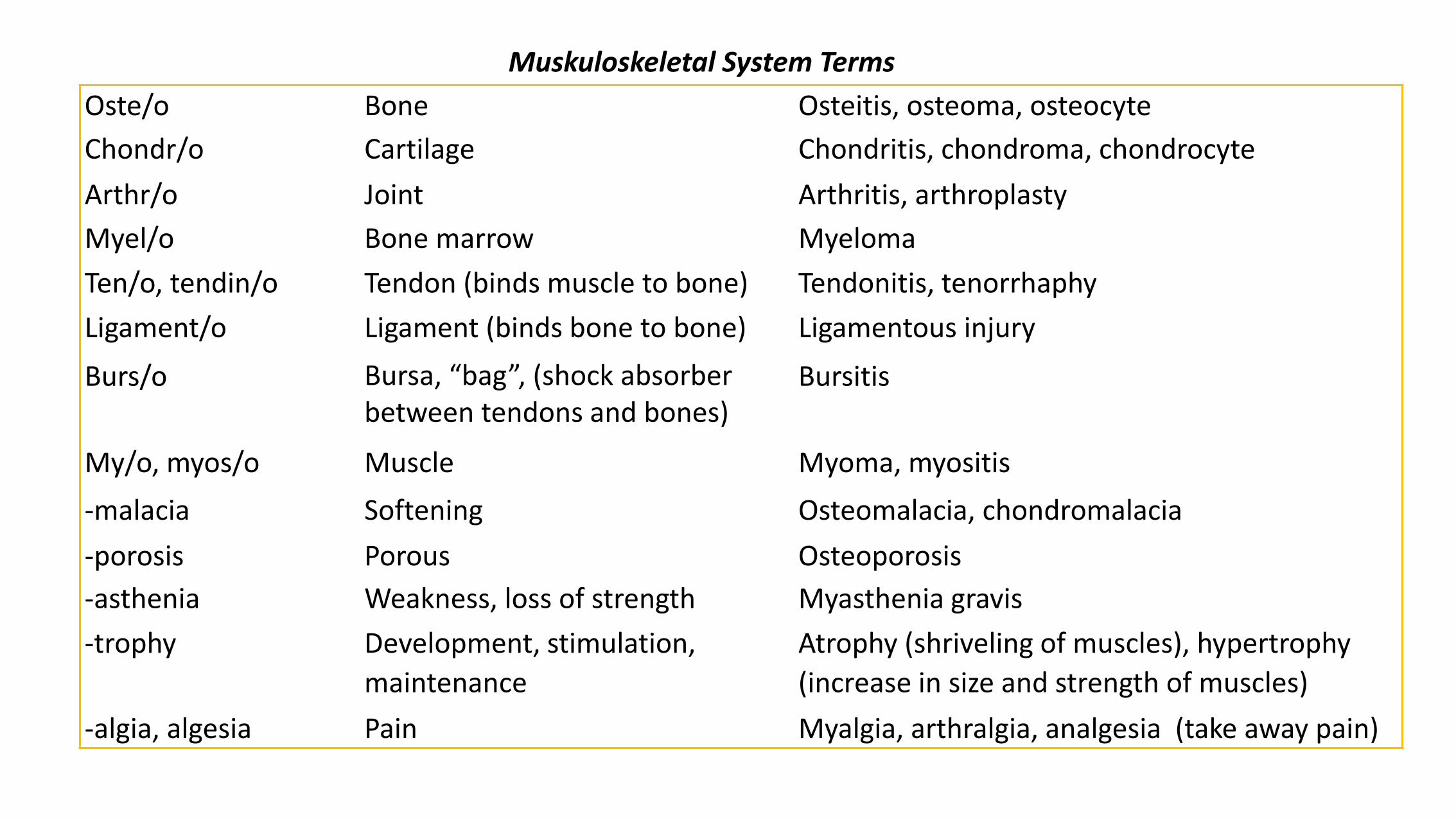
Muskuloskeletal System Terms
Oste/o Bone Osteitis, osteoma, osteocyte
Chondr/o Cartilage Chondritis, chondroma, chondrocyte
Arthr/o Joint Arthritis, arthroplasty
Myel/o Bone marrow Myeloma
Ten/o, tendin/o Tendon (binds muscle to bone) Tendonitis, tenorrhaphy
Ligament/o Ligament (binds bone to bone) Ligamentous injury
Burs/o Bursa, “bag”, (shock absorber between tendons and bones)
Bursitis
My/o, myos/o Muscle Myoma, myositis
-malacia Softening Osteomalacia, chondromalacia
-porosis Porous Osteoporosis
-asthenia Weakness, loss of strength Myasthenia gravis
-trophy Development, stimulation,
maintenance
Atrophy (shriveling of muscles), hypertrophy
(increase in size and strength of muscles)
-algia, algesia Pain Myalgia, arthralgia, analgesia (take away pain)

DIAGNOSTIC, PROCEDURAL AND LABORATORY TERMSDiagnosing bone and muscle ailments often involves taking x-rays, scans or radiographs or performing internal examinations to determine if an abnormality is present.
Link to Learning: Pain is the most common symptom of musculoskeletal disorders.
Please read: Des Moines University: Musculoskeletal System Procedures; Merck
Manual: Medical History and Physical Examination in Musculoskeletal Disorders;
and Englewood orthopedic associates: Common Tests.
PATHOLOGICAL TERMS
Musculoskeletal disorders (MSDs) are injuries or pain in the body’s joints, ligaments, muscles, nerves, tendons, and structures that support the limbs neck and back. MSDs can arise from a sudden exertion (e.g., lifting a heavy object), or they can arise from making the same motions repeatedly (repetitive strain), or from repeated exposure to force, vibration, or awkward posture. Injuries and pain in the musculoskeletal system caused by acute traumatic events like a car accident or fall are not considered musculoskeletal disorders. MSDs can affect many different parts of the body including upper and lower back, neck, shoulders and extremities (arms, legs, feet, and hands).
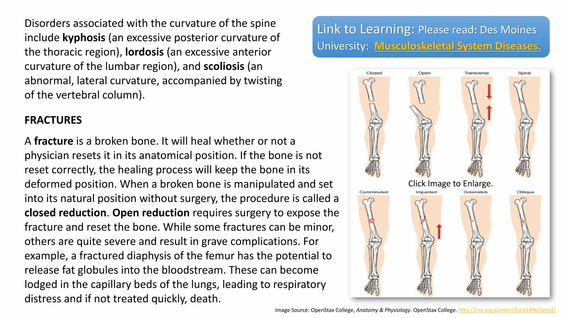
Disorders associated with the curvature of the spine include kyphosis (an excessive posterior curvature of the thoracic region), lordosis (an excessive anterior curvature of the lumbar region), and scoliosis (an abnormal, lateral curvature, accompanied by twisting of the vertebral column).
Link to Learning: Please read: Des Moines
University: Musculoskeletal System Diseases.
FRACTURES
A fracture is a broken bone. It will heal whether or not a physician resets it in its anatomical position. If the bone is not reset correctly, the healing process will keep the bone in its deformed position. When a broken bone is manipulated and set into its natural position without surgery, the procedure is called a closed reduction. Open reduction requires surgery to expose the fracture and reset the bone. While some fractures can be minor, others are quite severe and result in grave complications. For example, a fractured diaphysis of the femur has the potential to release fat globules into the bloodstream. These can become lodged in the capillary beds of the lungs, leading to respiratory distress and if not treated quickly, death.
Image Source: OpenStax College, Anatomy & Physiology. OpenStax College. http://cnx.org/content/col11496/latest/
Click Image to Enlarge.

Fractures are classified by their complexity, location, and other features. Common types of fractures are transverse, oblique, spiral, comminuted, impacted, greenstick, open (or compound), and closed (or simple). Healing of fractures begins with the formation of a hematoma, followed by internal and external calli. Osteoclasts resorb dead bone, while osteoblasts create new bone that replaces the cartilage in the calli. The calli eventually unite, remodeling occurs, and healing is complete.
Image Source: OpenStax College, Anatomy & Physiology. OpenStax College. http://cnx.org/content/col11496/latest/
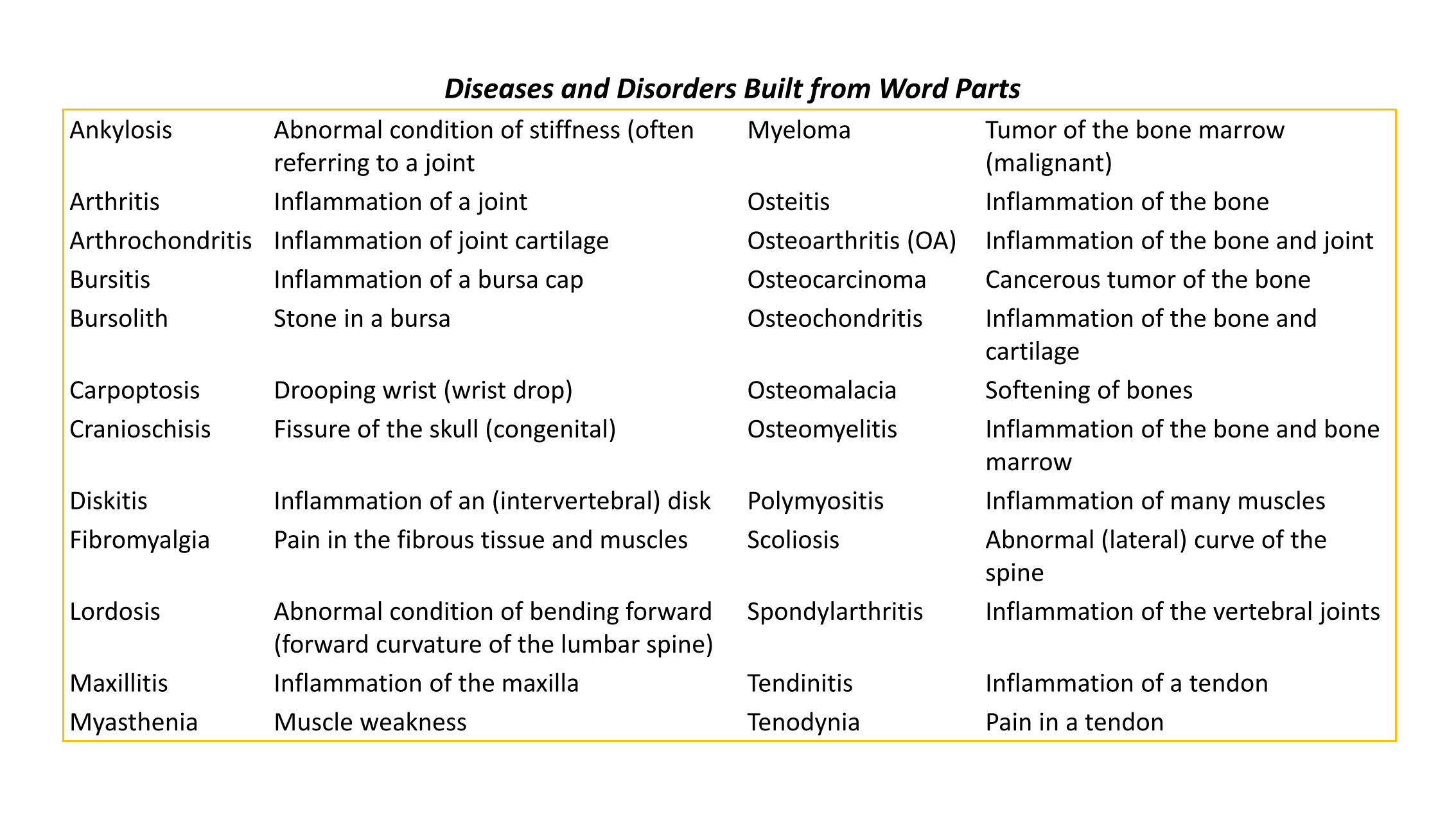
Ankylosis Abnormal condition of stiffness (often referring to a joint
Myeloma Tumor of the bone marrow (malignant)
Arthritis Inflammation of a joint Osteitis Inflammation of the bone
Arthrochondritis Inflammation of joint cartilage Osteoarthritis (OA) Inflammation of the bone and joint
Bursitis Inflammation of a bursa cap Osteocarcinoma Cancerous tumor of the bone
Bursolith Stone in a bursa Osteochondritis Inflammation of the bone and cartilage
Carpoptosis Drooping wrist (wrist drop) Osteomalacia Softening of bones
Cranioschisis Fissure of the skull (congenital) Osteomyelitis Inflammation of the bone and bone marrow
Diskitis Inflammation of an (intervertebral) disk Polymyositis Inflammation of many muscles
Fibromyalgia Pain in the fibrous tissue and muscles Scoliosis Abnormal (lateral) curve of the spine
Lordosis Abnormal condition of bending forward (forward curvature of the lumbar spine)
Spondylarthritis Inflammation of the vertebral joints
Maxillitis Inflammation of the maxilla Tendinitis Inflammation of a tendon
Myasthenia Muscle weakness Tenodynia Pain in a tendon
Diseases and Disorders Built from Word Parts
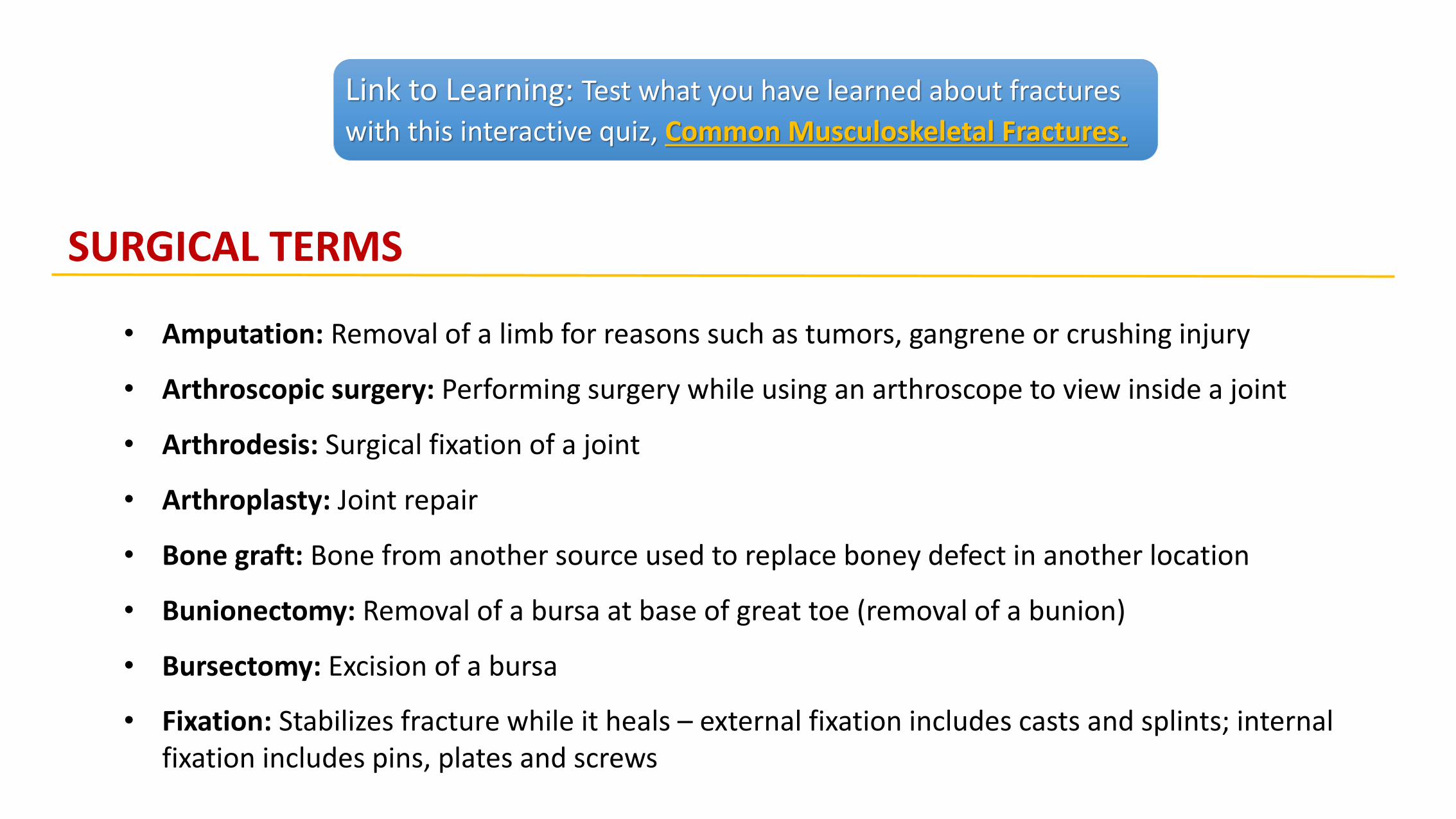
Link to Learning: Test what you have learned about fractures
with this interactive quiz, Common Musculoskeletal Fractures.
SURGICAL TERMS
• Amputation: Removal of a limb for reasons such as tumors, gangrene or crushing injury
• Arthroscopic surgery: Performing surgery while using an arthroscope to view inside a joint
• Arthrodesis: Surgical fixation of a joint
• Arthroplasty: Joint repair
• Bone graft: Bone from another source used to replace boney defect in another location
• Bunionectomy: Removal of a bursa at base of great toe (removal of a bunion)
• Bursectomy: Excision of a bursa
• Fixation: Stabilizes fracture while it heals – external fixation includes casts and splints; internal fixation includes pins, plates and screws
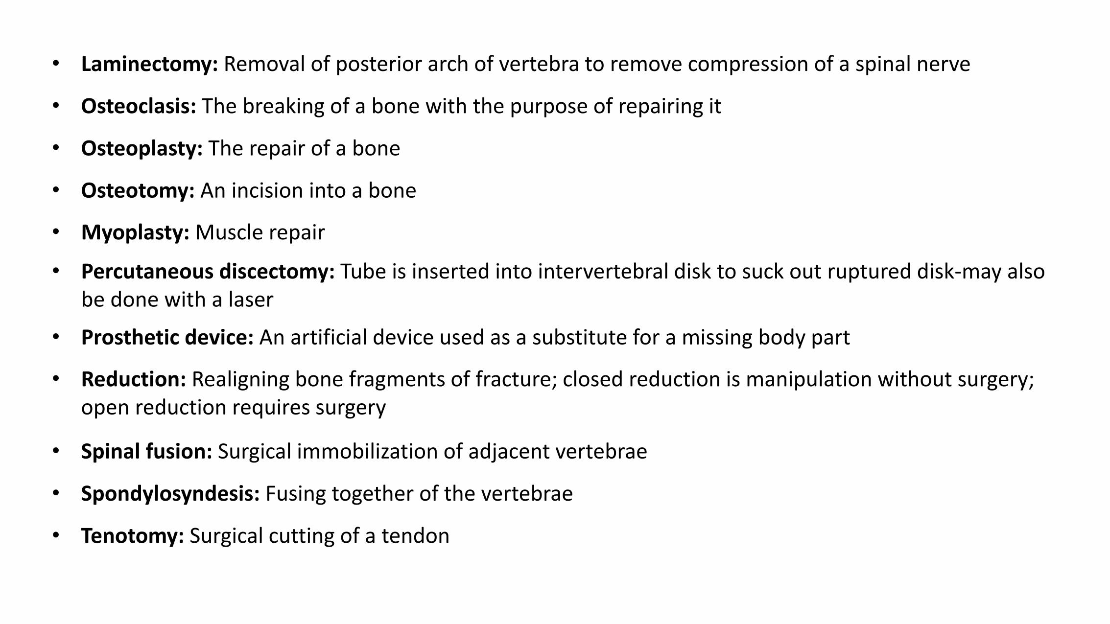
• Laminectomy: Removal of posterior arch of vertebra to remove compression of a spinal nerve
• Osteoclasis: The breaking of a bone with the purpose of repairing it
• Osteoplasty: The repair of a bone
• Osteotomy: An incision into a bone
• Myoplasty: Muscle repair
• Percutaneous discectomy: Tube is inserted into intervertebral disk to suck out ruptured disk-may also be done with a laser
• Prosthetic device: An artificial device used as a substitute for a missing body part
• Reduction: Realigning bone fragments of fracture; closed reduction is manipulation without surgery; open reduction requires surgery
• Spinal fusion: Surgical immobilization of adjacent vertebrae
• Spondylosyndesis: Fusing together of the vertebrae
• Tenotomy: Surgical cutting of a tendon
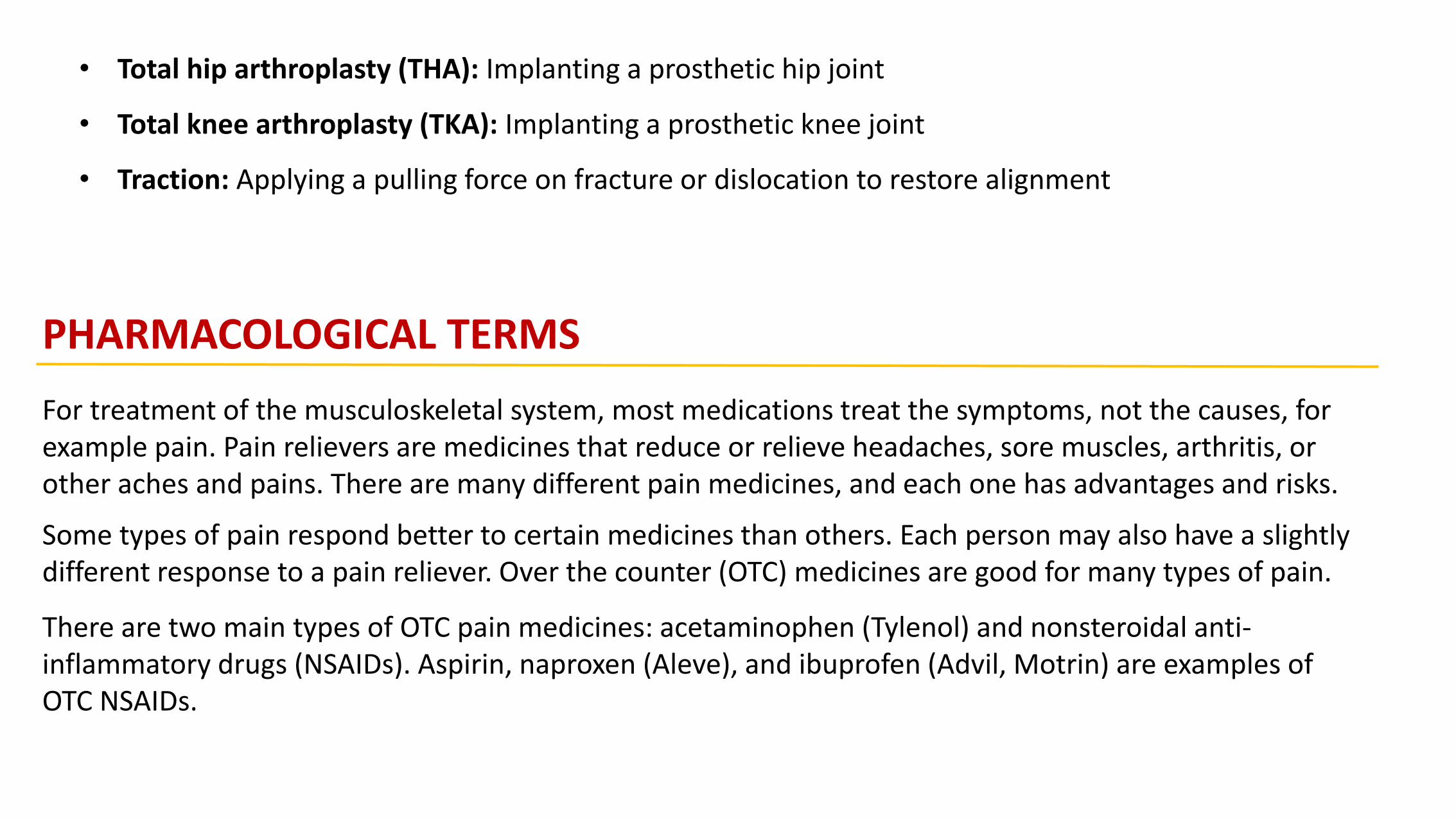
• Total hip arthroplasty (THA): Implanting a prosthetic hip joint
• Total knee arthroplasty (TKA): Implanting a prosthetic knee joint
• Traction: Applying a pulling force on fracture or dislocation to restore alignment
PHARMACOLOGICAL TERMS
For treatment of the musculoskeletal system, most medications treat the symptoms, not the causes, for example pain. Pain relievers are medicines that reduce or relieve headaches, sore muscles, arthritis, or other aches and pains. There are many different pain medicines, and each one has advantages and risks.
Some types of pain respond better to certain medicines than others. Each person may also have a slightly different response to a pain reliever. Over the counter (OTC) medicines are good for many types of pain.
There are two main types of OTC pain medicines: acetaminophen (Tylenol) and nonsteroidal anti-inflammatory drugs (NSAIDs). Aspirin, naproxen (Aleve), and ibuprofen (Advil, Motrin) are examples of OTC NSAIDs.

If OTC medicines don't relieve the pain, a doctor may prescribe something stronger. Many NSAIDs are also available at higher prescription doses. The most powerful pain relievers are narcotics. They are very effective, but they can sometimes have serious side effects. Because of the risks, a person must use them only under a doctor's supervision.
Analgesic: agent that relieves pain
Anti-Inflammatory (corticosteroid): agent that reduces inflammation
Muscle relaxant: agent that relaxes the muscles and relieves stiffness
Narcotic: agent that relieves pain by binding receptors in the brain which blocks the feeling of
pain-also called opioid pain relievers
Nonsteroidal (NSAID): agent that reduces inflammation without the use of steroids

AK Above knee L Left
BK Below the knee L1-L5 Lumbar vertebrae
BMD Bone mineral density OA Osteoarthritis
C1-C7 Cervical vertebrae P Phosphorus
Ca Calcium R Right
CTS Carpal tunnel syndrome RA Rheumatoid arthritis
EMG Electromyogram ROM Range of motion
FX Fracture T1-T2 Thoracic vertebrae
IM Intramuscular TMJ Temporomandibular joint
Common Abbreviations
Link to Learning: Please review the following: Des Moines University: Musculoskeletal
System Medical Record, to practice reading medical terminology relating to this system.
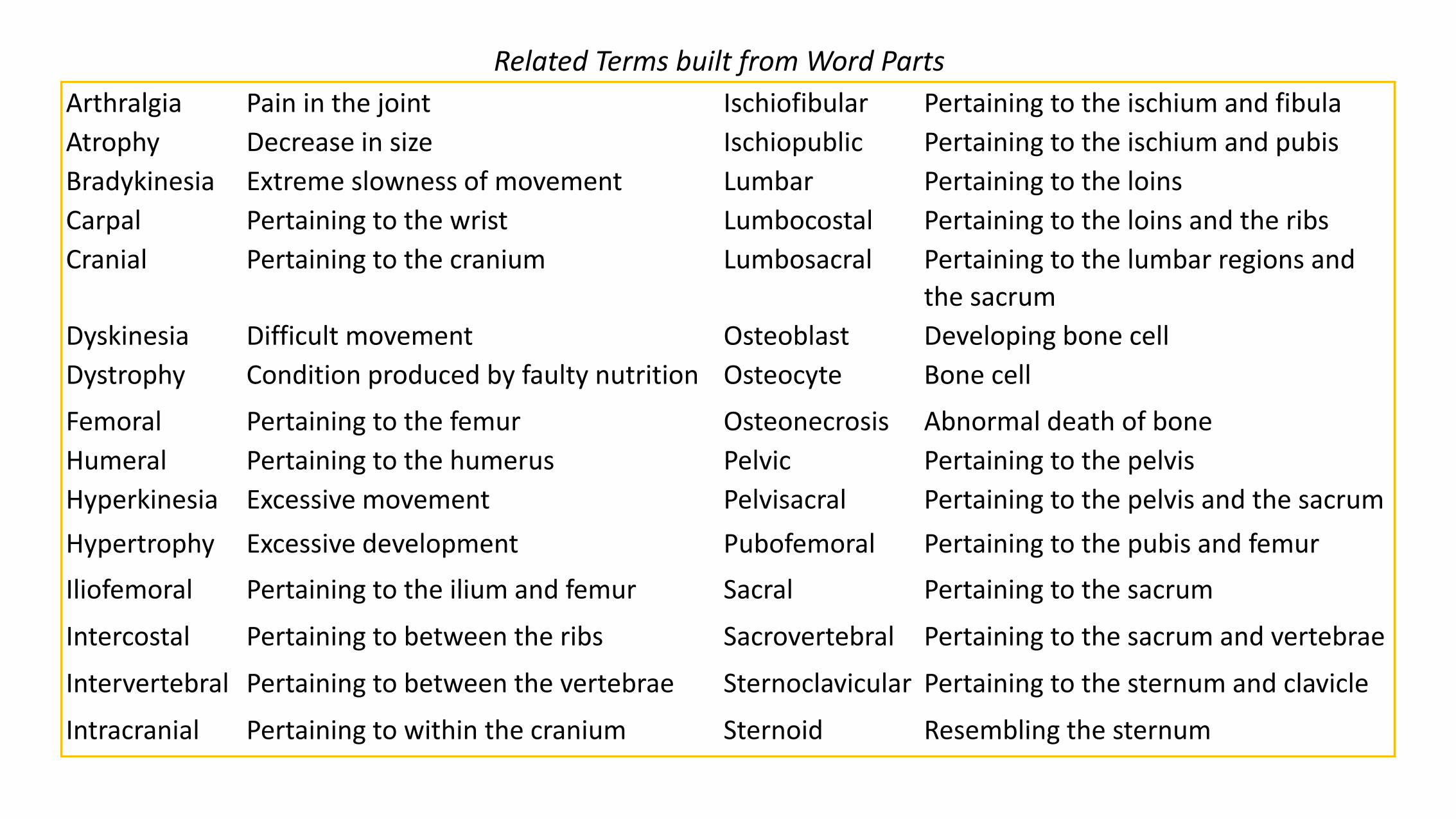
Arthralgia Pain in the joint Ischiofibular Pertaining to the ischium and fibula
Atrophy Decrease in size Ischiopublic Pertaining to the ischium and pubis
Bradykinesia Extreme slowness of movement Lumbar Pertaining to the loins
Carpal Pertaining to the wrist Lumbocostal Pertaining to the loins and the ribs
Cranial Pertaining to the cranium Lumbosacral Pertaining to the lumbar regions and
the sacrum
Dyskinesia Difficult movement Osteoblast Developing bone cell
Dystrophy Condition produced by faulty nutrition Osteocyte Bone cell
Femoral Pertaining to the femur Osteonecrosis Abnormal death of bone
Humeral Pertaining to the humerus Pelvic Pertaining to the pelvis
Hyperkinesia Excessive movement Pelvisacral Pertaining to the pelvis and the sacrum
Hypertrophy Excessive development Pubofemoral Pertaining to the pubis and femur
Iliofemoral Pertaining to the ilium and femur Sacral Pertaining to the sacrum
Intercostal Pertaining to between the ribs Sacrovertebral Pertaining to the sacrum and vertebrae
Intervertebral Pertaining to between the vertebrae Sternoclavicular Pertaining to the sternum and clavicle
Intracranial Pertaining to within the cranium Sternoid Resembling the sternum
Related Terms built from Word Parts
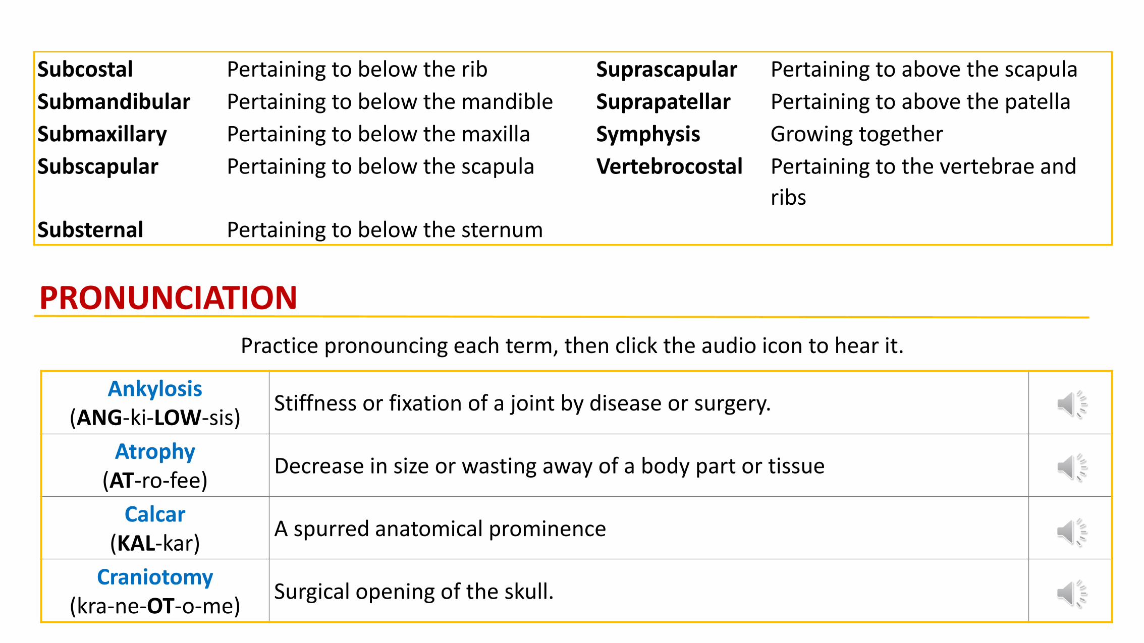
Subcostal Pertaining to below the rib Suprascapular Pertaining to above the scapula
Submandibular Pertaining to below the mandible Suprapatellar Pertaining to above the patella
Submaxillary Pertaining to below the maxilla Symphysis Growing together
Subscapular Pertaining to below the scapula Vertebrocostal Pertaining to the vertebrae and
ribs
Substernal Pertaining to below the sternum
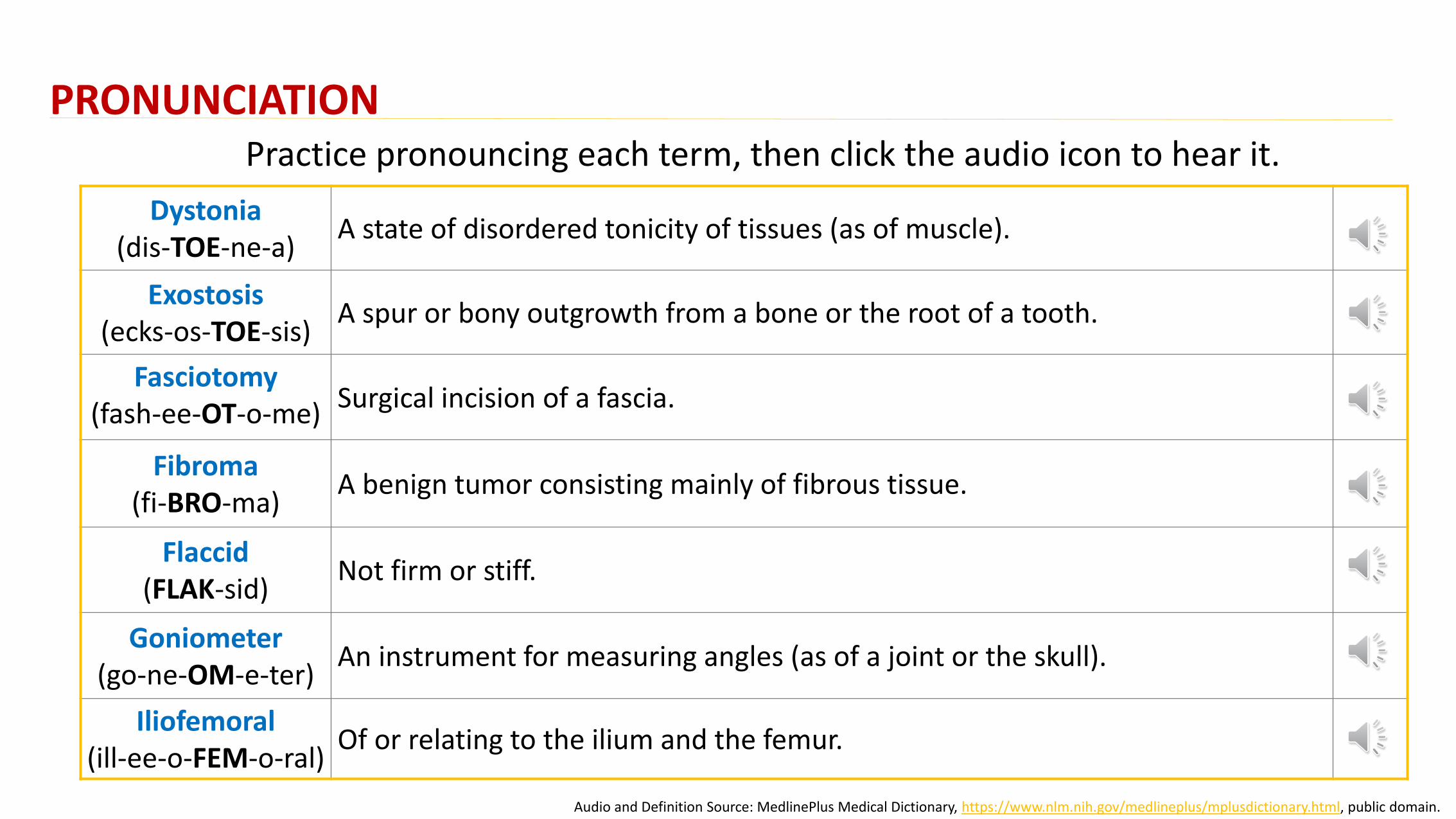
PRONUNCIATIONPractice pronouncing each term, then click the audio icon to hear it.
Ankylosis(ANG-ki-LOW-sis)
Stiffness or fixation of a joint by disease or surgery.
Atrophy(AT-ro-fee)
Decrease in size or wasting away of a body part or tissue
Calcar(KAL-kar)
A spurred anatomical prominence
Craniotomy(kra-ne-OT-o-me)
Surgical opening of the skull.
PRONUNCIATIONPractice pronouncing each term, then click the audio icon to hear it.
Dystonia(dis-TOE-ne-a)
A state of disordered tonicity of tissues (as of muscle).
Exostosis(ecks-os-TOE-sis)
A spur or bony outgrowth from a bone or the root of a tooth.
Fasciotomy(fash-ee-OT-o-me)
Surgical incision of a fascia.
Fibroma(fi-BRO-ma)
A benign tumor consisting mainly of fibrous tissue.
Flaccid(FLAK-sid)
Not firm or stiff.
Goniometer(go-ne-OM-e-ter)
An instrument for measuring angles (as of a joint or the skull).
Iliofemoral(ill-ee-o-FEM-o-ral)
Of or relating to the ilium and the femur.
Audio and Definition Source: MedlinePlus Medical Dictionary, https://www.nlm.nih.gov/medlineplus/mplusdictionary.html, public domain.
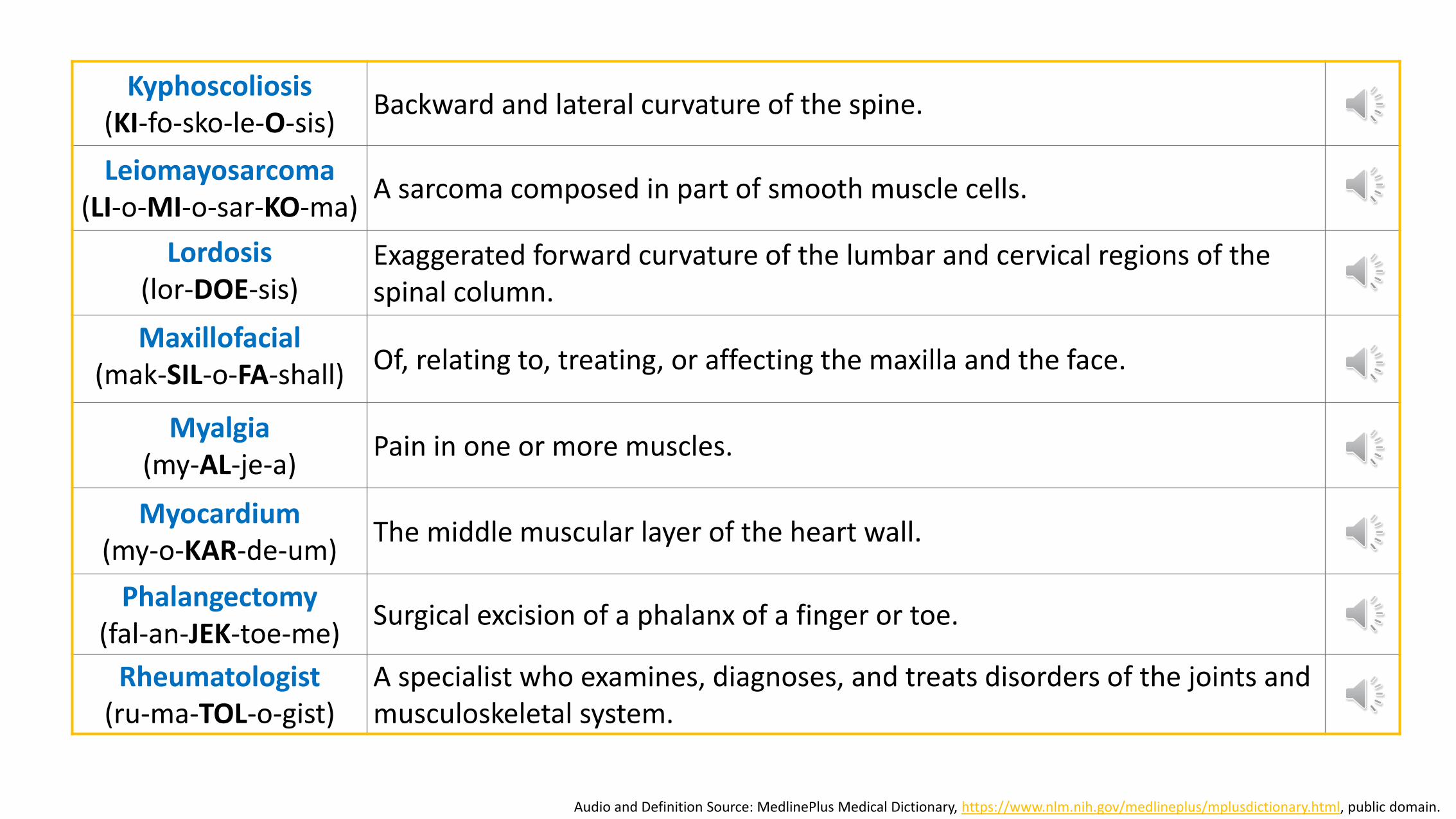
Kyphoscoliosis(KI-fo-sko-le-O-sis)
Backward and lateral curvature of the spine.
Leiomayosarcoma(LI-o-MI-o-sar-KO-ma)
A sarcoma composed in part of smooth muscle cells.
Lordosis(lor-DOE-sis)
Exaggerated forward curvature of the lumbar and cervical regions of the spinal column.
Maxillofacial(mak-SIL-o-FA-shall) Of, relating to, treating, or affecting the maxilla and the face.
Myalgia(my-AL-je-a)
Pain in one or more muscles.
Myocardium(my-o-KAR-de-um)
The middle muscular layer of the heart wall.
Phalangectomy(fal-an-JEK-toe-me)
Surgical excision of a phalanx of a finger or toe.
Rheumatologist(ru-ma-TOL-o-gist)
A specialist who examines, diagnoses, and treats disorders of the joints and musculoskeletal system.
Audio and Definition Source: MedlinePlus Medical Dictionary, https://www.nlm.nih.gov/medlineplus/mplusdictionary.html, public domain.
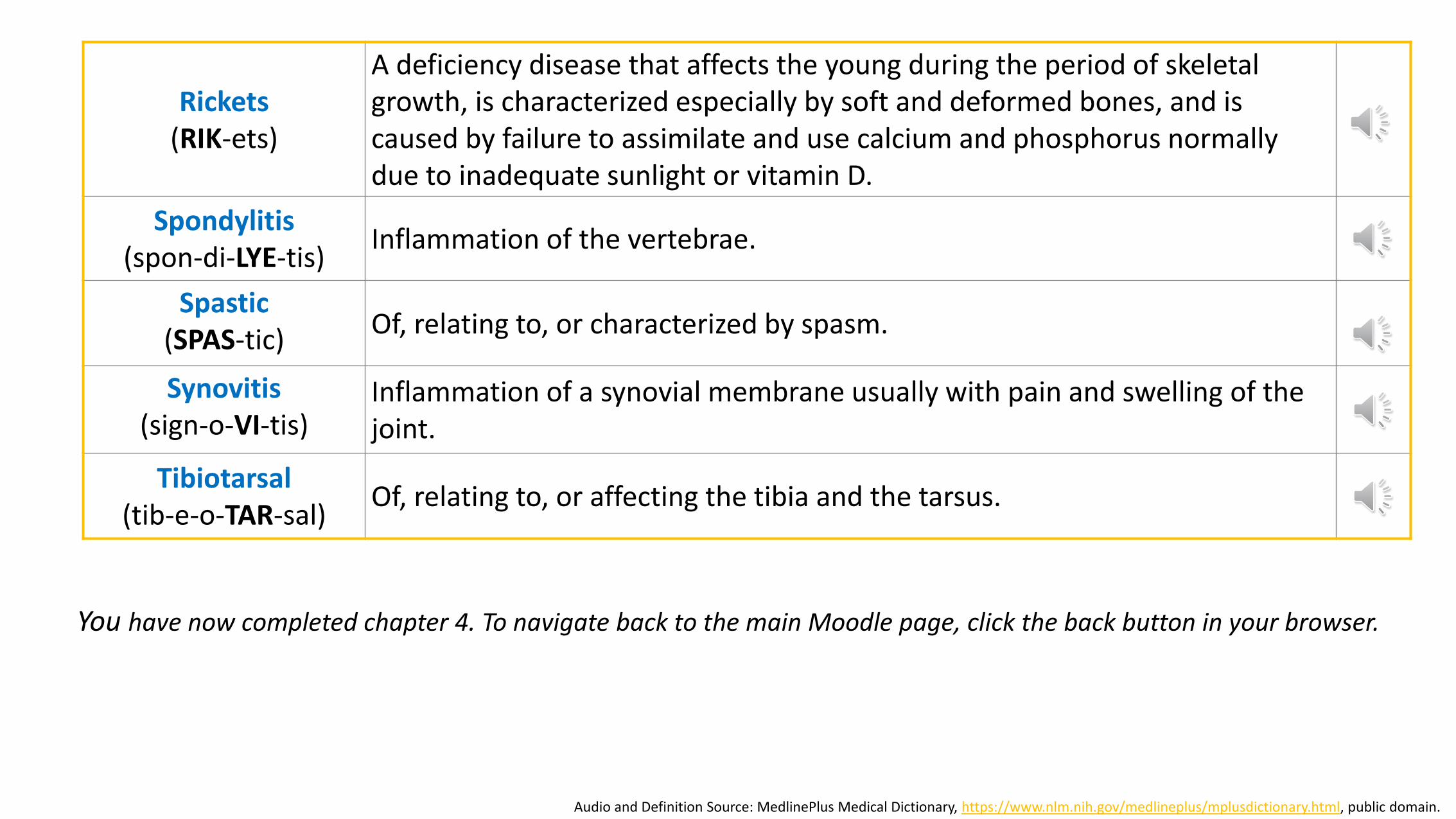
Rickets(RIK-ets)
A deficiency disease that affects the young during the period of skeletal growth, is characterized especially by soft and deformed bones, and is caused by failure to assimilate and use calcium and phosphorus normally due to inadequate sunlight or vitamin D.
Spondylitis(spon-di-LYE-tis)
Inflammation of the vertebrae.
Spastic(SPAS-tic)
Of, relating to, or characterized by spasm.
Synovitis(sign-o-VI-tis)
Inflammation of a synovial membrane usually with pain and swelling of the joint.
Tibiotarsal(tib-e-o-TAR-sal)
Of, relating to, or affecting the tibia and the tarsus.
Audio and Definition Source: MedlinePlus Medical Dictionary, https://www.nlm.nih.gov/medlineplus/mplusdictionary.html, public domain.
You have now completed chapter 4. To navigate back to the main Moodle page, click the back button in your browser.