cigna medical coverage policies – radiology musculoskeletal imaging · · 2016-02-18cigna...
TRANSCRIPT

©2016 eviCore healthcare Musculoskeletal Imaging Guidelines
Cigna Medical Coverage Policies – Radiology
Musculoskeletal Imaging Effective February 19, 2016
Instructions for use The following coverage policy applies to health benefit plans administered by Cigna. Coverage policies are intended to provide guidance in interpreting certain standard Cigna benefit plans and are used by medical directors and other health care professionals in making medical necessity and other coverage determinations. Please note the terms of a customer’s particular benefit plan document may differ significantly from the standard benefit plans upon which these coverage policies are based. For example, a customer’s benefit plan document may contain a specific exclusion related to a topic addressed in a coverage policy. In the event of a conflict, a customer’s benefit plan document always supersedes the information in the coverage policy. In the absence of federal or state coverage mandates, benefits are ultimately determined by the terms of the applicable benefit plan document. Coverage determinations in each specific instance require consideration of: 1. The terms of the applicable benefit plan document in effect on the date of service 2. Any applicable laws and regulations 3. Any relevant collateral source materials including coverage policies 4. The specific facts of the particular situation Coverage policies relate exclusively to the administration of health benefit plans. Coverage policies are not recommendations for treatment and should never be used as treatment guidelines. This evidence-based medical coverage policy has been developed by eviCore, Inc. Some information in this coverage policy may not apply to all benefit plans administered by Cigna. CPT® (Current Procedural Terminology) is a registered trademark of the American Medical Association (AMA). CPT® five digit codes, nomenclature and other data are copyright 2016 American Medical Association. All Rights Reserved. No fee schedules, basic units, relative values or related listings are included in the CPT® book. AMA does not directly or indirectly practice medicine or dispense medical services. AMA assumes no liability for the data contained herein or not contained herein.

V.18.0 Effective 2/19/2016 – MSK Imaging
MUSCULOSKELETAL IMAGING GUIDELINES
Musculoskeletal Imaging Guidelines MS-1~General Guidelines 3 MS-2~Imaging Techniques 4 MS-3~3D Rendering 5
DISEASE/ INJURY CATEGORY (Alphabetical Order) MS-4~Avascular Necrosis (AVN)/Osteonecrosis 6 MS-5~Fracture and Dislocation 7 MS-6~Foreign Body 8 MS-7~Ganglion Cysts 9 MS-8~Gout, Pseudogout and Crystal Deposition Disease 9 MS-9~Infection/Osteomyelitis 10 MS-10~Soft Tissue Mass(es) and Lesion of Bone 11 MS-11~Muscle/Tendon Unit Injuries/Disease 13 MS-12~Osteoarthritis 13 MS-13~Osteochondritis Dissecans 14 MS-14~Osteoporosis 15 MS-15~Rheumatoid Arthritis and Inflammatory Arthritis 16 MS-16~Total Joint Prosthesis 17 MS-17 Leg Length Discrepancy 18
ANATOMICAL AREAS MS-18~Anatomical Area Tables – General Information 18 MS-19~SHOULDER 19 MS-20~ELBOW 22 MS-21~WRIST 23 MS-22~HAND 25 MS-23~PELVIS 26 MS-24~HIP 27 MS-25~KNEE 30 MS-26~ANKLE 33 MS-27~FOOT 36
Page 2 of 38
V.18.0 Effective 2/19/2016 – MSK Imaging
MUSCULOSKELETAL IMAGING GUIDELINES
MS-1~GENERAL GUIDELINES
A clinical evaluation within 60 days is required before advanced imaging can be considered and should include a relevant history and physical examination, appropriate laboratory studies, and non-advanced imaging modalities. o Other meaningful contact (telephone call, electronic mail or messaging) by an
established patient can substitute for a face-to-face clinical evaluation. o A “clinical diagnosis” for many musculoskeletal bone, joint and soft tissue pain,
and injury disorders are based on a relevant history, physical examination, and plain X-ray.
Prior to advanced imaging consideration, an X-ray must be performed after the current episode of symptoms started or changed for all musculoskeletal conditions, unless otherwise noted in the guidelines.
Physician-directed non-surgical care may include rest, ice, compression, and elevation (R.I.C.E), non-steroidal anti-inflammatories (NSAIDs), narcotic and non-narcotic analgesic medications, oral or injectable corticosteroids, viscosupplementation injections; a physician-directed home exercise program, cross-training, and/or physical medicine, or immobilization by splinting/casting/bracing.
Orthopedic specialist evaluation can be helpful in determining the need for advanced imaging. o The need for repeat advanced imaging should be carefully considered and may not
be indicated if prior imaging has been performed. o Serial advanced imaging, whether CT or MRI, for surveillance of healing or
recovery from musculoskeletal disease is not supported by the medical evidence in the majority of musculoskeletal conditions.
Computer-Assisted Musculoskeletal Surgical Navigation Procedures: The Category III code used to describe computer-assisted navigation in orthopedic
surgery with CT/MRI image guidance is: +0055T. o Computer-assisted navigation (CAN) in orthopedic procedures describes the use of
computer-enabled tracking systems to facilitate alignment in a variety of surgical procedures and verification of an intended implant placement.
o Code +0055T is intended to be used in addition to the code for the primary surgical procedure.
o CT/MRI imaging acquisition for preoperative planning, in the absence of written payor instructions, is not to be reported with a diagnostic CT or MRI code
Page 3 of 38

V.18.0 Effective 2/19/2016 – MSK Imaging
MUSCULOSKELETAL IMAGING GUIDELINES
MS-2~Imaging Techniques
Plain X-Ray Should be done prior to advanced imaging in all musculoskeletal conditions/disorders,
unless otherwise noted in the guidelines, to rule out those situations that do not often require advanced imaging, such as osteoarthritis, acute/healing fracture, dislocation, osteomyelitis, acquired/congenital deformities, and tumors of bone amenable to biopsy or radiation therapy (in known metastatic disease), etc.
MRI or CT Magnetic Resonance Imaging (MRI) is often the preferred advanced imaging modality
in musculoskeletal conditions because it is superior in imaging the soft tissues and can also define physiological processes in some instances [e.g. edema, loss of circulation (AVN), and increased vascularity (tumors)].
Computed Tomography (CT) is better at imaging bone and joint anatomy; thus, it is useful for studying complex fractures (particularly of the joints), dislocations, and assessing delayed union or non-union of fractures if plain X-rays are equivocal. CT may be the procedure of choice in patients who cannot have MRI, such as those with pacemakers.
Contrast Issues Most musculoskeletal imaging (MRI or CT) is without contrast, except for the
following: o Tumors and osteomyelitis (without and with contrast) o MRI arthrography (with contrast only) o MRI for rheumatoid arthritis (contrast as requested)
Positron Emission Tomography (PET) At the present time, there is inadequate evidence to support the medical necessity of
PET for the routine assessment of musculoskeletal disorders. It should be considered experimental or investigational and will be forwarded to Medical Director Review.
See also: MS-16~Total Joint Prosthesis References 1. ACR Appropriateness Criteria, Musculoskeletal Imaging topics. 2. ACR—SPR—SSR Practice Guideline for the performance of radiography of the extremities.
Amended 2014. 3. Hsu, W. and T. M. Hearty (2012). Radionuclide Imaging in the Diagnosis and Management of
Orthopaedic Disease. Journal of the American Academy of Orthopaedic Surgeons 20(3): 151-159. 4. Kayser R, Mahlfeld K, Heyde CE. Partial rupture of the proximal Achilles tendon: a differential
diagnostic problem in ultrasound imaging. Br J Sports Med. 2005; 39:838-842.
Page 4 of 38
V.18.0 Effective 2/19/2016 – MSK Imaging
MUSCULOSKELETAL IMAGING GUIDELINES
MS-3~3D RENDERING
Indications for musculoskeletal 3-D image post-processing for preoperative planning when conventional imaging is insufficient for: o Complex fractures/dislocations (comminuted or displaced) of any joint o Spine fractures, pelvic/acetabulum fractures, intra-articular fractures
The code assignment for 3-D rendering depends upon whether the 3-D post-processing is performed on the scanner workstation (CPT®76376) or on an independent workstation (CPT®76377). o 2-D reconstruction (i.e., reformatting axial images into the coronal plane) is
considered part of the tomography procedure, is not separately reportable, and does not meet the definition of 3-D rendering.
o It is not appropriate to report 3-D rendering in conjunction with CTA and MRA because those procedure codes already include the postprocessing.
o In addition to the term “3-D,” the following terms may also be used to describe 3-D post-processing: maximum intensity projection (MIP) shaded surface rendering volume rendering
The 3-D rendering codes require concurrent supervision of image post processing 3-D manipulation of volumetric data set and image rendering.
Page 5 of 38

V.18.0 Effective 2/19/2016 – MSK Imaging
DISEASE/ INJURY CATEGORY (ALPHABETICAL ORDER)
MS-4~Avascular Necrosis (AVN)/Osteonecrosis
MS-4.1 AVN MRI without contrast when suspected AVN/osteonecrosis, with negative plain X-
ray(s)
Patients with acute lymphoblastic leukemia and known or suspected osteonecrosis should be imaged according to guidelines in: PACONC-3.2 Acute Lymphoblastic Leukemia
Known or suspected osteonecrosis in long-term cancer survivors should be imaged according to guidelines in: PACONC-19.4 Osteonecrosis in Long Term Cancer Survivors
Practice Notes Ficat Stage 0, I, II for suspected AVN of the hip; Lichtman Stage 1, 2 for suspected AVN of the lunate; Cruess Stage I, II for suspected AVN of the humeral head; Modified Ficat and Arlet Stage 0, 1, 2 for suspected AVN of the talus; Modified Ficat and Arlet Stage 0, I, II for suspected AVN of the distal femur.
References 1. ACR Appropriateness Criteria
®, Chronic Hip Pain, 2011.
2. Calder JD, Hine AL, Pearse MF, Revell PA. The relationship between osteonecrosis of the proximal femur identified by MRI and lesions proven by histological examination. J Bone Joint Surg Br. 2008, 90:154-158.
3. Karantanas AH, Drakonaki EE. The role of MR imaging in avascular necrosis of the femoral head. Semin Musculoskelet Radiol. 2011; 15(3):281-300.
Page 6 of 38

V.18.0 Effective 2/19/2016 – MSK Imaging
DISEASE/ INJURY CATEGORY (ALPHABETICAL ORDER)
MS-5~Fracture and Dislocation
MS-5.1 Acute CT or MRI without contrast is appropriate, if one of the following is present:
o Complex (comminuted or displaced) fracture/dislocation on plain film CT is preferred unless it is associated with neoplastic disease when MRI
without/with contrast is preferred unless MRI contraindicated o Suspected osteochondral fracture can also be considered for MRI arthrogram, or
CT arthrogram
MS-5.2 Occult/Stress/Insufficiency Fracture/Stress Reaction and Shin Splints For suspected hip, femur, tibia, calcaneous, periprosthetic, sacral, navicular , or
scaphoid occult/stress/insufficiency fractures, MRI or CT without contrast can be performed if the initial evaluation of history, physical exam and either plain X-ray or bone scan fail to establish a definitive diagnosis.
MRI or CT without contrast can be performed for all other suspected occult/stress/insufficiency fractures if repeat plain X-rays remain negative after a minimum of 2 weeks of conservative treatment.
MS-5.3 Other Indications CT or MRI without contrast is appropriate after recent (within 30 days) plain X-ray if
one of the following is present: o Concern for delayed union or non-union of fracture or joint fusions o As part of pre-operative evaluation for a planned surgery of a complex
fracture/dislocation o For stress reaction, advanced imaging is not medically necessary for surveillance
for “return to play” decisions of a stress reaction identified on an initial imaging study.
o For suspected shin splints, MRI of the lower leg without contrast (CPT®73718) is appropriate if failure of a 6 week trial of conservative treatment.
References 1. ACR Appropriateness Criteria®, Stress (fatigue/insufficiency) fracture, including sacrum, excluding
other vertebrae, 2011. 2. ACR Appropriateness Criteria®, Chronic hip pain, 2011. 3. ACR Appropriateness Criteria®, Acute hand and wrist trauma, 2008. 4. ACR Appropriateness Criteria®, Chronic ankle pain, 2009. 5. Harris GD and Hughes BC. Deciphering your patient’s leg pain. Emerg Med 2006; 38(6):24-30.
Page 7 of 38

V.18.0 Effective 2/19/2016 – MSK Imaging
6. Daffner RH, Weissman BN, Appel M, Bancroft L. et al. ACR Appropriateness Criteria®
, Stress
(fatigue/insufficiency) fracture, including sacrum, excluding other vertebrae. 2011. 7. Greene WB (Ed.). Essentials of Musculoskeletal Care. 3
rd Ed. Rosemont, IL, American Academy of
Orthopaedic Surgeons, 2005, pp.568-570. 8. Galbraith RM, Lavallee ME. Medial tibial stress syndrome: conservative treatment options. Curr
Rev Muscuolskelet Med. 2009; 2:127-133. 9. Boks SS, Vroegindeweij D, Kroes BW, Bernsen RMD et al. MRI follow-up of posttraumatic bone
bruises of the knee in general practice. AJR. 2007; 189: 556-562.
MS-6~FOREIGN BODY
MS-6.1 Foreign Body – General X-ray is the initial imaging study for foreign bodies in the foot.
o Ultrasound examination (CPT®76882) is the preferred imaging modality for non-radiopaque foreign bodies in the foot.
o MRI (contrast as requested) can be approved after plain X-rays rule out the presence of radiopaque foreign bodies.
o X-ray can be used after removal of the foreign body to confirm removal. References 1. ACR Appropriateness Criteria®, Acute Trauma to the Foot, 2010. 2. Chan C, Salam GA. Splinter removal. Am Fam Physician.2003; 67(12):2557-2562. 3. Friedman DI, Forti RJ, Wall SP, Crain EF. The utility of bedside ultrasound and patient perception
in detecting soft tissue foreign bodies in children. Pediatr Emerg Care. 2005; 21(8):487-492. 4. Peterson JJ, Bancroft LW, Kransdorf MJ. Wooden foreign bodies: imaging appearance. AJR 2002;
178(3):557-562.
Page 8 of 38
V.18.0 Effective 2/19/2016 – MSK Imaging
DISEASE/ INJURY CATEGORY (ALPHABETICAL ORDER)
MS-7~GANGLION CYSTS
MS-7.1 Ganglion Cysts – General MRI without contrast is appropriate for occult ganglions (smaller cysts that remain
hidden under the skin; suspected, but not palpable on physical examination) or cysts/masses in atypical anatomic locations. o Advanced imaging is not indicated for ganglions that can be diagnosed by history
and physical examination.
References 1. Rubin DA, Weissman BN, Appel M, Arnold E. ACR Appropriateness Criteria®: Chronic Wrist Pain.
Last review date 2012. 2. Freire V, Guerini H, Campagna R, Moutounet L et al. Imaging of hand and wrist cysts: a clinical
approach. AJR, 2012; 199: W618-W628. 3. Vo P, Wright T, Hayden F, Dell P, et al. Evaluating dorsal wrist pain: MRI diagnosis of occult
dorsal wrist ganglion. J Hand Surg Am. 1995; 20: 667-670.
MS-8~Gout/Calcium Pyrophosphate Deposition Disease (CPPD) (Pseudogout/Chondrocalcinosis)
MS-8.1 Gout General Early stages of gout can be diagnosed clinically since radiographic findings are not present early in the disease course.
MRI without and with contrast is indicated for gouty tophus, which can mimic an infectious or neoplastic process
MS-8.2 CPPD (pseudogout /chondrocalcinosis) General CPPD can often be diagnosed from plain X-rays; advanced is generally not necessary
References 1. Hsu CY, Shih TT, Huang KM, Chen PQ, Sheu JJ, Li YW. Tophaceous gout of the spine: MR
imaging features. Clin Radiol. 2002; 57(10):919. 2. Schumacher HR Jr, Becker MA, Edwards NL, Palmer WE, et al. Magnetic resonance imaging in the
quantitative assessment of gouty tophi. Int J Clin Pract. 2006; 60(4):408. 3. McQueen FM, Doyle A, Reeves Q, Gao A. Bone erosions in patients with chronic gouty arthropathy
are associated with tophi but not bone oedema or synovitis: new insights from a 3 T MRI study. Rheumatology. 2014; 53: 95-103.
Page 9 of 38

V.18.0 Effective 2/19/2016 – MSK Imaging
4. Dore RK. Gout: What primary care physicians want to know. J Clin Rheumatol. 2008;14(5 Suppl):S47-S54.
5. Eggebeen AT. Gout: an update. Am Fam Physician. 2007; 76(6):801-808. 6. Burns C, Wortmann RL. Chapter 44. Gout. In: Imboden JB, Hellmann DB, Stone JH, eds.
CURRENT Diagnosis & Treatment: Rheumatology. 3rd ed. New York: McGraw-Hill; 2013. http://www.accessmedicine.com/content.aspx?aID=57273972. Accessed July 14, 2015.
MS-9~Infection/Osteomyelitis
MS-9.1 Infection – General MRI without and with contrast after plain X-ray(s) and:
o Plain film(s) are negative and soft tissue or bone infection (osteomyelitis) is not responding to surgical or non-surgical care; or
o Plain film(s) are positive for osteomyelitis, and the extent of infection into the soft tissues and any skip lesions require evaluation
CT without and with contrast can replace an MRI: o To assess the extent of bony destruction from osteomyelitis; CT can guide
treatment decisions. o For pre-operative planning o If MRI is contraindicated
References 1. Green WB (Ed.). Essentials of Musculoskeletal Care. 3rd Ed. Rosemont, IL, American Academy of
Orthopaedic Surgeons, 2005, p.918. 2. Staheli LT. Fundamentals of Pediatric Orthopedics. 4th Ed. Philadelphia, Lippincott Williams &
Wilkins, 2008, pp.110-111. 3. ACR Appropriateness Criteria®, Suspected Osteomyelitis-Diabetic Patient, 2012.
MS-9.2 Septic Joint Analysis of joint fluid is most often sufficient to diagnose a septic joint. An MRI of
the joint, without/with contrast is appropriate when standard or image guided arthrocentesis is contraindicated or is unsuccessful and the clinical documentation satisfies all of the following criteria: o History and Physical examination findings[Any of the following]
Development of an acutely hot and swollen joint (< 2 weeks) Decreased range of motion due to pain Documented fever
o Laboratory tests [One of the following] Leukocytosis Elevated ESR or C-reactive protein Analysis of the joint fluid is non-diagnostic
o Plain xray of the joint Page 10 of 38

V.18.0 Effective 2/19/2016 – MSK Imaging
Reference 1. Coakley G, Mathews C, Field M, et al. BSR & BHPR, BOA, RCGP and BSAC guidelines for
management of the hot swollen joints in adults. Rheumatology. 2006; 45:1039-1041.
DISEASE/ INJURY CATEGORY (ALPHABETICAL ORDER)
MS-10~SOFT TISSUE MASS or LESION OF BONE
MS-10.1 Soft Tissue Mass History and physical exam should include: location, size, duration, growing or stable,
solid/cystic, fixed/not fixed to the bone, discrete or ill-defined, painful.
Plain X-ray should be performed initially, which could further determine the indication for the requested or other advanced imaging. If nondiagnostic, these initial X-rays can provide complementary information if advanced imaging is indicated.
MRI without and with contrast or without contrast is appropriate for: o Soft tissue mass(es) o Known or suspected soft tissue mass in a patient with a cancer predisposition
syndrome if a recent ultrasound is inconclusive. Plain X-ray is not required for
these patients. See: PACONC-2~Cancer Predisposition Syndromes and Screening Strategies
MRI hip without and with (CPT®73723) contrast and ultrasound (CPT®76881) are both appropriate for the diagnosis of pseudotumors surrounding metal-on-metal (MoM) hip prostheses. One of these two imaging modalities can be approved, but not both.
Advanced imaging is not indicated for: o Subcutaneous lipoma with no surgery planned o Ganglia (See MS-7) o Sebaceous cyst o Soft tissue mass(es) that has been stable for >/= 1 year
MS-10.2 Lesion of Bone History and physical exam should include assessment of the following: location; size;
duration; progressing or stable; solid/cystic; fixed/not fixed to the bone; discrete or ill-
defined; painful. Complete radiograph of the entire bone, containing the lesion of bone,
are required prior to consideration of advanced imaging.
Many benign bone tumors have a characteristic appearance on plain X-ray and advanced imaging is not necessary. MRI without and with contrast and/or CT without and with contrast may be indicated if one of the following applies: o Imaging requested for preoperative planning
Page 11 of 38

V.18.0 Effective 2/19/2016 – MSK Imaging
o Diagnosis uncertain based on plain X-ray appearance
MRI without and with contrast or without contrast is appropriate when plain X-ray reveals an osteochondroma with clinical concern of malignant transformation.
For Paget’s Disease, MRI without and with contrast can be considered if the diagnosis (based on plain X-rays and laboratory studies) is in doubt or if malignant degeneration is suspected (occurs in up to 10% of the cases).
Practice Notes The medical necessity and appropriateness or type of advanced imaging studies for lesions of bone are often best determined through consultation with an orthopaedic surgical oncologist.
References 1. ACR Practice Guideline. ACR-SSR Practice Guideline for the Performance and Interpretation of
Magnetic Resonance Imaging (MRI) of Bone and Soft Tissue Tumors. Revised 2010. 2. ACR Appropriateness Criteria
®, Soft tissue masses, 2009. 3. ACR Appropriateness Criteria
®, Primary bone tumors, 2009. 4. Schneider D, Hofmann MR, Peterson JA. Diagnosis and treatment of Paget's Disease of Bone. Am
Fam Physician. 2002; 65:2069-2072. 5. Theodorou DJ, Theodorou SJ, Kakitsubata Y. Imaging of Paget Disease of bone and its
musculoskeletal complications: review. AJR, 2012; 196: S64-S75.
Page 12 of 38
V.18.0 Effective 2/19/2016 – MSK Imaging
DISEASE/ INJURY CATEGORY (ALPHABETICAL ORDER)
MS-11~Muscle/Tendon Unit Injuries/Diseases
MS-11.1 Muscle/Tendon Unit Injuries/Diseases – General MRI without contrast can be considered for a suspected partial tendon rupture of a
specific (named) tendon
MRI is NOT needed for muscle belly strains/muscle tears
MRI without contrast can be performed on complete tendon ruptures for pre-operative planning (for example, Achilles tendon ruptures, posterior tibial tendon rupture, humeral attachment of the pectoralis major, proximal and distal biceps tendon rupture)
References 1. ACR Appropriateness Criteria
®, Chronic ankle pain, 2009.
2. Greene WB (Ed.). Essentials of Musculoskeletal Care. 3rd Ed. Rosemont, IL, Academy of Orthopaedic Surgeons, 2005, p.452.
3. Kayser R, Mahlfeld K, Heyde CE. Partial rupture of the proximal Achilles tendon: a differential diagnostic problem in ultrasound imaging. Br J Sports Med. 2005; 39:838-842.
MS-12~OSTEOARTHRITIS
MS-12.1 Osteoarthritis – General Plain X-rays are performed initially, which most often will reveal “characteristic joint
space narrowing and osteophytic spurring.”
CT without contrast is appropriate for preoperative planning in arthrodesis surgery and in joint replacement surgery when congenital, post-traumatic or otherwise acquired deformities. (See MS-1 and MS-3 for information on Computer-Assisted Musculoskeletal Surgical Navigation Procedures)
Pre-operative advanced imaging studies (e.g., CT, MRI) associated with customized joint replacement procedures are considered experimental, investigational or unproven.
MRI arthrogram or CT arthrogram is appropriate when joint sparing/salvage reconstructive surgery is planned for the following: o Suspected concomitant labral tear of the shoulder (see MS-19) o Suspected concomitant labral tear of the hip (see MS-24) o Suspected concomitant internal derangement of the knee (see MS-25) o Suspected concomitant rotator cuff tear of the shoulder (see MS-19)
Page 13 of 38

V.18.0 Effective 2/19/2016 – MSK Imaging
References 1. ACR Appropriateness Criteria
®, Chronic hip pain,2008.
2. Manek NJ and Lane NE. Osteoarthritis: Current concepts in diagnosis and management. Am Fam
Physician 2000 March;61(6):1795-1804. 3. Greene WB (Ed.). Essentials of Musculoskeletal Care. 3rd Ed. Rosemont,IL, American Academy of
Orthopaedic Surgeons, 2005, p. 84.
MS-13~OSTEOCHONDRITIS DISSECANS
MS-13.1 Osteochondritis Dissecans MRI or CT without contrast can be performed after plain X-ray(s):
o If osteochondral fracture fragment is displaced, or o To evaluate healing if follow-up plain X-rays are equivocal after 8 weeks of failed
conservative treatment
References 1. ACR Practice Guideline. ACR-SSR Practice Guideline for the Performance and Interpretation of
Pediatric Magnetic Resonance Imaging (MRI). Revised 2011. 2. ACR Appropriateness Criteria
®, Non traumatic knee pain, 2008. 3. ACR Practice Guideline. ACR-SSR Practice Guideline for the Performance and Interpretation of
Magnetic Resonance Imaging (MRI) of the elbow. Revised 2011.
Page 14 of 38

V.18.0 Effective 2/19/2016 – MSK Imaging
DISEASE/ INJURY CATEGORY (ALPHABETICAL ORDER)
MS-14~OSTEOPOROSIS
Quantitative CT (CPT®77078) can be approved for screening when DXA scanner is unavailable or known to be inaccurate for ANY of the following populations: o woman, age ≥65 years o woman, age <65 years whose 10-year fracture risk is equal to or greater than that
of a 65-year-old Caucasian woman without additional risk factors (i.e., a 9.3% 10-year risk for any osteoporotic fracture) as determined by FRAX score
o man, age >50 years with at least one factor related to an increased risk of osteoporosis (i.e., age > 70, low body weight, weight loss >10%, physical inactivity, corticosteroid use, androgen deprivation therapy, hypogonadism and previous fragility fracture)
Note: Repeat screening quantitative computed tomography (QCT) can be approved no sooner than every two years.
Quantitative CT scan (CPT®77078) can be approved for non-screening/monitoring when DXA scanner is unavailable or known to be inaccurate for ANY of the following circumstances: o multiple healed compression fractures o significant scoliosis o advanced arthritis of the spine due to increased cortical sclerosis often with large
marginal osteophytes o follow-up in cases where QCT was the original study o obese patient over the weight limit of the dual-energy X-ray absorptiometry (DXA)
exam table Note: Repeat non-screening/monitoring QCT can be approved no earlier than one year following a change in treatment regimen, and only when the results will directly impact a treatment decision. References 1. American College of Radiology. Osteoporosis and Bone Mineral Density, ACR Appropriateness
Criteria, 2010. 2. American Association of Clinical Endocrinologists (AACE) Menopause Guidelines Revision Task
Force. American Association of Clinical Endocrinologists medical guidelines for clinical practice for the diagnosis and treatment of postmenopausal osteoporosis.
3. National Osteoporosis Foundation (NOF). Clinician’s guide to prevention and treatment of osteoporosis.
4. U.S. Preventive Services Task Force (USPSTF). Screening for osteoporosis. January 2011
Page 15 of 38

V.18.0 Effective 2/19/2016 – MSK Imaging
DISEASE/ INJURY CATEGORY (ALPHABETICAL ORDER)
MS-15~Rheumatoid Arthritis (RA) and Inflammatory Arthritis
Prior to advanced imaging, a physical exam and appropriate laboratory studies (for
example: Lyme titers, RA factor, sedimentation rate, C-reactive protein (CRP), and
antinuclear antibody (ANA)], joint fluid analysis and plain X-rays should be performed.
MRI without or with contrast is appropriate for the most symptomatic joint, or of the dominant hand or wrist, in the following situations: o When diagnosis is uncertain prior to initiation of drug therapy o To study the effects of treatment with disease modifying anti-rheumatic drug
(DMARD) therapy o To identify seronegative RA patients that might benefit from early DMARD
therapy o To determine change in treatment, such as:
Switching from standard DMARD therapy to tumor necrosis factor (TNF) therapy
Changing to a different TNF drug therapy, then one MRI (contrast as requested) of a single joint can be performed
Addition of other treatments, including joint injections
MRI should NOT be considered for routine follow-up of treatment
MS-15.2 Pigmented Villonodular Synovitis (PVNS) MRI extremity, any joint, without contrast, or CT arthrography if MRI contraindicated
References 1. Rubin DA, Weissman BN, Appel M, Arnold E, Bencardino JT, Fries IB, Hayes CW, Hochman MG,
Jacobson JA, Luchs JS, Math KR, Murphey MD, Newman JS, Scharf SC, Small KM, Expert Panel on Musculoskeletal Imaging. ACR Appropriateness Criteria® chronic wrist pain. [Online publication]. Reston (VA): American College of Radiology (ACR); 2012. 13 p.
2. Boutry N, Morel M, Flipo RM, Demondion X, Cotten A. Early rheumatoid arthritis: a review of MRI and sonographic findings. AJR. 2007; 189:1502-1509.
3. Murphey MD, Rhee JH, Lewis RB, Fanburg-Smith JC. Pigmented villonodular synovitis: radiologic-pathologic correlation. Radiographics. 2008; 28:1493-1518.
4. Conaghan P, Edmonds J, Emery P, et al. Magnetic resonance imaging in rheumatoid arthritis: summary of OMERACT activities, current status, and plans. Journal of Rheumatology 2001; 28(5):1158-1161.
5. Ostergaard M, McQueen FM, Bird P, et al. Magnetic resonance imaging in rheumatoid arthritis--advances and research priorities. Journal of Rheumatology 2005; 32(12):2462-2464.
6. The use of MRI in early RA. Rheumatology. 2008; 47(11):1597-1599.
Page 16 of 38
V.18.0 Effective 2/19/2016 – MSK Imaging
7. Gossec L, Fautrel B, Pham T, et al. Structural evaluation in the management of patients with rheumatoid arthritis: development of recommendations for clinical practice based on published evidence and expert opinion. Joint Bone Spine. 2005; 72:229-234.
8. Cohen SB, Potter H, Deodhar A, et al. Extremity magnetic resonance imaging in rheumatoid arthritis: updated literature review. Arthritis Care & Research. 2011; 63(5):660-665.
9. Singh JA, Furst DE, Bharat A, et al. 2012 update of the 2008 American College of Rheumatology recommendations for the use of disease-modifying antirheumatic drugs and biologic agents in the treatment of rheumatoid arthritis. Arthritis Care & Research. 2012; 64(5):625-639.
10. Saag KG, Teng GG, Patkar NM, Anuntiyo J, et al. American College of Rheumatology 2008 recommendations for the use of nonbiologic and biologic disease-modifying antirheumatic drugs in rheumatoid arthritis. Arthritis & Rheumatism (Arthritis Care & Research), 2008; 59:762-784.
MS-16~TOTAL JOINT PROSTHESIS
MS-16.1 Total Joint Prosthesis - General CT or MRI without contrast is appropriate with a high suspicion for a periprosthetic
fracture and a negative plain X-ray. See also MS-5.2
MRI hip without contrast (CPT®73721) and ultrasound (CPT® 76881) are both appropriate for the diagnosis of ALVAL (aseptic lymphocytic-dominated vasculitis-associated lesion) pseudotumors surrounding metal-on-metal (MoM) hip prostheses. One of these two imaging modalities can be approved, but not both. See also MS-10.1.
Note: PET is under investigation, but also has decreased specificity because it is positive
in most cases of aseptic loosening. According to Love et al. (2004),“F-FDG imaging is
less accurate than, and is not a suitable replacement for, leukocyte/marrow imaging
(bone scan with Indium labeled WBC’s) for diagnosing infection of the failed joint
replacement”
References 1. ACR Appropriateness Criteria: Imaging after total hip arthroplasty, 2015.
https://acsearch.acr.org/docs/3094200/Narrative 2. Toms AD, Davidson D, Masri BA, Duncan CP. Management of peri-prosthetic infection in total
joint arthroplasty. J Bone Joint Surg Br. 2006; 88(2):149-155. 3. Love C, Marwin SE, Tomas MB, et al. Diagnosing infection in the failed joint replacement: A
comparison of coincidence detection 18F-FDG and 111In-labeled leukocyte/99mTc-sulfur colloid marrow imaging. J Nucl Med 2004; 45(11):1864-1871. ACR Appropriateness Criteria, Imaging after
total knee arthroplasty, 2011. 4. Love C, Marwin SE, Tomas MB, Krauss ES, et al. Diagnosing infection in the failed joint
replacement. A comparison of coincidence detection 18F-FDG and 111In-Labeled leukocyte/99mTc-sulfur colloid marrow imaging. J Nucl Med. 2004; 45(11):1864-1871.
Page 17 of 38
V.18.0 Effective 2/19/2016 – MSK Imaging
DISEASE/ INJURY CATEGORY (ALPHABETICAL ORDER)
MS-17~LIMB LENGTH DISCREPANCY
MS-17.1 Limb Length Discrepancy Requests will be sent to Medical Director review. Either plain radiographic or “CT
scanogram,” both reported with CPT®77073, is appropriate to radiographically evaluate limb length discrepancy due to congenital anomalies, acquired deformities, growth plate (physeal injuries or surgery), or inborn errors of metabolism.
Reference 1. Leitzes A, Potter HG, Amaral T, et. al. Reliability and accuracy of MRI scanogram in the evaluation
of limb length discrepancy. Journal of Pediatric Orthopaedics. 2005; 25(6):747-749.
MS-18~Anatomical Area Tables – General Information
The imaging guidelines for each anatomical area are presented in table format. The table below includes a description of how each column header should be utilized for each guideline MS-19 through MS-27.
Condition X-Ray? Conservative Treatment
Advanced Imaging Comments
Patient’s condition
Is an initial plain X-ray required before advanced imaging can be approved?
(Yes or No)
Is a failure of 6 weeks of provider-guided conservative treatment, within the
past 12 weeks, required?
(Yes or No)
The appropriate advanced imaging indicated for this
condition. In some scenarios, advanced imaging may not be
indicated.
Additional comments related to
the condition.
Page 18 of 38
V.18.0 Effective 2/19/2016 – MSK Imaging
ANATOMIC AREAS
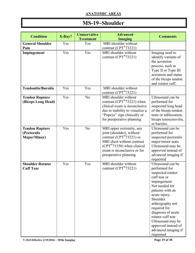
MS-19~Shoulder
Condition X-Ray? Conservative Treatment
Advanced Imaging Comments
General Shoulder Pain
Yes Yes MRI shoulder without contrast (CPT®73221)
Impingement Yes Yes MRI shoulder without contrast (CPT®73221)
Imaging used to identify variants of the acromion process, such as Type II or Type III acromion and status of the biceps tendon and rotator cuff.
Tendonitis/Bursitis Yes Yes MRI shoulder without contrast (CPT®73221)
Tendon Rupture (Biceps Long Head)
Yes No MRI shoulder without contrast (CPT®73221) when clinical exam is inconclusive due to inability to visualize a “Popeye” sign clinically or for preoperative planning
Ultrasound can be performed for suspected long head of the biceps tendon tears or subluxation, biceps tenosynovitis, or bursitis.
Tendon Rupture (Pectoralis Major/Minor)
Yes No MRI upper extremity, any joint (shoulder), without contrast (CPT®73221) or MRI chest without contrast (CPT®71550) when clinical exam is inconclusive or for preoperative planning
Ultrasound can be performed for suspected pectoralis major/minor tears Ultrasound may be approved instead of advanced imaging if requested
Shoulder Rotator Cuff Tear
Yes Yes MRI shoulder without contrast (CPT®73221)
Ultrasound can be performed for suspected rotator cuff tear or impingement. Not needed for patients with an acute injury. Shoulder arthrography not required for diagnosis of acute rotator cuff tear Ultrasound may be approved instead of advanced imaging if requested
Page 19 of 38
V.18.0 Effective 2/19/2016 – MSK Imaging
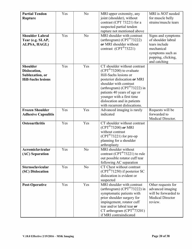
Partial Tendon Rupture
Yes No MRI upper extremity, any joint (shoulder), without contrast (CPT 73221) for a suspected partial tendon rupture not mentioned above
MRI is NOT needed for muscle belly strains/muscle tears
Shoulder Labral Tear (e.g. SLAP, ALPSA, HAGL)
Yes No MRI shoulder with contrast (arthrogram) (CPT®73222) or MRI shoulder without contrast (CPT®73221)
Signs and symptoms of shoulder labral tears include mechanical symptoms such as popping, clicking, and catching
Shoulder Dislocation, Subluxation, or Hill-Sachs lesions
Yes Yes CT shoulder without contrast (CPT®73200) to evaluate Hill-Sachs lesions or posterior dislocation or MRI shoulder with contrast (arthrogram) (CPT®73222) in patients 40 years of age or younger with a first time dislocation and in patients with recurrent dislocations.
Frozen Shoulder Adhesive Capsulitis
Yes Yes Advanced imaging is rarely indicated
Requests will be forwarded to Medical Director.
Osteoarthritis Yes Yes CT shoulder without contrast (CPT®73200) or MRI without contrast (CPT®73221) for pre-op planning for a shoulder arthroplasty
Acromiclavicular (AC) Separation
Yes No MRI shoulder without contrast (CPT®73221) to rule out possible rotator cuff tear following AC separation
Sternoclavicular (SC) Dislocation
Yes No CT Chest without contrast (CPT®71250) if posterior SC dislocation is evident or suspected
Post-Operative Yes Yes MRI shoulder with contrast (arthrogram) (CPT®73222) in symptomatic patients with prior shoulder surgery for impingement, rotator cuff tear and/or labral tear or CT arthrogram (CPT®73201) if MRI contraindicated
Other requests for advanced imaging will be forwarded to Medical Director review.
Page 20 of 38

V.18.0 Effective 2/19/2016 – MSK Imaging
References 1. McDonald LS, Dewing CB, Shupe PG, Provencher MT. Disorders of the proximal and distal
aspects of the biceps muscle. J Bone Joint Surg. 2013; 95:1235-1245. 2. Woodward TW, Best TM. The painful shoulder: Part II. Acute and chronic disorders. Am Fam
Physician. 2000; 61(11):3291-3300. 3. ACR Appropriateness Criteria®, Acute shoulder pain, 2010. 4. Bradley M, Tung G, Green A. Overutilization of shoulder magnetic resonance imaging as a
diagnostic screening tool in patients with chronic shoulder pain. J Shoulder Elbow Surgery. 2005; 14(3):233-237.
5. Fongemie AE, Buss DD, Rolnick SJ. Management of shoulder impingement syndrome and rotator cuff tears. Am Fam Physician. 1998; 57(4):667-674.
6. Greene WB (Ed.). Essentials of Musculoskeletal Care. 3rd Ed. Rosemont, IL, American Academy of Orthopaedic Surgeons, 2005, p.212.
7. Wheeless CR. Sternoclavicular Joint Injury, Updated January 3, 2013. http://www.wheelessonline.com/ortho/sternoclavicular_joint_injury.
8. Petersen SA, Murphy TP. The timing of rotator cuff repair for the restoration of function. Journal of Shoulder and Elbow Surgery, 2011; 20(1):62-68.
9. Hovelius L, Olofsson A, Sandstrom B, Augustini BG, et al. Nonoperative treatment of primary anterior shoulder dislocation in patients forty years of age and younger: a prospective twenty-five year follow-up. J Bone Joint Surg. 2008; 90:945-52.
10. Streubel PN, Krych AJ, Simone JP, Dahm DL, et al. Anterior glenohumeral instability: a pathology-based surgical treatment strategy. J Am Acad Orthop Surg. 2014; 22:283-294.
11. Major NM, Browne J, Domzalski T, Cothran RL, Helms CA. Evaluation of the glenoid labrum with 3-T MRI: is intraarticular contrast necessary. AJR. 2011; 196: 1139-1144.
12. Magee T. 3-T MRI of the shoulder: is MR arthrography necessary? AJR. 2009:192: 86-92. 13. Werner BC, Brockmeier SF, Miller MD. Etiology, diagnosis, and management of failed SLAP
repair. J Am Acad Orthop Surg. 2014; 22:554-565. 14. Rehman A, Robinson P. Sonographic evaluation of injuries of the pectoralis muscles. AJR, 2005;
184:1205-1211.
Page 21 of 38

V.18.0 Effective 2/19/2016 – MSK Imaging
ANATOMIC AREAS
MS-20~ELBOW
Condition X-Ray? Conservative Treatment
Advanced Imaging Comments
General Elbow Pain Yes Yes MRI elbow without contrast (CPT®73221)
Tendonitis Yes Yes MRI elbow without contrast (CPT®73221)
Lateral or Medial Epicondylitis-Tendonitis (Tennis Elbow)
Yes Yes MRI elbow without contrast (CPT®73221) following a recent X-ray, recent diagnostic MSK ultrasound, after 6 months of conservative treatment or for preop planning
All other requests will be forwarded to Medical Director review.
Osteochondritis Dissecans
Yes Yes MRI elbow without contrast (CPT®73221) or CT elbow(CPT®73200) without contrast
See also MS-13
Ruptured Biceps Insertion at Elbow
Yes No MRI elbow without contrast (CPT®73221) when clinical exam is inconclusive or for preoperative planning
Ruptured Triceps Insertion at Elbow
Yes No MRI upper extremity, any joint (elbow), without contrast (CPT®73221) when clinical exam is inconclusive or for preoperative planning
Partial Tendon Rupture
Yes No MRI upper extremity, any joint (elbow) without contrast (CPT®73221) for a suspected partial tendon rupture of a tendon not otherwise specified
MRI is NOT
needed for muscle belly strains/muscle tears
Trauma Yes No MRI elbow without contrast (CPT®73221) or CT without contrast (CPT®73200) when surgery is being considered
Ulnar Collateral Ligament (UCL) Tear
Yes No MRI elbow arthrogram (CPT®73222) following acute or repetitive elbow trauma
Post-Operative Yes Yes CT upper extremity (elbow) (CPT®73200) in symptomatic post-op patients following surgical treatment of complex fractures or MRI elbow without contrast (CPT®73221) in symptomatic post-op patients following soft-tissue surgery.
Other requests for advanced imaging will be forwarded to Medical Director review.
Page 22 of 38
V.18.0 Effective 2/19/2016 – MSK Imaging
References 1. McDonald LS, Dewing CB, Shupe PG, Provencher MT. Disorders of the proximal and distal aspects
of the biceps muscle. J Bone Joint Surg. 2013; 95:1235-1245. 2. Torp-Pedersen TE, Torp-Pedersen ST, Qvistgaard E, et al. Effect of glucocorticosteroid injections in
tennis elbow verified on colour Doppler ultrasonography: evidence of inflammation. Br J Sports
Med. 2008; 42(12):978-982. 3. Johnson GW, Cadwallader K, Scheffel SB, Epperly TD.Treatment of lateral epicondylitis. Am Fam
Physician. 2007; 76(6):843-848. 4. Greene WB (Ed.). Essentials of Musculoskeletal Care. 3rd Ed. Rosemont, IL, American Academy of
Orthopaedic Surgeons, 2005, pp. 279-280. 5. ACR Appropriateness Criteria®
, Chronic elbow pain, 2011. 6. Griffith JF, Roebuck DJ, Cheng JCY, et al. Acute elbow trauma in children: Spectrum of injury
revealed by MR imaging not apparent on radiographs. AJR. 2001; 176:53-60. 7. Bruce JR, Andrews JR. Ulnar collateral ligament injuries in the throwing athlete. J Am Acad Orthop
Surg. 2014; 22:315-325.
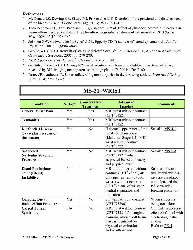
MS-21~WRIST
Condition X-Ray? Conservative Treatment
Advanced Imaging Comments
General Wrist Pain Yes Yes MRI wrist without contrast (CPT®73221)
Tendonitis Yes Yes MRI wrist without contrast (CPT®73221)
Kienböck’s Disease (avascular necrosis of the lunate)
Yes No If normal appearance of the lunate on plain X-ray (Lichtman Stage 1,2), MRI wrist without contrast (CPT®73221)
See also MS-4.1
Suspected Navicular/Scaphoid Fracture
Yes No MRI wrist without contrast (CPT®73221) when suspected based on history and physical exam
See also MS-5.2
Distal Radioulnar Joint (DRUJ) Instability
Yes No MRI of both wrists without contrast (CPT®73221) or CT upper extremity (both wrists) without contrast (CPT®73200) of wrists in neutral supination and pronation
Standard PA and true lateral wrist X-rays are mandatory with clenched fist PA view with forearm pronation.
Complex Distal Radius/Ulna Fracture
Yes No CT wrist without contrast (CPT®73200)
When surgery is being considered
Carpal Tunnel Syndrome
No No MRI wrist without contrast (CPT®73221) for surgical planning when a soft tissue mass is identified on physical examination and/or ultrasound
Clinical diagnosis is often confirmed with electrodiagnostic studies. Refer to PN-2
Page 23 of 38

V.18.0 Effective 2/19/2016 – MSK Imaging
Intrinsic Ligament/Triangular Fibrocartilage Complex (TFCC) Injuries
Yes Yes MRI wrist with contrast (arthrogram) (CPT®73222)
Complete Rupture of a Specific Named Tendon Not Otherwise Specified
Yes No MRI upper extremity, any joint (wrist), without contrast (CPT®73221) for preoperative planning
Partial Tendon Rupture
Yes No MRI upper extremity, any joint (wrist), without contrast (CPT®73221) for a suspected partial tendon rupture of a tendon not otherwise specified
MRI is NOT needed for muscle belly strains/muscle tears
Post-Operative Yes Yes CT upper extremity (wrist) without contrast (CPT®73200) in symptomatic patients following surgery for navicular/scaphoid fractures and complex distal radius/ulna fractures or MRI wrist with contrast (arthrogram) (CPT®73222) in symptomatic patients following DRUJ or TFCC surgery
Other requests for advanced imaging will be forwarded to Medical Director review.
References 1. Bruno MA, Weissman BN, Kransdorf MJ, Adler R et al. ACR Appropriateness Criteria®: Acute
Hand and Wrist Trauma. Last review date 2013. 2. Rubin DA, Weissman BN, Appel M, Arnold E. ACR Appropriateness Criteria®: Chronic Wrist
Pain. Last review date 2012. 3. Hayter CL, Gold SL, Potter HG. Magnetic resonance imaging of the wrist: bone and cartilage injury.
J Magn Reson Imaging. 2013; 37(5):1005-19. 4. Pruitt DL, Gilula LA, Manske PR, Vannier MW: Computed tomography scanning with image
reconstruction in evaluation of distal radius fractures. J Hand Surg Am. 1994; 19(5):720-727. 5. Magee T. Comparison of 3-T MRI and arthroscopy of intrinsic wrist ligament and TFCC tears.
AJR. 2009:192: 80-85. 6. Lee RK, Ng AW, Tong CS, Griffith JF, Tse WL, Wong C, Ho PC. Intrinsic ligament and triangular
fibrocartilage complex tears of the wrist: comparison of MDCT arthrography, conventional 3-T MRI, and MR arthrography. Skeletal Radiol. 2013,42:1277-85.
7. Pahwa S, Srivastava DN, Sharma R, Gamanagatti S, Kotwal PP, Sharma V. Comparison of conventional MRI and MR arthrography in the evaluation wrist ligament tears: A preliminary experience. Indian J Radiol Imaging. 2014. 3:259-67.
Page 24 of 38

V.18.0 Effective 2/19/2016 – MSK Imaging
ANATOMICAL AREAS
MS-22~HAND
Condition X-Ray? Conservative Treatment
Advanced Imaging Comments
General Hand Pain Yes Yes MRI upper extremity, other than joint (Hand) without contrast (CPT®73218) or MRI upper extremity, any joint (Hand), without contrast (CPT®73221)
Tendonitis Yes Yes MRI upper extremity (other than hand joint) without contrast (CPT®73218) or MRI upper extremity, any joint without contrast (CPT 73221)
Occult Fracture Yes No Advanced imaging guided by MS-5
See also MS-5.1
Complex Fracture Yes No CT upper extremity (hand) without contrast (CPT®73218) when plain X-ray shows a complex fracture
Ulnar Collateral Ligament (UCL) Thumb Injury
Yes No MRI upper extremity, any joint, (CPT®73221), if rule out for Stener lesion or complete tear of UCL of the thumb MCP joint
Also called “Gamekeepers Thumb;” Ultrasound (CPT®76882) can be performed instead of advanced imaging if requested for Stener Lesion
Complete Rupture of a Specific Named Tendon not Otherwise Specified
Yes No MRI upper extremity, other than joint (hand) without contrast (CPT®73218) for pre-operative planning
Partial Tendon Rupture
Yes No MRI Upper Extremity, other than joint (hand), without contrast (CPT®73218) for a suspected partial tendon rupture of a tendon not otherwise specified
MRI is NOT needed for muscle belly strains/muscle tears
Post-Operative Yes Yes CT upper extremity (elbow) without contrast (CPT®73200) or MRI upper extremity (elbow) without contrast (CPT® 73221) in symptomatic post-op patients following surgical treatment for
Other requests for advanced imaging will be forwarded to Medical Director review.
Page 25 of 38
V.18.0 Effective 2/19/2016 – MSK Imaging
complex hand fractures or MRI upper extremity, any joint (elbow), without contrast (CPT® 73221) in symptomatic post-op patients following soft-tissue surgery.
References 1. Bruno MA, Weissman BN, Kransdorf MJ, Adler R et al. ACR Appropriateness Criteria®: Acute
Hand and Wrist Trauma. Revised 2013. 2. Hayter CL, Gold SL, Potter HG. Magnetic resonance imaging of the wrist: Bone and cartilage
injury. J Magn Reson Imaging. 2013; 37(5):1005-19.
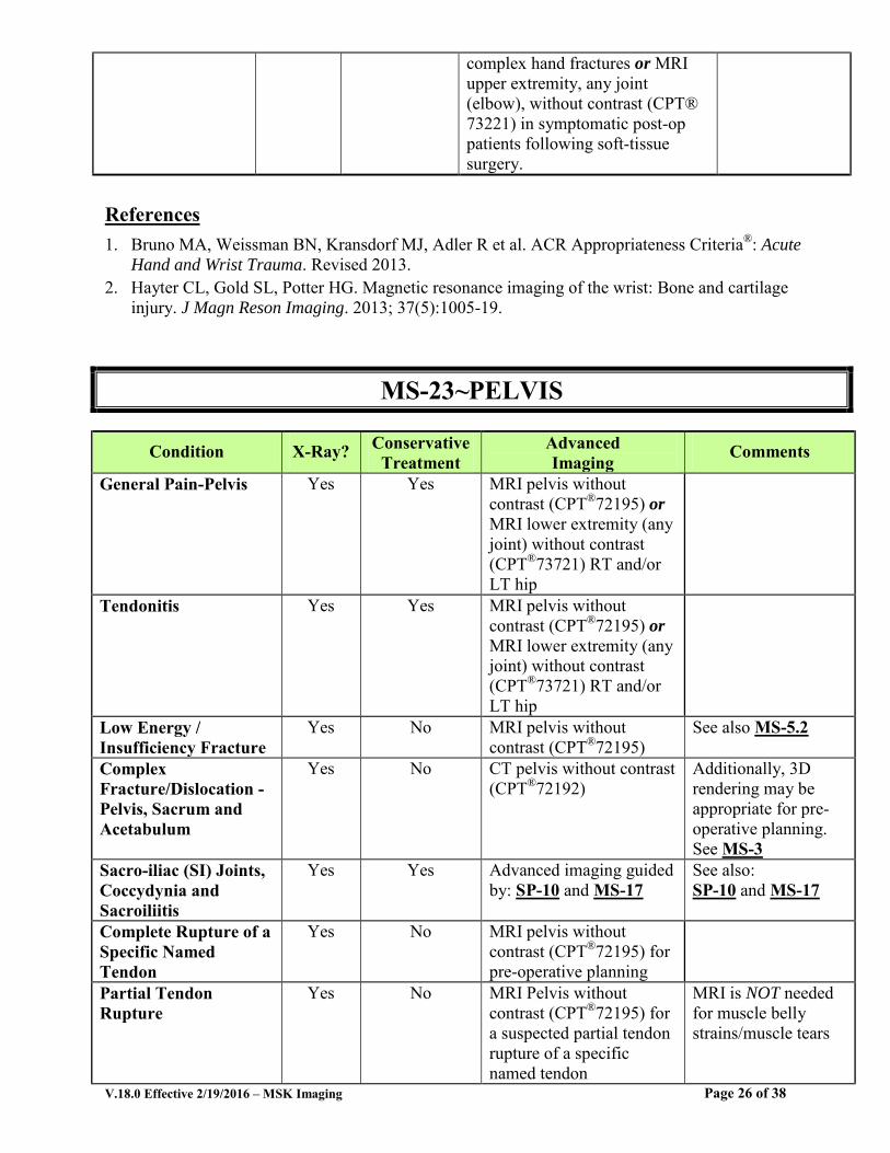
MS-23~PELVIS
Condition X-Ray? Conservative Treatment
Advanced Imaging Comments
General Pain-Pelvis Yes Yes MRI pelvis without contrast (CPT®72195) or MRI lower extremity (any joint) without contrast (CPT®73721) RT and/or LT hip
Tendonitis Yes Yes MRI pelvis without contrast (CPT®72195) or MRI lower extremity (any joint) without contrast (CPT®73721) RT and/or LT hip
Low Energy / Insufficiency Fracture
Yes No MRI pelvis without contrast (CPT®72195)
See also MS-5.2
Complex Fracture/Dislocation -Pelvis, Sacrum and Acetabulum
Yes No CT pelvis without contrast (CPT®72192)
Additionally, 3D rendering may be appropriate for pre-operative planning. See MS-3
Sacro-iliac (SI) Joints, Coccydynia and Sacroiliitis
Yes Yes Advanced imaging guided by: SP-10 and MS-17
See also: SP-10 and MS-17
Complete Rupture of a Specific Named Tendon
Yes No MRI pelvis without contrast (CPT®72195) for pre-operative planning
Partial Tendon Rupture
Yes No MRI Pelvis without contrast (CPT®72195) for a suspected partial tendon rupture of a specific named tendon
MRI is NOT needed for muscle belly strains/muscle tears
Page 26 of 38
V.18.0 Effective 2/19/2016 – MSK Imaging
Post-Operative Yes Yes CT pelvis without contrast (CPT®72192) in symptomatic patients following surgery for complex pelvic ring/acetabular fractures
Other requests for advanced imaging will be forwarded to Medical Director review.
References 1. Daffner RH, Weissman BN, Appel M, Bancroft L. et al. ACR Appropriateness Criteria®, Stress
(fatigue/insufficiency) fracture, including sacrum, excluding other vertebrae. 2011. 2. Mehta S, Auerbach JD, Born CT, Chin KR. Sacral fractures. J Am Acad Orthop Surg. 2006; 14:656-
665.
MS-24~HIP
Condition X-Ray? Conservative Treatment
Advanced Imaging Comments
General Hip Pain Yes Yes MRI lower extremity, any joint (hip) without contrast(CPT®73721)
Tendonitis Yes Yes MRI lower extremity, any joint (hip) without contrast (CPT®73721)
Hip Abductor Tear/Avulsion
Yes No MRI hip without Contrast (CPT®73721)
Ultrasound (CPT®76882) can be approved as an alternative to MRI
Complete Rupture of a Specific Named Tendon
Yes No MRI Lower Extremity, any joint (hip) without contrast (CPT®73721) for pre-operative planning
Partial Tendon Rupture
Yes No MRI Lower Extremity, any joint (hip) without contrast (CPT®73721) for a suspected partial tendon rupture of a specific named tendon not otherwise specified
MRI is NOT needed for muscle belly strains/muscle tears
Metal-On-Metal (MoM) Hip Prostheses
Yes No MRI hip without and with contrast (CPT®73723) or Ultrasound (CPT®76881) for diagnosis of pseudotumors
Either MRI or Ultrasound (CPT®76881) can be approved, not both. See also MS-17
Suspected Occult Hip Fracture
Yes No MRI hip without contrast (CPT®73721); or CT hip without contrast (CPT®73700) when suspected and X-ray is negative for fracture
See also MS-5.2
Page 27 of 38
V.18.0 Effective 2/19/2016 – MSK Imaging
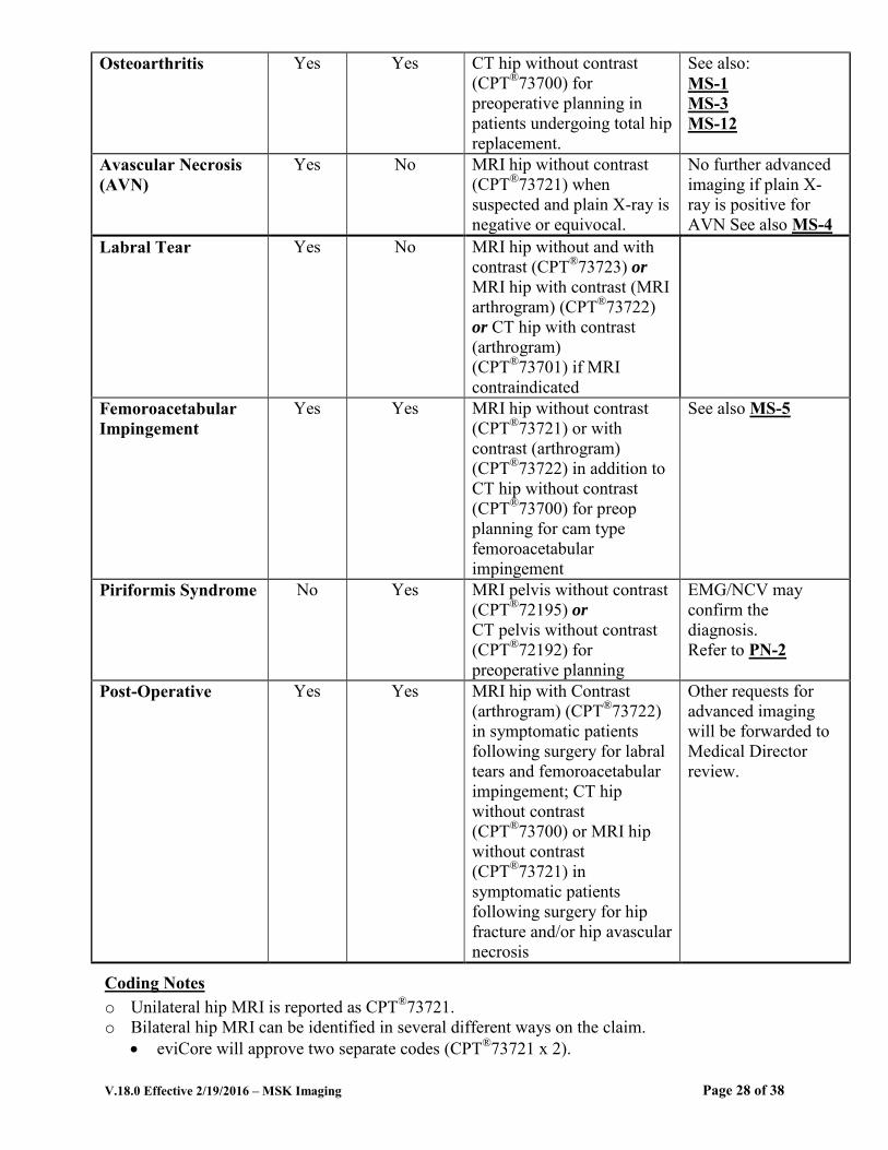
Osteoarthritis Yes Yes CT hip without contrast (CPT®73700) for preoperative planning in patients undergoing total hip replacement.
See also: MS-1 MS-3 MS-12
Avascular Necrosis (AVN)
Yes No MRI hip without contrast (CPT®73721) when suspected and plain X-ray is negative or equivocal.
No further advanced imaging if plain X-ray is positive for AVN See also MS-4
Labral Tear Yes No MRI hip without and with contrast (CPT®73723) or MRI hip with contrast (MRI arthrogram) (CPT®73722) or CT hip with contrast (arthrogram) (CPT®73701) if MRI contraindicated
Femoroacetabular Impingement
Yes Yes MRI hip without contrast (CPT®73721) or with contrast (arthrogram) (CPT®73722) in addition to CT hip without contrast (CPT®73700) for preop planning for cam type femoroacetabular impingement
See also MS-5
Piriformis Syndrome No Yes MRI pelvis without contrast (CPT®72195) or CT pelvis without contrast (CPT®72192) for preoperative planning
EMG/NCV may confirm the diagnosis. Refer to PN-2
Post-Operative Yes Yes MRI hip with Contrast (arthrogram) (CPT®73722) in symptomatic patients following surgery for labral tears and femoroacetabular impingement; CT hip without contrast (CPT®73700) or MRI hip without contrast (CPT®73721) in symptomatic patients following surgery for hip fracture and/or hip avascular necrosis
Other requests for advanced imaging will be forwarded to Medical Director review.
Coding Notes o Unilateral hip MRI is reported as CPT®73721. o Bilateral hip MRI can be identified in several different ways on the claim.
eviCore will approve two separate codes (CPT®73721 x 2).
Page 28 of 38

V.18.0 Effective 2/19/2016 – MSK Imaging
References 1. ACR Appropriateness Criteria®, Chronic hip pain, 2011. 2. ACR Appropriateness Criteria®, Avascular necrosis of the hip, 2009. 3. Greene WB (Ed.). Essentials of Musculoskeletal Care. 2nd Ed. Rosemont, IL, American Academy of
Orthopaedic Surgeons, 2001,p. 295. 4. Greene WB (Ed.). Essentials of Musculoskeletal Care. 3rd Ed. Rosemont, IL, American Academy of
Orthopaedic Surgeons, 2005, pp.433-436; 438-440. 5. Manek NJ and Lane NE. Osteoarthritis: Current concepts in diagnosis and management. Am Fam
Physician. 2000; 61(6):1795-1804. 6. Papadoupoulos EC and Kahn SN. Piriformis syndrome and low back pain: a new classification and
review of the literature. Orthop Clin North Am 2004 Jan; 35(1):65-71. 7. Reurink G, Sebastian, et al. Reliability and Validity of Diagnostic Acetabular Labral Lesions with
Magnetic Resonance Arthrography. J Bone Joint Surg A. 2012; 94(181): 1643-1648. 8. Steinbach LS, Palmer WE, Schweitzer ME. MR Arthrography. RadioGraphics 2002; 22:1223-1246. 9. Daffner RH, Weissman BN, Appel M, Bancroft L.et al. ACR Appropriateness Criteria®, Stress
(fatigue/insufficiency) fracture, including sacrum, excluding other vertebrae. 2011.
Page 29 of 38

V.18.0 Effective 2/19/2016– MSK Imaging
ANATOMICAL AREAS
MS-25~KNEE
Condition X-Ray? Conservative Treatment
Advanced Imaging
Comments
General Knee Pain Yes Yes MRI lower extremity, any joint (knee) without contrast (CPT®73721)
Tendonitis Yes Yes MRI lower extremity, any joint (knee) without contrast (CPT®73721)
Complex Knee Fracture
Yes No CT knee without contrast (CPT®73700)
See also MS 5
Meniscus Tear Yes No MRI knee without contrast (CPT®73721) In absence of conservative treatment, at least 2 of following 4 criteria must be met: 1) Positive McMurray’s or positive Thessaly test 2) twisting or acute injury of the knee 3) locked knee/inability to fully extend the knee, or 4) knee effusion
Ligament Tear Yes No MRI knee without contrast (CPT®73721) if any of the following signs are positive in comparison to the normal knee:
-Anterior drawer -Lachman -Pivot shift test -Posterior drawer -Posterior sag test -Valgus stress test; -Varus stress test
Osteoarthritis Yes Yes MRI knee without contrast (CPT®73721) when clinical presentation consistent with symptomatic meniscus tear; CT knee without contrast (CPT®73700) for preoperative planning for knee arthroplasty when congenital, post-traumatic or otherwise acquired deformities exist of the patella, distal femur and/or proximal tibia
Additionally, 3D rendering may be appropriate for pre-operative planning. See also: MS-1 MS-3 MS-12
Patellar Dislocation Yes No MRI knee without contrast (CPT®73721) with acute knee
Page 30 of 38
V.18.0 Effective 2/19/2016 – MSK Imaging
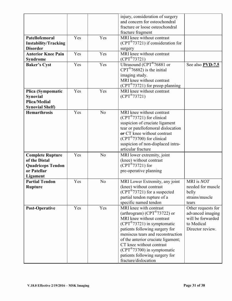
injury, consideration of surgery and concern for osteochondral fracture or loose osteochondral fracture fragment
Patellofemoral Instability/Tracking Disorder
Yes Yes MRI knee without contrast (CPT®73721) if consideration for surgery
Anterior Knee Pain Syndrome
Yes Yes MRI knee without contrast (CPT®73721)
Baker’s Cyst Yes Yes Ultrasound (CPT®76881 or CPT®76882) is the initial imaging study. MRI knee without contrast (CPT®73721) for preop planning
See also PVD-7.5
Plica (Sympomatic Synovial Plica/Medial Synovial Shelf)
Yes Yes MRI knee without contrast (CPT®73721)
Hemarthrosis Yes No MRI knee without contrast (CPT®73721) for clinical suspicion of cruciate ligament tear or patellofemoral dislocation or CT knee without contrast (CPT®73700) for clinical suspicion of non-displaced intra-articular fracture
Complete Rupture of the Distal Quadriceps Tendon or Patellar Ligament
Yes No MRI lower extremity, joint (knee) without contrast (CPT®73721) for pre-operative planning
Partial Tendon Rupture
Yes No MRI Lower Extremity, any joint (knee) without contrast (CPT®73721) for a suspected partial tendon rupture of a specific named tendon
MRI is NOT
needed for muscle belly strains/muscle tears
Post-Operative Yes Yes MRI knee with contrast (arthrogram) (CPT®73722) or MRI knee without contrast (CPT®73721) in symptomatic patients following surgery for meniscus tears and reconstruction of the anterior cruciate ligament; CT knee without contrast (CPT®73700) in symptomatic patients following surgery for fracture/dislocation
Other requests for advanced imaging will be forwarded to Medical Director review.
Page 31 of 38

V.18.0 Effective 2/19/2016 – MSK Imaging
References 1. Harrison BK, Abell BE, Gibson TW. The Thessaly test for detection of meniscal tears: validation of
a new physical examination technique for primary care medicine. Clin J Sport Med, 2009; 19: 9-12. 2. Landewé RBM, Günther KP, Lukas C, et al. EULAR/EFFORT recommendations for the diagnosis
and initial management of patients with acute or recent onset swelling of the knee. Ann Rheum Dis. 2010; 69:12-19.
3. Johnson MW. Acute knee effusions: a systematic approach to diagnosis. Am Fam Physician. 2000; 61(8):2391-2400.
4. ACR Appropriateness Criteria, Nontraumatic knee pain, 2008. 5. Sung-Jae Kim, Byoung-Yoon Hwang, Duck-Hyun Choi, Yu-Mei. J Bone Joint Surg A. 2012;
94(16):e118 1-7. 6. Kannus P, Järvinen M. Nonoperative treatment of acute knee ligament injuries. A review with
special reference to indications and methods. Sports Med.1990; 9(4):244-260. 7. Manek NJ and Lane NE. Osteoarthritis: Current concepts in diagnosis and management. Am Fam
Physician. 2000; 61(6):1795-1804. 8. Greene WB (Ed.). Essentials of Musculoskeletal Care. 3rd Ed. Rosemont, IL, American Academy of
Orthopaedic Surgeons, 2005, p.84; 541-545. 9. Lee IS, Choi JA, Kim TK, et al. Reliability analysis of 16-MDCT in preoperative evaluation of total
knee arthroplasty and comparison with intraoperative measurements. AJR. 2006; 186(6):1778-1782. 10. Morrissey RT and Weinstein SL (Eds.). Lovell and Winter’s Pediatric Orthopaedics. 6th Ed.
Philadelphia, Uppinortt Williams and Wilkins, p.1413. 11. Woolson ST, Harris AHS, Wagner DW, Giori NJ. Component alignment during total knee
arthroplasty with use of standard or custom instrumentation. Journal of Bone and Joint Surgery, 2014; 96:366-372.
12. Vance K, Meredick R, Schweitzer ME, Lubowitz JH. Magnetic resonance imaging of the postoperative meniscus. Arthroscopy, 2009; 25: 522-30.
13. Magee T, Shapiro M, Williams D. Prevalence of meniscal radial tears of the knee revealed by MRI after surgery. AJR, 2004; 184: 931-936.
14. Meyers AB, Haims AH, Menn K, Moukaddam H. Imaging of anterior cruciate ligament repair and its complications. AJR, 2010; 194: 476-484.
Page 32 of 38

V.18.0 Effective 2/19/2016 – MSK Imaging
ANATOMICAL AREAS
MS-26~ANKLE
Condition X-Ray? Conservative Treatment
Advanced Imaging Comments
General Ankle Pain Yes Yes MRI ankle without contrast (CPT®73721)
Complex Fracture Yes No CT ankle without contrast (CPT®73700)
Ankle Sprain, Including Avulsion Fracture
Yes Yes MRI ankle without contrast (CPT®73721) or CT without contrast (CPT®73700)
High Ankle Sprain Yes No MRI ankle without contrast (CPT®73721)
Suspected Osteochondral Injury
Yes No MRI ankle without contrast (CPT®73721) or MRI ankle with contrast (arthrogram) (CPT®73722) or CT ankle with contrast (arthrography) (CPT®73701)
Anterior Impingement Posterior Impingement (e.g., Os Trigonum Syndrome)
Yes No MRI ankle without contrast (CPT®73721)or MRI ankle with contrast (arthrogram) (CPT®73722), or CT ankle with contrast (arthrogram) (CPT®73701)
Ultrasound (CPT®76881 or 76882) may be approved instead of advanced imaging if requested.
Anterior-Lateral Impingement
Yes No MRI ankle without contrast (CPT®73721)or MRI ankle with contrast (arthrogram) (CPT®73722), or CT ankle with contrast (arthrogram) (CPT®73701)
Tendonitis Yes Yes MRI ankle without contrast (CPT®73721) for suspected posterior tibial dysfunction, peroneal tendon or subluxation, Achilles tendonitis
Ultrasound (CPT®76881 or 76882) may be approved instead of advanced imaging if requested.
Ruptured Achilles Tendon
Yes No MRI ankle without contrast (CPT®73721) for preoperative evaluation
Ultrasound (CPT®76881 or CPT®76882) for preoperative evaluation may be approved instead of advanced imaging if requested.
Continued . . .
Page 33 of 38

V.18.0 Effective 2/19/2016 – MSK Imaging
ANATOMICAL AREAS
MS-26~ANKLE Continued . . .
Condition X-Ray? Conservative Treatment
Advanced Imaging Comments
Complete Rupture -Tear of a Specific Named Tendon Yes No
MRI lower extremity, any joint (ankle) without contrast (CPT®73721) for pre-operative planning
Partial Tendon Rupture
Yes No
MRI lower extremity, any joint (ankle), without contrast (CPT®73721) for a suspected partial tendon rupture of a specific named tendon
MRI is NOT needed for muscle belly strains/muscle tears
Instability Yes Yes
MRI ankle without contrast (CPT®73721) or
MRI ankle with contrast (arthrogram) (CPT®73722) for preoperative evaluation
Post-Operative Yes Yes
MRI ankle without contrast (CPT®73721) in symptomatic patients following surgery for ligament/tendon injuries; CT ankle without contrast (CPT®73700) for symptomatic patients following surgery for complex fractures
Other requests for advanced imaging will be forwarded to Medical Director review.
One Study/Area Only In foot and ankle advanced imaging, studies are frequently ordered of both areas. This is unnecessary since ankle MRI will image from above the ankle to the mid- metatarsal area. Only one CPT® code should be reported.
Page 34 of 38
V.18.0 Effective 2/19/2016 – MSK Imaging
References 1. ACR Appropriateness Criteria®
, Chronic ankle pain, 2009. 2. Wolfe MW, Uhl TL, McClusky LC. Management of ankle sprains. Am Fam Physician 2001 Jan;
63(1):93-104. 3. Greene WB (Ed.). Essentials of Musculoskeletal Care. 3rd Ed. Rosemont, IL, American Academy of
Orthopedic Surgeons, 2005, pp.593-596; 606-609; 683. 4. Bergkvist D, Astrom I, Josefsson PO, Dahlberg LE. Acute Achilles Tendon Rupture: A
Questionnaire Follow-up of 487 Patients. J Bone Joint Surg Am, 2012 Jul 03; 94(13): 1229-1233. 5. Hartgerink P, Fessell DP, Jacobson JA, et al. Full- versus partial-thickness Achilles tendon tears:
sonographic accuracy and characterization in 26 cases with surgical correlation. Radiology 2001; 220:406-412.
6. Jones MP, Riaz JK, Smith RLC. Surgical Interventions for Treating Acute Achlles Tendon Rupture: Key Findings from a Recent Cochrane Review. J Bone Joint Surg Am, 2012 Jun 20; 94(12):e88 1-6.
7. Vaseenon T, Amendola A. Update on anterior ankle impingement. Current Reviews in
Musculoskeletal Medicine, 2012; 5:140-150. 8. Talusan PG, Toy J, Perez J, Milewski MD, Reach JS. Anterior ankle impingement: diagnosis and
treatment. J Am Acad Orthop Surg, 2014; 22: 333-339. 9. Nault ML, Kocher MS, Micheli LJ. Os Trigonum Syndrome. J Am Acad Orthop Surg, 2014; 22:545-
553. 10. Peace KAL, Jillier JC, Hulme A, Healy JC. MRI features of posterior ankle impingement syndrome
in ballet dancers: a review 25 cases. Clinical Radiology, 2004; 59:1025-1033
Page 35 of 38
V.18.0 Effective 2/19/2016 – MSK Imaging
ANATOMICAL AREAS
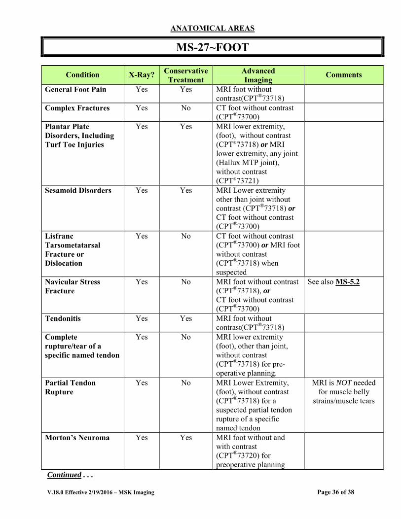
MS-27~FOOT
Condition X-Ray? Conservative Treatment
Advanced Imaging Comments
General Foot Pain Yes Yes MRI foot without contrast(CPT®73718)
Complex Fractures Yes No CT foot without contrast (CPT®73700)
Plantar Plate Disorders, Including Turf Toe Injuries
Yes Yes MRI lower extremity, (foot), without contrast (CPT®73718) or MRI lower extremity, any joint (Hallux MTP joint), without contrast (CPT®73721)
Sesamoid Disorders Yes Yes MRI Lower extremity other than joint without contrast (CPT®73718) or CT foot without contrast (CPT®73700)
Lisfranc Tarsometatarsal Fracture or Dislocation
Yes No CT foot without contrast (CPT®73700) or MRI foot without contrast (CPT®73718) when suspected
Navicular Stress Fracture
Yes No MRI foot without contrast (CPT®73718), or CT foot without contrast (CPT®73700)
See also MS-5.2
Tendonitis Yes Yes MRI foot without contrast(CPT®73718)
Complete rupture/tear of a specific named tendon
Yes No MRI lower extremity (foot), other than joint, without contrast (CPT®73718) for pre-operative planning.
Partial Tendon Rupture
Yes No MRI Lower Extremity, (foot), without contrast (CPT®73718) for a suspected partial tendon rupture of a specific named tendon
MRI is NOT needed for muscle belly
strains/muscle tears
Morton’s Neuroma Yes Yes MRI foot without and with contrast (CPT®73720) for preoperative planning
Continued . . .
Page 36 of 38

V.18.0 Effective 2/19/2016 – MSK Imaging
ANATOMICAL AREAS
MS-27~FOOT Continued . . .
Condition X-Ray? Conservative Treatment
Advanced Imaging Comments
Plantar Fasciitis Yes Yes MRI foot without contrast (CPT®73718) for preoperative planning
Non-Surgical Conservative
treatment is for 6 months or more.
Suspected Plantar Fascia Rupture or Tear
Yes Yes MRI foot without contrast (CPT®73718)
Calcaneal Insufficiency/Stress Fracture
Yes No MRI foot without contrast (CPT®73718)
See also MS 5.2
Diabetic Foot Infection
Yes No MRI foot without and with contrast (CPT®73720) for suspected osteomyelitis or deep infection when plain X-ray is negative
See also MS 9.1
Tarsal Tunnel Syndrome
Yes Yes MRI ankle without contrast (CPT®73721) or CT without contrast (CPT®73700) for pre-op planning if mass/lesion is suspected as etiology of entrapment
Tarsal Coalition Yes Yes MRI ankle without contrast (CPT®73721) or CT without contrast (CPT®73700) for preoperative planning
Sinus Tarsi Syndrome
Yes Yes MRI ankle without contrast (CPT®73721) if diagnosis is unclear or for preoperative evaluation.
Post-Operative Yes Yes MRI foot without contrast (CPT®73718) in symptomatic patients following surgery for conditions including the tendons, ligaments and plantar plate CT foot without contrast (CPT®73700) in symptomatic patients following surgery for complex fractures, sesamoid fractures and subtalar arthrodesis
Other requests for advanced imaging
will be forwarded to Medical Director
review.
Page 37 of 38

V.18.0 Effective 2/19/2016 – MSK Imaging
References 1. Greene WB (Ed.). Essentials of Musculoskeletal Care 3rd Ed. Rosemont, IL, American Academy of
Orthopaedic Surgeons, 2005, pp.619-622; 667-671; 681-684; 697-699; 700-702 2. ACR Appropriateness Criteria®, Chronic foot pain, 2008. 3. ACR Appropriateness Criteria®, Stress (fatigue/insufficiency) fracture, including sacrum, excluding
other vertebrae, 2011. 4. Needell S and Cutler J. Morton neuroma imaging. eMedicine, April 11, 2011,
http://emedicine.medscape.com/article/401417-overview. Accessed November 7, 2012. 5. Morton’s Neuroma. MDGuidelines™. http://www.mdguidelines.com/mortons-neuroma. Accessed
November 7, 2012. 6. Berquist TH (Ed.). Radiology of the Foot and Ankle. 2nd Ed. Philadelphia, Lippincott, 2000, pp.155-
156. 7. Bouché R. Sinus Tarsi syndrome. American Academy of Podiatric Sports Medicine.
http://www.aapsm.org/sinus_tarsi_syndrome.html. Accessed May 9, 2011 November 7, 2012. 8. D Resnick, Internal Derangements of Joints 2006: Imaging-Arthroscopic Correlation. Washington,
DC, Oct.31- Nov. 4, 2006 9. Doty JF, Coughlin MJ. Metatarsophalangeal joint instability of the lesser toes and plantar plate
deficiency. J Am Acad Orthop Surg, 2014; 22:235-245. 10. Lareau CR, Sawyer GA, Wang JH, DiGiovanni CW. Plantar and medial heel pain: diagnosis and
management. J Am Acad Orthop Surg, 2014; 22:372-380. 11. Sung, W, Weil L Jr, Weill LS Sr, Rolfes RJ: Diagnosis of plantar plate injury by magnetic resonance
imaging with reference to Intraoperative findings. Journal of Foot Ankle Surgery 2012; 51 (5): 570-574.
Page 38 of 38