chapter 4 – prokaryotic profilesfpm/bio205/sp-08/chapter-04.pdfchapter 4 – prokaryotic profiles...
TRANSCRIPT
1
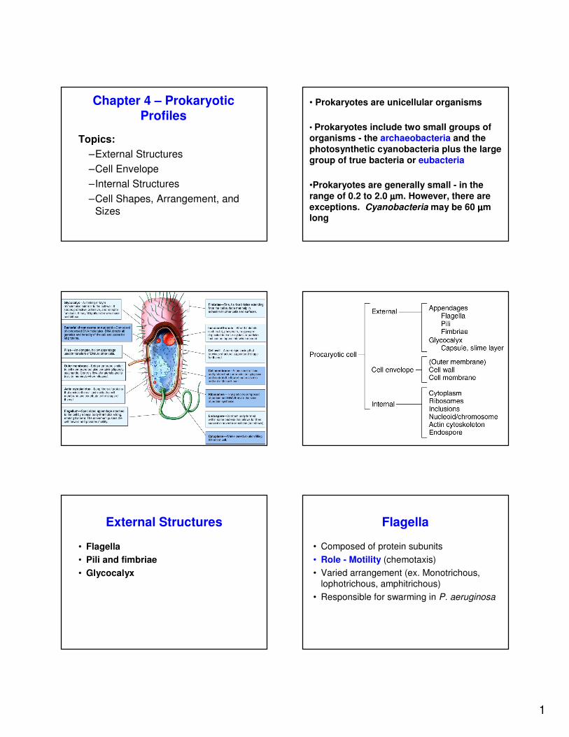
Chapter 4 – Prokaryotic Profiles
Topics:
–External Structures
–Cell Envelope
–Internal Structures
–Cell Shapes, Arrangement, and
Sizes
• Prokaryotes are unicellular organisms
• Prokaryotes include two small groups of organisms - the archaeobacteria and the
photosynthetic cyanobacteria plus the large
group of true bacteria or eubacteria
•Prokaryotes are generally small - in the
range of 0.2 to 2.0 µµµµm. However, there are
exceptions. Cyanobacteria may be 60 µµµµm long
External Structures
• Flagella
• Pili and fimbriae
• Glycocalyx
Flagella
• Composed of protein subunits
• Role - Motility (chemotaxis)
• Varied arrangement (ex. Monotrichous, lophotrichous, amphitrichous)
• Responsible for swarming in P. aeruginosa
2
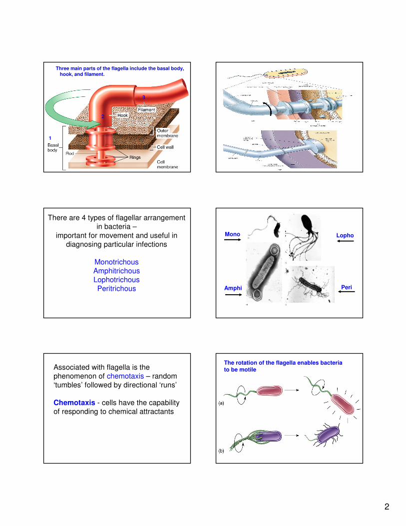
Three main parts of the flagella include the basal body,hook, and filament.
1
2
3
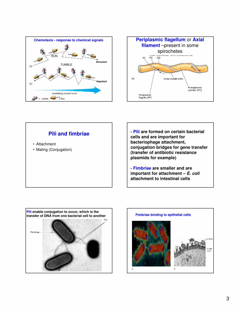
There are 4 types of flagellar arrangement
in bacteria –
important for movement and useful in
diagnosing particular infections
Monotrichous
Amphitrichous
Lophotrichous
Peritrichous
Mono
Amphi
Lopho
Peri
Associated with flagella is the
phenomenon of chemotaxis – random
‘tumbles’ followed by directional ‘runs’
Chemotaxis - cells have the capability
of responding to chemical attractants
The rotation of the flagella enables bacteria to be motile
3
Chemotaxis - response to chemical signals
RUN
TUMBLE
Periplasmic flagellum or Axial filament –present in some
spirochetes
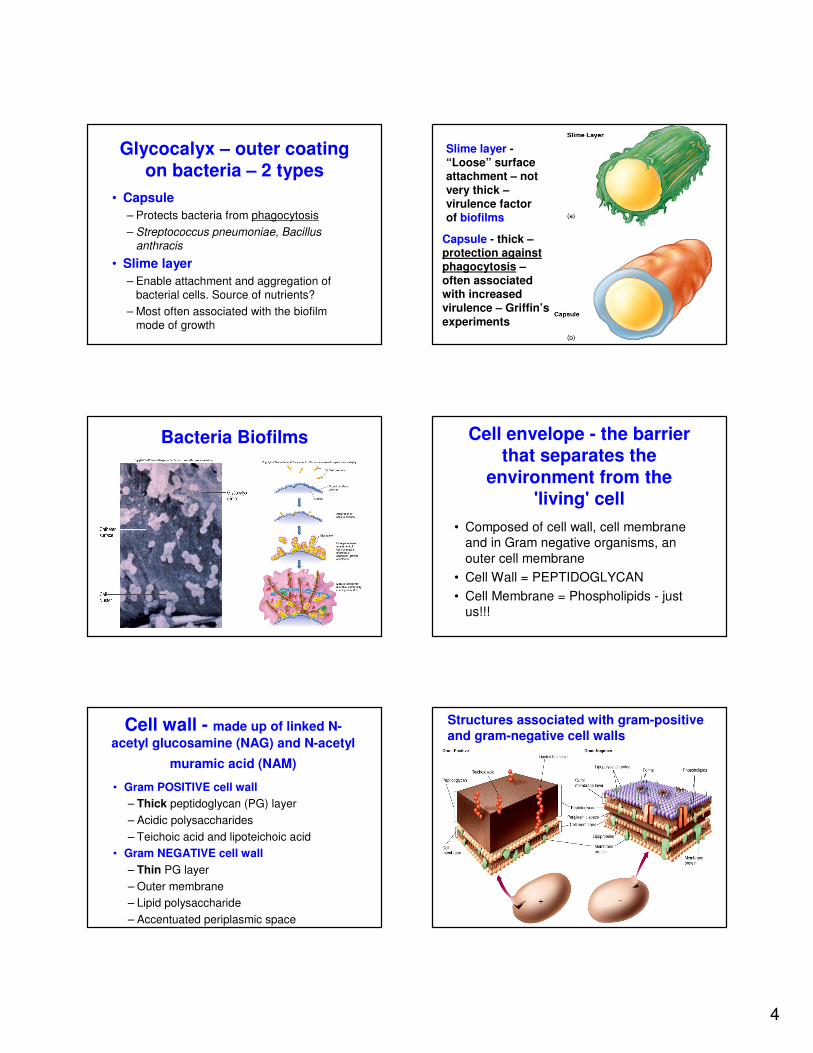
Pili and fimbriae
• Attachment
• Mating (Conjugation)
- Pili are formed on certain bacterial
cells and are important for
bacteriophage attachment,
conjugation bridges for gene transfer
(transfer of antibiotic resistance
plasmids for example)
- Fimbriae are smaller and are
important for attachment – E. coli
attachment to intestinal cells
Pili enable conjugation to occur, which is the transfer of DNA from one bacterial cell to another Fimbriae binding to epithelial cells
4
Glycocalyx – outer coating
on bacteria – 2 types
• Capsule
– Protects bacteria from phagocytosis
– Streptococcus pneumoniae, Bacillus anthracis
• Slime layer
– Enable attachment and aggregation of bacterial cells. Source of nutrients?
– Most often associated with the biofilm mode of growth
Slime layer -“Loose” surface attachment – not
very thick –virulence factor of biofilms
Capsule - thick –protection against phagocytosis –
often associated with increased virulence – Griffin’s
experiments
Bacteria Biofilms Cell envelope - the barrier that separates the
environment from the 'living' cell
• Composed of cell wall, cell membrane and in Gram negative organisms, an
outer cell membrane
• Cell Wall = PEPTIDOGLYCAN
• Cell Membrane = Phospholipids - just us!!!
Cell wall - made up of linked N-
acetyl glucosamine (NAG) and N-acetyl
muramic acid (NAM)
• Gram POSITIVE cell wall
– Thick peptidoglycan (PG) layer
– Acidic polysaccharides
– Teichoic acid and lipoteichoic acid
• Gram NEGATIVE cell wall
– Thin PG layer
– Outer membrane
– Lipid polysaccharide
– Accentuated periplasmic space
Structures associated with gram-positive
and gram-negative cell walls
5
- Teichoic acid consisting of glycerol, phosphates
and ribitol is found in polymers in gram-positives.
- Outer membrane - found primarily in Gram
negatives - lipopolysaccharide (LPS) is a major component - also called endotoxin - lipid A is a major component of LPS and causes the toxic
events of fever and blood vessel dilation observed in Gram-negative infections.
- Periplasmic space - a gap between the cell membrane and the cell wall - particularly evident in Gram negative bacteria.
Cartoon of the NAG and NAM polymers
- Layers of alternating NAM and NAG
- Linkage between NAM from one layer to the NAM of the other one
Linkage of two polymer chains through NAM in Gram positive bacteria
Both NAG & NAM have tetrapeptides
GRAM NEGATIVE
GRAM POSITIVE
Mycobacteria
Nontypical Cell Walls
- Mycobacteria- Non Gram positive or Negative- Increased amounts of LIPIDS- Special staining ���� ACID-FAST STAINING
No cell wall = No Peptidoglycan
• Cell membrane contain sterols for stability
- classical example is Mycoplasma - a
common cause of atypical pneumonia
- on agar, Mycoplasma looks like a 'fried egg'
6
Scanning electron micrograph of Mycoplasma pneumoniae
Cell Membrane
• Phospholipid bilayer and integral proteins
• Mycoplasma – STEROLS
• Function:
1) Selective permeability
2) Energy reactions
3) Synthesis of molecules
Internal Structures
• Cytoplasm
• Genetic structures
• Endospore
Cytoplasm
• Gelatinous solution containing water
(70-80%), nutrients, proteins, and genetic material.
• Presence of ACTIN-like filaments = Cytoskeleton
Genetic material and structures
• Single circular bacterial chromosome
• Nucleoid
• PLASMIDS – Independent circular DNA structures
• Ribosomes - 'structures' that have
multiple components - responsible for protein synthesis
Prokaryotic Ribosome
7
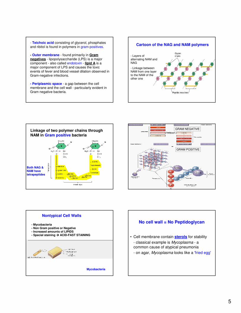
Storage Bodies
- NUTRITIONAL SOURCE – Glycogen,
Starch, β-hydroxybutyrate
- Gas vesicles
During nutrient depleted conditions, some bacteria (vegetative cell) form into an
endospore in order to survive
- Specific endospore
staining techniques often make the
endosporeslook like a “safety pin”
- Bacillus and Clostridium
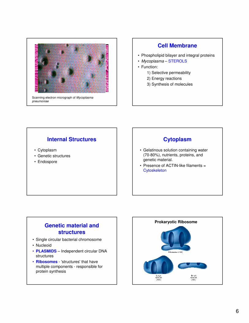
Endosporulation - a survival mechanism for lean times
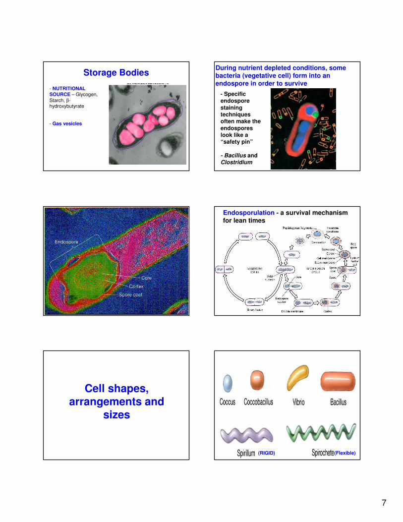
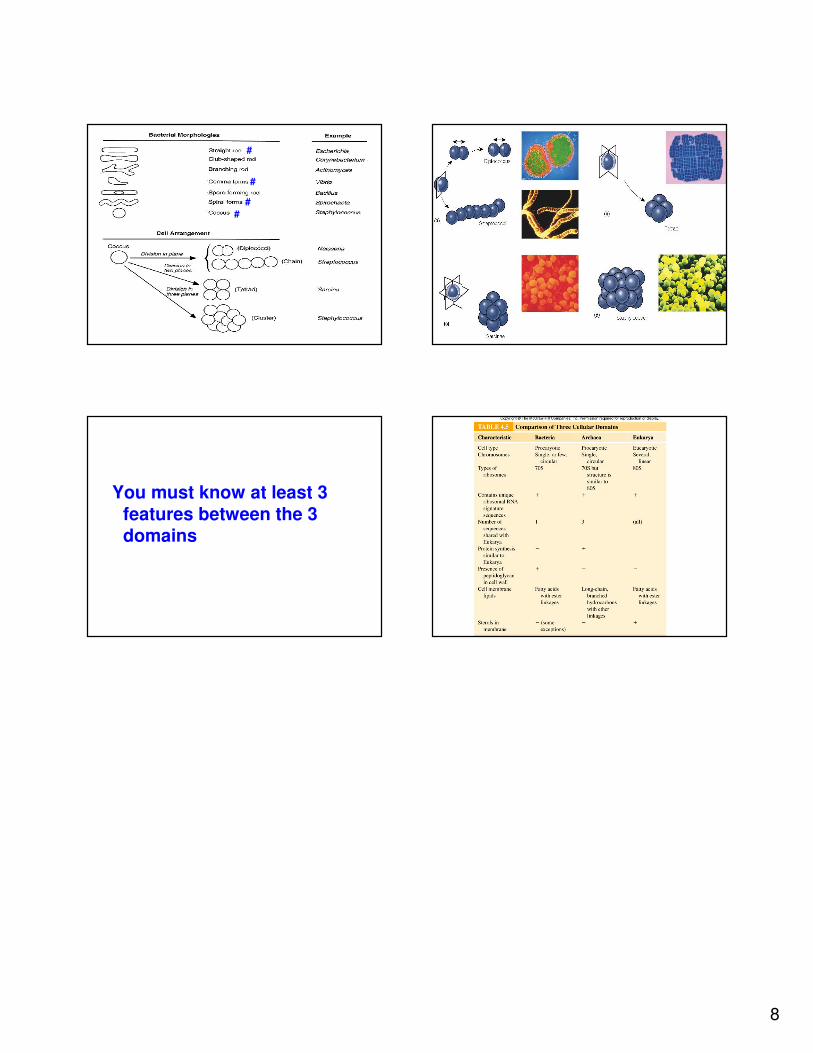
Cell shapes, arrangements and
sizes
(RIGID) (Flexible)
8
#
#
#
#
You must know at least 3
features between the 3 domains
Table 4.5