studying oral biofilms
TRANSCRIPT
STUDY OF
ORAL BIOFILMS
MEDICAL DEPARTMENT DENTAID
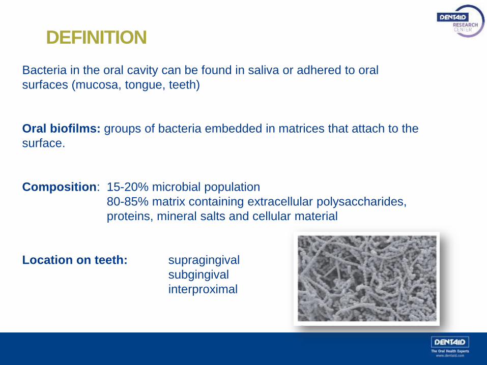
DEFINITION
Bacteria in the oral cavity can be found in saliva or adhered to oral
surfaces (mucosa, tongue, teeth)
Oral biofilms: groups of bacteria embedded in matrices that attach to the
surface.
Composition: 15-20% microbial population
80-85% matrix containing extracellular polysaccharides,
proteins, mineral salts and cellular material
Location on teeth: supragingival
subgingival
interproximal
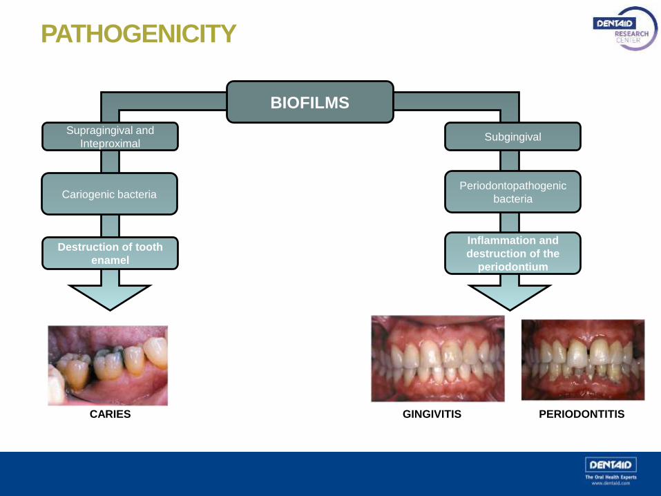
PATHOGENICITY
BIOFILMS
Supragingival and
Inteproximal
Cariogenic bacteria
Destruction of tooth
enamel
Subgingival
Inflammation and
destruction of the
periodontium
Periodontopathogenic
bacteria
CARIES GINGIVITIS PERIODONTITIS

Aggregatibacter actinomycetemcomitans
Porphyromonas gingivalis
Prevotella intermedia
Fusobacterium nucleatum
Tannerella forsythia
Campylobacter rectus
Eikenella corrodens
Parvimonas micra
Selenomonas spp
Eubacterium spp
Treponema spp
700 bacterial species are present in oral biofilm
BACTERIAL DIVERSITY IN THE ORAL CAVITY

BIOFILM FORMATION
Formation of acquired pellicle
with salivary proteins on the
enamel
1
Adhesion of primary colonisers:
gram-positive bacilli and cocci (S.
sanguis, S. oralis,…) and growth
2
Fusobacterium nucleatum
joins in: facilitating adhesion
by other bacteria (gram-
negative, anaerobes…)
3
4 Tertiary colonisers. Increased
complexity: gram-negative, strict
anaerobes
4 μm4 μm

BIOFILM PROPERTIES
When bacteria grow in the form of a biofilm, they work together as a
bacterial community, which gives them the following properties:
Physiological heterogeneity
Increased phenotypic resistance
Quorum sensing (interbacterial communication)
Adaptive capacity
Resistance to antimicrobial agents

IMPORTANCE OF THE STUDY OF BIOFILMS
Isolated bacteria (planktonic bacteria) behave differently and
have different properties than bacteria that are organised in
biofilms.
These different properties give them increased resistance to
antiseptics and greater pathogenicity.
It is necessary to study the behaviour of bacteria in biofilms
to therefore prevent the oral diseases they may cause.
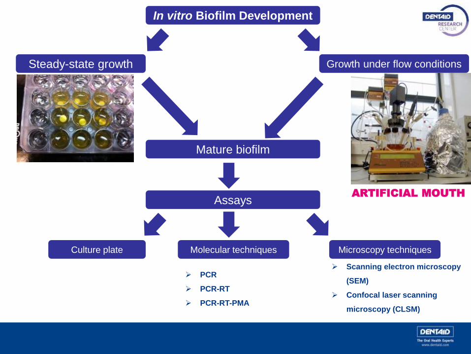
In vitro Biofilm Development
Steady-state growth Growth under flow conditions
Mature biofilm
Culture plate Molecular techniques Microscopy techniques
PCR
PCR-RT
PCR-RT-PMA
Scanning electron microscopy
(SEM)
Confocal laser scanning
microscopy (CLSM)
AssaysARTIFICIAL MOUTH

IN VITRO BIOFILM DEVELOPMENT
Microtiter plate
Steady state growth
This is performed on microtiter plates. Oral inocula are used
(from saliva or subginvigal plaque) or a selection of supra and/or
subgingival bacterial.
The idea is to simulate the physiological conditions of the mouth
environment (pH, temp, anaerobiosis).
The mouth’s characteristic dynamism cannot be recreated:
saliva flow and crevicular flow to which bacteria are subjected.
This dynamism determines the structural properties of the
biofilm.
Studies show that a biofilm’s structure and properties vary
depending on whether growth is under steady state or flow
conditions.

Growth under flow conditions
- Study model for in vitro formation of multispecies oral biofilms under conditions similar to
reality, thanks to the flow system
- Similar growth to what we would observe in the oral cavity
ARTIFICIAL MOUTH
AFTER 4 DAYS
INCUBATION
to obtain a mature
oral biofilm
Bioreactor
IN VITRO BIOFILM DEVELOPMENT
Culture medium
(Food source)
6 bacterial
species
that tipically form a
subgingival biofilm
Hydroxyapatite discs
In conditions that simulate
real life:
Flow system,37ºC,anaerobiosis

ASSAYS
Different assays are performed on hydroxyapatite discs
containing the mature biofilms, in order to assess their
behaviour.
These assays mainly involve the use of antiseptics and
are aimed at evaluating their efficacy and/or biofilm
resistance.
Assays are also performed on the extracellular matrix
(80% of the biofilm), which has a great impact on the
biofilm properties such as resistance to antiseptics.

CULTURES
Anaerobic chamber
Culture plate
- The mature treated biofilm is plated on the culture media appropriate for its growth.
- Depending on the metabolic characteristics of the species, different culture media are used.
- Then, the microbial population that has grown is assessed (quantitatively and qualitatively).

MOLECULAR TECHNIQUES
PCR (polymerase chain reaction)
These are DNA extraction techniques. They make it possible
to amplify the bacterial DNA of a sample:
- PCR: Qualitative. Detects DNA through gel
eletrophoresis signalling
- PCR-RT (real time PCR): quantifies the product of this
amplification as it is synthesised with fluorochromes
- PCR-RT-PMA (PCR-RT with propidium monoazide).
When PMA is added, DNA from live and dead bacteria
can be distinguished to thereby assess antiseptic
efficacy

MICROSCOPY TECHNIQUES
Confocal Laser Scanning Microscopy (CLSM)
• Increased resolution
• Ability to penetrate biofilm
• Imaging with extremely thin optical sections.
• Single focal plane can be achieved .
• Live and dead bacterial populations can be identified and quantified:
ASSESSMENT OF ANTISEPTIC EFFICACY

Confocal Laser Scanning Microscopy images
CHX 0,12% + CPC 0,05%
PERIO·AIDNegative control
Confocal laser microscopy images of an oral biofilm developed in vitro. Syto9 marked microorganisms (live) are shown in green
and propidium iodide marked microorganisms (dead) are shown in red.
The image on the left is a negative control and the one on the right is the same bacterial population after treatment with 0.12%
Chlorhexidine + 0.05% CPC (Perio·Aid Tratamiento)
EVALUATING THE RESULTS–
MICROSCOPY TECHNIQUES
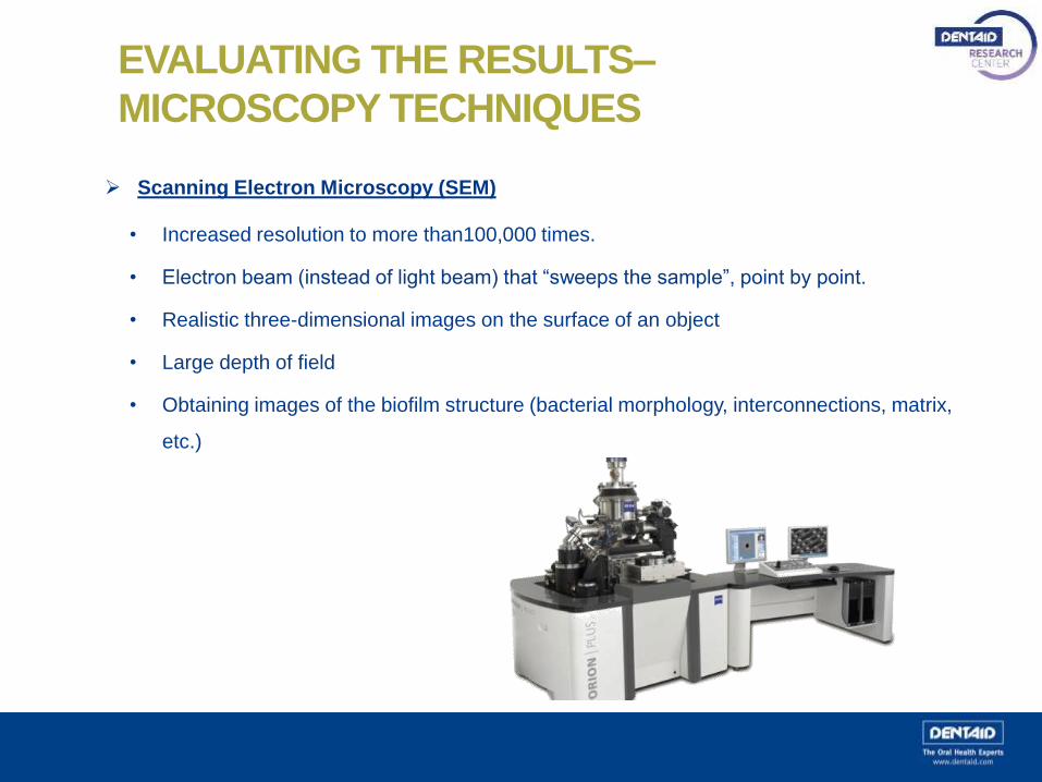
Scanning Electron Microscopy (SEM)
• Increased resolution to more than100,000 times.
• Electron beam (instead of light beam) that “sweeps the sample”, point by point.
• Realistic three-dimensional images on the surface of an object
• Large depth of field
• Obtaining images of the biofilm structure (bacterial morphology, interconnections, matrix,
etc.)

Scanning Electron Microscopy images
Different micrographs taken with SEM pertaining to mono- and multi-species oral biofilms grown in
the Microbiology Laboratory at Dentaid Research Center.

@dentaid_news