chapter 2 space biology
TRANSCRIPT
45G. Clément, Fundamentals of Space Medicine, Space Technology Library 23, DOI 10.1007/978-1-4419-9905-4_2, © Springer Science+Business Media, LLC 2011
Chapter 2
Space Biology
Gravity provides a directional stimulus that plays an important role in basic life processes in the cell, such as biosynthesis, membrane exchange, and cell growth and development. It is likely that the growth and development of plants are determined by hormones, whose transport is also influenced by gravity. Will these functions develop normally when deprived of the gravitational stimulus? This chapter will review the fundamental questions raised in the space environment in the areas of gravitational biology, developmental biology, plant biology, and radiobiology. For more details, the readers are referred to the book Fundamentals of Space Biology by Clément and Slenzka [2006, Springer] (Figure 2.1).
2.1. What is life?
It is generally admitted that, for scientific purposes, an object must meet six criteria to be considered alive: (1) movement (even plants move: stems shoot upward, flowers open and close, and leaves follow the movement of the Sun); (2) organization (animals and plants have organs, whose structure is nearly identical within the same species); (3) homeostasis (the ability to maintain constant conditions within the body); (4) energy (all living things absorb and use energy); (5) reproduction; and (6) growth (during the growth process, cells not only increase in number but they also develop into different types of cells that are needed to form the organs and tissues of the new individual) [DuTemple, 2000].
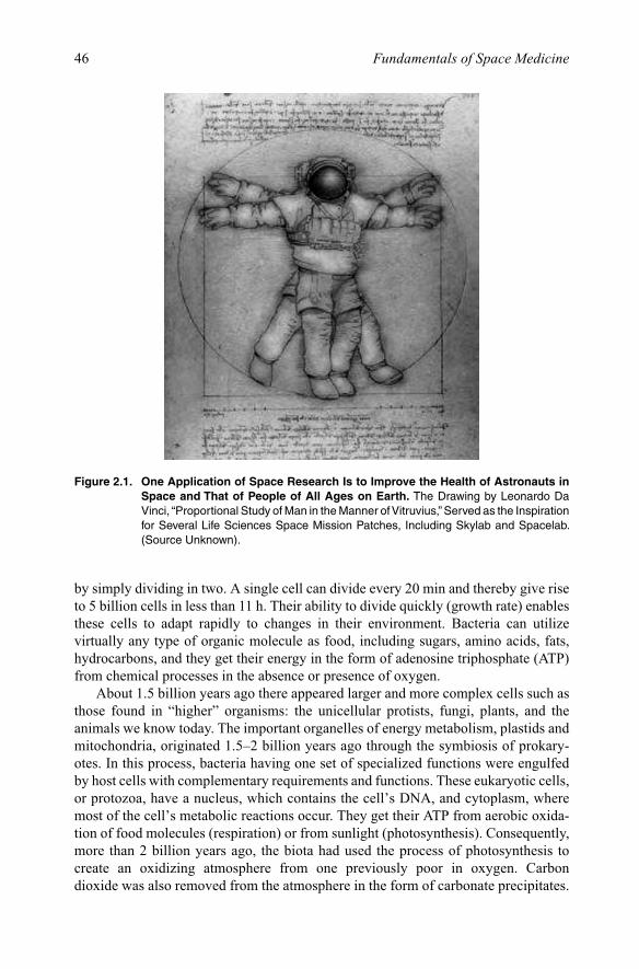
2.1.1. Life on EarthPlanet Earth is thought to be 4.6 billion years old. The first life form appeared about 4 billion years ago by the spontaneous aggregation of molecules that rapidly evolved into microscopic, relatively simple cells. Over the following millennia, these primi-tive cells evolved into at least 10 million different species, which represent Earth’s existing biological diversity. All organisms, including animals, plants, fungi, and an untold collection of microbial species, have their common ancestral roots within these earliest life forms (Figure 2.2).
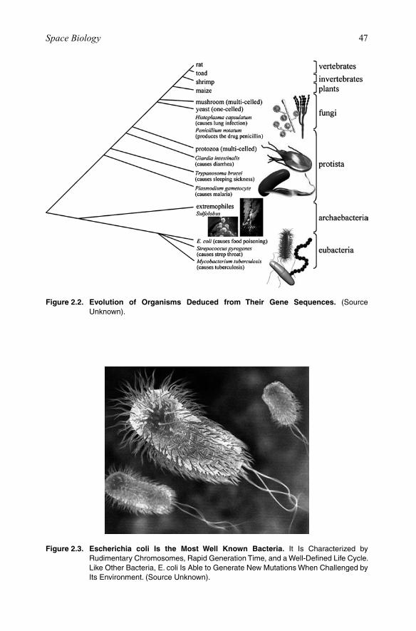
Chemical and fossil evidence indicates that life on Earth as we know it today evolved by natural selection from a few simple cells, called prokaryotes because they lacked nuclei. The earliest prokaryotes probably already had mechanisms that allowed them to replicate their genetic information, encoded in nucleic acids, and to express this information by translation into various proteins. Typical prokaryotic cells are bac-teria (Figure 2.3). They are small, with relatively simple internal structures containing deoxyribonucleic acid (DNA), proteins, and small molecules. They replicate quickly
46 Fundamentals of Space Medicine
by simply dividing in two. A single cell can divide every 20 min and thereby give rise to 5 billion cells in less than 11 h. Their ability to divide quickly (growth rate) enables these cells to adapt rapidly to changes in their environment. Bacteria can utilize virtually any type of organic molecule as food, including sugars, amino acids, fats, hydrocarbons, and they get their energy in the form of adenosine triphosphate (ATP) from chemical processes in the absence or presence of oxygen.
About 1.5 billion years ago there appeared larger and more complex cells such as those found in “higher” organisms: the unicellular protists, fungi, plants, and the animals we know today. The important organelles of energy metabolism, plastids and mitochondria, originated 1.5–2 billion years ago through the symbiosis of prokary-otes. In this process, bacteria having one set of specialized functions were engulfed by host cells with complementary requirements and functions. These eukaryotic cells, or protozoa, have a nucleus, which contains the cell’s DNA, and cytoplasm, where most of the cell’s metabolic reactions occur. They get their ATP from aerobic oxida-tion of food molecules (respiration) or from sunlight (photosynthesis). Consequently, more than 2 billion years ago, the biota had used the process of photosynthesis to create an oxidizing atmosphere from one previously poor in oxygen. Carbon dioxide was also removed from the atmosphere in the form of carbonate precipitates.
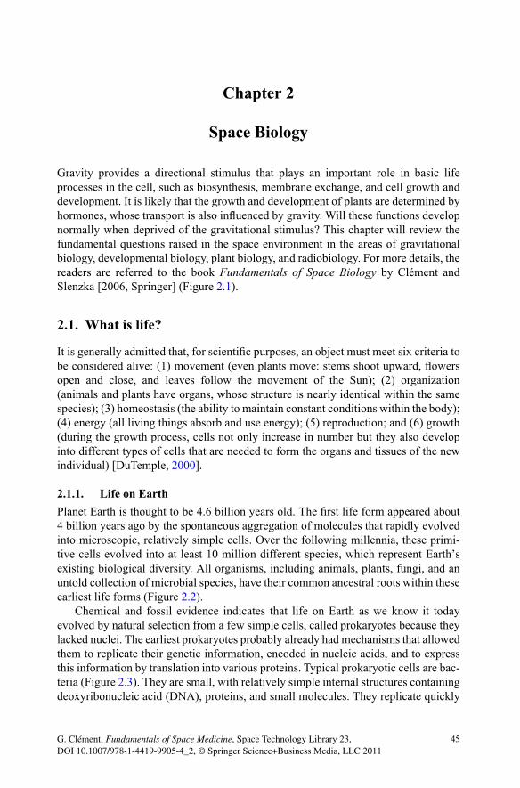
Figure 2.1. One Application of Space Research Is to Improve the Health of Astronauts in Space and That of People of All Ages on Earth. The Drawing by Leonardo Da Vinci, “Proportional Study of Man in the Manner of Vitruvius,” Served as the Inspiration for Several Life Sciences Space Mission Patches, Including Skylab and Spacelab. (Source Unknown).
47Space Biology
Figure 2.2. Evolution of Organisms Deduced from Their Gene Sequences. (Source Unknown).
Figure 2.3. Escherichia coli Is the Most Well Known Bacteria. It Is Characterized by Rudimentary Chromosomes, Rapid Generation Time, and a Well-Defined Life Cycle. Like Other Bacteria, E. coli Is Able to Generate New Mutations When Challenged by Its Environment. (Source Unknown).
48 Fundamentals of Space Medicine
A myriad of bacteria, mollusks, corals, and other organisms contributed to vast lime-stone deposits and continue to do so today. With these and other processes, Earth’s biosphere has transformed a once sterile planet, intermediate in character between Venus and Mars, into the living planet we now enjoy.
Bacteria have been detected or isolated from many hostile environments on Earth, including the dry, extremely cold surfaces and interstices of rocks in the dry valleys of the Antarctic, hot environments associated with submarine and terrestrial volcanoes and geothermal systems, and deep subsurface sediments and aquifers. Investigations in extreme terrestrial environments are in their infancy, and we still know little about either most of the organisms inhabiting these environments, also called extremophiles, or in many cases the geochemistry and geophysics of the environments themselves.
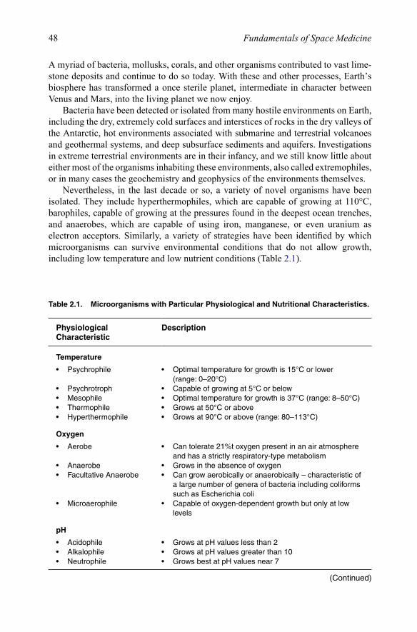
Nevertheless, in the last decade or so, a variety of novel organisms have been isolated. They include hyperthermophiles, which are capable of growing at 110°C, barophiles, capable of growing at the pressures found in the deepest ocean trenches, and anaerobes, which are capable of using iron, manganese, or even uranium as electron acceptors. Similarly, a variety of strategies have been identified by which microorganisms can survive environmental conditions that do not allow growth, including low temperature and low nutrient conditions (Table 2.1).
Table 2.1. Microorganisms with Particular Physiological and Nutritional Characteristics.
Physiological Characteristic
Description
Temperature
• Psychrophile • Optimaltemperatureforgrowthis15°Corlower (range:0–20°C)
• Psychrotroph • Capableofgrowingat5°Corbelow• Mesophile • Optimaltemperatureforgrowthis37°C(range:8–50°C)• Thermophile • Growsat50°Corabove• Hyperthermophile • Growsat90°Corabove(range:80–113°C)
Oxygen
• Aerobe • Cantolerate21%toxygenpresentinanairatmosphereand has a strictly respiratory-type metabolism
• Anaerobe • Growsintheabsenceofoxygen• FacultativeAnaerobe • Cangrowaerobicallyoranaerobically–characteristicof
a large number of genera of bacteria including coliforms such as Escherichia coli
• Microaerophile • Capableofoxygen-dependentgrowthbutonlyatlowlevels
pH
• Acidophile • GrowsatpHvalueslessthan2• Alkalophile • GrowsatpHvaluesgreaterthan10• Neutrophile • GrowsbestatpHvaluesnear7
(Continued)
49Space Biology
Physiological Characteristic
Description
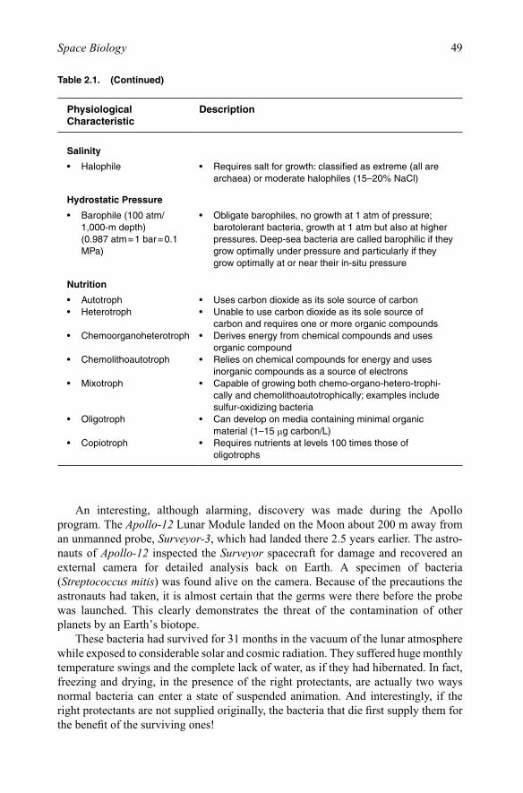
Salinity
• Halophile • Requiressaltforgrowth:classifiedasextreme(allarearchaea)ormoderatehalophiles(15–20%NaCl)
Hydrostatic Pressure
• Barophile(100atm/ 1,000-mdepth)(0.987atm=1bar=0.1MPa)
• Obligatebarophiles,nogrowthat1atmofpressure;barotolerantbacteria,growthat1atmbutalsoathigherpressures. Deep-sea bacteria are called barophilic if they grow optimally under pressure and particularly if they grow optimally at or near their in-situ pressure
Nutrition
• Autotroph • Usescarbondioxideasitssolesourceofcarbon• Heterotroph • Unabletousecarbondioxideasitssolesourceof
carbonandrequiresoneormoreorganiccompounds• Chemoorganoheterotroph • Derivesenergyfromchemicalcompoundsanduses
organic compound• Chemolithoautotroph • Reliesonchemicalcompoundsforenergyanduses
inorganic compounds as a source of electrons• Mixotroph • Capableofgrowingbothchemo-organo-hetero-trophi-
callyandchemolithoautotrophically;examplesincludesulfur-oxidizingbacteria
• Oligotroph • Candeveloponmediacontainingminimalorganicmaterial(1–15mgcarbon/L)
• Copiotroph • Requiresnutrientsatlevels100timesthoseofoligotrophs
Table 2.1. (Continued)
An interesting, although alarming, discovery was made during the Apollo program. The Apollo-12 Lunar Module landed on the Moon about 200 m away from an unmanned probe, Surveyor-3, which had landed there 2.5 years earlier. The astro-nauts of Apollo-12 inspected the Surveyor spacecraft for damage and recovered an external camera for detailed analysis back on Earth. A specimen of bacteria (Streptococcus mitis) was found alive on the camera. Because of the precautions the astronauts had taken, it is almost certain that the germs were there before the probe was launched. This clearly demonstrates the threat of the contamination of other planets by an Earth’s biotope.
These bacteria had survived for 31 months in the vacuum of the lunar atmosphere while exposed to considerable solar and cosmic radiation. They suffered huge monthly temperature swings and the complete lack of water, as if they had hibernated. In fact, freezing and drying, in the presence of the right protectants, are actually two ways normal bacteria can enter a state of suspended animation. And interestingly, if the right protectants are not supplied originally, the bacteria that die first supply them for the benefit of the surviving ones!

50 Fundamentals of Space Medicine
Likewise, spores of the Bacillus bacteria were found during the summer of 2000 in salt crystals buried 600 m below ground at a cavern in New Mexico. When they were extracted from the crystals in a laboratory and placed in a nutrient solution, the microorganisms revived and began to grow. These bacteria had survived in a state of suspended animation for 250 million years. Until now, the world’s oldest living survi-vors were thought to be 25–40 million-year-old bacteria spores discovered in a bee preserved in amber. Traditionally, endospore and cyst development were considered the principal mechanisms for long-term survival by microorganisms, but it is now clear that many microorganisms have mechanisms for long-term survival that do not involve spore or cyst formation.
2.1.2. Life on MarsWithout exception, life in Earth’s biosphere is carbon-based and is organized within a phase boundary or membrane that envelops reacting biomolecules. Every documented terrestrial cellular life form is a self-replicating entity that has genetic information in the form of nucleic acid polymers (DNA) coding for proteins. Biologically active systems require at a minimum liquid water, carbon, nitrogen, phosphate, sulfur, various metals, and a source of energy either in the form of solar radiation or from chemosynthetic processes.
The conditions that nurtured early self-replicating systems and their transition into microbial cells are speculative. In contrast, it is much easier to model the early stages of evolution. Origins-of-life experiments have outlined the synthesis of the basic building blocks of life, including amino acids, nucleotides, and simple polypeptides and polynucleotides. Yet creation of self-sustaining, self-replicating biological enti-ties capable of evolution has not yet been achieved in the laboratory. Even if success-ful, this achievement would not necessarily mimic how life started on Earth or in other parts of the universe.
For life to originate, the presence of liquid water and a source of usable free energy are necessities. The synthesis and polymerization of basic organic building blocks of life on Earth eventually led to self-replicating nucleic acids coding for proteins, but the earliest replicating systems were not necessarily composed of amino acids and nucleotides. If extraterrestrial biological systems exist, their modes of information storage, retrieval, and processing and their enzymatic activity may not be identical to those of biological entities on Earth. Understanding this prebiotic evolution is one of the major goals of the astrobiology program, which is the study of biology of the early Earth and elsewhere in the universe.
In the search for extraterrestrial life, microbes are far more likely than multi- cellular organisms to retain viability on small Solar System bodies because they can adapt to a much wider range of environmental conditions. As mentioned already, single-cell organisms such as bacteria have infiltrated virtually every corner of Earth’s biosphere and still constitute the bulk of Earth’s biomass. They grow in temperate marine and terrestrial settings, within other microbial or multi-cellular organisms, in deep subsurface niches, and in extreme environments that would be lethal for other life forms. They often influence geochemical reactions within the biosphere and frequently play key roles in food chains and complex ecosystems.
51Space Biology
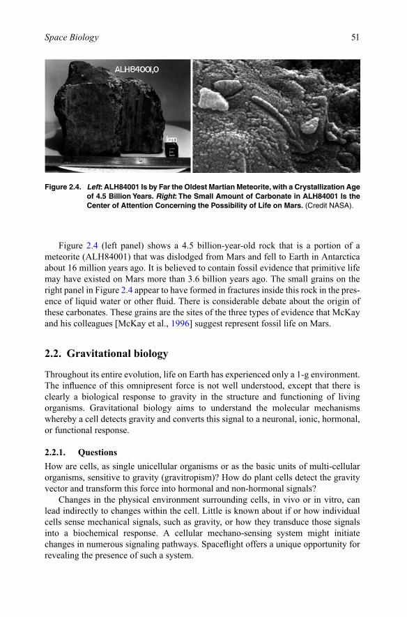
Figure 2.4 (left panel) shows a 4.5 billion-year-old rock that is a portion of a meteorite (ALH84001) that was dislodged from Mars and fell to Earth in Antarctica about 16 million years ago. It is believed to contain fossil evidence that primitive life may have existed on Mars more than 3.6 billion years ago. The small grains on the right panel in Figure 2.4 appear to have formed in fractures inside this rock in the pres-ence of liquid water or other fluid. There is considerable debate about the origin of these carbonates. These grains are the sites of the three types of evidence that McKay and his colleagues [McKay et al., 1996] suggest represent fossil life on Mars.
2.2. Gravitational biology
Throughout its entire evolution, life on Earth has experienced only a 1-g environment. The influence of this omnipresent force is not well understood, except that there is clearly a biological response to gravity in the structure and functioning of living organisms. Gravitational biology aims to understand the molecular mechanisms whereby a cell detects gravity and converts this signal to a neuronal, ionic, hormonal, or functional response.
2.2.1. QuestionsHow are cells, as single unicellular organisms or as the basic units of multi-cellular organisms, sensitive to gravity (gravitropism)? How do plant cells detect the gravity vector and transform this force into hormonal and non-hormonal signals?
Changes in the physical environment surrounding cells, in vivo or in vitro, can lead indirectly to changes within the cell. Little is known about if or how individual cells sense mechanical signals, such as gravity, or how they transduce those signals into a biochemical response. A cellular mechano-sensing system might initiate changes in numerous signaling pathways. Spaceflight offers a unique opportunity for revealing the presence of such a system.
Figure 2.4. Left: ALH84001 Is by Far the Oldest Martian Meteorite, with a Crystallization Age of 4.5 Billion Years. Right: The Small Amount of Carbonate in ALH84001 Is the Center of Attention Concerning the Possibility of Life on Mars. (Credit NASA).
52 Fundamentals of Space Medicine
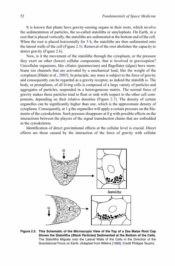
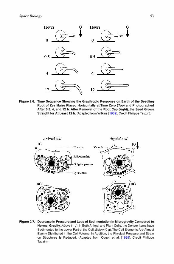
It is known that plants have gravity-sensing organs in their roots, which involve the sedimentation of particles, the so-called statoliths or amyloplasts. On Earth, in a root that is placed vertically, the statoliths are sedimented at the bottom end of the cell. When the root is placed horizontally for 3 h, the statoliths are then sedimented onto the lateral walls of the cell (Figure 2.5). Removal of the root abolishes the capacity to detect gravity (Figure 2.6).
Now, is it the movement of the statoliths through the cytoplasm, or the pressure they exert on other (lower) cellular components, that is involved in graviception? Unicellular organisms, like ciliates (paramecium) and flagellates (algae) have mem-brane ion channels that are activated by a mechanical load, like the weight of the cytoplasm [Häder et al., 2005]. In principle, any mass is subject to the force of gravity and consequently can be regarded as a gravity receptor, as indeed the statolith is. The body, or protoplasm, of all living cells is composed of a large variety of particles and aggregates of particles, suspended in a heterogeneous matrix. The normal force of gravity makes these particles tend to float or sink with respect to the other cell com-ponents, depending on their relative densities (Figure 2.7). The density of certain organelles can be significantly higher than one, which is the approximate density of cytoplasm. Consequently, at 1 g the organelles will apply a certain pressure on the fila-ments of the cytoskeleton. Such pressure disappears at 0 g with possible effects on the interactions between the players of the signal transduction chains that are embedded in the cytoskeleton.
Identification of direct gravitational effects at the cellular level is crucial. Direct effects are those caused by the interaction of the force of gravity with cellular
Figure 2.5. This Schematic of the Microscopic View of the Top of a Zea Maize Root Cap Shows the Statoliths (Black Particles) Sedimented at the Bottom of the Cells. The Statoliths Migrate onto the Lateral Walls of the Cells in the Direction of the GravitationalForceonEarth.(AdaptedfromWilkins[1989]. Credit Philippe Tauzin).
53Space Biology
Figure 2.6. Time Sequence Showing the Gravitropic Response on Earth of the Seedling Root of Zea Maize Placed Horizontally at Time Zero (Top) and Photographed After 0.5, 4, and 12 h. After Removal of the Root Cap (right), the Seed Grows Straight for At Least 12 h. (AdaptedfromWilkins[1989]. Credit Philippe Tauzin).
Figure 2.7. Decrease in Pressure and Loss of Sedimentation in Microgravity Compared to Normal Gravity. Above(1g):inBothAnimalandPlantCells,theDenserItemshaveSedimented to the Lower Part of the Cell. Below (0 g): The Cell Elements Are Almost Evenly Distributed in the Cell Volume. In Addition, the Physical Pressure and Strain on Structures Is Reduced. (Adapted from Cogoli et al. [1989]. Credit Philippe Tauzin).

54 Fundamentals of Space Medicine
structures and organelles or by its absence, respectively. Indirect effects are those caused by changes in the cell microenvironment under altered gravitational condi-tions. Indirect effects may be due to the absence of convection and sedimentation at 0 g that causes a change of the distribution of nutrients and of waste products around the cells [Cogoli, 2006].
In a world of molecules embedded in fluids and loaded with electrical charges dominated by viscosity and electrostatic forces, gravity is an extremely weak force. However, the impact of gravity may not be negligible in biological systems that are not static, but in a non-equilibrium status. In a biological process consisting of many subsequent steps, the principle of “small cause/large effect” applies, by which a small perturbation of one of the steps is sufficient to provoke dramatic changes downstream, as predicted by the bifurcation theory described Tabony et al. [2002].
All living systems react in one way or another to changes of the environmental parameters such as temperature, illumination, pressure, concentrations of nutrients, or activators/inhibitors. Gravity is a mechanical force. Change of the gravitational envi-ronment, i.e., changes of the forces acting on the cell, is a significant environmental change. It should therefore be no surprise that single cells also react and adapt to changes from 1 to 0 g conditions [Cogoli, 2006].
Important changes such as the loss of sedimentation, density-driven convection and hydrostatic pressure are occurring in a weightless cell culture. For a cell immersed in a fluid, as it is the case in a culture, this is a completely new situation. First, in 1 g, mammalian cells sediment within a few minutes to the bottom of the flask, where many of them may spread and adhere. In 0 g, instead, cells remain in suspension. Going from 1 to 0 g is a change from a two – to a three-dimensional environment and has a remarkable impact on cell interactions, cell movements, and, due to the lack of a substratum on which to spread and adhere, on cell shape.
Second, density-driven convection, which is due to changes in the concentration of nutrients and waste products in the medium, does not occur in microgravity, thus preventing mechanical diffusion. Thermodynamic diffusion is not affected, however.
Third, a new convection, predicted at the beginning of the twentieth century by Marangoni and not detectable at 1 g, becomes relevant in micro-gravity. The lack of buoyancy prevents gas bubbles, like the CO2 bubbles developed by the metabolism of cells, to rise to the surface of a culture, thus favoring the formation of larger bubbles in the middle of the liquid phase rather than a separation of the liquid and gas phases.
The physiology of the cell may also be influenced by gravity. While passive trans-port of small molecules through the lipid bilayer is governed by diffusion (a gravity-independent process), active transport of ions and charged molecules, in which protein channels and transient membrane invaginations are involved, may be influenced by gravity. The balanced exchange of ions and molecules through cell membranes might be sensitive to gravity. The same may hold for membrane turnover, a basic process in cell life, and for intercellular diffusion of substances of varying molecular weight.
Gravity may also play a role in intercellular transport processes. In fact exothermic metabolic processes generate continuously warmer micro-regions that are less dense than the neighborhood. Thus, thermal convections are produced by gravity with consequent ultra-structural rearrangements. Such convections are obvi-ously absent in microgravity.
55Space Biology
Also, the energy turnover in the cells can be influenced by gravity. Gravity causes an uneven distribution of the organelles that gives rise to a torque capable to modify the shape and the structure of the cell. Energy is required to maintain its shape against gravity. In microgravity, such energy may be saved for other processes, such as prolif-eration or biosynthesis.
Finally, free-swimming cells consume energy to swim against gravity to avoid sedimentation. Such energy is not required at 0 g.
To investigate these phenomena, research programs in the biological sciences and biotechnology have focused on three primary areas of interest: (a) separation physics aimed at providing improved resolution and sensitivity in preparative and bioanalytical techniques; (b) cell biology, cell function, and cell-cell interactions; and (c) physical chemistry of biological macromolecules and their interactions, including studies of pro-tein crystal growth directed at supporting crystallographic structure determinations.
In the field of biotechnology, for example, the absence of convection and sedimen-tation can help the separation and isolation of biological specimens. The increase in surface tension will improve transport processes, and consequently secretion and growth. The objective is to cultivate proteins (hormones, enzymes, antibodies) and cells that secrete a medically valuable substance. The purified product would be returned to Earth for medical use, product characterization, or improvement of ground-based separation techniques. However, this process is now challenged by ground-based computer graphics models, and by genetic-engineering techniques, like the cloning process, that are much less expensive than experiments in space.
2.2.2. Results of space experimentsWhen gravity is altered, biological changes are observed even when cells are isolated from the whole organism and grown in culture (in vitro). Physical scientists predicted this would not occur because gravity is an extremely weak force compared with the other fundamental physical forces acting on or within cells. However, spaceflight results suggest that microgravity may alter the characteristics of cultured cells. Most cells flown in space have either been suspended in an aqueous medium or attached to an extra-cellular matrix bathed by an aqueous medium.
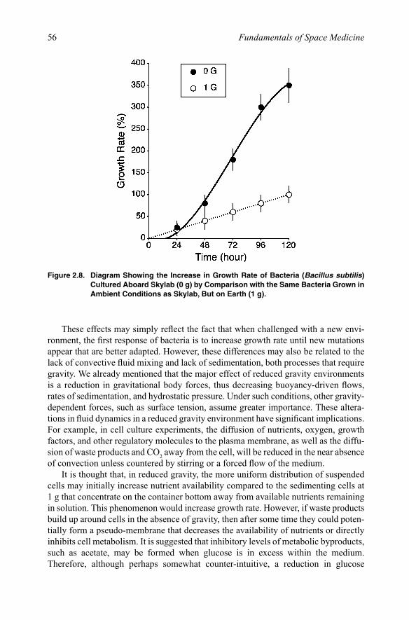
2.2.2.1. Suspended culturesMany space missions have flown bacteria in experimental cultures. The first cultures of Escherichia coli flew on board the U.S. Biosatellite-2 in 1967. Mattoni et al. [1971] reported that after a 45-h orbital flight, the flight populations grew significantly faster than the ground controls. Another bacteria, Bacillus subtilis, was found to exhibit an increased duration of exponential growth, and an approximate doubling of final cell population density compared to ground controls [Klaus et al., 1997] (Figure 2.8).
Planel et al. [1994, 2004] discovered an increased resistance of E. coli and Staphylococcus aureus to antibiotics when cultured in several experiments in space. The effect was attributed to an increase of the thickness of the cell membrane, observed in electron micrographs, with consequent decrease in the membrane permeability. These results suggest that humans are at greater risk in space, given that there may be larger populations of bacteria in a confined environment, which are, moreover, less sensitive to antibiotics.
56 Fundamentals of Space Medicine
These effects may simply reflect the fact that when challenged with a new envi-ronment, the first response of bacteria is to increase growth rate until new mutations appear that are better adapted. However, these differences may also be related to the lack of convective fluid mixing and lack of sedimentation, both processes that require gravity. We already mentioned that the major effect of reduced gravity environments is a reduction in gravitational body forces, thus decreasing buoyancy-driven flows, rates of sedimentation, and hydrostatic pressure. Under such conditions, other gravity-dependent forces, such as surface tension, assume greater importance. These altera-tions in fluid dynamics in a reduced gravity environment have significant implications. For example, in cell culture experiments, the diffusion of nutrients, oxygen, growth factors, and other regulatory molecules to the plasma membrane, as well as the diffu-sion of waste products and CO2 away from the cell, will be reduced in the near absence of convection unless countered by stirring or a forced flow of the medium.
It is thought that, in reduced gravity, the more uniform distribution of suspended cells may initially increase nutrient availability compared to the sedimenting cells at 1 g that concentrate on the container bottom away from available nutrients remaining in solution. This phenomenon would increase growth rate. However, if waste products build up around cells in the absence of gravity, then after some time they could poten-tially form a pseudo-membrane that decreases the availability of nutrients or directly inhibits cell metabolism. It is suggested that inhibitory levels of metabolic byproducts, such as acetate, may be formed when glucose is in excess within the medium. Therefore, although perhaps somewhat counter-intuitive, a reduction in glucose
Figure 2.8. Diagram Showing the Increase in Growth Rate of Bacteria (Bacillus subtilis) Cultured Aboard Skylab (0 g) by Comparison with the Same Bacteria Grown in Ambient Conditions as Skylab, But on Earth (1 g).
57Space Biology
availability actually may be beneficial to cell growth. Also, local toxic byproducts could become concentrated on the bottom of the 1-g container with cells in increased proximity to each other. Such a process could limit cell growth. Thus, changes in bacteria and possibly other cells during spaceflight may be related to alterations in the microenvironment surrounding non-motile cells, e.g., the equilibrium of extra-cellular mass-transfer processes governing nutrient uptake and waste removal. Such changes appear to be typical indirect effects of gravity caused by changes of the microenviron-ment of the cells.
The current view is that a “cumulative” response resulting from reduced gravity may be responsible for the observed effects at the level of the single cell. Earlier pre-dictions suggesting that no effect of spaceflight should be expected were more focused on the physical inability of gravity to elicit an immediate or “direct” response from organisms of such small mass. Rather than a “direct” response, reduced gravity is suspected to initiate a cascade of events: the altered physical force leads to an altered chemical environment, which in turn gives rise to an altered physiological response [Klaus, 1998].
Saccharomyces cerevisiae, the yeast used to bake bread and cakes, is a highly appreciated organism in the study of several aspects of eukaryotic cell, like signal transduction, genetic expression, and adaptation to environmental stress. It has the great advantage of being resistant to rough environmental conditions like freezing or lack of nutrients. It also has biological properties and behavior analogous to those of mammalian cells that are, by contrast, much more sensitive to the environment and therefore much more difficult to keep alive in space experiments. The analogy with mammalian cells permits the investigation of crucial biological processes, including cancer in yeast cells. In addition, yeast is widely used in biotechnological processes, in particular in genetic engineering. Therefore, it is not surprising that yeast cells have been extensively chosen for experiments in space.
With the increasing interest in bioprocessing in space the requirement for sophis-ticated cell culture and tissue engineering facilities, also known as bioreactors, to be installed in space laboratories was obvious. Space bioreactors were first developed using yeast cells that are easy to cultivate and to preserve instead of delicate and sensi-tive mammalian cells. Now that the instrumentation has proven adequate, the experi-mentation with mammalian cells and tissue can begin.
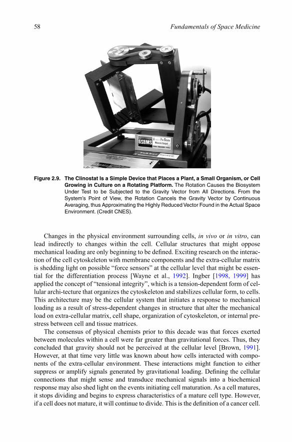
2.2.2.2. Attached cellsEarly results with cultured cells from muscles or bones suggest that spaceflight induces a wide variety of responses. For example, delayed differentiation and changes in the cytoskeleton, nuclear morphology, and gene expression have been reported for bone cells [Hughes-Fulford and Lewis, 1996]. Muscle fibers cultured in space were 10–20% thinner (i.e., atrophied) compared with ground controls due to a decrease in protein synthesis rather than an increase in protein degradation [Vandenburgh et al., 1999]. Interestingly, the atrophy of isolated muscle fibers in culture was very similar to the amount of muscle atrophy reported in flight animals (see Chapter 5, Section 5.3). These data from bone and muscle cells suggest that spaceflight affects adherent cells and tissues even when isolated from systemic factors. The same results were obtained during ground-based studies using clinostats (Figure 2.9).
58 Fundamentals of Space Medicine
Changes in the physical environment surrounding cells, in vivo or in vitro, can lead indirectly to changes within the cell. Cellular structures that might oppose mechanical loading are only beginning to be defined. Exciting research on the interac-tion of the cell cytoskeleton with membrane components and the extra-cellular matrix is shedding light on possible “force sensors” at the cellular level that might be essen-tial for the differentiation process [Wayne et al., 1992]. Ingber [1998, 1999] has applied the concept of “tensional integrity”, which is a tension-dependent form of cel-lular archi-tecture that organizes the cytoskeleton and stabilizes cellular form, to cells. This architecture may be the cellular system that initiates a response to mechanical loading as a result of stress-dependent changes in structure that alter the mechanical load on extra-cellular matrix, cell shape, organization of cytoskeleton, or internal pre-stress between cell and tissue matrices.
The consensus of physical chemists prior to this decade was that forces exerted between molecules within a cell were far greater than gravitational forces. Thus, they concluded that gravity should not be perceived at the cellular level [Brown, 1991]. However, at that time very little was known about how cells interacted with compo-nents of the extra-cellular environment. These interactions might function to either suppress or amplify signals generated by gravitational loading. Defining the cellular connections that might sense and transduce mechanical signals into a biochemical response may also shed light on the events initiating cell maturation. As a cell matures, it stops dividing and begins to express characteristics of a mature cell type. However, if a cell does not mature, it will continue to divide. This is the definition of a cancer cell.
Figure 2.9. The Clinostat Is a Simple Device that Places a Plant, a Small Organism, or Cell Growing in Culture on a Rotating Platform. The Rotation Causes the Biosystem Under Test to be Subjected to the Gravity Vector from All Directions. From theSystem’s Point of View, the Rotation Cancels the Gravity Vector by Continuous Averaging,thusApproximatingtheHighlyReducedVectorFoundintheActualSpaceEnvironment. (Credit CNES).

59Space Biology
The maturation process may be triggered by multiple factors, including loads placed on the extra-cellular matrix during different phases of development.
In summary, flight experiments suggest that gravity, quite likely, is perceived by cells through physical changes both in the aqueous medium surrounding cells in culture and in cellular structures that oppose or sense mechanical loads. Exactly how the gravity signal is then transduced to cellular functions is yet to be determined. The answer to this question is not only relevant to understanding the fundamental pro-cesses in normal cell physiology, but also in the patho-physiology of certain diseases, such as age-related bone loss, cancer, or immune disorders [Bouillon et al., 2001].
2.2.2.3. Threshold for gravity perceptionThe changes in the swimming behavior of ciliates and flagellates, which presumably compensate part of the changes in the cell physical properties in 0 g (e.g., sedimenta-tion, thermal convection) can be measured for calculation of the sensitivity to gravity perception [Machemer et al., 1991]. In 1992, a sophisticated slow rotating centrifuge microscope, called NIZEMI (for Niedergeschwindigkeit Zentrifuge Mikroskop) devel-oped by the German Space Agency, measured the minimal in-flight acceleration that was able to induce a graviceptive response in microorganisms. The following accel-eration threshold were obtained: Paramecium, 0.35 g; Euglena, 0.16 and 0.12 g; and Loxodes, less than 0.15 g. Interestingly, the results were similar when the cells were subjected either to increasing or decreasing accelerations, and the effect was indepen-dent of the previous exposure to microgravity up to 12 days, although the cells under-went several division cycles.
Because the organelles used for gravity-sensing mechanisms in these organisms show some analogy to the statoliths in plants and the otoliths in humans and other vertebrates, the results of these studies on threshold for gravity perception are of fun-damental importance for determining the optimal level of artificial gravity for long-duration human missions.
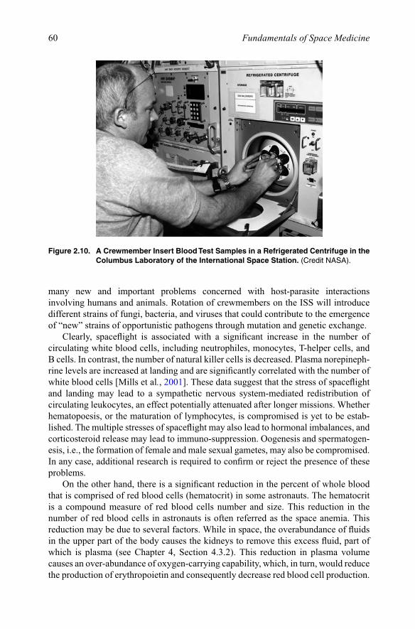
2.2.2.4. Human blood cellsAlthough the reports to date are conflicting, some indicate that a microgravity environment may compromise the immune system function. These investigations are carried out on cultures of lymphocytes prepared on the ground and tested in space, and with whole-blood samples taken from the crew and tested in-flight, respectively (Figure 2.10). Cogoli et al. [1980] reported that cultures of human lymphocytes subjected to microgravity responded to concanavalin A, a lymphocyte stimulating agent, 90% less than ground-based controls. This is a standard test used to evaluate the competence of peripheral blood lymphocytes to multiply when stimulated with this agent. Studies on the astronauts of the first four space shuttle flights revealed that the lymphocyte responses to photohemagglutinin, another lymphocyte stimulating agent, were reduced from 18% to 61% of normal following spaceflight. It has been suggested that the above changes were due to stress-related effects, but this should be studied further.
These studies are important because, as was discussed earlier, the concentrations of microorganisms in space vehicles may be significantly higher than normal. The conditions associated with space travel, space stations, and planetary colonies raise
60 Fundamentals of Space Medicine
many new and important problems concerned with host-parasite interactions involving humans and animals. Rotation of crewmembers on the ISS will introduce different strains of fungi, bacteria, and viruses that could contribute to the emergence of “new” strains of opportunistic pathogens through mutation and genetic exchange.
Clearly, spaceflight is associated with a significant increase in the number of circulating white blood cells, including neutrophiles, monocytes, T-helper cells, and B cells. In contrast, the number of natural killer cells is decreased. Plasma norepineph-rine levels are increased at landing and are significantly correlated with the number of white blood cells [Mills et al., 2001]. These data suggest that the stress of spaceflight and landing may lead to a sympathetic nervous system-mediated redistribution of circulating leukocytes, an effect potentially attenuated after longer missions. Whether hematopoesis, or the maturation of lymphocytes, is compromised is yet to be estab-lished. The multiple stresses of spaceflight may also lead to hormonal imbalances, and corticosteroid release may lead to immuno-suppression. Oogenesis and spermatogen-esis, i.e., the formation of female and male sexual gametes, may also be compromised. In any case, additional research is required to confirm or reject the presence of these problems.
On the other hand, there is a significant reduction in the percent of whole blood that is comprised of red blood cells (hematocrit) in some astronauts. The hematocrit is a compound measure of red blood cells number and size. This reduction in the number of red blood cells in astronauts is often referred as the space anemia. This reduction may be due to several factors. While in space, the overabundance of fluids in the upper part of the body causes the kidneys to remove this excess fluid, part of which is plasma (see Chapter 4, Section 4.3.2). This reduction in plasma volume causes an over- abundance of oxygen-carrying capability, which, in turn, would reduce the production of erythropoietin and consequently decrease red blood cell production.
Figure 2.10. A Crewmember Insert Blood Test Samples in a Refrigerated Centrifuge in the Columbus Laboratory of the International Space Station. (Credit NASA).
61Space Biology
This process would be favored by the fact that muscles lose mass and thus require less oxygen. However, it is also possible that the over-abundance of oxygen-carrying capacity in the blood is responsible for an increase in the destruction rate of red blood cells. Finally, as we will see in Chapter 5, as astronauts lose calcium in their bones, the structure and function of the bone and its marrow may change and may result in a decrease in red blood cell production.
2.2.3. Bioprocessing in spaceResearch in biotechnology relies on the manipulation of cells of living organisms. The purpose of these manipulations is to produce useful molecules, natural or artificial, in useful quantities, to develop new organisms or new biological molecules for specific uses, or to improve yields of plant and animal products through genetic alteration. Recombinant techniques, for example, make it possible to produce natural or artificially mutated versions of proteins exhibiting a wide range of activities and uses, scientific and medical, in large quantities. The techniques essential to these manipulations are applied in aqueous environments and are subject to fluid dynamics and transport processes.
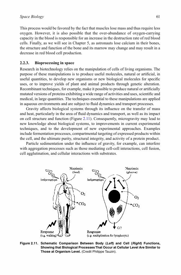
Gravity affects biological systems through its influence on the transfer of mass and heat, particularly in the area of fluid dynamics and transport, as well as its impact on cell structure and function (Figure 2.11). Consequently, microgravity may lead to new knowledge about biological systems, to improvements in current experimental techniques, and to the development of new experimental approaches. Examples include fermentation processes, compartmental targeting of expressed products within the cell, and the ultimate purity, structural integrity, and activity of a protein product.
Particle sedimentation under the influence of gravity, for example, can interfere with aggregation processes such as those mediating cell-cell interactions, cell fusion, cell agglutination, and cellular interactions with substrates.
Figure 2.11. Schematic Comparison Between Body (Left) and Cell (Right) Functions, Showing that Biological Processes That Occur at Cellular Level Are Similar to Those at Organism Level. (Credit Philippe Tauzin).
62 Fundamentals of Space Medicine
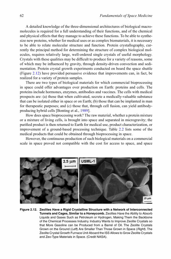
A detailed knowledge of the three-dimensional architectures of biological macro-molecules is required for a full understanding of their functions, and of the chemical and physical effects that they manage to achieve these functions. To be able to synthe-size new proteins, whether for medical uses or as complex biomaterials, it is necessary to be able to relate molecular structure and function. Protein crystallography, cur-rently the principal method for determining the structure of complex biological mol-ecules, requires relatively large, well-ordered single crystals of useful morphology. Crystals with these qualities may be difficult to produce for a variety of reasons, some of which may be influenced by gravity, through density-driven convection and sedi-mentation. Protein crystal growth experiments conducted on board the space shuttle (Figure 2.12) have provided persuasive evidence that improvements can, in fact, be realized for a variety of protein samples.
There are two types of biological materials for which commercial bioprocessing in space could offer advantages over production on Earth: proteins and cells. The proteins include hormones, enzymes, antibodies and vaccines. The cells with medical prospects are: (a) those that when cultivated, secrete a medically-valuable substance that can be isolated either in space or on Earth; (b) those that can be implanted in man for therapeutic purposes; and (c) those that, through cell fusion, can yield antibody-producing hybrid cells [Bonting et al., 1989].
How does space bioprocessing work? The raw material, whether a protein mixture or a mixture of living cells, is brought into space and separated in microgravity; the purified product is then returned to Earth for medical use, product characterization, or improvement of a ground-based processing technique. Table 2.2 lists some of the medical products that could be obtained through bioprocessing in space.
However, the continuous production of such biological materials on a commercial scale in space proved not compatible with the cost for access to space, and space
Figure 2.12. Zeolites Have a Rigid Crystalline Structure with a Network of Interconnected Tunnels and Cages, Similar to a Honeycomb.ZeolitesHavetheAbilitytoAbsorbLiquidsandGasesSuchasPetroleumorHydrogen,MakingThemtheBackboneof the Chemical Processes Industry. Industry Wants to Improve Zeolite Crystals so that More Gasoline can be Produced from a Barrel of Oil. The Zeolite Crystals Grown on the Ground (Left) Are Smaller Than Those Grown in Space (Right). The ZeoliteCrystalGrowthFurnaceUnitAboardtheISSAllowstoGrowZeoliteCrystalsand Zeo-Type Materials in Space. (Credit NASA).
63Space Biology
bioprocessing remains marginal today. Furthermore, ground-based genetic engineer-ing in mammalian or human embryo cells is now a very strong alternative to space bioprocessing, together with purification methods such as affinity or immuno-affinity chromatography and high-pressure liquid chromatography. Also, alternatives to X-ray crystallography are emerging, using physical and mathematical models and computer graphics, that are equally useful in determining the three-dimensional structure of proteins.
2.3. Development biology
The major goal for developmental biology is to determine whether any organism can develop from fertilization through the formation of viable gametes (reproductive cells) in the next generation, i.e., from egg to egg, in the microgravity and radiation environ-ment of space. In the event that normal development does not occur, the priority is to determine which period of development is most sensitive to microgravity.
2.3.1. QuestionsCan higher plants and animals be propagated through several generations in the space environment? Although many embryos orient their cleavage planes relative to the gravity vector, we do not understand whether gravity, per se, is essential to gameto-genesis, fertilization, implantation in animals, organogenesis, or development of normal sensory-motor responses. Given the effects of microgravity exposure on bone, muscle, and vestibular function, there is some doubt whether vertebrates can develop normally in space.
The amphibian has been used as a model for many experiments on embryonic development in space [Souza et al., 1995]. In Xenopus laevis, the South African three-clawed frog, for example, the unfertilized egg has a polarized structure because of an
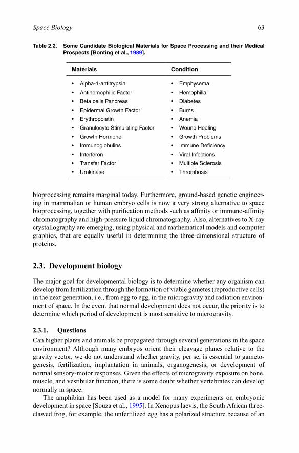
Table 2.2. Some Candidate Biological Materials for Space Processing and their Medical Prospects [Bonting et al., 1989].
Materials Condition
• Alpha-1-antitrypsin • Emphysema
• AntihemophilicFactor • Hemophilia
• BetacellsPancreas • Diabetes
• EpidermalGrowthFactor • Burns
• Erythropoietin • Anemia
• GranulocyteStimulatingFactor • WoundHealing
• GrowthHormone • GrowthProblems
• Immunoglobulins • ImmuneDeficiency
• Interferon • ViralInfections
• TransferFactor • MultipleSclerosis
• Urokinase • Thrombosis
64 Fundamentals of Space Medicine
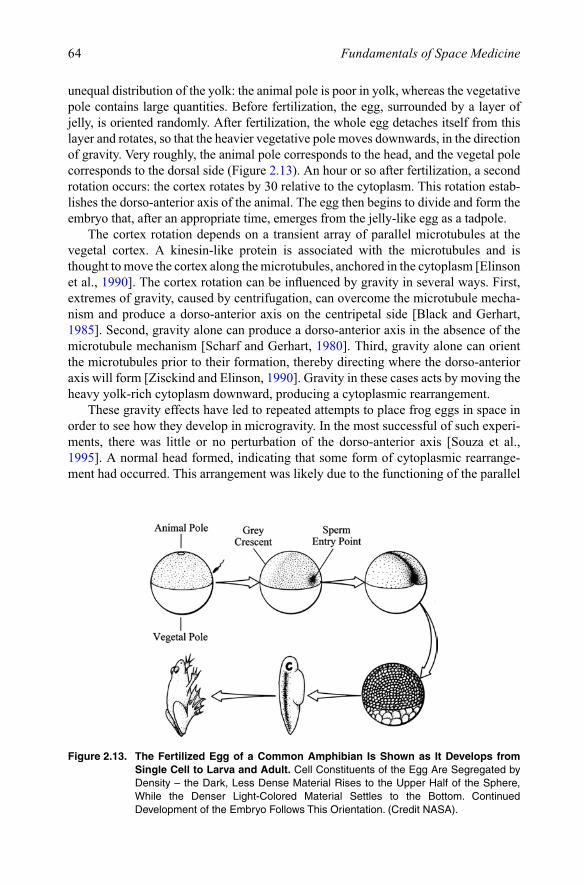
unequal distribution of the yolk: the animal pole is poor in yolk, whereas the vegetative pole contains large quantities. Before fertilization, the egg, surrounded by a layer of jelly, is oriented randomly. After fertilization, the whole egg detaches itself from this layer and rotates, so that the heavier vegetative pole moves downwards, in the direction of gravity. Very roughly, the animal pole corresponds to the head, and the vegetal pole corresponds to the dorsal side (Figure 2.13). An hour or so after fertilization, a second rotation occurs: the cortex rotates by 30 relative to the cytoplasm. This rotation estab-lishes the dorso-anterior axis of the animal. The egg then begins to divide and form the embryo that, after an appropriate time, emerges from the jelly-like egg as a tadpole.
The cortex rotation depends on a transient array of parallel microtubules at the vegetal cortex. A kinesin-like protein is associated with the microtubules and is thought to move the cortex along the microtubules, anchored in the cytoplasm [Elinson et al., 1990]. The cortex rotation can be influenced by gravity in several ways. First, extremes of gravity, caused by centrifugation, can overcome the microtubule mecha-nism and produce a dorso-anterior axis on the centripetal side [Black and Gerhart, 1985]. Second, gravity alone can produce a dorso-anterior axis in the absence of the microtubule mechanism [Scharf and Gerhart, 1980]. Third, gravity alone can orient the microtubules prior to their formation, thereby directing where the dorso-anterior axis will form [Zisckind and Elinson, 1990]. Gravity in these cases acts by moving the heavy yolk-rich cytoplasm downward, producing a cytoplasmic rearrangement.
These gravity effects have led to repeated attempts to place frog eggs in space in order to see how they develop in microgravity. In the most successful of such experi-ments, there was little or no perturbation of the dorso-anterior axis [Souza et al., 1995]. A normal head formed, indicating that some form of cytoplasmic rearrange-ment had occurred. This arrangement was likely due to the functioning of the parallel
Figure 2.13. The Fertilized Egg of a Common Amphibian Is Shown as It Develops from Single Cell to Larva and Adult. Cell Constituents of the Egg Are Segregated by Density–theDark,LessDenseMaterialRisestotheUpperHalfoftheSphere,While the Denser Light-Colored Material Settles to the Bottom. Continued DevelopmentoftheEmbryoFollowsThisOrientation.(CreditNASA).
65Space Biology
microtubule mechanism. One possibility is that gravity-induced rearrangement is an evolutionarily primitive mechanism, which substitutes for the microtubule mecha-nism. If there were any frogs lacking the microtubule mechanism, their eggs would be interesting objects to put in space: the hypothesis is that the dorso-anterior axis would be altered in the resulting space tadpoles.
Developmental biology also includes all aspects of the life span of an organism, from fertilization through aging. Topics of research include gamete production, fertil-ization, embryogenesis, implantation (in mammals), the formation of organs (organo-genesis), and postnatal development (changes after birth). The role of gravity in these processes is entirely unknown. For example, we don’t know if cell division (mitosis) and the orientation of bilateral symmetry are influenced by gravity.
It is known that at some point after fertilization, different in diverse organisms, cells become committed to developing along a certain pathway. This restriction in fate is called determination. During early cell divisions in most animal embryos, there are gradual restrictions in developmental potentiality. This is not the case in plants. Sooner or later in all animals the cells in the embryo can usually give rise only to a certain tissue or organ. They have lost their plural potentialities. This second process of development is differentiation, a term that designates the processes whereby the dif-ferences that were “determined” become manifest. The mechanisms by which the determination and differentiation occur at the right time to produce the normal organ-isms is called the formation of pattern, i.e., not only do they realize their fates, but they do so in the correct place at the correct time.
The formation of the various tissues and organs, or organogenesis, not only spans several developmental stages, but also continues after birth or hatching and into the natal period. For each organ system, there appears to be a critical period during which development can be disrupted by relatively small environmental stresses. The systems affected by weightlessness in the adult, e.g., vestibular apparatus, bone metabolism, and the formation of blood cells, might suffer more severe and more permanent effects if the gravity stimulus were withdrawn during the appropriate stage of organogenesis. This would be similar to the results of the experiments indicating that the receptors of the visual system, their neural connections, and the visual cortex develop abnormally in animals raised in complete darkness [Imbert, 1979].
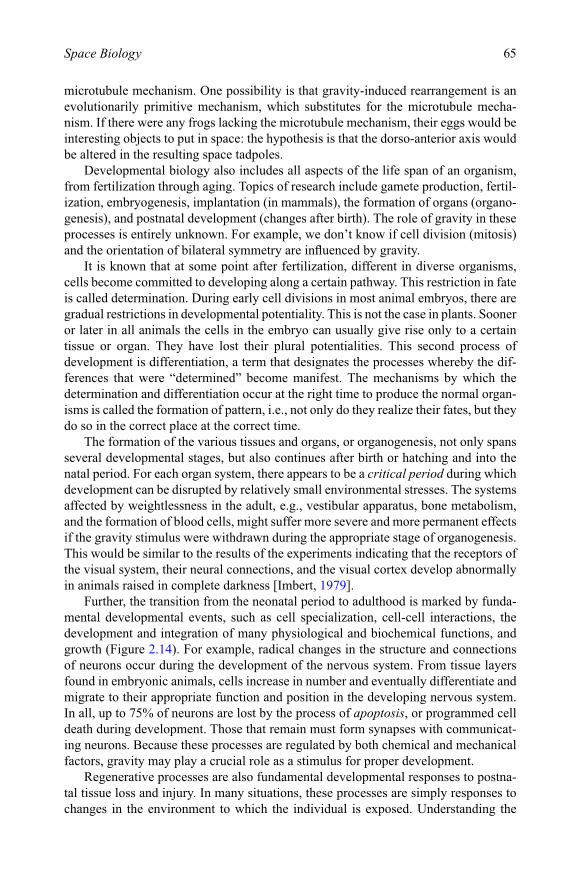
Further, the transition from the neonatal period to adulthood is marked by funda-mental developmental events, such as cell specialization, cell-cell interactions, the development and integration of many physiological and biochemical functions, and growth (Figure 2.14). For example, radical changes in the structure and connections of neurons occur during the development of the nervous system. From tissue layers found in embryonic animals, cells increase in number and eventually differentiate and migrate to their appropriate function and position in the developing nervous system. In all, up to 75% of neurons are lost by the process of apoptosis, or programmed cell death during development. Those that remain must form synapses with communicat-ing neurons. Because these processes are regulated by both chemical and mechanical factors, gravity may play a crucial role as a stimulus for proper development.
Regenerative processes are also fundamental developmental responses to postna-tal tissue loss and injury. In many situations, these processes are simply responses to changes in the environment to which the individual is exposed. Understanding the
66 Fundamentals of Space Medicine
role of gravity not only in ontogeny, the development of the individual, but also in phylogeny, which is the evolution of species, justifies the studies on various species in space for successive generations over long periods of time. By acting on this external factor, would it then be possible to modify the blueprints contained in the genome and change some characters of the species? In other words, would we all become bone-less, jellyfish-like organisms, after many generations in space?
2.3.2. Results of space experimentsDiverse organisms have been subjected to microgravity for varying periods of time. The results of these studies have been inconsistent. Both normal and abnormal devel-opments have been observed, depending on the organism and the stage of develop-ment at which the material was subjected to microgravity. Moreover, in the study of embryonic material in particular, most experiments have by necessity been performed with eggs that were fertilized on the ground, well before orbital flight, so that the criti-cal g-sensitive time period immediately after fertilization was spent at 1 g. Also, in many experiments, the other environmental factors, such as launch and re-entry forces, atmosphere, and radiation level, were not adequately controlled.
Figure 2.14. Comparison Between the Embryonic Development of Fish, Salamander, Chick, and Human (from Left to Right, Respectively). The Early Stages (Drawn to Scale) Are Closely Similar Among Species. The Later Stages (Not Drawn to Scale) Are More Divergent. (Source Unknown).

67Space Biology
2.3.2.1. InvertebratesBecause aquatic species normally live in a neutrally buoyant environment, they should be less susceptible to microgravity than terrestrial species. However, it has been shown that the formation of skeletal hard parts (shells, spicules) that involve calcium carbon-ate is altered during development in microgravity. By studying the sea scallop calcifi-cation process, for example, scientists hope to learn more of the mechanics behind bone density loss in humans during long-duration spaceflight (see Chapter 5, Section 5.5.2), a problem closely related to osteoporosis here on Earth.
Sea urchins are a long-standing, widely used model for studying the biology of fertilization. Common genetic origins, or homologies, between the sea urchin system and mammalian systems make the sea urchin a good model for obtaining basic infor-mation that can point to important questions to be addressed by studying mammalian systems. Sea urchin sperm also provides the added benefit of survivability; these ani-mals are able to tolerate delays that sometimes occur with flight research. A series of experiments carried out in space using the ESA BioRack facility indicated that micro-gravity caused an increase in sperm motility. However it has not been demonstrated if this increase in motility allows the sperm to get to the eggs more quickly and fertilize better [Tash et al., 2001].
Jellyfish serve as excellent subjects for research on gravity-sensing mechanisms because their specialized gravity-sensing organs have been well characterized by biologists. Jellyfish Ephyrae that developed in microgravity had significantly more abnormal arm numbers as compared with 1-g flight (centrifuged) and ground controls. As compared to controls, Ephyrae that developed in space showed abnormalities in swimming behavior when tested postflight. However, the mean numbers of statoliths and pulses per minute as determined postflight did not differ significantly from con-trols. Ephyrae that were flown after developing on Earth tended to show changes in their gravity-sensing organs too. Studies on gravity threshold conducted in the onboard centrifuge revealed that more than 50% of the animals convert to Earth-like swim-ming behavior upon exposure to 0.3 g. The swimming behavior of both Ephyrae hatched on Earth and in microgravity showed that they had difficulty orienting them-selves in space [Souza et al., 2000].
Experiments on the fruit fly Drosophila melanogaster during a 4-day Vostok mis-sion revealed that mating is possible without gravity, and that developmental pro-cesses and morphogenesis were normal in microgravity. Nematodes Caenorhabditis elegans successfully reproduced twice in space and generated thousands of offspring [Nelson et al., 1995]. However, the mating activity of males of the parasitic wasp Habrobracon was severely disrupted, and the capability for their eggs to hatch in orbit was decreased. Studies on gypsy moth have been performed to study the effect of microgravity on the diapause cycle. Diapause is the dormant period in an insect life cycle when it is undergoing development into its next phase. Results show that micro-gravity shortens the diapause cycle of gypsy moths and leads to the emergence of larvae that are sterile. The capability to produce sterile larvae may lead to the develop-ment of a natural form of pest control.
According to the laws of aerodynamics, insects cannot produce enough lift pres-sure to fly. The mechanism whereby they achieve flight must involve unsteady flows interacting with the dynamically changing wing surfaces. Interestingly, experiments

68 Fundamentals of Space Medicine
carried out on insects in space have shown that larvae of fruit fly that developed in space did not learn to fly and preferred to float without beating their wings. Wing abnormalities and mutations have also been reported in floor beetle when examined after spaceflight. Similarly, honeybees were unable to fly normally and tumbled in weightlessness with no wing beat.
Perhaps the most famous space experiment using invertebrates is the one carried out on Skylab in 1973 to ascertain whether two common cross spiders (Arachnous diadematus) spin webs differently in microgravity. Because the spider senses its own weight when constructing the web to determine the required amount of silk to make the web, it was thought that gravity played an important role in the construction of the web. Studies were carried out in space during Skylab, Spacelab, and ISS missions. Results showed that during their first attempt in space, the webs were different from ground controls, but later the webs were nearly identical [Summerlin, 1977]. However, although the spiders did not spin their web patterns differently (Figure 2.15), it seems that the threads themselves were different.
Figure 2.15. Left: The Common Spider Produces a Web of Nearly Concentric Circles Each Day at Approximately the Same Time. The Web Is Constructed in a Very Orderly Fashion, Starting with a Bridge and Frame (a–d), and Axial Threads (e–i). Spiral Emanating from the Hub Is Constructed Next (j–l) [Summerlin, 1977]. Right: We Proposed an ISS Experiment Using Agelena Labyrinthica, Which Produces a Sheet Web with a Three-Dimensional Funnel Shaped Retreat Spun Above It. Each Segment of the Spider Housing Will be Illuminated by a Sheet Laser and Recorded by Dual Cameras for Three-Dimensional Analysis. The Spider Housing Will be Mounted on Rails and Will Automatically Move Toward the Cameras After Each Picture Is Taken.
69Space Biology
The spiders used in these previous space studies were orb-weavers, whose webs were mostly two-dimensional, i.e., the upper and the lower part in orb-webs differ only in shape and not in fundamental structure, as it is the case in three-dimensional webs. We have proposed an ISS experiment in which spiders that build three- dimensional web structures would be flown. Based on recent discoveries that percep-tion of depth and height is altered in microgravity and that there is an alteration in the mental representation of physical space when the gravitational reference is removed [Clément and Reschke, 2008] we hypothesize that the spiders will behave like astro-nauts during exposure to microgravity. Consequently, the shape and the speed for building three-dimensional webs should be affected during early exposure to micro-gravity. But after longer exposure the animals should use other strategies to build the same three-dimensional webs as they do on Earth. In fact, it is even possible that the webs built in space after complete adaptation would be the most perfect of three-dimensional structures. A detailed analysis of the strategies used by the spiders to build these perfect webs and their final design will be extremely useful for arachnolo-gists, architects, artists, and engineers.
There is a strong interest on the part of industry in advanced composite materials. Spider silk is an ultra-lightweight fiber that combines enormous tensile strength with elasticity. Each fiber can stretch 40% of its length and absorb a hundred times as much energy as steel without breaking. Spiders have specialized rear legs, which are capable of applying the sticky silk without adhering to it. Engineers would like to develop systems that mimic the action of these legs, which are known in engineering as an “end-effector”.
An experiment is also planned to use scorpions onboard the ISS. It is known that the circadian patterns in animals and humans are also influenced by activities such as food intake and locomotion. The exposure of scorpions to microgravity will help to analyze entraining and coupling mechanisms of biological clocks and will contribute to the analysis of disturbances of clock systems in humans, by fully automatic mea-surement of physiological parameters with circadian patterns, which include locomo-tion, eye movements, O2 consumption and cardio-vascular activity. Scorpions represent an interesting animal model because they can tolerate a complete lack of food and water for more than 6 months without nutritional care. The animals will be connected to sensors and electrodes and exposed to microgravity, 1-g, and different light regimes [Wilson, 2003].
Snails Biomphalaria glabrata also flew on several occasions onboard space shuttle and ISS missions. On orbit video recording revealed that the snails were eas-ily dislodged from the aquarium wall, while on Earth they spent most of their time attached to the walls. Once separated from the wall they floated through the water, which gave them the chance to contact other snails in orbit. As these snails are her-maphrodites, mating pairs were often seen floating attached to one another. After the spacecraft landed, embryos of all developmental stages were present [Marxen et al., 2001].
2.3.2.2. Lower vertebratesNo vertebrates have ever been raised from conception to sexual maturity in the absence of gravity. No birds or reptiles have bred on orbit, although fertilized chicken and
70 Fundamentals of Space Medicine
quail eggs have flown on several occasions. Young chick embryos have survived. Quail eggs that were fertilized on the ground have hatched on the Mir space station, but yielded hatchlings that were disoriented1 and would not or could not spontane-ously feed [Jones, 1992].
Studies of sea urchins, fish, frogs, and newts [Dournon et al., 2001; Moody and Golden, 1999] indicate that fertilization can occur in space, but in these cases the gametes had been developed while the organism was on Earth. In most of these stud-ies, however, mating and insemination was performed on the ground before launch. Inseminated females store the sperm in a compartment of the body called spermatoth-eca and use the sperm cells at the moment of egg deposition. The advantage of this approach is that the time of fertilization and therefore the age of embryos can pre-cisely be determined by the experimenter.
This type of fertilization was successfully performed in salamanders (Pleurodeles waltl) and newts (Cynops pyrrhogaster) onboard Spacelab, Mir, and the ISS [Izumi-Kurotani and Kiyomoto, 2003]. The female newts keep spermatozoa in their cloacae ready to fertilize eggs after hormonal stimulation of ovulation. Egg laying then occurs within 24–48 h. The presence of spermatozoa in the perivitelline space and of sper-matic spots on the surface of the eggs in microgravity can be considered as a proof that the development of embryos is not based on parthenogenesis. During these exper-iments, about 56% of eggs were successfully fertilized. By comparison, the ground experiments revealed a ratio of 51%, suggesting that occurrence of egg fertilization was not affected by microgravity [Aimar et al., 2000]. Using the same method, in-flight fertilization in house crickets Acheta domesticus was performed onboard the ISS in 2005. After the flight, embryos were recovered, suggesting that eggs could develop for 8 days in microgravity.
Female frogs were sent into space and induced to shed eggs that were then artificially inseminated. As already mentioned, the eggs did not rotate, even though the cortex did, and yet, surprisingly, the tadpoles emerged and appeared normal. There were abnormalities noted at the cellular level though. After returning to Earth, the tadpoles metamorphosed and matured into normal frogs. Subsequent embryonic stud-ies revealed that the cleavage rhythm during development appeared normal, yet some morphological changes occurred in frog embryos and tadpoles (Figure 2.16). The embryo had a thicker blastula roof that should have created abnormalities in the tad-pole, but no deformations appeared, suggesting plasticity of the embryo [Souza et al., 1995; Duprat et al., 1998].
Another interesting finding was that the tadpoles did not inflate their lungs during spaceflight. Earth or 1-g space (centrifuged) tadpoles swam to the surface, gulped air, and expanded their lungs within 2–3 days of hatching. Air bubbles were present in the
1 When a cosmonaut took a hatchling from its habitat, the chick appeared content as long as it was held. But once released, the bird first flapped its wings for orientation and began to spin like a ballerina, then kicked its legs, causing it to tumble like a spinning ball. The cosmonaut noted that the chick would fix its eyes on the cosmonaut while trying to orient in space. When placed in their habitat, the chicks had difficulty flying to their perch to eat, and, unlike the adults, had difficulty grasping the perch for stability when eating. The hatchlings ate normally only when held by the crew and, thus, did not survive. By contrast, adult quails adapted quickly to the space environment. They soared, rather than flapping their wings, and held onto their perch for stability when eating.

71Space Biology
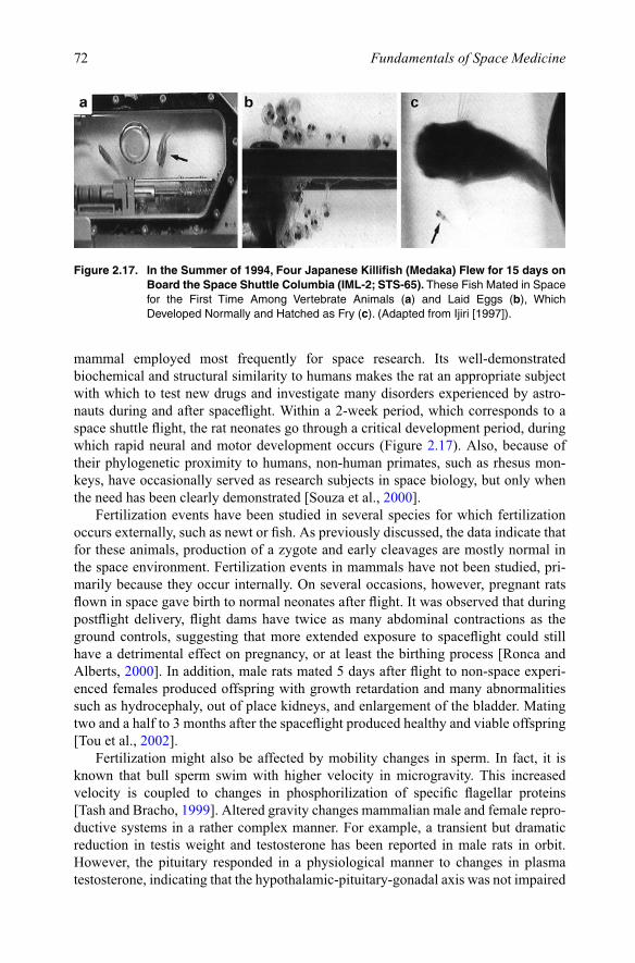
tadpole aquatic habitat on orbit, yet the tadpoles did not inflate their lungs while in microgravity. Two possible explanations for these flight findings include lack of direc-tional cues and increased influence of surface tension that may make it more difficult for an orbit-born tadpole to burst through a bubble and gulp air. The tadpoles returned to Earth within 2–3 days of emerging from the egg, and the lungs appeared normal by the time the tadpoles were 10-day old [Wassersug, 2001]. One investigation has sug-gested that gametes formed in space are normal [Ijiri, 1997]. In this experiment, Medaka fish mated freely in microgravity and the subsequent developmental steps were similar in flight and ground-control fish. Newly laid eggs formed a cluster on the belly of the female fish (Figure 2.17). After detachment from the female’s body, young fish hatched in microgravity and swam normally both in space and after returning to Earth. Back on the ground, the offspring produced healthy second-generation animals.
These studies produced multiple important findings. They show that vertebrates can be induced to ovulate in space and that rotation of fertilized eggs is not required for normal development in space. Long-duration microgravity exposure studies on the ISS revealed that larvae were able to regulate the morphological changes that occur during developmental in microgravity. The vertebrate embryo is very adaptive and the system is plastic, yet the long-term fate of the animal throughout its life in space remains unknown.
2.3.2.3. MammalsWhen investigations address human adaptation to spaceflight and its health implica-tions, the use of other mammalian species often becomes necessary. The rat is the
Figure 2.16. Comparison in the Development of Amphibian Eggs from Pleurodele Newt on Earth and in Space. There Are Clear Abnormalities in Orbit, Such as Larger Sillons andOddNumberofCells,intheFlightSpecimensbyComparisonwiththeGroundControls.(AdaptedfromGualandris-Parisotetal.[2002]).
72 Fundamentals of Space Medicine
mammal employed most frequently for space research. Its well-demonstrated biochemical and structural similarity to humans makes the rat an appropriate subject with which to test new drugs and investigate many disorders experienced by astro-nauts during and after spaceflight. Within a 2-week period, which corresponds to a space shuttle flight, the rat neonates go through a critical development period, during which rapid neural and motor development occurs (Figure 2.17). Also, because of their phylogenetic proximity to humans, non-human primates, such as rhesus mon-keys, have occasionally served as research subjects in space biology, but only when the need has been clearly demonstrated [Souza et al., 2000].
Fertilization events have been studied in several species for which fertilization occurs externally, such as newt or fish. As previously discussed, the data indicate that for these animals, production of a zygote and early cleavages are mostly normal in the space environment. Fertilization events in mammals have not been studied, pri-marily because they occur internally. On several occasions, however, pregnant rats flown in space gave birth to normal neonates after flight. It was observed that during postflight delivery, flight dams have twice as many abdominal contractions as the ground controls, suggesting that more extended exposure to spaceflight could still have a detrimental effect on pregnancy, or at least the birthing process [Ronca and Alberts, 2000]. In addition, male rats mated 5 days after flight to non-space experi-enced females produced offspring with growth retardation and many abnormalities such as hydrocephaly, out of place kidneys, and enlargement of the bladder. Mating two and a half to 3 months after the spaceflight produced healthy and viable offspring [Tou et al., 2002].
Fertilization might also be affected by mobility changes in sperm. In fact, it is known that bull sperm swim with higher velocity in microgravity. This increased velocity is coupled to changes in phosphorilization of specific flagellar proteins [Tash and Bracho, 1999]. Altered gravity changes mammalian male and female repro-ductive systems in a rather complex manner. For example, a transient but dramatic reduction in testis weight and testosterone has been reported in male rats in orbit. However, the pituitary responded in a physiological manner to changes in plasma testosterone, indicating that the hypothalamic-pituitary-gonadal axis was not impaired
Figure 2.17. In the Summer of 1994, Four Japanese Killifish (Medaka) Flew for 15 days on Board the Space Shuttle Columbia (IML-2; STS-65).TheseFishMatedinSpacefor the First Time Among Vertebrate Animals (a) and Laid Eggs (b), Which DevelopedNormallyandHatchedasFry(c).(AdaptedfromIjiri[1997]).

73Space Biology
by spaceflight. So, spermatogenesis was not reduced. Examination of the ovaries of postpartum rats flown in space during 9–20 days of gestation showed no effect on ovarian weight or number follicles [Tou et al., 2002]. The physiological mechanisms for reproduction are obviously intact in microgravity, despite of modifications of some components of the complete system.
As for the early period of development, the effects of microgravity on nervous system development were considered in only a few animal species and specific tracts. While these effects on the early formation of the nervous system were mainly based on studies in the aquatic animals, axonal growth and dendritic morphology related to functions such as equilibrium control and control of circadian activity, respectively were also studied in rats.
Rat embryos exposed to microgravity during the period when the vestibular sys-tem starts to become functional, showed delayed development compared to controls. In particular, 3 h after shuttle landing, central projections from the gravisensing organs receptors to the medial vestibular nucleus were more immature than in the controls [Bruce, 2003]. This result suggests that gravity is required for appropriate synaptic development and fine-tuning of the projections from the gravity sensing receptors to the central nervous system. These observations were supplemented by studies of neonate rats during the 16-day Neurolab STS-90 mission, which revealed an absence of connections into the vestibular nuclei from the cerebellum, the main control center for balance and coordination of movement [Raymond et al., 2003]. Recent studies have also revealed that microgravity affected the retinas of neonatal rats, probably by degeneration of cells or parts of individual cell types [Tombrain-Tink and Barnstable, 2005].
The force of gravity may influence events underlying the postnatal development of motor function in rats, similar to those noted in hatchling quail. Such effects most likely depend on the age of the animal, duration of the altered gravitational loading, and the specific motor function. The effect of microgravity on muscle mass and func-tion occurs in less than 1 week [Tischler et al., 1993]. The ossification of skeletal bones of fetuses of female rats flown in space during their pregnancy was arrested. However, during the 1-g re-adaptation period, the reduced ossification of the embryos was over-compensated, and newborns from this mission were ahead of the controls. Exposure of bone and bone cell cultures originating from mammals to microgravity is a widely used tool for understanding the underlying mechanisms of bone formation. Nevertheless, the basic mechanisms of the modifications in developing bones in microgravity are poorly understood. Isolated fetal mouse long bones experience no change in relative length increase and collagen synthesis induced by microgravity. Instead, a decreased mineralization, as well as a decrease in glucose consumption and an increase in calcium release is seen [Van Loon et al., 1995].
Like morphology, all physiological functions in organisms, as well as their behav-ior, experience modifications during development. The righting response from a supine posture to a prone posture is a good experimental model to test maturation of vestibular function. Beside the vestibular system, tactile cues from contact with a solid surface, as well as proprioceptive cues from muscle spindles and tendons contribute to a successful righting response. To separate the contribution of vestibular from other sensory inputs, the righting response can be studied during water immersion, i.e., the

74 Fundamentals of Space Medicine
animal is positioned in the supine position in a water-filled container and then released. Righting behavior in the absence of tactile cues revealed clear response deficits in neonates that underwent prenatal development in space (Figure 2.18). Exposure to microgravity during postnatal periods of life significantly retarded the development of this righting behavior [Ronca, 2003].
Walton [1998] also reported differences in swimming behavior and locomotion in neonatal rats when the musculo-skeletal system did not bear weight during critical times of development. The results from the 17-day Neurolab shuttle mission showed that neonatal rats flown in space exhibited altered locomotor behavioral development that persisted for the 1-month recovery period, and that righting reflex strategies were still abnormal 5 months after return to Earth.
One interesting feature of sensory, neuronal, and motor systems is the existence of critical periods during their development. The concept of critical period during devel-opment goes back to studies performed by Nobel prizes laureates Hubel and Wiesel [1962] on the visual system in kitten. Deprivation is the preferred scientific method to study the existence and duration of critical periods. Consequently, every long-lasting change in the environment may have its specific critical period. In general, three cri-teria must be fulfilled to define a development period as “critical”: (a) the developing system must be susceptible to a specific environmental modification; (b) the extent of modification must be related to age, and in particular to a well-defined period of devel-opment; and (c) the modification must persist for long periods of postnatal life or even permanently. In space studies, only the first two criteria were observed; indeed, long-duration effects of irreversibility were rarely noted.
Other results from space studies indicated delayed development of certain nerve connections to muscles. The connections returned to normal after return to Earth, yet
Figure 2.18. This Cartoon Shows the Sequence of Body Movements During the Righting Response in Water by Neonatal Rats Raised on Earth (Synchronous) or Exposed to Microgravity (Flight). (Credit Philippe Tauzin).
75Space Biology
fibers in hind limb muscles did not reach normal size even after a month back on Earth. The data suggest that biomechanical loading of limbs during early development may be essential for innervation of muscles. Another mechanism, however, may be at work: besides the lack of loading during critical times, there is also the possibility that adaptive changes in the vestibular system, particularly the reduction in descending otolith input required to maintain muscle tone (see H-reflex data in Chapter 3, Section 3.3.2), modify the nerve-to-muscle connections [Ronca and Alberts, 1997].
To date, relatively little neurobehavioral research has been done in microgravity with vertebrates, juvenile or adult. This is partly because the habitats for raising them in space did not exist and because the study of vertebrates up to sexual maturity requires longer exposure to microgravity [Wassersug, 2001]. The ISS will now pro-vide both capabilities. Physiological experiments using implanted electrodes in fish, rats, or rhesus monkeys have provided interesting data on the adaptive changes in the neuro-vestibular system, for example (see Chapter 3, Section 3.2.1). However these experiments are limited to constrained or caged animals, which do not experience motion in microgravity, making it difficult to draw a comparison with adaptive changes in astronauts. The response of animals to free-fall is astonishingly diverse, as shown by the observations made on frogs, lizards, and snakes in parabolic flight [Wassersug, 2001]. Other observations of animals placed in microgravity after vestibular lesions prove interesting for understanding the role of gravity in the process of recovery of balance function.
In conclusion, short-duration exposure to the space environment does not signifi-cantly affect the embryonic development, although interesting and unexplained changes occur during embryogenesis and early development. However, because the animals in most of these studies were only partially adapted to the space environment due to the short duration of the flight, it is possible that long-duration exposure will have more significant effects. The opportunity to conduct development studies onboard the ISS will leave room for much investigation.
2.4. Plant biology
2.4.1. QuestionsOn Earth, plant roots as a rule grow downward toward gravity, while stems grow up and away from gravity, a phenomenon known as gravitropism (see Figure 1.3). Circumnutation, i.e., the successive bowing or bending in different directions of the growing tip of the stems and roots, might also due in part to gravity. By studying plants in microgravity on board spacecraft, biologists seek to understand how plants respond to gravity at microscopic and macroscopic levels. Also, plants respond to environmental stimuli such as light, temperature, and magnetic or electric fields. These responses are masked on Earth by the overriding response of plants to gravity. In addition, any exploration strategy that includes a long-term sustained human presence in space absolutely requires the ability to continuously grow and reproduce various plant species over multiple generations for food production and closed environmental life support system.
76 Fundamentals of Space Medicine
2.4.2. Results of space experimentsA large variety of plants with short life spans have flown in space: algae, carrots, anise, pepper, wheat, pine, oat, mung beans, cress, lentils, corn, soybeans, lettuce, cucumbers, maize, sunflowers, peas, cotton, onion, nutmeg, barley, spindle trees, flax, orchids, gladiolas, daylilies, and tobacco. Because of this wide variety, for the most part, observations on plants exposed to microgravity have been anecdotal. It has been demonstrated repeatedly that plants do grow in microgravity. However, whether plants can grow and develop normally over several generations remains to be determined.
2.4.2.1. GraviceptionFor research purposes, the gravitational response, as with any stimulus response, has been divided into three steps: (a) stimulus perception: how a plant senses gravity; (b) signal transduction: how the plant transfers this knowledge into action; and (c) the response or resulting action: differential cell elongation or differential growth that results in the root or shoot bending in a new direction.
Where does the response occur? In roots we have already seen that perception occurs in the root cap (see Figures 2.5 and 2.6). The sensing mechanism underlying a plant’s ability to orient its organs in a gravitational field seems to involve the sedimen-tation of intracellular particles known as statoliths. The statoliths each consist of a number of starch grains surrounded by two membranes, the structure being termed an amyloplast. With the movement of statoliths, the cell receives a mechanical stimulus. How a cell transfers this mechanical stimulus into a chemical signal is still of great debate. One hypothesis is that as the statoliths “fall”, they come to rest on other organ-elles such as the endoplasmic reticulum or plasma membrane, thus exerting pressure on the organelle that results in the opening of ion channels and the release of ions such as calcium that initiate the signal transduction pathway [Perbal et al., 1997].
Transmission of the stimulus to the reaction zone, i.e., the bending of the root, could occur because of a change in the flow of the plant hormone auxin. How gravity regulates auxin transport remains unknown. Nevertheless, auxin typically flows in a fixed direction, from the shoot towards the root. After flowing down to the root tips, auxin begins to flow in the opposite direction, as if making a U-turn, along the roots. If the root is tilted relative to gravity, the concentration of auxin increases in the lower part of the elongation zone in the root, causing a differential growth between the lower part and the upper part.2 As a result, the root bends downward (Figure 2.19).
In microgravity, statoliths do not settle within the root cap cells (Figure 2.20), so gravity is not perceived, nor is asymmetric auxin distribution induced. Microgravity experiments on board sounding rockets, the space shuttle, and the ISS have shown that growth direction was indeed uncontrolled in microgravity, and some roots even extended in the same direction as the aerial stems (Figure 2.21).
Onboard centrifuge experiments have demonstrated that seedlings grown in space required a dose of 20–30 g s (gravity time seconds), or less than 0.1 g for 200–300 s,
2 Interestingly, phototropism in shoots seems to obey to the same mechanism. Light would stimulate the movement of auxin away from the light source. The increased supply of auxin to cells opposite the light source would cause them to elongate more than the cells on the same side as the light source.
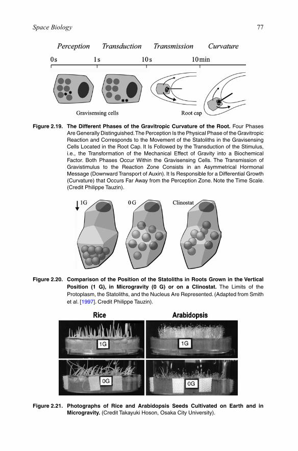
77Space Biology
Figure 2.19. The Different Phases of the Gravitropic Curvature of the Root.FourPhasesAre Generally Distinguished. The Perception Is the Physical Phase of the Gravitropic Reaction and Corresponds to the Movement of the Statoliths in the Gravisensing CellsLocatedintheRootCap.ItIsFollowedbytheTransductionoftheStimulus,i.e., the Transformation of the Mechanical Effect of Gravity into a Biochemical Factor. Both Phases OccurWithin the Gravisensing Cells.TheTransmission ofGravistimulus to the Reaction Zone Consists in an Asymmetrical HormonalMessage(DownwardTransportofAuxin).ItIsResponsibleforaDifferentialGrowth(Curvature)thatOccursFarAwayfromthePerceptionZone.NotetheTimeScale.(Credit Philippe Tauzin).
Figure 2.20. Comparison of the Position of the Statoliths in Roots Grown in the Vertical Position (1 G), in Microgravity (0 G) or on a Clinostat. The Limits of the Protoplasm, the Statoliths, and the Nucleus Are Represented. (Adapted from Smith etal.[1997]. Credit Philippe Tauzin).
Figure 2.21. Photographs of Rice and Arabidopsis Seeds Cultivated on Earth and in Microgravity.(CreditTakayukiHoson,OsakaCityUniversity).
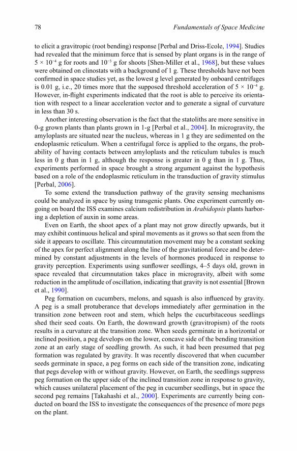
78 Fundamentals of Space Medicine
to elicit a gravitropic (root bending) response [Perbal and Driss-Ecole, 1994]. Studies had revealed that the minimum force that is sensed by plant organs is in the range of 5 × 10⁻⁴ g for roots and 10−3 g for shoots [Shen-Miller et al., 1968], but these values were obtained on clinostats with a background of 1 g. These thresholds have not been confirmed in space studies yet, as the lowest g level generated by onboard centrifuges is 0.01 g, i.e., 20 times more that the supposed threshold acceleration of 5 × 10⁻⁴ g. However, in-flight experiments indicated that the root is able to perceive its orienta-tion with respect to a linear acceleration vector and to generate a signal of curvature in less than 30 s.
Another interesting observation is the fact that the statoliths are more sensitive in 0-g grown plants than plants grown in 1-g [Perbal et al., 2004]. In microgravity, the amyloplasts are situated near the nucleus, whereas in 1 g they are sedimented on the endoplasmic reticulum. When a centrifugal force is applied to the organs, the prob-ability of having contacts between amyloplasts and the reticulum tubules is much less in 0 g than in 1 g, although the response is greater in 0 g than in 1 g. Thus, experiments performed in space brought a strong argument against the hypothesis based on a role of the endoplasmic reticulum in the transduction of gravity stimulus [Perbal, 2006].
To some extend the transduction pathway of the gravity sensing mechanisms could be analyzed in space by using transgenic plants. One experiment currently on-going on board the ISS examines calcium redistribution in Arabidopsis plants harbor-ing a depletion of auxin in some areas.
Even on Earth, the shoot apex of a plant may not grow directly upwards, but it may exhibit continuous helical and spiral movements as it grows so that seen from the side it appears to oscillate. This circumnutation movement may be a constant seeking of the apex for perfect alignment along the line of the gravitational force and be deter-mined by constant adjustments in the levels of hormones produced in response to gravity perception. Experiments using sunflower seedlings, 4–5 days old, grown in space revealed that circumnutation takes place in microgravity, albeit with some reduction in the amplitude of oscillation, indicating that gravity is not essential [Brown et al., 1990].
Peg formation on cucumbers, melons, and squash is also influenced by gravity. A peg is a small protuberance that develops immediately after germination in the transition zone between root and stem, which helps the cucurbitaceous seedlings shed their seed coats. On Earth, the downward growth (gravitropism) of the roots results in a curvature at the transition zone. When seeds germinate in a horizontal or inclined position, a peg develops on the lower, concave side of the bending transition zone at an early stage of seedling growth. As such, it had been presumed that peg formation was regulated by gravity. It was recently discovered that when cucumber seeds germinate in space, a peg forms on each side of the transition zone, indicating that pegs develop with or without gravity. However, on Earth, the seedlings suppress peg formation on the upper side of the inclined transition zone in response to gravity, which causes unilateral placement of the peg in cucumber seedlings, but in space the second peg remains [Takahashi et al., 2000]. Experiments are currently being con-ducted on board the ISS to investigate the consequences of the presence of more pegs on the plant.
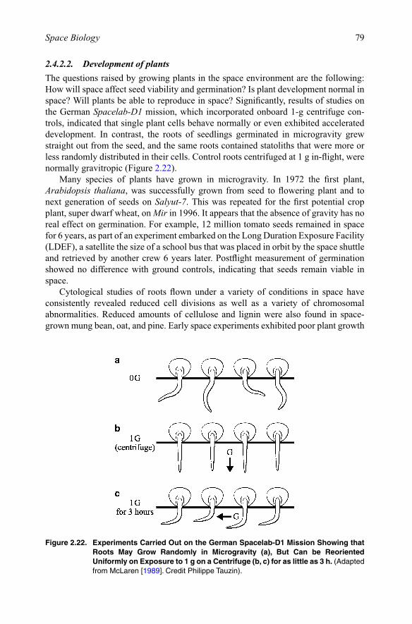
79Space Biology
2.4.2.2. Development of plantsThe questions raised by growing plants in the space environment are the following: How will space affect seed viability and germination? Is plant development normal in space? Will plants be able to reproduce in space? Significantly, results of studies on the German Spacelab-D1 mission, which incorporated onboard 1-g centrifuge con-trols, indicated that single plant cells behave normally or even exhibited accelerated development. In contrast, the roots of seedlings germinated in microgravity grew straight out from the seed, and the same roots contained statoliths that were more or less randomly distributed in their cells. Control roots centrifuged at 1 g in-flight, were normally gravitropic (Figure 2.22).
Many species of plants have grown in microgravity. In 1972 the first plant, Arabidopsis thaliana, was successfully grown from seed to flowering plant and to next generation of seeds on Salyut-7. This was repeated for the first potential crop plant, super dwarf wheat, on Mir in 1996. It appears that the absence of gravity has no real effect on germination. For example, 12 million tomato seeds remained in space for 6 years, as part of an experiment embarked on the Long Duration Exposure Facility (LDEF), a satellite the size of a school bus that was placed in orbit by the space shuttle and retrieved by another crew 6 years later. Postflight measurement of germination showed no difference with ground controls, indicating that seeds remain viable in space.
Cytological studies of roots flown under a variety of conditions in space have consistently revealed reduced cell divisions as well as a variety of chromosomal abnormalities. Reduced amounts of cellulose and lignin were also found in space-grown mung bean, oat, and pine. Early space experiments exhibited poor plant growth
Figure 2.22. Experiments Carried Out on the German Spacelab-D1 Mission Showing that Roots May Grow Randomly in Microgravity (a), But Can be Reoriented Uniformly on Exposure to 1 g on a Centrifuge (b, c) for as little as 3 h. (Adapted fromMcLaren[1989]. Credit Philippe Tauzin).

80 Fundamentals of Space Medicine
and altered development: plants died in transition from vegetative to flowering stage or plants flowered, but were abnormal. Beginning in 1993, a series of experiments on the space shuttle and then on Mir was initiated to examine this problem. It now appears that early abnormalities in plant reproduction could be caused by the toxic effect of ethylene, rather than spaceflight factors, and that seed size was diminished possibly because storage and utilization of reserves are modified in absence of gravity [Musgrave et al., 1997].
Arabidopsis thaliana has been successfully grown from seed-to-seed on ISS (see Figure 2.21). During a 2-month growth period, the plants progressed from seed hydra-tion to germination, vegetative, and reproductive stages, producing mature seeds. Ninety percent of the seeds germinated in space, although only 70% of the plants grew to maturity. Some of the seeds that were harvested from the plants that were grown in microgravity were planted in a ground study. These seeds produced typical plants without any visible abnormalities [Link et al., 2003]. Soybeans were also grown from seed to seed for the first time in space. Biomass production in the space seeds was approximately 4% larger than ground controls. Flight and grounds controls produced nearly identical numbers of seeds, but the space seeds were larger on average. Scientists found that the seeds that were produced in space were healthy, the germina-tion rates were comparable to those on Earth, and no major morphological differences were evident.
Brassica rapa (field mustard) and Triticum aestivum (super dwarf wheat) plants were germinated and grown in a plant growth chamber over several growth cycles on ISS. By the end of the experiment, the plant growth chamber produced a total of eight harvests, seven primings, and a plant tissue archive of more than 300 plants. In-flight progress of plant growth is monitored through image collection; harvested plants are frozen or fixed for later analysis on the ground [Morrow et al., 2004]. Seed protein was significantly lower in the ISS material. Also, microscopy of immature seeds fixed on ISS showed embryos to be at a range of developmental stages, while ground con-trol embryos had all reached the same stage of development. These differences could be attributable to differences in water delivery or reduced gas exchange due to lack of convection (Figure 2.23).
Plants continue to conduct photosynthesis and transpiration in space. However, some studies indicate a decreased ability to do so. Chloroplasts can have their internal structure disorganized, and their starch stores depleted. When plants were able to produce seeds in space, either additional light was needed versus on Earth or it was noted that oxygen production from photosynthesis was reduced. These results suggest that the microgravity environment may affect the flavor and nutritional quality of produces grown in space [Musgrave et al., 2005].
The growth and development of the dwarf wheat plants on the ISS was similar to the growth and development of plants on Earth. Analysis of the plants indicated that the microgravity-grown plants were 10% taller than plants grown on Earth. In 0 g, by comparison with 1-g control, the growth of the primary root and its apical (up-down) dominance over the secondary roots were reduced (Figure 2.24). Also, the growth of plant organs in space seems characterized by changes in the orientation of stem and leaves and secondary roots, more adventitious roots, and faster growth of secondary roots. The morphology of the primary root is not strongly modified. Experiments in

81Space Biology
space should be done to confirm that the reduced apical dominance results from a change in the hormonal content in roots. Once again Arabidopsis harboring a gene responsible for auxin depletion will be useful to analyze auxin distribution in space grown seedlings.
Figure 2.23. Astronaut Peggy A. Whitson Displays a First Crop of Soybeans Growing Inside the Advanced Astro-Culture Unit on Board the ISS. ThisExperimentIsUsedtoDetermineOptimalTimeforCross-PollinationandHarvesting.(CreditNASA).
Figure 2.24. Summary of the Effects of Microgravity on the Development and Growth of Plant Roots and Shoots.(AdaptedfromPerbal[2001]).
82 Fundamentals of Space Medicine
The cell cycle has been intensively studied in plants in the last decade [Inzé, 2005]. Plant molecular biologists now have the opportunity to use many molecular tools to analyze plant growth in space [Paul and Ferl, 2002]. We are close to understanding the causes of the changes in the development of plants in space. Many pioneering experi-ments have been done in space without monitoring gas composition, temperature, humidity, so that the conclusion of their authors must be questioned since plants are very sensitive to external factors. More clear-cut results have been obtained on board the ISS, because dedicated facilities providing onboard 1-g controls and better culture conditions have been developed. We know that the reproductive phase is complete in microgravity when the culture conditions (gas and liquid exchanges) are adequate. However, whether or not a seedling growing from the beginning in microgravity and across multiple generations can flower and produce normal seeds that can lead to normal plants remains a matter of debate. The experience gained from the past studies will be useful for the future.
2.5. Radiation biology
The broad spectrum of radiation encountered in space goes from extreme ultraviolet radiation, X-rays and high-energy particles such as electrons, neutrons, protons, to heavy ions such as iron. This section will be limited to a description of the ionizing radiation encountered in LEO and its biological effects as revealed by the biology experiments performed during space missions to date. A more complete description of the space radiation environment can be found in Eckart [1996]. The issues of radiation from the medical perspective will be discussed in greater detail in Chapter 8.
2.5.1. Ionized radiation in spaceThe sources of radiation during space missions are diverse:
(a) Cosmic radiation includes galactic radiation from supernova explosions as well as radiation of solar origin associated with solar flares (Figure 2.25). The primary galactic radiation present outside the Earth’s atmosphere is composed of about 85% protons (with a hydrogen nucleus), 12% alpha particles (with a helium nucleus), and 1.5% of heavy ion particles with high charge and energy. These high-energy particles interact with the nuclei of the nitrogen and oxygen atoms of the atmosphere, resulting in a highly complex secondary radiation, which irradi-ates the whole surface of the globe.
(b) The solar particle radiation consists of 95% protons. High surface doses may be experienced, but dosage levels rapidly decline with the depth of material penetrated.
(c) Space missions that include travel within or through the Van Allen belts also add a third source of radiation. The Van Allen belts consist of protons and electrons trapped by the geomagnetic field. A phenomenon of special importance for mis-sions in LEO is the South Atlantic Anomaly, in which the particles are drawn closer to the Earth than at other regions of the globe due to the asymmetry of the geomagnetic field.
(d) Finally, additional radiation is created in high-energy collisions of primary particles with spacecraft materials (Figure 2.26).
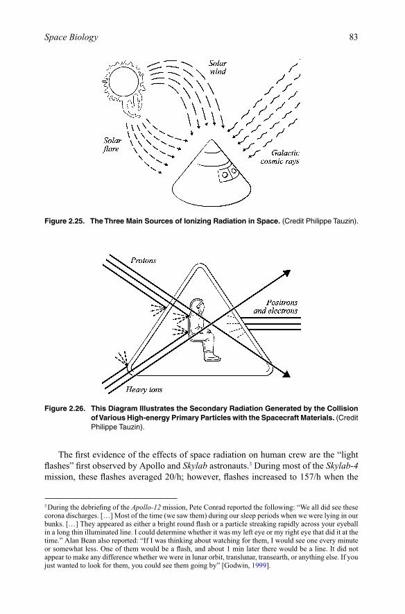
83Space Biology
The first evidence of the effects of space radiation on human crew are the “light flashes” first observed by Apollo and Skylab astronauts.3 During most of the Skylab-4 mission, these flashes averaged 20/h; however, flashes increased to 157/h when the
3 During the debriefing of the Apollo-12 mission, Pete Conrad reported the following: “We all did see these corona discharges. […] Most of the time (we saw them) during our sleep periods when we were lying in our bunks. […] They appeared as either a bright round flash or a particle streaking rapidly across your eyeball in a long thin illuminated line. I could determine whether it was my left eye or my right eye that did it at the time.” Alan Bean also reported: “If I was thinking about watching for them, I would see one every minute or somewhat less. One of them would be a flash, and about 1 min later there would be a line. It did not appear to make any difference whether we were in lunar orbit, translunar, transearth, or anything else. If you just wanted to look for them, you could see them going by” [Godwin, 1999].
Figure 2.25. The Three Main Sources of Ionizing Radiation in Space. (Credit Philippe Tauzin).
Figure 2.26. This Diagram Illustrates the Secondary Radiation Generated by the Collision of Various High-energy Primary Particles with the Spacecraft Materials. (Credit Philippe Tauzin).

84 Fundamentals of Space Medicine
Skylab orbit passed over the center of the so-called “South Atlantic Anomaly”. The flashes are believed to be due to high-energy heavy particles of cosmic rays and have been reproduced in humans on Earth by exposure to high-energy ionizing particles. Three explanations have been proposed to explain the phenomenon of light flashes seen by crewmembers: (a) an emission of photons by high-energy particles slowed by fluid in the eye (Cerenkov radiation); (b) a light generated by these particles ionizing fluid in the eye; or (c) an artificial light stimulus caused by these particles impacting retinal sensors in the eye [Pinsky et al., 1975].
2.5.2. Biological effects of radiationAll space agencies agree that the effects of space radiation, especially on the non-dividing cells of the retina and central nervous system, must be assessed before long-duration human missions beyond Earth’s magnetosphere are attempted. However, most of the biological effects of radiation remain largely unknown. The mechanisms of ionizing radiation impacts on cells are either direct, with particles impacting a vital target molecule and directly transferring their energy, or indirect, with particles impacting other molecules (e.g., water) to yield longer-lasting, very-reactive free radi-cals (with unpaired electron).
The biological effects of protons are fairly well understood. Through bombard-ment of spacecraft material, protons produce neutrons. These neutrons, upon colliding with a hydrogen nucleus, liberate their energy. Because living organisms contain many hydrogen-rich compounds, such as proteins, fat, and water (70%), they are most likely to be affected. However, the half-life of neutrons is only 11 min, after which they decay naturally to protons and electrons. Early dosimetry performed on Skylab and Russian space stations indicates that the flux of neutrons is probably not signifi-cant. The major space hazard comes from the highly charged energetic particles.
From ground-based experience (such as radiotherapy or nuclear explosion) it is known that the early, acute effects of radiation include skin effects (burns), eye lens opacification (cataract), graying of hair, immune system suppression (higher risk of infection), and the loss of non-dividing cells. Late effects include cancer in blood- forming organs (bone marrow, thyroid, lung, stomach, colon, and bladder) and genetic effects, which arise from, cell transformation (chromosome aberrations and translocations).
At the cellular level, when DNA strands break, non-rejoined breaks can lead to cell death, whereas incorrectly rejoined breaks can lead to mutation. The temporal and spatial characteristics of the radiation energy determine the quantity and quality of damage. Single-strand breaks can normally repair. However, double-strand breaks with close single hits or a high-density energy hit do not repair. Cells in mitosis are the most vulnerable. High-energy particles, with a high capacity to transfer their energy along the path, can generate significant percentage of double-strand DNA breaks. The effects are widespread and can lead to the death of numerous cells along the path. In addition, it is difficult to protect the vehicle and its inhabitants from these particles, even with shielding [Tobias and Todd, 1974].
Most results in space radiology have been obtained during short-duration space missions in LEO However, some observations were made on specimens flown on board free-flying satellites for several years, as well as on the ISS. The biological

85Space Biology
systems investigated include bacterial spores, plant seeds, and animal eggs, which were alternately sandwiched between nuclear track detectors allowing both a precise determination of the biological region penetrated by the particles and a determination of the charge and energy of each particle passing through. Chromosome damage and abnormalities were seen. In general, seeds are less sensitive than developing embryos or growing plants, which may be because their cells are not actively dividing. It is difficult to determine if these effects in lower organisms will lead to tumor induction, shortening of life, or chromosome aberration in organisms with longer life spans [Planel et al., 1994].
Several studies have been performed to try and determine which radiation is the most damaging, or even whether the damage was solely due to radiation at all. Some studies show that standard radioprotectant chemicals like cysteine, aminoethyliothio-urea, and 5-methoxytryptamine don’t stop the damage. This might indicate that the low-energy, indirect radiation is not primarily at fault. However, on some of the flights for which damage was found, the duration was short enough that galactic cosmic radiation dosages were rather low. Of course high-energy particles remain a possible threat, but some of the chromosomal damage and abnormalities could also be attrib-uted to other environmental factors, like microgravity. On the other hand, experiments with protozoa and bacteria suggest that small doses of radiation may actually be ben-eficial because small doses elicit stress responses and have been shown to increase DNA repair [Planel et al., 1987; Hammond et al., 1999].
Microlesions of cultured retina cells induced by single heavy ions were first dis-covered via spaceflight experiments. These findings initiated biological investigations using particle accelerators on Earth. However, the results of ground – and space-based studies are often conflicting. For example, a higher number of mutations were observed in biological systems (e.g., larvae of Drosophila) exposed to an “artificial” radiation source while in orbit compared to ground controls. This difference suggests the pos-sible existence of a combined effect of radiation and microgravity, the repair of radi-ation-induced lesions being altered in space [Planel et al., 1985].
This illustrates the difficulty of differentiating between the effects of the several factors inevitably present during spaceflight. For this reason, some biological effects, and their protection, can be studied only in space. An experiment on board the ISS utilizes a phantom torso, i.e., part-dummy, part-dosimeter-imbedded mock-up of a human’s upper body, minus a set of arms, built to determine the effects of radiation on the human body (Figure 2.27). Dosimeters are mounted at critical organ-tissue loca-tions within the dummy where critical organs are located: the head, the heart, the liver and kidneys. The dosimeters record the level of radiation received as a function of time. Other instruments mounted on the outside of the ISS measure the spectrum of particles that first hit the ISS shielding. As they go through the station wall and the dummy, the radiation is modified. The secondary radiation may have a different effect on tissue than the primary radiation. So the radiation spectrum is measured before and after it hits the dummy. The information gained from this and subsequent experiments (see Chapter 8, Section 8.3.5) will help determine the best types of materials and methods for shielding human crew.
For human mission to Mars, considerably better quantitative data on low-energy transfer radiation dose rates beyond the magnetosphere are still required. In particular,
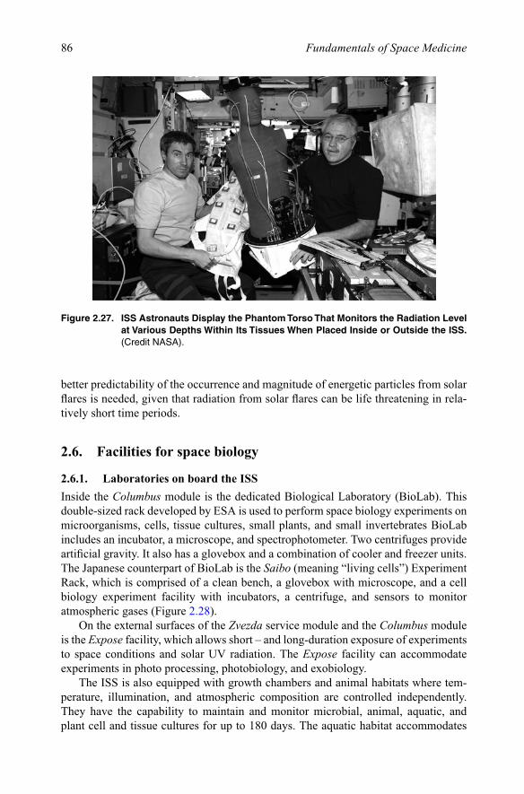
86 Fundamentals of Space Medicine
better predictability of the occurrence and magnitude of energetic particles from solar flares is needed, given that radiation from solar flares can be life threatening in rela-tively short time periods.
2.6. Facilities for space biology
2.6.1. Laboratories on board the ISSInside the Columbus module is the dedicated Biological Laboratory (BioLab). This double-sized rack developed by ESA is used to perform space biology experiments on microorganisms, cells, tissue cultures, small plants, and small invertebrates BioLab includes an incubator, a microscope, and spectrophotometer. Two centrifuges provide artificial gravity. It also has a glovebox and a combination of cooler and freezer units. The Japanese counterpart of BioLab is the Saibo (meaning “living cells”) Experiment Rack, which is comprised of a clean bench, a glovebox with microscope, and a cell biology experiment facility with incubators, a centrifuge, and sensors to monitor atmospheric gases (Figure 2.28).
On the external surfaces of the Zvezda service module and the Columbus module is the Expose facility, which allows short – and long-duration exposure of experiments to space conditions and solar UV radiation. The Expose facility can accommodate experiments in photo processing, photobiology, and exobiology.
The ISS is also equipped with growth chambers and animal habitats where tem-perature, illumination, and atmospheric composition are controlled independently. They have the capability to maintain and monitor microbial, animal, aquatic, and plant cell and tissue cultures for up to 180 days. The aquatic habitat accommodates
Figure 2.27. ISS Astronauts Display the Phantom Torso That Monitors the Radiation Level at Various Depths Within Its Tissues When Placed Inside or Outside the ISS. (Credit NASA).

87Space Biology
small fresh water organisms to support egg generation studies for examination at all life stages. Animal habitats are capable of housing up to six rats or a dozen mice. These habitats are compatible with another compartment accommodating pregnant mice and subsequently their offspring from birth through weaning. Plant units are able to support plant specimens of various heights through all stages of growth and devel-opment. The insect habitat supports drosophiles and other insects for multigenera-tional studies and radiation biology. Egg incubators support the incubation and development of small reptilian and avian eggs prior to hatching.
These units include:
(a) The NASA Advanced Biological Research System (ABRS) has two chambers to grow a variety of biological organisms, including plants, microorganisms, and small arthropods (insects and spiders).
(b) The Biotechnology Specimen Temperature Controller (BSTC) can grow and maintain mammalian cell cultures in microgravity.
(c) The European Modular Cultivation System (EMCS) is being used for multi-gen-eration experiments and studies of gravitational effects on early development and growth in seeds and plants and other small organisms, such as worms and fruit flies.
(d) The Eosteo Bone Culture System of the CSA provides the right conditions to grow bone cells in microgravity.
(e) With two aquariums, automatic feeding systems, LED lights to generate day/night cycle, and CCD cameras for observations, the Japanese Space Exploration Agency (JAXA) Aquatic Habitat (AQH) enables a variety of breeding experi-ments in space with small freshwater fish, such as Medaka or zebrafish.
Figure 2.28. A Crewmember Conducts a Biology Experiment Facility in the Saibo rack in the Kibo Laboratory of the ISS. (Credit NASA).

88 Fundamentals of Space Medicine
(f) For studies on organ function to embryonic development of mammals is the Mice Drawer System (MDS) developed by ASI and NASA. Research conducted with the MDS is an analog to the human research program, but allowing better focus at microscopic level.
(g) Finally, the LADA Greenhouse is used for growing plants in the Russian segment of the ISS. Since its launch in 2002, it has supported a series of experiments on fundamental plant biology and space farming, growing multiple generations of sweet peas, wheat, tomatoes, and lettuce (for the enjoyment of the crew!).
2.6.2. BioprocessingThe NASA Commercial Generic Bioprocessing Apparatus (CGBA) provides pro-grammable, accurate temperature control – from cold stowage to a customizable incu-bator – and can be used in a wide variety of biological studies, such as protein crystal growth, small insect habitats, plant development, antibiotic-producing bacteria, and cell culture studies.
ESA also uses the Protein Crystallization Diagnostics Facility (PCDF) to study the protein crystal growth conditions by way of non-intrusive optical techniques like Dynamic Light Scattering (DLS), Mach-Zehnder Interferometry (MZI), and classical microscopy. Understanding how crystals grow in purely diffuse conditions helps define the best settings to get organic crystals as perfect as possible. These crystals are then preserved and analyzed on Earth or on-board to deduce the three-dimensional shape of proteins.
2.6.3. Storage and operations2.6.3.1. FreezersFreezers allow freezing storage, and transportation of samples collected on ISS for later return to Earth. There are three ESA-built and NASA-operated freezers called Minus Eighty-Degree Laboratory Freezer for ISS (MELFI) which each have a volume of 175 L of samples stored at temperatures ranging from +4°C to as low as −80°C. A NASA General Laboratory Active Cryogenic ISS Equipment Refrigerator (GLACIER) also serves as an on-orbit ultra-cold freezer (as low as −165°C) and has a volume of 11.4 L. These freezers have special “damping” mechanisms that ensure that this microgravity level is undisturbed by human or machine activity. Smaller devices, with a volume of 4.2 L and temperatures ranging from −20°C to 48.5°C, can be used either as a freezer, refrigerator, or incubator. The Kriogem-3 M is a Russian refrigerator-incubator for stowage of biological samples.
2.6.3.2. GloveboxesPeriodic sampling is performed automatically or by the onboard crew, in such a way as to leave the remaining material undisturbed. Sampling is obviously carefully planned and minimized to preclude vibrations and other unwanted gravitational forces. Gloveboxes provide an enclosed workspace used for performing experiments and handling research organisms. They provide containment of experiments, and a safe environment for research with liquids, combustion, and hazardous materials on board the ISS (Figure 2.29). The laboratory-fixed ESA/NASA Microgravity Science

89Space Biology
Glovebox (MSG) ensures that hazardous materials do not float about the cabin. Crewmembers access the work area through ports equipped with rugged, sealed gloves. A video system and data downlinks allow for control of the enclosed experi-ments from the ground. A Portable Glove Box (PGB) is a smaller glovebox that can be transported around ISS and used to provide two levels of containment for experi-ments in any laboratory module. Three levels of containment can even be achieved by placing the PGB inside the larger volume of the MSG.
2.6.3.3. CentrifugesSeveral on-board centrifuges are provided within the facilities described above. These centrifuges provide a 1-g control for microgravity experiments. However, a missing tool for space biology on board the ISS is a multi-purpose centrifuge that would pro-vide the capability to explore a range of gravity levels between 10−6 and 1 g in order to study gravity thresholds for certain phenomena. This multi-gravity research facility would help determine the optimal parameters for artificial gravity in humans.
A Centrifuge Accommodation Facility (CAF) was originally designed planned for flying on board the ISS. Although the flight model was built and ready to launch, the project was cancelled. This NASA-JAXA facility could provide artificial gravity ranging from 10−6 to 2 g. Appropriate incubators and growth chambers were provided for cells, simple organisms, plants, and animals. Some habitats would have had the experimental capability of selectable gravity levels of up to 2 g by being mounted on a 2.5-m diameter centrifuge. Other habitats were equipped with internal centrifuges,
Figure 2.29. An Astronaut Works on a Biology Experiment Inside the Microgravity Science Glovebox (MSG) in the Columbus Laboratory of the ISS. (Credit NASA).
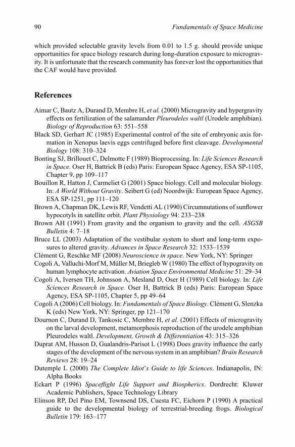
90 Fundamentals of Space Medicine
which provided selectable gravity levels from 0.01 to 1.5 g. should provide unique opportunities for space biology research during long-duration exposure to micrograv-ity. It is unfortunate that the research community has forever lost the opportunities that the CAF would have provided.
References
Aimar C, Bautz A, Durand D, Membre H, et al. (2000) Microgravity and hypergravity effects on fertilization of the salamander Pleurodeles waltl (Urodele amphibian). Biology of Reproduction 63: 551–558
Black SD, Gerhart JC (1985) Experimental control of the site of embryonic axis for-mation in Xenopus laevis eggs centrifuged before first cleavage. Developmental Biology 108: 310–324
Bonting SJ, Brillouet C, Delmotte F (1989) Bioprocessing. In: Life Sciences Research in Space. Oser H, Battrick B (eds) Paris: European Space Agency, ESA SP-1105, Chapter 9, pp 109–117
Bouillon R, Hatton J, Carmeliet G (2001) Space biology. Cell and molecular biology. In: A World Without Gravity. Seibert G (ed) Noordwijk: European Space Agency, ESA SP-1251, pp 111–120
Brown A, Chapman DK, Lewis RF, Vendetti AL (1990) Circumnutations of sunflower hypocotyls in satellite orbit. Plant Physiology 94: 233–238
Brown AH (1991) From gravity and the organism to gravity and the cell. ASGSB Bulletin 4: 7–18
Bruce LL (2003) Adaptation of the vestibular system to short and long-term expo-sures to altered gravity. Advances in Space Research 32: 1533–1539
Clément G, Reschke MF (2008) Neuroscience in space. New York, NY: SpringerCogoli A, Valluchi-Morf M, Müller M, Briegleb W (1980) The effect of hypogravity on
human lymphocyte activation. Aviation Space Environmental Medicine 51: 29–34Cogoli A, Iversen TH, Johnsson A, Mesland D, Oser H (1989) Cell biology. In: Life
Sciences Research in Space. Oser H, Battrick B (eds) Paris: European Space Agency, ESA SP-1105, Chapter 5, pp 49–64
Cogoli A (2006) Cell biology. In: Fundamentals of Space Biology. Clément G, Slenzka K (eds) New York, NY: Springer, pp 121–170
Dournon C, Durand D, Tankosic C, Membre H, et al. (2001) Effects of microgravity on the larval development, metamorphosis reproduction of the urodele amphibian Pleurodeles waltl. Development, Growth & Differentiation 43: 315–326
Duprat AM, Husson D, Gualandris-Parisot L (1998) Does gravity influence the early stages of the development of the nervous system in an amphibian? Brain Research Reviews 28: 19–24
Dutemple L (2000) The Complete Idiot’s Guide to life Sciences. Indianapolis, IN: Alpha Books
Eckart P (1996) Spaceflight Life Support and Biospherics. Dordrecht: Kluwer Academic Publishers, Space Technology Library
Elinson RP, Del Pino EM, Townsend DS, Cuesta FC, Eichorn P (1990) A practical guide to the developmental biology of terrestrial-breeding frogs. Biological Bulletin 179: 163–177
91Space Biology
Gualandris-Parisot L, Husson D, Bautz A, Durand D, et al. (2002) Effects of space environment on the embryonic development up to hatching of salamander eggs fertilized and developed during orbital flights. Biological Science in Space
Godwin R (1999) Apollo 12 NASA Mission Reports. Burlington, Canada: Apogee Books, CG Publishing Inc
Häder D-P, Hemmersbach R, Lebert M (2005) Gravity and the Behaviour of Unicellular Organisms. Cambridge, MA: Cambridge University Press
Hammond TG, Lewis FC, Goodwin TJ, Linnehan RM, et al. (1999) Gene expression in space. Nature Medicine 5: 359
Hubel DH, Wiesel TN (1962) Receptive fields, binocular interaction and functional architecture in the cat’s visual cortex. Journal of Physiology 160: 106–164
Hughes-Fulford M, Lewis M (1996) Effects of microgravity on osteoblast growth activation. Experimental Cell Research 224: 103–109
Ijiri K (1997) Explanations for a video version of the first vertebrate mating in space: A fish story. Biological Science in Space 11: 153–167
Imbert M (1979) Development of the visual system: Role of early experience. Journal of Physiology 75: 207–217
Ingber DE (1998) The architecture of life. Scientific American 278: 48–57Ingber DE (1999) How cells (might) sense microgravity. FASEB Journal 13: S3-S15Inzé D (2005) Green light for the cell cycle. Embryology Journal 24: 657–662Izumi-Kurotani A, Kiyomoto M (2003) Morphogenesis and gravity in a whole
amphibian embryo and in isolated blastomeres of sea urchins. Advances in Space Biology and Medicine 9: 83–99
Jones TA (1992) Gravity and the ontogeny of animals. The Physiologist 35: S77–79Klaus D, Simske S, Todd P, Stodieck L (1997) Investigation of space flight effects on
Escherichia coli and a proposed model of underlying physical mechanisms. Microbiology 143: 449–455
Klaus D (1998) Microgravity and its implication for fermentation technology. Trends in Biotechnology 16: 369–373
Link BM, Durst SJ, Zhou W, Stankovic B (2003) Seed-to-seed growth of Arabidopsis thaliana on the International Space Station. Advances in Space Research 31: 2237–2243
Machemer H, Machemer-Röhnisch S, Bräucker R, Takahashi K (1991) Gravikinesis in Paramecium: Theory and isolation of a physiological response to the natural gravity vector. Journal of Comparative Physiology A168: 1–12
Mattoni RHT, Keller EC, Ebersold WT, Eiserling FA, Romig WR (1971) Induction of lysogenic Bacteria in the space environment. In: The Experiments of Biosatellite II. Saunders J (ed). Washington DC: NASA, NASA SP-204, pp 309–324
Marxen JC, Reelsen O, Becker W (2001) Embryonic development of the freshwater snail Biomphalaria glabrata under mg conditions (STS-89 mission). Journal of Gravitational Physiology 8: 29–36
McKay DS, Gibson EK, Thomas-Keprta KL, Vali H, et al. (1996) Search for life on Mars: Possible relic biogenic activity in Martian meteorite ALH84001. Science 273: 924–930
McLaren A (1989) Developmental biology. In: Life Sciences Research in Space. Oser H, Battrick B (eds) Paris: European Space Agency, ESA SP-1105, Chapter 3, pp 31–36

92 Fundamentals of Space Medicine
Mills PJ, Meck JV, Waters WW, D’Aunno D, Ziegler MG (2001) Peripheral leukocyte subpopulations and catecholamine levels in astronauts as a function of mission duration. Psychosomatic Medicine 63: 886–890
Moody SA, Golden C (1999) Developmental Biology Research in Space: Anticipating the International Space Station. Recommendations from the September 1999 Developmental Biology Workshop sponsored by the International Space Life Sciences Working Group in Woods Hole, MA
Morrow RC, Iverson JT, Richter RC, Stadler JJ (2004) Biomass Production System (BPS) technology validation test results. Transactions Journal of Aerospace 1: 1061–1070
Musgrave ME, Kuang A, Tuominem LK, et al. (2005) Seed storage reserves and glu-cosinolates in Brassica rapa L. grown on the International Space Station. Journal of the American Society for Horticultural Science 130: 848–856
Musgrave ME, Kuang A, Porterfield DM (1997) Plant reproduction in spaceflight environments. Gravitational Space Biology Bulletin 10: 83–90
Nelson GA, Schubert WW, Kazarians GA, Richards GF, et al. (1995) Genetic and molecular dosimetry of HZE radiation. In: Biorack on Spacelab IML-1. Mattock C (ed) Noorwijk: European Space Agency, ESA SP-1162, pp 41–50
Paul AL, Ferl RJ (2002) Molecular aspects of stress-gene regulation during space-flight. Journal of Plant Growth & Regulation 21: 166–176
Perbal G (2001) The role of gravity in plant development. In: A World Without Gravity. Fitton B, Battrick B (eds) Noordwijk: ESA Publications Division, ESA SP-1251, pp 121–136
Perbal G, Driss-Ecole D (1994) Sensitivity to gravistimulus of lentil seedling roots grown in space during the IML-1 mission of Spacelab. Physiology Plant 70: 119–126
Perbal G, Driss-Ecole D, Tewinkel M, Volkmann D (1997) Statocyte polarity and gravisensitivity in seedling roots grown in microgravity. Planta 203: S57–S62
Perbal G, Lefranc A, Jeune B, Driss-Ecole D (2004) Mechanotransduction in root gravity sensing cells. Physiology of Plant 120: 303–311
Perbal G (2006) Plant development in microgravity. In: Fundamentals of Space Biology. Clément G, Slenzka K (eds) New York, NY: Springer, pp 227–290
Pinsky LS, Osborne WZ, Hoffman RA, Bailey JV (1975) Light flashes observed by astronauts on Skylab 4. Science 188: 928–930
Planel H, Tixador R, Richoilley G, Gasset G, Templier J (1985) Respective role of microgravity and cosmic rays on Paramecium tetraurelia cultured aboard Salyut 6. Acta Astronautica 12: 443–446
Planel H et al. (1987) Influence on cell proliferation of background radiation or expo-sure to very low, chronic gamma radiation. Health Physics 52: 571–578
Planel H et al. (1994) Influence of a long duration exposure, 69 months, to the space flight factors in Artemia cysts, tobacco and rice seeds. Advances in Space Research 14: 31–32
Raymond J, Demêmes D, Blanc E, Dechesne CJ (2003) Development of the vestibu-lar system in microgravity. In: The Neurolab Spacelab Mission: Neuroscience
93Space Biology
Research in Space. JC Buckey, JL Homick (eds) NASA Johnson Space Center, Houston: NASA SP-2003-535, pp 143–149
Ronca AE, Alberts JR (1997) Altered vestibular function in fetal and newborn rats gestated in space. Journal of Gravitational Physiology 4: P63-P66
Ronca AE, Alberts JR (2000) Physiology of a microgravity environment selected contribution: Effects of spaceflight during pregnancy on labor and birth at 1 G. Journal of Applied Physiology 89: 849–854
Ronca A (2003) Mammalian development in space. In: Developmental Biology Research in Space. Marthy HJ (ed) Amsterdam: Elsevier Science BV, pp 217–251
Scharf SR, Gerhart JC (1980) Determination of the dorsal-ventral axis in eggs of Xenopus laevis: complete rescue of UV-impaired eggs by oblique orientation before first cleavage. Developmental Biology 79: 181–198
Shen-Miller J, Hinchman R, Gordon SA (1968) Threshold for georesponse to accel-eration in gravity-compensated Avena seedlings. Plant Physiology 43: 338–344
Smith JD, Todd P, Staehelin LA (1997) Modulation of statolith mass and grouping in white clover (Trifolium repens) growth in 1-g, microgravity and on the clinostat. Plant Journal 12: 1361–1373
Souza KA, Black SD, Wassersug RJ (1995) Amphibian development in the virtual absence of gravity. Proceeding of the National Academy of Sciences 92: 1975–1978
Souza K, Theridge G, Callahan PX (eds) (2000) Life Into Space. Space Life Sciences Experiments. Ames Research Center, Kennedy Space Center, 1991–1998. Life Sciences Division, NASA Ames Research Center, and Moffetts Field, CA: NASA SP-2000-534
Summerlin LB (ed) (1977) Skylab, Classroom in Space. NASA Marshall Spaceflight Center, Huntsville, AL: NASA SP-401
Tabony J, Glade N, Papaseit C, Demongeot J (2002) Microtubule self-organization and its gravity dependence. In: Advances in Space Biology and Medicine. Cogoli A (ed) Chapter 8, Amsterdam: Elsevier, pp 19–58
Takahashi H, Kamada M, Yamazaki Y, Fujii N et al. (2000) Morphogenesis in cucum-ber seedlings is negatively controlled by gravity. Planta 210: 515–518
Tash JS, Kim S, Schuber M, Seibt D, Kinsey WH (2001) Fertilization of sea urchin eggs and sperm motility are negatively impacted under low hypergravitational forces significant to spaceflight. Biology of Reproduction 65: 1224–1231
Tash JS, Bracho GE (1999) Microgravity alters protein phosphorylation changes dur-ing initiation of sea urchin sperm motility. FASEB Journal Suppl 13: S43–S54
Tischler ME, Hendriksen EJ, Munoz KA, Stump CS, et al. (1993) Spaceflight on STS-48 and Earth-based unweighting produce similar effects on skeletal muscle of young rats. Journal of Applied Physiology 74: 2161–2165
Tobias CA, Todd P (1974) Space Radiation Biology and Related Topics. New York: Academic Press
Tombrain-Tink J, Barnstable CJ (2005) Space shuttle flight environment induces degeneration in the retina of rat neonates. Gravitational Space Biology 18: 97–98

94 Fundamentals of Space Medicine
Tou J, Ronca A, Grindeland R, Wade C (2002) Models to study gravitational biology of mammalian reproduction. Biology of Reproduction 67: 1681–1687
Van Loon JJ, Bervoets DJ, Burger EH, et al. (1995) Decreased mineralization and increased calcium release in insolated fetal mouse long bones under near weight-lessness. Journal Bone Mineral Research 10: 550–557
Vandenburgh H, Chromiak J, Shansky J, Del Tatto M, Lemaire J (1999) Space travel directly induces skeletal muscle atrophy. FASEB Journal 13: 1031–1038
Walton K (1998) Postnatal development under conditions of simulated weightlessness and spaceflight. Brain Research Reviews 28: 25–34
Wayne R, Staves MP, Leopold AC (1992) The contribution of the extracellular matrix to gravisensing in characean cells. Journal of Cellular Science 101: 611–623
Wassersug RJ (2001) Vertebrate biology in microgravity. American Scientist 89: 46–53
Wilkins MB (1989) Plant biology. In: Life Sciences Research in Space. Oser H, Battrick B (eds) Paris: European Space Agency, ESA SP-1105, Chapter 4, pp 37–48
Wilson A, ed. (2003) European Utilisation Plan for the International Space Station. Nordwijk: ESA Publications Division, ESA SP-1270
Zisckind N, Elinson RP (1990) Gravity and microtubules in dorsoventral polarization of the Xenopus egg. Development and Growth Differentiation 32: 575–581

http://www.springer.com/978-1-4419-9904-7