chapter 2 expression of erythropoietin and its … · chapter 2 expression of erythropoietin and...
TRANSCRIPT
Chapter 2
EXPRESSION OF ERYTHROPOIETIN AND ITS RECEPTOR IN THE CENTRAL NERVOUS SYSTEM
^Hugo H. Marti and ^Christian Bauer ^Institutfur Physiologie und Pathophysiologie, Universitdt Heidelberg, Im Neuenheimer Feld 326, D-69120 Heidelberg, Germany; ^Physiologisches Institut, Universitdt Zurich, Winterthurerstrasse 190, CH-8057 Zurich, Switzerland
Abstract: Erythropoietin (EPO) is a glycoprotein that is produced mainly by interstitial fibroblasts in the kidney. Released into the circulation, EPO makes its way to the bone marrow where it regulates red cell production by preventing apoptosis of erythroid progenitor cells. Recently, EPO has emerged as a multifunctional growth factor that plays a significant role in the nervous system. Both EPO and its receptor are expressed throughout the brain in glial cells, neurons and endothelial cells. Brain-derived EPO is upregulated by hypoxia, and expression of both EPO and its receptor are specifically modulated during cerebral ischemia. EPO has potent neuroprotective properties in vivo and in vitro and appears to act in a dual way by directly protecting neurons from ischemic damage and by stimulating endothelial cells, and thus supporting the growth of new blood vessels. EPO eventually also modulates inflammatory responses. Thus, hypoxically upregulated EPO is a naturally self-regulated physiological protective mechanism in the mammalian brain, especially during ischemia. As EPO is also a clinically extremely well studied and tolerated compound, its use in stroke patients is tempting.
Keywords: neuron; astrocyte; microglia; endothelial cell; HIF-1; VEGF; IGF-1; angiogenesis; neuroprotection; apoptosis; hypoxia; ischemia; stroke.
1. INTRODUCTION
Clinicians, who are treating patients suffering from the anemia of End-Stage Renal Failure with recombinant erythropoietin (EPO), have often reported an improvement of the cognitive function of their patients (reviewed by Ehrenreich and Siren, 2001a). However, it was never quite
16 Chapter 2
clear if this enhancement of cognitive function is the result of an increase in the oxygen transport capacity of the blood leading to an improved oxygenation of the brain, or if EPO has a direct effect on brain cells by itself This latter assumption was largely disregarded because EPO was not thought to cross the blood-brain barrier, due to its large size (30 kDa) and its many negative charges (Davis et al., 1987;Recny et al., 1987). Indeed, results from various studies indicated that endogenous kidney-derived EPO only gets access into the brain after breakdown of the blood-brain barrier (Marti et al., 1997;Buemi et al., 2000). Nonetheless, recent evidence suggests that high amounts of recombinant EPO can attain the brain in a number of experimental settings (Brines et al., 2000;Juul et al., 2004), a finding that may explain the beneficial effects on cognitive function seen in these patients.
These observations led to two interesting questions that concern the presence of EPO and EPO receptors in the brain. First, as EPO mediates its effects through binding to its cognate receptor, EPO receptor should be expressed at the site of action in the central nervous system (CNS) to enable EPO to elicit biological functions. Second, if EPO receptors are naturally occurring in the CNS, one has to postulate that EPO is endogenously produced in the brain itself to activate these receptors, on the assumption that kidney-derived EPO does not cross the blood-brain barrier under physiological conditions. Indeed, it was demonstrated that EPO receptors are widely distributed in the mammalian brain, and that the expression of EPO mRNA and EPO protein largely coincides with the occurrence of EPO receptor mRNA and protein (reviewed by Marti and Bemaudin, 2003;Genc et al., 2004;Marti, 2004). The upregulation of brain EPO in a large number of experimental conditions associated with tissue hypoxia is well documented, and comprise many mammalian species including mice, rats, monkeys, and humans (Marti et al., 1996;Marti et al., 2000;Siren et al., 2001;Gencetal.,2004).
In this chapter we will first deal with the normal pOi gradients within the brain. We will next consider those areas of the CNS that produce EPO and carry EPO receptors, and will finally have a look at the physiology of EPO function in the CNS, as an actor on neurons, glial cells and endothelial cells, including the action of EPO on the architecture of brain vessels.
2. OXYGEN GRADIENTS WITHIN THE BRAIN AND EXPRESSION OF HIF-la AND HIF-2 a
The brain exhibits a high rate of oxygen consumption, comprising some 20 % of the normal oxygen consumption at rest. Within the brain, the p02
2. EXPRESSION OF EPO AND ITS RECEPTOR IN THE CNS 17
profiles are low and non-uniform, ranging from 2 mmHg in the fornical part of the pons all the way up to 25-35 mm Hg in the cerebral cortex and 20 mmHg in the hippocampus (reviewed by Erecinska and Silver, 2001). The brain is therefore particularly sensitive towards hypoxia. Accordingly, a marked hypoxic induction of the transcription factors HIF-1 (hypoxia-inducible factor 1) (Bergeron et al., 1999) and HIF-2 (Wiesener et al., 2003) in the brain can be readily observed. Both HIF-1 and HIF-2 are dimers, having in common a constitutively expressed P-chain, yet their individual a-subunits (HIF-1 a, HIF-2 a) are inducible, e.g. by hypoxia.
These transcription factors direct many hypoxic responses such as the production of EPO, the Vascular Endothelial Growth Factor (VEGF), and the glucose transporter Glut-1. Classically, the regulation of these transcription factors occurs through an immediate oxygen-dependent proteolysis of their respective alpha subunits, HIF-1 a and HIF-2a (reviewed by Schofield and Ratcliffe, 2004;Sharp and Bemaudin, 2004). HIF-1 a is ubiquitously expressed, in the brain especially in parenchymal cells like the neurons, whilst HIF-2a is more confined to non-parenchymal cells i.e., endothelial and glial cells (Wiesener et al., 2003). These findings indicate that redundancy in the induction and thus funcfion of HIF-1 a and of HIF-2a is limited and that both factors may have complementary functions in a coordinated transcriptional response to hypoxia.
The notion for an oxygen-dependent regulation of HIF-1 a and HIF-2a holds certainly true for a more immediate hypoxic reactions, i.e. minutes to two days, as compared to conditions of global brain hypoxia lasting more than one week. Under such chronic conditions, the Insulin-Like Growth Factor 1 (IGF-1) apparently adopts the role of a signal that keeps HIF-1 a (and possibly HIF-2a) elevated despite the fact that the hypoxic marker EF5 does not indicate tissue hypoxia (Chavez and LaManna, 2002). This upregulation of HIF-1 a by IGF-1 is accompanied by an induction of some of the classical HIF-1 target genes e.g. EPO, VEGF and Glut-1 in the brain (Chavez and LaManna, 2002). In keeping with these observations are the results obtained by Masuda and co-workers (Masuda et al., 1997) who assessed a hypoxia-independent stimulation of EPO mRNA by IGF-1 in primary cultures of brain astrocytes.
All in all, these results strongly suggest that IGF-1 and hypoxia enhance two distinct and independent mechanisms of activation of HIF-1 and its targets genes, and may thereby account for the neuronal rescue by IGF-1 found in conditions of post-ischemic brain injury (Guan et al., 2003). This assumption holds true, despite of the fact that the induction of HIF-1 a by IGF-1 has not yet been clearly elucidated at the molecular level. In addition, recent results, demonstrating synergistic cooperative actions of EPO and IGF-1 that confer acute neuroprotection on cultured neurons indicate yet

li Chapter 2
another level of complex interaction between theses two cytokines (Digicaylioglu et al, 2004).
3. EXPRESSION OF EPO AND EPO RECEPTORS IN THE CNS
It came to a surprise, when it was found that both EPO and EPO receptor are constitutively expressed in the brain of rats and mice (Tan et al., 1992;Digicaylioglu et al., 1995;Marti et al., 1996), as it was largely held that the two entities of the "EPO System" are disconnected, one residing in the kidney (the EPO production machinery), and the other one in the bone marrow (the EPO receptor machinery). It soon turned out that these two entities of the EPO system are located within one and the same organ, even within one single cell, not only in the brain of rodents, but in the brain of monkeys and humans as well (Marti et al. 1996) (Fig. 2-1).
EPO-R-C
monkey h u m a n
5£ m 3
K m I k .
o
£ 3 m n m m
E m
m SI o a. >.
E m o o a
m
m
3 o
CD 1 3
ds
o o
X m I k .
o o
m a E S m m o m B £
2!
O c f- a iz CO c c
o o m o a
^^^^^^^^^^^^^^^^^^m^i^^^i^^^^^^^t^i^msi^^^^^^^^^^^^^^.
Figure 2-1. EPO receptor mRNA in different regions of the monkey and human brain. One microgram of total RNA was analyzed by RT-PCR specific for primate EPO receptor (EPOR). The RT-PCR product for primate EPO receptor is 485 bp. No RT, no reverse transcriptase added; positive control, human EPO receptor cDNA. (Reproduced by permission of Blackwell Publishing Ltd. from Marti et al., 1996).
In the monkey brain EPO expression was detected in all areas investigated, including various cortical areas, the cerebellum, the hypothalamus, the hippocampus, and the caudate nucleus. In the few

2. EXPRESSION OF EPO AND ITS RECEPTOR IN THE CNS 19
samples of the human brain that became available at the time, EPO mRNA along with immunoreactive EPO was found in the temporal cortex and the hippocampus (Marti et al., 1996;Siren et al., 2001). The detection of the "EPO System", i.e. EPO and its receptor, in the human brain immediately attracted a broader attention, particularly in view of the fact that EPO also occurs in the cerebrospinal fluid of human adults (Marti et al., 1997) and neonates (Juul et al., 1997).
The next question concerns the cell type(s) that produce EPO, and carry EPO receptors. It was soon detected that not only astrocytes that are in close contact both with neurons and brain capillaries produce EPO (Masuda et al., 1994;Marti et al., 1996) but that neuronal cells themselves are a source of brain-derived EPO, as was shown both in vitro and in vivo (Bemaudin et al., 1999;Bemaudin et al., 2000;Siren et al., 2001). Expression of EPO receptor mRNA and protein was demonstrated in the brain of mouse, rat, monkey and humans (Digicaylioglu et al., 1995;Marti et al., 1996;Liu et al., 1997), and the apposite analysis at the cellular level revealed that neurons and astrocytes carry the EPO receptor (Bemaudin et al., 1999;Bemaudin et al., 2000;Siren et al., 2001). In addition, it was shown that neurons, astrocytes and the microglia of human origin express EPO receptor mRNA in primary cultures of the respective cell type (Nagai et al., 2001). Of particular note is the fact that yet another glia cell type, the oligodendrocytes, was found to be devoid of EPO mRNA and of EPO receptor mRNA in purified cultures of adult human origin whilst immature oligodendrocytes isolated from embryonic rat brain were shown to produce EPO, as well as its receptor (Nagai et al., 2001).
Apart from neurons and glial cells, there is yet another cell type that deserves mention. These are the vascular endothelial cells that are one of the targets for EPO in the CNS, and suggest therefore a role for EPO in the vascular response to brain injury. This notion is supported by the fact that a strong immunoreactivity of the EPO receptor was found to be associated with brain endothelial cells (Brines et al., 2000), and that the astrocytic processes that surround brain capillaries carry a dense population of the EPO receptor (Brines et al., 2000). These in vivo findings are supported by in vitro analyses of endothelial cells derived from the human umbilical cord (Anagnostou et al., 1994) and rat brain microvessels (Yamaji et al., 1996) which clearly demonstrated that EPO receptors are also associated with brain endothelial cells. All of these findings are compatible with the idea that neuronal cells, glial cells and endothelial cells form an interactive meshwork that promotes the formation of new blood vessels in the brain in situations of an imminent ischemia.

20 Chapter 2
In summary, EPO as well as EPO receptors are expressed in various parts of the CNS. Table 2-1 presents an overview on the cellular sites of EPO and EPO receptor expression in the CNS.
Table 2-1. Cellular expression of EPO and its receptor in the CNS of rodents and men Protein EPO EPOR
Rodents Humans Rodents Humans
Neurons Astro Micrgl Oligodc Ec
In vitro + + + n.a. -
In vivo + + n.a. n.a. ?
In vitro -+ --n.a.
In vivo + + n.a. n.a. 9
In vitro + + n.a. + +
In vivo + + n.a. n.a. +
In vitro + + +
-+
In vivo + + n.a. n.a. +
Astro, astrocytes; Micrgl; microglia; Oligodc, oligrodendrocytes; Ec, endothelial cells; +, expression detected; -, no expression; n.a., not analysed; ?, not proven. (Adapted from Marti, 2004).
We will now regard in more detail the "EPO Network" in the brain taking in consideration neurons, glial cells and brain vascular endothelial cells.
NEURONAL CELLS: PHYSIOLOGY OF EPO
A high expression of both EPO and EPO receptors on neurons is found in those areas of the brain that are known to be particularly susceptible towards acute hypoxia (Lipton, 1999), e.g. the telencephalon (endbrain) and the hippocampus (Digicaylioglu et al., 1995). This exceptional sensitivity towards hypoxic insult is reflected by the high expression of synapses that use glutamate as a transmitter in those areas of the brain that are concerned with the storage and the retrieval of memory contents (Tang et al., 1999;Miu et al., 2004). In view of the fact that EPO protects neuronal cells from glutamate "overflow " (Morishita et al., 1997), the following scheme may be envisioned regarding the physiological counterbalance between glutamate and EPO in the brain: i) EPO decreases the number and/or the sensitivity of glutamate receptors of the NMDA type on the extracellular surface of neuronal cells, ii) EPO decreases glutamate release from neurons, iii) EPO increases glutamate uptake in presynaptic terminals and astrocytes, and iv) EPO increases the release of y-amino butyric acid (GABA) that has an inhibitory effect on the postsynaptic structures of those neurons that use glutamate as a transmitter. Future research will show which of these possibilities is effective. In this way, EPO may be regarded as a "natural" modifier of glutamate action that helps to physiologically buffer the excitatory action of glutamate, thereby preventing the occurrence of a local
2. EXPRESSION OF EPO AND ITS RECEPTOR IN THE CNS 21
glutamate excitotoxicity, and/or EPO may also contribute to a modification of the NMDA receptor-mediated consolidation of memory contents (Tang et al., 1999).
All of the neuronal structures mentioned above carry EPO receptors that are necessary and sufficient to transmit the EPO "signal" into the cell. It is interesting to note in this connection that EPO is involved in more complex neurological functions like synaptic transmission (Weber et al., 2002), better performance in pain and fear paradigms (Campana and Myers, 2003 ;Miu et al., 2004), in cognitive function (Ehrenreich and Siren, 2001a) and in spatial navigation performance (Sadamoto et al., 1998). There is evidence that EPO stimulates neuronal function and viability via activation of calcium channels (Assandri et al., 1999) and release of neurotransmitters (Koshimura et al., 1999;Yamamoto et al., 2002). Although the precise molecular mechanisms that account for these findings are so far poorly understood, it has become clear that EPO possesses a clear-cut neurotrophic activity: EPO augments choline acetyltransferase activity in primary cultured mouse septal neurons and promotes regeneration of septal cholinergic neurons in adult rats which had undergone fimbria-fornix transections (Konishi et al., 1993). Recently, it was also demonstrated that addition of EPO enhances survival and dopaminergic differentiation of CNS precursor cells in vitro (Studer et al., 2000) reminiscent of its survival function during erythropoiesis in the bone marrow. Furthermore, hypoxia-induced EPO appears to act directly on forebrain neural stem cells, promoting the production of neuronal progenitors suggesting that EPO is involved in neurogenesis after hypoxia (Shingo et al., 2001). All these results suggest that EPO is a neurotrophic factor that supports both differentiation and growth of neurons during development.
It should also be noted that not only the neuronal network in the brain profits from locally released EPO, but that the same notion is valid for the spinal cord. In this part of the CNS it was shown that a-motoneurons of the spinal cord benefit from the application of EPO in conditions of a spinal cord ischemic injury by the very same mechanisms that were described for the more centrally located neurons (Celik et al., 2002).
So far, we have mainly considered the interaction of the "EPO system" with neuronal cells, and will now turn to a population of cells that outnumbers the neurons by a factor 10, namely the glial cells.

22 Chapter 2
5. GLIAL CELLS: DEFINITIONS AND CONCEPTS REGARDING EPO
The term "glia" is derived from the Greek word glia, which Uterally means "glue", and indeed the glial cells have long been regarded as a cellular glue that hold the neuronal elements together. No statement could be less true in regard to a cell population that has very diverse physiological functions, as glia cells are involved in virtually every aspect of neural function (Zhang, 2001).
Let us now consider these functions in more detail: Largely the glial cells can be divided in microglia and macroglia that have diverse functions. The microglia is concerned with the recognition and processing of foreign antigens, and would thereby qualify as an immune competent cell type that identifies any foreign material entering the brain. In addition, the microglia has interesting functions in the recruitment of neuronal stem cells (Zhang, 2001;Doetsch, 2003). Considering that EPO receptor expression was shown in cultures of human microglial cells (Nagai et al., 2001), it remains a challenging task for future research activities if EPO can "convince" the microglia to recruit ever more neuronal stem cells. Furthermore, EPO can act as an anti-inflammatory cytokine during cerebral ischemia by reducing the recruitment of microglia into the infarcted area (Villa et al., 2003) and, thus, play an immunomodulatory role in the CNS.
The macroglia on the other hand, may be divided in two major cell types: the star-shaped astrocytes, the term being derived from the Greek word astron, meaning star, and the oligodendrocytes from oligos, meaning few. Oligodendrocytes are myelin-forming cells that wrap the axons, and provide the basis for a rapid transmission of action potentials from the central to the peripheral part of the CNS. In certain diseases, this function is lost, for example in multiple sclerosis. Under the assumption that EPO promotes growth and differentiation of embryonic oligodendrocytes, the hypothesis can be put forward that the "EPO system" supports the myelinization of the growing axons in the embryonic CNS (Sugawa et al., 2002). This idea tallies nicely with the developmental regulation of EPO and its receptor in the embryonic and fetal brain of animals and humans, and suggests therefore a widespread role of the "EPO system" in the maturation of the mammalian CNS (Juul, 2002).
Astrocytes on the other hand provide important structural, metabolic and trophic support to neurons. They encase the brain, interact with endothelial cells to form the blood-brain barrier, absorb neurotransmitters, maintain extracellular ion homeostasis and secrete growth factors, cytokines and
2. EXPRESSION OF EPO AND ITS RECEPTOR IN THE CNS 23
components of the extracellular matrix. Astrocytes are further involved in the formation and stabilization of synapses and the modulation of synaptic efficacy (Doetsch, 2003). Despite of their functional importance, the experimental evidence obtained so far - regarding the effects of the "EPO system" on the more specific function of glial cells - is pretty sparse. Nonetheless, EPO was shown to effectively support the development of astrocytes in a concentration-dependent manner (Sugawa et al., 2002). In this way, astrocytes may thus be physiologically involved in the EPO-induced modulation of excitatory postsynaptic glutamate responses as well (Kawakamietal.,2001).
None of these functions in neurons or glial cells can be maintained without an appropriate blood flow feeding these cells with oxygen and nutrients. Ideally, regulation of blood flow should thus also be geared by EPO. Recent insights suggest that blood vessels and nerves have much more in common than was originally anticipated (reviewed by Carmeliet, 2003). They use similar principles to differentiate, grow and navigate towards their targets. Moreover, the vascular and the nervous systems cross-talk by using similar signals and factors. These factors include VEGF, angiopoietins, ephrins, platelet-derived growth factors, and EPO. Therefore, we will next consider the maintenance of blood vessels in the brain that are under the direction of EPO and other oxygen-controlled cytokines.
6. EPO AND THE ARCHITECTURE OF BRAIN VESSELS
Formation and remodeling of new blood vessels (vascular morphogenesis) is governed by three processes, vasculogenesis, angiogenesis and arteriogenesis. Vasculogenesis is defined as the differentiation of mesodermal progenitor cells (angioblasts) into endothelial cells in situ where they subsequently aggregate and form a primary vascular plexus (Risau and Flamme, 1995). Vasculogenesis occurs primarily during embryonic development. Angiogenesis is defined as the formation of new blood vessels by sprouting of endothelial cells from pre-existing vessels or by intravascular subdivision (intussuscepfion) (Risau, 1997). Angiogenesis further refines the primitive embryonic vascular plexus and includes remodeling, a process that transforms the relatively uniformly sized vasculature into the network of small and large vessels, that finally undergoes maturation by recruiting perivascular cells, such as smooth muscle cells and pericytes. Angiogenesis is an important process during embryogenesis but also occurs in the adult in response to altered metabolic requirements, e.g. it can be triggered by hypoxia. Finally, arteriogenesis is

24 Chapter 2
the rapid proliferation of pre-existing collateral vessels that occurs in ischemic tissue (for a review see Buschmann and Schaper, 1999). EPO could be involved in all three processes, acting on endothelial cells, vascular smooth muscle cells and even cardiomyocytes (reviewed by Smith et al., 2003).
Originally, EPO was shown to act on the mitogenesis and chemotaxis of endothelial cells derived from the human umbilical vein and bovine adrenal capillaries (Anagnostou et al., 1994). It was then shown that vessel outgrowth of rat aortic rings was stimulated by EPO (Carlini et al., 1995) suggesting that EPO has angiogenic properties. Indeed, neovascularization in vivo is stimulated in the endometrium after EPO injection into the mouse uterine cavity (Yasuda et al., 1998). In the brain, EPO induces a dose-dependent mitogenic activity on brain capillary endothelial cells (Yamaji et al., 1996).
With regard to endothelial functions of EPO, it is noteworthy that endothelial cells and hematopoietic cells are derived from the same mesenchymal precursor, the so-called hemangioblast (Risau, 1997). This may explain why endothelial cells carry the EPO receptor and can be stimulated by EPO (Ribatti et al., 1999;Yasuda et al., 1998). Very recently, it was shown that EPO is a potent physiologic stimulus for endothelial progenitor cell mobilization and EPO stimulates postnatal neovascularization (Heeschen et al., 2003). In erythroid precursor cells and neurons, EPO is a survival factor. The same notion seems to hold true for endothelial cells; EPO prevents cell injury and DNA fragmentation (Chong et al., 2002). EPO might also influence endothelial cells indirectly through the activation of the VEGF/VEGF receptor system. Interestingly, it was shown that EPO-induced proliferation of bovine aortic and glomerular endothelial cells was prevented by a specific anti-VEGF antibody (Nitta et al., 1999;Victoria et al., 1998). Thus, EPO might exert its function on endothelial cells via activation of VEGF receptors. Indeed, mRNA expression for both the VEGF receptor-1 and VEGF receptor-2 was upregulated in aortic cells after EPO pretreatment (Victoria et al., 1998).
It thus appears likely that increased expression of EPO and its receptor in blood vessels during cerebral ischemia in mice (Bemaudin et al., 1999) as well as in humans (Siren et al., 2001) contributes to new vessel growth in the tissue area suffering from hypoxia. Hypoxia- or ischemia-induced EPO thus stimulates new vessel growth enabling the transport of more red blood cells and thereby increasing the amount of oxygen delivered to the hypoxic tissue which in turn counteracts the detrimental effects of hypoxia on neurons.

2. EXPRESSION OF EPO AND ITS RECEPTOR IN THE CNS 25
7. PATHOPHYSIOLOGY OF EPO: CONTROLLING THE AFTERMATH OF TISSUE HYPOXIA
EPO has been shown to be neuroprotective in the brain after exposure to a variety of insuhs, including cerebral ischemia, head injury, seizures and experimental autoimmune encephalomyelitis (reviewed in Marti, 2004). In 1998, Sasaki and colleagues provided good evidence that endogenous brain-derived EPO is crucial for neuronal survival in vivo. Infusion of a soluble EPO receptor into the brain of gerbils, submitted to a mild ischemia that did not produce neuronal damage by itself, resulted in neuronal cell death in the hippocampus (Sakanaka et al., 1998). These results clearly indicate that brain-derived EPO is an endogenous protective agent for neurons against mild forms of tissue hypoxia and ischemia. Thus, the "EPO system" serves as an endogenous system to protect brain cells from damage caused by intermittent episodes of hypoxia. Along this line, EPO has been implicated in the mechanisms of ischemic tolerance or preconditioning. Preconditioning means that practically any stimulus capable of causing injury to a tissue can, when applied below the threshold level of damage, activate endogenous protective mechanisms and thus potentially lessen the impact of subsequent, more severe insults (reviewed in Dimagl et al., 2003). In models of ischemic preconditioning both in vitro and in vivo, hypoxia-induced EPO release from astrocytes can inhibit hypoxia-induced apoptosis in neurons (Ruscher et al., 2002) and thus provide stroke tolerance (Prass et al., 2003).
The fact that a differential temporal and cellular modulation of the "EPO system" by ischemia is also detected in human brain tissue (Siren et al., 2001) indicates that EPO may have a beneficial effect for the treatment of stroke patients. Results from a first clinical phase I / phase II study are indeed promising. Intravenous high-dose recombinant EPO in a total of 53 stroke patients was well tolerated and associated with an improvement in clinical outcome at one month (Ehrenreich et al., 2002). Currently, a larger multi centre study is under way.
8. SUMMARY
A primary goal, not only for stroke patients but also for other diseases of the CNS, is to protect neural function. Imitation of brain endogenous protective mechanisms may be the key to future successful approaches to neuroprotection; therefore activation and mimicry of endogenous mechanisms can be expected to be efficient and well tolerated (Ehrenreich
26 Chapter 2
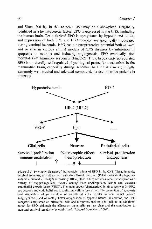
and Siren, 2001b). In this respect, EPO may be a showpiece. Originally identified as a hematopoietic factor, EPO is expressed in the CNS, including the human brain. Brain-derived EPO is upregulated by hypoxia and IGF-1, and expression of both EPO and EPO receptor are specifically modulated during cerebral ischemia. EPO has a neuroprotective potential both in vitro and in vivo in various animal models of CNS diseases by inhibition of apoptosis in neurons and inducing angiogenesis. EPO eventually also modulates inflammatory responses (Fig. 2-2). Thus, hypoxically upregulated EPO is a naturally self-regulated physiological protective mechanism in the mammalian brain, especially during ischemia. As EPO is also a clinically extremely well studied and tolerated compound, its use in stroke patients is tempting.
Hypoxia/ischemia IGF-1
HlF-l (HlF-2)
VEGF Epo
Glial cells
Survival, proliferation immune modulation
I
Neurons
Neurotrophic effects neuroprotection
f i
Endothelial cells
Survival proliferation angiogenesis
Figure 2-2. Schematic diagram of the possible actions of EPO in the CNS. Tissue hypoxia, cerebral ischemia, as well as the Insulin-like Growth Factor-1 (IGF-1) activate the hypoxia-inducible factor-1 (HIF-1) (and possibly HIF-2), that in turn activates gene transcription of a variety of oxygen-regulated factors, among them erythropoietin (EPO) and vascular endothelial growth factor (VEGF). The main targets (characterized by thick arrows) for EPO are neurons and endothelial cells, conferring cellular protection. The prevention of apoptosis and stimulation of proliferation of endothelial cells, results in new vessel growth (angiogenesis), and ultimately better oxygenation of hypoxic tissues. In addition, the EPO receptor is expressed on microglial cells and astrocytes, making glial cells to an additional target for EPO, although the effects on these cells are less clear and the contribution to neuronal survival remains to be established. (Adapted from Marti, 2004).
2. EXPRESSION OF EPO AND ITS RECEPTOR IN THE CNS 27
All these results support the idea that EPO acts in the CNS by a variety of mechanisms in neurons, endothelial cells and glial cells. Stimulation of new vessel growth (angiogenesis) leads to a better tissue oxygenation in the brain, in addition to its erythropoietic effect resulting in an increased oxygen carrying capacity of the blood. EPO also modulates electrophysiological and inflammatory responses, making it to an attractive neurotrophic and neuroprotective factor. Thus - coming back to the initial clinical observation - cognitive function might be indeed improved by a direct action of EPO within the CNS.
REFERENCES
Anagnostou A, Liu Z-Y, Steiner M, Chin K, Lee ES, Kessimian N, Noguchi CT (1994) Erythropoietin-receptor mRNA expression in human endothelial cells. Proc Natl Acad Sci USA 91:3974-3978.
Assandri R, Egger M, Gassmann M, Niggli E, Bauer C, Forster I, Gorlach A (1999) Erythropoietin modulates intracellular calcium in a human neuroblastoma cell line. J Physiol (London) 516:343-352.
Bergeron M, Yu AY, Solway KE, Semenza GL, Sharp PR (1999) Induction of hypoxia-inducible factor-1 (HIF-1) and its target genes following focal ischaemia in rat brain. Eur J Neurosci 11:4159-4170.
Bemaudin M, Marti HH, Roussel S, Divoux D, Nouvelot A, MacKenzie ET, Petit E (1999) A potential role for erythropoietin in focal permanent cerebral ischemia in mice. J Cereb Blood Flow Metab 19:643-651.
Bemaudin M, Bellail A, Marti HH, Yvon A, Vivien D, Duchatelle I, MacKenzie ET, Petit E (2000) Neurons and astrocytes express EPO mRNA: oxygen-sensing mechanisms that involve the redox-state of the brain. Glia 30:271-278.
Brines ML, Ghezzi P, Keenan S, Agnello D, de Lanerolle NC, Cerami C, Itri LM, Cerami A (2000) Erythropoietin crosses the blood-brain barrier to protect against experimental brain injury. Proc Natl Acad Sci USA 97:10526-10531.
Buemi M, Allegra A, Corica F, Floccari F, D'Avella D, Aloisi C, Calapai G, lacopino G, Frisina N (2000) Intravenous recombinant erythropoietin does not lead to an increase in cerebrospinal fluid erythropoietin concentration. Nephrol Dial Transplant 15:422-423.
Buschmann I, Schaper W (1999) Arteriogenesis versus angiogenesis: two mechanisms of vessel growth. News Physiol Sci 14:121-125.
Campana WM, Myers RR (2003) Exogenous erythropoietin protects against dorsal root ganglion apoptosis and pain following peripheral nerve injury. Eur J Neurosci 18:1497-1506.
Carlini RG, Reyes AA, Rothstein M (1995) Recombinant human erythropoietin stimulates angiogenesis in vitro. Kidney Int 47:740-745.
Carmeliet P (2003) Blood vessels and nerves: common signals, pathways and diseases. Nature Rev Genet 4:710-720.
Celik M, Gokmen N, Erbayraktar S, Akhisaroglu M, Konakc S, Ulukus C, Gene S, Gene K, Sagiroglu E, Cerami A, Brines M (2002) Erythropoietin prevents motor neuron apoptosis and neurologic disability in experimental spinal cord ischemic injury. Proc Natl Acad Sci USA 99:2258-2263.

28 Chapter 2
Chavez JC, LaManna JC (2002) Activation of hypoxia-inducible factor-1 in the rat cerebral cortex after transient global ischemia: potential role of insulin-like growth factor-1. J Neurosci 22:8922-8931.
Chong ZZ, Kang JQ, Maiese K (2002) Erythropoietin is a novel vascular protectant through activation of Aktl and mitochondrial modulation of cysteine proteases. Circulation 106:2973-2979.
Davis JM, Arakawa T, Strickland TW, Yphantis DA (1987) Characterization of recombinant human erythropoietin produced in Chinese hamster ovary cells. Biochemistry 26:2633-2638.
Digicaylioglu M, Bichet S, Marti HH, Wenger RH, Rivas LA, Bauer C, Gassmann M (1995) Localization of specific erythropoietin binding sites in defined areas of the mouse brain. ProcNatl Acad Sci USA 92:3717-3720.
Digicaylioglu M, Garden G, Timberlake S, Fletcher L, Lipton SA (2004) Acute neuroprotective synergy of erythropoietin and insulin-like growth factor I. Proc Natl Acad Sci USA 101:9855-9860.
Dimagl U, Simon RP, Hallenbeck JM (2003) Ischemic tolerance and endogenous neuroprotection. Trends Neurosci 26:248-254.
Doetsch F (2003) The glial identity of neural stem cells. Nature Neurosci 6:1127-1134. Ehrenreich H, Siren AL (2001a) Benefits of recombinant human erythropoietin on cognitive
function. Erythropoiesis: new dimensions in the treatment of anaemia 11:35-40. Ehrenreich H, Siren AL (2001b) Neuroprotection - what does it mean? - what means do we
have? Eur Arch Psyciatry Clin Neurosci 251:149-151. Ehrenreich H, Hasselblatt M, Dembowski C, Cepek L, Lewczuk P, Stiefel M, Rustenbeck
HH, Breiter N, Jacob S, Knerlich F, Bohn M, Poser W, Rtither E, Kochen M, Gefeller 0, Gleiter C, Wessel TC, De Ryck M, Itri L, Prange H, Cerami A, Brines M, Siren AL (2002) Erythropoietin therapy for acute stroke is both safe and beneficial. Mol Med 8:495-505.
Erecinska M, Silver lA (2001) Tissue oxygen tension and brain sensitivity to hypoxia. Respir Physiol 128:263-276.
Gene S, Koroglu TF, Gene K (2004) Erythropoietin and the nervous system. Brain Res 1000:19-31.
Guan J, Bennet L, Gluckman PD, Gunn AJ (2003) Insulin-like growth factor-1 and post-ischemic brain injury. Prog Neurobiol 70:443-462.
Heeschen C, Aicher A, Lehmann R, Fichtlscherer S, Vasa M, Urbich C, Mildner-Rihm C, Martin H, Zeiher AM, Dimmeler S (2003) Erythropoietin is a potent physiologic stimulus for endothelial progenitor cell mobilization. Blood 102:1340-1346.
Juul SE, Harcum J, Li Y, Christensen RD (1997) Erythropoietin is present in the cerebrospinal fluid of neonates. J Pediatr 130:428-430.
Juul S (2002) Erythropoietin in the central nervous system, and its use to prevent hypoxic-ischemic brain damage. Acta Paediatr Suppl 91:36-42.
Juul SE, McPherson RJ, Farrell FX, Jolliffe L, Ness DJ, Gleason CA (2004) Erytropoietin concentrations in cerebrospinal fluid of nonhuman primates and fetal sheep following high-dose recombinant erythropoietin. Biol Neonate 85:138-144.
Kawakami M, Sekiguchi M, Sato K, Kozaki S, Takahashi M (2001) Erythropoietin receptor-mediated inhibition of exocytotic glutamate release confers neuroprotection during chemical ischemia. J Biol Chem 276:39469-39475.
Konishi Y, Chui D-H, Hirose H, Kunishita T, Tabira T (1993) Trophic effect of erythropoietin and other hematopoietic factors on central cholinergic neurons in vitro and in vivo. Brain Res 609:29-35.
Koshimura K, Murakami Y, Sohmiya M, Tanaka J, Kato Y (1999) Effects of erythropoietin on neuronal activity. J Neurochem 72:2565-2572.
2. EXPRESSION OF EPO AND ITS RECEPTOR IN THE CNS 29
Lipton P (1999) Ischemic cell death in brain neurons. Physiol Rev 79:1431-1568. Liu C, Shen K, Liu ZY, Noguchi CT (1997) Regulated human erythropoietin receptor
expression in mouse brain. J Biol Chem 272:32395-32400. Marti HH, Wenger RH, Rivas LA, Straumann U, Digicaylioglu M, Henn V, Yonekawa Y,
Bauer C, Gassmann M (1996) Erythropoietin gene expression in human, monkey and murine brain. Eur J Neurosci 8:666-676.
Marti HH, Gassmann M, Wenger RH, Kvietikova I, Morganti-Kossmann MC, Kossmann T, Trentz O, Bauer C (1997) Detection of erythropoietin in human liquor: Intrinsic erythropoietin production in the brain. Kidney Int 51:416-418.
Marti HH, Bemaudin M, Petit E, Bauer C (2000) Neuroprotection and angiogenesis: A dual role of erythropoietin in brain ischemia. News Physiol Sci 15:225-229.
Marti HH, Bemaudin M (2003) Function of erythropoietin in the brain. In: Erythropoietin: molecular biology and clinical use (Jelkmann W, ed), ppl95-215. Johnson City (TN): FP Graham Publishing Co.
Marti HH (2004) Erythropoietin and the hypoxic brain. J Exp Biol 207: 3233-3242. Masuda S, Okano M, Yamagishi K, Nagao M, Ueda M, Sasaki R (1994) A novel site of
erythropoietin production: oxygen-dependent production in cultured rat astrocytes. J Biol Chem 269:19488-19493.
Masuda S, Chikuma M, Sasaki R (1997) Insulin-like growth factors and insulin stimulate erythropoietin production in primary cultured astrocytes. Brain Res 746:63-70.
Miu AC, Olteanu AI, Chis I, Heilman RM (2004) Have no fear, erythropoietin is here: erythropoietin protects fear conditioning performances after functional inactivation of the amygdala. Behav Brain Res: in press.
Morishita E, Masuda S, Nagao M, Yasuda Y, Sasaki R (1997) Erythropoietin receptor is expressed in rat hippocampal and cerebral cortical neurons, and erythropoietin prevents in vitro glutamate-induced neuronal death. Neuroscience 76:105-116.
Nagai A, Nakagawa E, Choi HB, Hatori K, Kobayashi S, Kim SU (2001) Erythropoietin and erythropoietin receptors in human CNS neurons, astrocytes, microglia, and oligodendrocytes grown in culture. J Neuropathol Exp Neurol 60:386-392.
Nitta K, Uchida K, Kimata N, Honda K, Kobayashi H, Kawashima A, Yumura W, Nihei H (1999) Recombinant human erythropoietin stimulates vascular endothelial growth factor release by glomerular endothelial cells. Eur J Pharmacol 373:121-124.
Prass K, Scharff A, Ruscher K, Lowl D, Muselmann C, Victorov I, Kapinya K, Dimagl U, Meisel A (2003) Hypoxia-induced stroke tolerance in the mouse is mediated by erythropoietin. Stroke 34:1981-1986.
Recny MA, Scoble HA, Kim Y (1987) Structural characterization of natural human urinary and recombinant DNA-derived erythropoietin. Identification of des-arginine 166 erythropoietin. J Biol Chem 262:17156-17163.
Ribatti D, Presta M, Vacca A, Ria R, Giuliani R, Dell'Era P, Nico B, Roncali L, Dammacco F (1999) Human erythropoietin induces a pro-angiogenic phenotype in cultured endothelial cells and stimulates neovascularization in vivo. Blood 93:2627-2636.
Risau W, Flamme I (1995) Vasculogenesis. Annu Rev Cell Dev Biol 11:73-91. Risau W (1997) Mechanisms of angiogenesis. Nature 386:671-674. Ruscher K, Freyer D, Karsch M, Isaev N, Megow D, Sawitzki B, Priller J, Dimagl U, Meisel
A (2002) Erythropoietin is a paracrine mediator of ischemic tolerance in the brain: evidence from an in vitro model. J Neurosci 22:10291-10301.
Sadamoto Y, Igase K, Sakanaka M, Sato K, Otsuka H, Sakaki S, Masuda S, Sasaki R (1998) Erythropoietin prevents place navigation disability and cortical infarction in rats with permanent occlusion of the middle cerebral artery. Biochem Biophys Res Commun 253:26-32.

30 Chapter 2
Sakanaka M, Wen TC, Matsuda S, Masuda S, Morishita E, Nagao M, Sasaki R (1998) In vivo evidence that erythropoietin protects neurons from ischemic damage. Proc Natl Acad Sci USA 95:4635-4640.
Schofield CJ, Ratcliffe PJ (2004) Oxygen sensing by HIF hydroxylases. Nature Rev Mol Cell Biol 5:343-354.
Sharp FR, Bemaudin M (2004) HIFl and oxygen sensing in the brain. Nature Rev Neurosci 5:437-448.
Shingo T, Sorokan ST, Shimazaki T, Weiss S (2001) Erythropoietin regulates the in vitro and in vivo production of neuronal progenitors by mammalian forebrain neural stem cells. J Neurosci 21:9733-9743.
Siren AL, Knerlich F, Poser W, Gleiter CH, Bruck W, Ehrenreich H (2001) Erythropoietin and erythropoietin receptor in human ischemic/hypoxic brain. Acta Neuropathol 101:271-276.
Smith KJ, Bleyer AJ, Little WC, Sane DC (2003) The cardiovascular effects of erythropoietin. Cardiovasc Res 59:538-548.
Studer L, Csete M, Lee SH, Kabbani N, Walikonis J, Wold B, McKay R (2000) Enhanced proUferation, survival, and dopaminergic differentiation of CNS precursors in lowered oxygen. J Neurosci 20:7377-7383.
Sugawa M, Sakurai Y, Ishikawa-Ieda Y, Suzuki H, Asou H (2002) Effects of erythropoietin on gUal cell development; oligodendrocyte maturation and astrocyte proliferation. Neurosci Res 44:391-403.
Tan CC, Eckardt K-U, Firth JD, Ratcliffe PJ (1992) Feedback modulation of renal and hepatic erythropoietin mRNA in response to graded anemia and hypoxia. Am J Physiol 263:F474-F481.
Tang YP, Shimizu E, Dube GR, Rampon C, Kerchner GA, Zhuo M, Liu G, Tsien JZ (1999) Genetic enhancement of learning and memory in mice. Nature 401:63-69.
Victoria M, Arroyo A, Castilla MA, Pacheco FRG, Tan D, Riesco A, Casado S, Caramelo C (1998) Role of vascular endothelial growth factor on erythropoietin-related endothelial cell proliferation. J Am Soc Nephrol 9:1998-2004.
Villa P, Bigini P, Mennini T, Agnello D, Laragione T, Cagnotto A, Viviani B, Marinovich M, Cerami A, Coleman TR, Brines M, Ghezzi P (2003) Erythropoietin selectively attenuates cytokine production and inflammation in cerebral ischemia by targeting neuronal apoptosis. J Exp Med 198:971-975.
Weber A, Maier RF, Hoffmann U, Grips M, Hoppenz M, Aktas AG, Heinemann U, Obladen M, Schuchmann S (2002) Erythropoietin improves synaptic transmission during and following ischemia in rat hippocampal slice cultures. Brain Res 958:305-311.
Wiesener MS, Jiirgensen JS, Rosenberger C, Scholze CK, Horstrup JH, Wamecke C, Mandriota S, Bechmann I, Frei UA, Pugh CW, Ratcliffe PJ, Bachmann S, Maxwell PH, Eckardt KU (2003) Widespread hypoxia-inducible expression of HIF-2a in distinct cell populations of different organs. FASEB J 17:271-273.
Yamaji R, Okada T, Moriya M, Naito M, Tsuruo T, Miyatake K, Nakano Y (1996) Brain capillary endothelial cells express two forms of erythropoietin receptor mRNA. Eur J Biochem 239:494-500.
Yamamoto M, Koshimura K, Kawaguchi M, Sohmiya M, Murakami Y, Kato Y (2002) Stimulating effect of erythropoietin on the release of dopamine and acetylcholine from the rat brain slice. Neurosci Lett 292:131-133.
Yasuda Y, Masuda S, Chikuma M, Inoue K, Nagao M, Sasaki R (1998) Estrogen-dependent production of erythropoietin in uterus and its implication in uterine angiogenesis. J Biol Chem 273:25381-25387.

2. EXPRESSION OF EPO AND ITS RECEPTOR IN THE CNS 31
Zhang SC (2001) Defining glial cells during CNS development. Nature Rev Neurosci 2:840-843.