a developmental transition in definitive erythropoiesis...
TRANSCRIPT

A developmental transition in definitiveerythropoiesis: erythropoietin expressionis sequentially regulated by retinoic acidreceptors and HNF4Takako Makita,1 Gabriela Hernandez-Hoyos,3,4 Tim Hung-Po Chen,1 Hong Wu,5
Ellen V. Rothenberg,3 and Henry M. Sucov1,2,6
Departments of 1Biochemistry and Molecular Biology and 2Cell and Neurobiology, Institute for Genetic Medicine,University of Southern California Keck School of Medicine, Los Angeles, California 90033, USA; 3Department of Biology,California Institute of Technology, Pasadena, California 91125, USA; 4Stowers Institute for Medical Research, Kansas City,Missouri 64110, USA; 5Department of Molecular and Medical Pharmacology, Howard Hughes Medical Institute, Universityof California, Los Angeles School of Medicine, Los Angeles, California 90095, USA
The cytokine erythropoietin (Epo) promotes erythropoietic progenitor cell proliferation and is required forerythropoietic differentiation. We have found that the Epo gene is a direct transcriptional target gene ofretinoic acid signaling during early erythropoiesis (prior to embryonic day E12.5) in the fetal liver. Mouseembryos lacking the retinoic acid receptor gene RXR� have a morphological and histological phenotype that iscomparable with embryos in which the Epo gene itself has been mutated, and flow cytometric analysisindicates that RXR�-deficient embryos are deficient in erythroid differentiation. Epo mRNA levels are reducedsubstantially in the fetal livers of RXR�−/− embryos at E10.25 and E11.25, and genetic analysis shows that theRXR� and Epo genes are coupled in the same pathway. We furthermore show that the Epo gene is retinoicacid inducible in embryos, and that the Epo gene enhancer contains a DR2 sequence that represents a retinoicacid receptor-binding site and a retinoic acid receptor transcriptional response element. However, unlikeEpo-deficient embryos that die from anemia, the erythropoietic deficiency in RXR�−/− embryos is transient;Epo mRNA is expressed at normal levels by E12.5, and erythropoiesis and liver morphology are normal byE14.5. We show that HNF4, like RXR� a member of the nuclear receptor family, is abundantly expressed infetal liver hepatocytes, and is competitive with retinoic acid receptors for occupancy of the Epo gene enhancerDR2 element. We propose that Epo expression is regulated during the E9.5–E11.5 phase of fetal livererythropoiesis by RXR� and retinoic acid, and that expression then becomes dominated by HNF4 activityfrom E11.5 onward. This transition may be responsible for switching regulation of Epo expression fromretinoic acid control to hypoxic control, as is found throughout the remainder of life.
[Key Words: Erythropoiesis; HNF4; retinoic acid regulation; RXR�; fetal liver; DR2]
Received December 6, 2000; revised version accepted February 7, 2001.
The generation of red blood cells is a necessary step invertebrate embryogenesis to progress from diffusion-lim-ited growth to circulatory system-mediated growth. Redblood cell production (erythropoiesis) involves the com-mitment of a pluripotent hematopoietic stem cell to theerythroid lineage, progression through erythroid BFU-Eand CFU-E progenitor cell stages, proerythroblast andseveral types of erythroblast stages, and ultimately cul-minating in the terminally differentiated erythrocyte.During development, the location of erythropoiesis
shifts from the yolk sac in early embryos, to the fetalliver in midgestation embryos, to the bone marrow andspleen in late embryos and in postnatal life. Yolk sacerythropoiesis is referred to as primitive erythropoiesis,and is contrasted with definitive erythropoiesis, whichoccurs in the fetal liver and throughout postnatal life. Inmouse embryos, yolk sac erythropoiesis initiates aroundembryonic day 7.5 (E7.5) and ceases around E12.5, al-though primitive erythrocytes persist for another 2 or3 d. Erythropoietic activity initiates in the fetal liveraround E9.5 (Houssaint 1981; Palis et al. 1999), concur-rent with the formation of the liver from the hepaticdiverticulum of the foregut, and ends late in gestationwhen bone marrow erythropoiesis is initiated. Thus,there is a brief window during which primitive and de-
6Corresponding author.E-MAIL [email protected]; FAX (323) 442-2764.Article and publication are at www.genesdev.org/cgi/doi/10.1101/gad.871601.
GENES & DEVELOPMENT 15:889–901 © 2001 by Cold Spring Harbor Laboratory Press ISSN 0890-9369/01 $5.00; www.genesdev.org 889
Cold Spring Harbor Laboratory Press on March 1, 2020 - Published by genesdev.cshlp.orgDownloaded from

finitive erythroid cells coexist. In a normal mouse em-bryo, 25% of the peripheral red blood cells at E12.5 arederived from the fetal liver, whereas at E14.5, 90% ofthese cells are of definitive origin (Mucenski et al. 1991;Trimborn et al. 1999).A number of cytokines are established to control the
erythroid differentiation process. Among these, the gly-coprotein erythropoietin (Epo) has two critical roles indefinitive erythropoiesis. First, Epo contributes to thesurvival and proliferation of definitive erythropoieticprogenitor cells in the fetal liver. Thus, in Epo−/− em-bryos (Wu et al. 1995) the CFU-E and proerythroblastpopulations are reduced several-fold in number. Second,Epo is essential for these cells to reach the terminal stepsof definitive erythropoiesis, as no fetal liver-derivederythroblasts or red blood cells form in Epo-deficient em-bryos. Epo is not required for primitive erythropoiesis, inthat mouse embryos lacking Epo support yolk sac pro-duction of red blood cells. Epo-deficient embryos die ofanemia at E13.5, as the yolk sac-derived red blood cellsthat sustained development from E7.5–E12.5 die out andare not replaced through definitive erythropoiesis. Theseembryos show a characteristic small and pale fetal liverat E12.5 and E13.5, a consequence of the absence of ter-minal erythropoiesis. In histological sections, Epo defi-ciency is manifest in a dramatic increase in cell death,indicative of the aforementioned role in erythroid cellsurvival. Mouse embryos lacking the Epo receptor (Wu etal. 1995) or the JAK2 (Neubauer et al. 1998; Parganas etal. 1998) component of the Epo receptor signal transduc-tion cascade have a comparable midgestation anemicphenotype.The primary physiological regulator of Epo expression
in late embryos and in postnatal stages is oxygen tension(Bunn et al. 1998). A mostly unknown hypoxia sensingmechanism (Zhu and Bunn 1999) results in the activityof the transcription factor HIF1 (hypoxia-inducible factor1), which binds to a defined sequence in the 3� enhancerof the Epo gene (Semenza et al. 1991) and initiates Epoexpression. In the fetal liver, Epo is expressed primarilyby hepatocytes (Koury et al. 1991), a property which isconserved in hepatocellular carcinoma cell lines such asHep3B and HepG2, in which Epo expression is inducedin response to hypoxia (Goldberg et al. 1987). Adjacent tothe HIF1-binding site in the mouse Epo 3� enhancer isthe sequence TGACCTCTTGACCC, which is known asa DR2 element because of the direct repeat of the hexa-meric sequence TGACC(C/T) spaced by two nucleo-tides. The Epo enhancer DR2 element substantially aug-ments hypoxic induction of Epo gene reporter constructsin transfected Hep3B cells, but is itself not responsiblefor responding to hypoxia (Blanchard et al. 1992). HNF4(hepatocyte nuclear factor 4) is currently believed to bethe primary factor that is responsible for Epo gene regu-lation through the DR2 element (Bunn et al. 1998).HNF4 is expressed in the fetal liver and postnatal kid-ney, the two major sites of Epo expression, and introduc-tion of an HNF4 expression construct in transfectedHeLa cells (which do not normally express HNF4) con-fers hypoxic inducibility to an Epo reporter gene (Galson
et al. 1995). HNF4 appears to function synergisticallywith HIF1 on the Epo enhancer by direct protein–proteininteraction and through the recruitment of transcrip-tional coactivators (Bunn et al. 1998).We have studied the biological function of the retinoic
acid receptor, which is comprised of a heterodimer ofRAR and RXR (Evans 1988). The RXR–RAR heterodimeris well established to bind to and to transactivatethrough generic DR2 elements; however, there has beenno prior evidence that retinoic acid or retinoic acid re-ceptors are involved in Epo gene expression. Thus, reti-noic acid treatment does not activate an Epo reportergene, nor alter hypoxic induction of the reporter gene, intransfected Hep3B cells (Blanchard et al. 1992). We havedescribed previously a loss-of-function mutation of theRXR� gene (Sucov et al 1994) that results in embryoniclethality at E14.5–E15.5 as a consequence of cardiac fail-ure and placental dysfunction. However, transiently atE11.5–E12.5 in RXR�−/− embryos, there is a deficiency inliver size and appearance that is strikingly similar to theappearance of Epo-deficient embryos. The RXR� fetalliver phenotype is corrected from E13.5, such that whenthese mutant embryos die of cardiac failure 1 or 2 d later,their livers appear normal, unlike Epo−/− embryos, whichdie at E13.5. In this study, we show that the initial ex-pression of the Epo gene in the fetal liver is regulated byretinoic acid receptors, and that the transient liver phe-notype in RXR�−/− embryos is a consequence of impairederythropoiesis. Furthermore, we show that RXR–RARand HNF4 are mutually competitive for activationthrough the Epo enhancer DR2 element, and propose amodel in which regulation of Epo gene expression in thefetal liver transitions from a retinoic acid- and RXR�/RAR-dependent mechanism to a HNF4- and hypoxia-de-pendent mechanism.
Results
A transient reduction in fetal liver erythropoiesisin RXR�-deficient embryos
The initial steps of liver morphogenesis occur normallyin RXR�-deficient embryos. Thus, in both wild-type andRXR�−/− embryos at E10.5, the endodermal hepatic cordshave become invaded by nonparenchymal mesoderm,and endothelial cells line all sinusoids (Fig. 1a–f). How-ever, over the next 2 d, the liver tissue of RXR�−/− em-bryos fails to expand normally. At E12.5, the liver is aprominent and externally visible organ in the wild-typeembryo, but in RXR�-deficient embryos is only margin-ally evident (Fig. 1g). Upon dissection, the mutant liversappeared to be organized properly, but contained ap-proximately fourfold less tissue than wild-type litter-mates (estimated by counting cell numbers, by measure-ment of weight, and by reconstruction of liver tissuevolume from histological sections). The penetrance ofthis phenotype at E12.5 was near 100% in inbred strainsand in a mixed background of C57Bl/6 and 129/Sv, andmore variable in other mixed strain backgrounds (datanot shown). No wild-type embryo was ever deficient in
Makita et al.
890 GENES & DEVELOPMENT
Cold Spring Harbor Laboratory Press on March 1, 2020 - Published by genesdev.cshlp.orgDownloaded from

the fetal liver in this regard. RXR�-deficient embryoswere not growth retarded at E12.5, and with the excep-tion of the liver deficiency, were otherwise mostly nor-
mal. The mutant embryos were also not obviously ane-mic, although this is almost certainly because of suffi-cient yolk sac-derived primitive erythropoiesis, which at
Figure 1. Liver morphology in RXR�−/− embryos. (a–f) Morphology of liver tissue at E10.5. Richardson’s stained Epon sections of awild-type embryo (1 µm) (a) and littermate RXR�−/− (d) embryo indicate two lobes of the developing liver flanking the stomach(s); theoverall morphology of the mutant tissue is normal. (b,c,e,f) Successively higher magnifications of a and d, respectively. (Asterisk)Regions of tissue damage caused by histology artifact. (g,h) External appearance of littermate embryos at E12.5 (g) and E14.5 (h). Thearrows point to the externally apparent fetal liver in wild-type (wt) embryos at E12.5, and to the absence of evident tissue in RXR�−/−
(ko) embryos at the same stage, whereas at E14.5, the fetal liver is prominent in embryos of both genotypes. An eye defect (Kastneret al. 1994) can be noted in mutant embryos at both stages. (i–n) Morphology of liver tissue at E12.5. Both 5 µ paraffin hematoxylinand eosin-stained sections (i,j,l,m) and 1 µ Richardson’s stained Epon sections (k,n) of different embryos are shown. Erythropoietic cells(E) and hepatocytes (H) are morphologically distinguishable in wild-type tissue, although because of section angle or stage of cell cycle,it is not possible to identify all cells by morphology. Note the widespread presence of pycnotic nuclei and cellular debris (horizontalarrows) in RXR�−/− tissue. It is uncertain as to what extent the debris is from erythroblasts or hepatocytes, but material from both celltypes may be represented. There is extensive vacuolization in the RXR�−/− liver tissue (vertical arrows) that is present, but to a muchlesser extent, in wild-type tissue, and may be an indication of impending cell death, or aberrant hepatocyte glycogen or lipid storage.Cellular debris and vacuolization are both seen in Epo−/− livers as well. Scale bars, b,e,j,k,m, and n, 100 µm; c and f, 20 µm.
Retinoic acid regulation of erythropoiesis
GENES & DEVELOPMENT 891
Cold Spring Harbor Laboratory Press on March 1, 2020 - Published by genesdev.cshlp.orgDownloaded from

E12.5 predominates peripheral circulation. Further-more, mutant embryos at E12.5 are not yet altered inplacental (Sapin et al. 1997) or cardiac (Dyson et al. 1995)function.In wild-type E12.5 embryos, differentiating erythropoi-
etic cells can be identified in liver tissue on the basis oftheir smaller size and highly condensed and deeply stain-ing nuclei (Fig. 1j,k). These cells are likely to be eryth-roblasts, as they are still nucleated, but are not stem orprogenitor cells, which are not strongly eosin positive.Furthermore, these cells can be distinguished from cir-culating erythrocytes, which at E12.5, are mostly yolksac derived and thus nucleated (primitive erythropoie-sis), as the latter are found within sinusoids and vesselsenclosed by endothelium. Hepatocytes are recognizableas larger cells with larger nuclei that stain more dif-fusely. The overall appearance of the normal tissue washighly uniform and organized.In contrast, in littermate embryos lacking RXR�, there
was a less compact organization in the mutant tissue(Fig. 1m,n). Extended regions of cell death were promi-nent, appearing as a loss of cellular structure with frac-tured nuclei, with extensive cellular vacuolization andwith extracellular and subcellular granular material ap-parent. Differentiating erythropoietic cells were lesscommon in the mutant fetal liver, suggestive of a defectin erythropoietic survival and/or differentiation. Because
erythropoiesis is a major aspect of the growth of the liverthrough midgestation, a deficiency in erythropoiesis is aplausible explanation for the observation that RXR�−/−
embryos are deficient in liver tissue at E12.5.The liver deficiency in RXR�−/− embryos is transient.
At E13.5, mutant embryos had externally visible livertissue, and at E14.5, the mass and volume of liver inRXR�−/− embryos was less than twofold reduced relativeto that of wild-type littermates, and was substantiallygreater than what was present 2 or 3 d earlier (Fig. 1h).Liver tissue from E14.5 mutant embryos was histologi-cally normal (data not shown; see also Sucov et al. 1994).The RXR�−/− background is lethal at this time because ofcardiac defects (Sucov et al. 1994).
Flow cytometric analysis of fetal liver populations
To obtain a more precise description of the RXR�-defi-cient liver phenotype, we quantitatively characterizedthe fetal liver cell population by flow cytometric analy-sis (Fig. 2). For this analysis, we used the cell surfacemarkers c-kit and TER119; c-kit expression is high inhematopoietic and erythroid progenitor cells and is re-duced and ultimately eliminated as differentiation en-sues, whereas TER119 is specifically expressed in eryth-roblasts and erythrocytes, but not in BFU-E and CFU-Epopulations (Ikuta et al. 1990; Kina et al. 2000). The
Figure 2. Flow cytometric analysis of fetalliver cell populations. (a) Distribution pro-files of cells expressing c-kit and TER119 areshown for littermate wild-type and RXR�−/−
embryos isolated at E12.25. The scale forboth axes is presented in log units. The des-ignations of regions R1–R4 was made on thebasis of the presence of troughs betweenpeaks. R1 includes progenitor cells, R2 in-cludes proerythroblasts, R3 representserythroblasts and erythrocytes, and R4 in-cludes hepatocytes and potentially othernonerythroid hematopoietic cells. Note theconcentration of cells in the R3 region in thewild-type embryo, and the disproportionatenumber of cells in the R1 and R2 regions inthe homozygous embryo. (b,c) Compiledrepresentation of cellular distributions, ex-pressed in terms of total cell number perfetal liver for each region. (b) Cellular distri-butions at E12.25. (c) Cellular distributionsat E14.25. (Solid bars) Wild-type plus hetero-zygous embryos; (hatched bars) RXR� ho-mozygous embryos; error bars, ±S.E.M.
Makita et al.
892 GENES & DEVELOPMENT
Cold Spring Harbor Laboratory Press on March 1, 2020 - Published by genesdev.cshlp.orgDownloaded from

population of cells that is positive for both markers isbelieved to be at the proerythroblast stage. The double-negative cell population includes hepatocytes and differ-entiated nonerythroid hematopoietic cells, although thelatter population is insignificant at E12.5 as almost allhematopoiesis is devoted to erythropoiesis at this stage.A depiction of the distribution of fetal liver cell popu-
lations (expressed in terms of normalized frequency per50,000 fetal liver cells) from a wild-type embryo atE12.25 and from a littermate RXR�−/− embryo with aprominent phenotype is shown in Figure 2a. In wild-typeembryos, the majority of cells were found in the c-kitnegative, TER119-positive category (region 3 [R3] in Fig.2a), representing erythroblasts and erythrocytes. How-ever, in mutant embryos, a disproportionate percentageof cells were found in regions 1 and 2, with a compen-satory reduction in the percentage of cells in the R3population. It should be noted that the R3 population inboth wild-type and mutant livers is contaminated byyolk sac-derived red blood cells within hepatic blood ves-sels, which are also c-kit negative and TER119 positive.Consequently, the impairment of definitive erythropoi-etic differentiation in mutant liver is even more severethan suggested by the data shown in Figure 2.We analyzed a total of 37 fetal liver samples from 5
different litters of embryos isolated at E12.25, represent-ing 13 wild-type, 11 heterozygous, and 13 RXR� homo-zygous embryos. The compiled results, expressed interms of the absolute number of cells in each categoryper fetal liver, are shown in Figure 2b. Because of thedecrease in total cell number in mutant embryos (3.4-fold average decrease for the 5 litters of Fig. 2), there wasa 2.4–2.6-fold decrease in the total number of cells in theR1, R2, and R4 populations, despite an increase in thenormalized frequency of cells in these categories. How-ever, the R3 population of differentiating erythroid cellswas compromised to a much greater degree (5.2-fold). Ofthe 3.4-fold reduction in total fetal liver cell number inmutant embryos relative to wild-type and heterozygousembryos, 90% of this reduction occurs in the hemato-poietic population (R1–R3), and 59% occurs specificallyin the erythrocyte (R3) population. As noted above, thisis a minimum estimate, because of the presence of con-taminating primitive erythrocytes in both samples.These results show a modest reduction in erythroid
progenitor cell populations, and a more severe block inerythropoietic differentiation, in RXR�−/− embryos atE12.25. An independent assessment of erythroid progeni-tor cells at E12.5 in a colony outgrowth assay indicated acomparable frequency of BFU-Es in wild-type (75 ± 18per 105 nucleated fetal liver cells) and mutant (67 ± 8)tissue, which is consistent with the flow cytometricanalysis. The cellular phenotype and the histological ap-pearance together indicate a defect in erythroid progeni-tor cell survival and differentiation. The reduction in thenonhematopoietic R4 cell population may be a second-ary consequence of the reduced number of erythroid pro-genitor cells and/or of the increased cell death present infetal liver tissue, or may represent some additional func-tion of RXR� in hepatocytes.
At E14.25, the RXR�-deficient fetal liver was histo-logically normal (data not shown), and contained nomore than a twofold reduction in total cell number. Flowcytometric analysis of E14.25 fetal liver samples indi-cated that all populations were fully or almost normal-ized at this developmental time, both in frequency andin absolute numbers (Fig. 2c), although there was still asmall residual decrease in the R3 population at this time.
Epo expression is reduced in RXR�-deficient fetal liver
Because the fetal liver phenotype of RXR�−/− embryos atE12.25–E12.5 is very similar to that of Epo-deficient em-bryos, we addressed whether a deficiency in Epo expres-sion might underlie the RXR�−/− phenotype. RNA wasisolated from fetal livers of individual littermate em-bryos at developmental stages through midgestation. Be-cause the amount of total RNA recovered per fetal liveris small, we used a commercially packaged quantitativeRT–PCR assay in which 18S ribosomal RNA serves as aninternal standard; the amount of 18S rRNA amplifica-tion product is attenuated by the inclusion of compe-timer oligonucleotides that are titrated to ensure thatthe level of amplification of the 18S rRNA and Epo prod-ucts are in a linear range. We found (Fig. 3a) that EpomRNA levels were reduced ninefold relative to wild-type embryos at E10.25 (the earliest time point sampled).However, at E12.25 there were equal levels of Epo mes-sage in knockout and wild-type liver samples (Fig. 3b).We anticipate that there is a lag of ∼2 or more d betweenthe onset of Epo gene expression and the phenotypic con-sequences of that expression, representing the time nec-essary for proliferation and differentiation of erythroidprogenitor cells (e.g., it takes 2–3 d for CFU-E erythroidprogenitor cells to differentiate in vitro). Thus, the sub-stantially reduced level of Epo mRNA expression inRXR�-deficient fetal liver at E10.25 is consistent with afailure in the survival, proliferation, and differentiationof erythroid progenitor cells as evaluated at E12.25,whereas normal Epo expression at E12.25 is consistentwith the phenotypic recovery and normal appearance ofthe fetal liver in RXR�−/− embryos at E14.25, as observed(Figs. 1 and 2). Note that the substantial reduction in Epoexpression at E10.25 occurs at a time when the overallmorphology of the mutant liver tissue is normal, andthat the normal expression at E12.25 occurs in tissuethat is highly dysmorphic but soon to recover (Fig. 1). Attimes between E10.25 and E12.25, we observed an inter-mediate level of Epo expression in RXR�−/− embryos (i.e.,a threefold reduction at E11.25) and an intermediatelevel of tissue dysmorphology (data not shown).To demonstrate that the deficiency of Epo expression
at E10.25 is a specific effect, the expression of othergenes was investigated. HNF3� is a transcription factorthat is expressed in definitive endoderm (plus other non-hepatic tissues), including hepatocytes from the onset ofhepatogenesis. Albumin is a structural protein gene thatis exclusively expressed in hepatocytes. As noted above,c-kit is expressed in hematopoietic stem and erythroidprogenitor cells. As shown in Figure 3c, all three genes
Retinoic acid regulation of erythropoiesis
GENES & DEVELOPMENT 893
Cold Spring Harbor Laboratory Press on March 1, 2020 - Published by genesdev.cshlp.orgDownloaded from

were comparably expressed in the E10.25 liver ofRXR�−/− embryos. Normal expression of c-kit also showsthat hematopoietic stem cells have colonized the liver ofRXR�−/− embryos at E10.25 in an apparently normalmanner, at least to the extent that c-kit is a reliablemarker of this population. Thus, the reduction in Epoexpression seen in RXR�−/− embryos is a specific effectand not an indirect consequence of globally altered fetalliver gene expression. Epo expression in the placenta andyolk sac, the two major non-hepatic sites of Epo expres-sion, was unaltered in RXR�−/− embryos (Fig. 3d), indi-cating that the phenotype is specific to the liver.
RXR� and Epo are epistatically related
The results described above suggested that the Epo genemight be downstream of RXR� in a common geneticpathway. To further confirm this model, we evaluatedembryos that were heterozygous at both the RXR� andEpo loci. Embryos that were heterozygous at either locusindividually were normal (see below), which indicatesthat fetal liver hematopoiesis is insensitive to a 50%reduction in the amount of RXR� or Epo protein. IfRXR� is a regulator of Epo expression prior to E12.5,
then reduction of Epo gene dosage by half, coupled witha reduced transactivation of the remaining allele by thereduced level of RXR�, might bring the level of Epo ex-pression to below a threshold level such that a pheno-type might result. In genetic terms, the evaluation ofsuch double heterozygotes (or trans-heterozygotes;Avery and Wasserman 1992) is a way of determining ifthe RXR� locus is a genetic enhancer of the Epo mutantphenotype.Epo−/+ mice were mated to RXR�−/+ animals to obtain
embryos at E12.0 that were either wild-type, single het-erozygotes, or double heterozygotes, from which fetalliver samples were analyzed as above by flow cytometrywith c-kit and TER119. These embryos were isolated ata slightly earlier stage relative to those described above(Fig. 2), on the basis of RNA analyses (Fig. 3a,b), whichsuggested the likelihood of a more significant responsewith younger embryos. In embryos singly heterozygousfor either the RXR� or the Epo gene mutations, there wasno significant decrease in total fetal liver cell number,and the distributions of the R1–R4 populations werecomparable with wild-type littermates. In contrast, indouble heterozygous embryos, there was a slightly lessthan twofold decrease in total fetal liver cell number,
Figure 3. Epo expression is specifically reduced in early RXR�−/− fetal liver and is retinoic acid inducible. Fetal liver samples wereanalyzed by relative RT–PCR, using 18S RNA as an internal standard. Varying the ratios of 18S primers ensures that a linear range isachieved for this product; for assays in which this titration is not shown, prior amplifications established the linear range of ampli-fication. (a) Analysis of fetal liver samples from E10.25 littermate embryos. (b) Analysis of fetal liver samples from littermate E12.25embryos. (c) Relative RT–PCR analysis of HNF3�, albumin, and c-kit expression in E10.25 and E12.25 fetal liver tissue. 18S RNAamplifications are not shown but were equal between corresponding lanes. Long and short exposures of the same gel for the albuminproduct are both shown to point out comparable amplification from wild-type and RXR�−/− samples at both time points. Quantitationof c-kit amplification relative to 18S RNA amplification indicated that at E12.25 there is a modest (1.6-fold) increase in the abundanceof this message in mutant tissue relative to wild type, which is consistent with flow cytometry analysis. (d) Epo expression in placentaand yolk sac of E10.25 wild-type and RXR�−/− embryos. (e) Analysis of fetal liver samples from E10.0 wild-type embryos cultured invitro for a further 9 h in the absence or presence of retinoic acid. (f) Epo expression in wild-type and RXR�−/− hepatocytes isolated fromE10.25 and E12.25 embryos and cultured without and with retinoic acid.
Makita et al.
894 GENES & DEVELOPMENT
Cold Spring Harbor Laboratory Press on March 1, 2020 - Published by genesdev.cshlp.orgDownloaded from
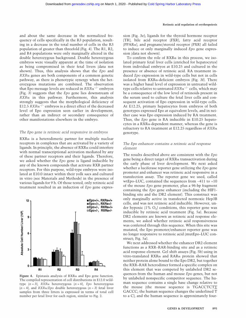
and about the same decrease in the normalized fre-quency of cells specifically in the R3 population, result-ing in a decrease in the total number of cells in the R3population of greater than threefold (Fig. 4). The R1, R2,and R4 populations were only marginally altered in thedouble heterozygous background. Double heterozygousembryos were visually apparent at the time of isolationas being compromised in their fetal livers (data notshown). Thus, this analysis shows that the Epo andRXR� genes are both components of a common geneticpathway, as there is phenotypic synergy when the het-erozygous mutations are combined. The observationthat Epo message levels are reduced in RXR�−/− embryos(Fig. 3) suggests that the Epo gene lies downstream ofRXR� in this pathway. Furthermore, this analysisstrongly suggests that the morphological deficiency ofE12.5 RXR�−/− embryos is a direct effect of the decreasedlevel of Epo expression seen earlier in development,rather than an indirect or secondary consequence ofother manifestations elsewhere in the embryo.
The Epo gene is retinoic acid responsive in embryos
RXR� is a heterodimeric partner for multiple nuclearreceptors in complexes that are activated by a variety ofligands. In principle, the absence of RXR� could interferewith normal transcriptional activation mediated by anyof these partner receptors and their ligands. Therefore,we asked whether the Epo gene is ligand inducible byany of the known compounds that activate RXR partnerreceptors. For this purpose, wild-type embryos were iso-lated at E10.0 intact within their yolk sacs and culturedin vitro (see Materials and Methods) in the presence ofvarious ligands for 9 h. Of those tested, only retinoic acidtreatment resulted in an induction of Epo gene expres-
sion (Fig. 3e); ligands for the thyroid hormone receptor(TR), bile acid receptor (FXR), fatty acid receptor(PPAR�), and pregnane/steroid receptor (PXR) all failedto induce or only marginally induced Epo gene expres-sion (data not shown).To confirm the role of RXR� in this process, we iso-
lated primary fetal liver cells (enriched for hepatocytes)from individual embryos at E10.25 and cultured in thepresence or absence of retinoic acid. RA treatment in-duced Epo expression in wild-type cells but not in cellsisolated from RXR�-deficient embryos (Fig. 3f). Therewas a higher basal level of expression in untreated wild-type cells relative to untreated RXR�−/− cells, whichmaybe a consequence of the low level of retinoids present inthe serum used to culture the fetal liver cells and con-sequent activation of Epo expression in wild-type cells.At E12.25, primary hepatocytes from embryos of bothgenotypes expressed Epo at equivalent levels, and in nei-ther case was Epo expression induced by RA treatment.Thus, the Epo gene is RA inducible in E10.25 hepato-cytes in a RXR�-dependent manner, whereas the gene isrefractory to RA treatment at E12.25 regardless of RXR�genotype.
The Epo enhancer contains a retinoic acid responseelement
The results described above are consistent with the Epogene being a direct target of RXR� transactivation duringthe early phase of liver development. We next askedwhether a luciferase reporter gene utilizing the Epo genepromoter and enhancer was retinoic acid responsive in atransfection assay. The reporter gene we used, calledwtEpo–LUC, contained the sequences from −411 to +10of the mouse Epo gene promoter, plus a 96-bp fragmentcontaining the Epo gene enhancer (including the HIF1-binding site and the DR2 element). This construct wasonly marginally active in transfected normoxic Hep3Bcells, and was not retinoic acid inducible. However, un-der hypoxic (1% O2) conditions, this reporter gene wasinducible by retinoic acid treatment (Fig. 5a). BecauseDR2 elements are known as retinoic acid response ele-ments, we asked whether retinoic acid responsivenesswas conferred through this sequence. When this site wasmutated, the Epo promoter/enhancer reporter gene wasno longer responsive to retinoic acid (mutEpo–LUC con-struct; Fig. 5a).We next addressed whether the enhancer DR2 element
functions as a RXR–RAR-binding site and as a retinoicacid response element. Gel shift assays (Fig. 5b) using invitro-translated RXR� and RAR� protein showed thatneither protein alone bound to the Epo DR2, but togetherthe RXR–RAR heterodimer formed a specific complex onthis element that was competed by unlabeled DR2 se-quences from the human and mouse Epo genes, but notby unlabeled nonspecific competitor sequence. The hu-man sequence contains a single base change relative tothe mouse (the mouse sequence is TGACCTCTTGACCC; the human sequence changes the underlined Tto a C), and the human sequence is approximately four-
Figure 4. Epistasis analysis of RXR� and Epo gene function.The compiled representation of cell distributions in E12.0 wild-type (n = 3), RXR� heterozygous (n = 6), Epo heterozygous(n = 4), and RXR�/Epo double heterozygous (n = 3) fetal liversamples from three litters is expressed in terms of total cellnumber per fetal liver for each region, similar to Fig. 2.
Retinoic acid regulation of erythropoiesis
GENES & DEVELOPMENT 895
Cold Spring Harbor Laboratory Press on March 1, 2020 - Published by genesdev.cshlp.orgDownloaded from

fold lower in affinity for RXR–RAR binding, consistentwith the known sequence requirements for DNA recog-nition by retinoic acid receptors.A reporter gene (4xDR2-TK-LUC) was prepared by in-
sertion of four copies of the mouse Epo DR2 elementinto a minimal thymidine kinase promoter–luciferaseconstruct. The element was multimerized to maximizeresponsiveness, a well-known requirement when theminimal TK promoter is used in transfection studies; wealso assayed a reporter construct containing a single DR2element with comparable, although less dramatic, re-sults (data not shown). In CV-1 cells, which express a
very low level of endogenous nuclear receptors, this re-porter gene was not basally active and was not retinoicacid inducible. However, when RXR and RAR expressionvectors were cotransfected into these cells, the reportergene was dramatically induced by retinoic acid treat-ment (Fig. 6a). Thus, as shown by gel shift and transfec-tion assays, the DR2 element in the Epo gene 3� en-hancer is a bonafide retinoic acid response element.
Competitive regulation of the Epo enhancer DR2element by RXR–RAR and HNF4
In Hep3B cells, the chromosomal Epo gene is inducibleby hypoxia (unlike in CV-1 cells), and therefore repre-sents a more authentic context for examination of Epogene expression. Hep3B cells express endogenous RXRand RAR (see below). In our initial transfection assays,the 4xDR2-TK-LUC reporter gene was only modestly in-duced by RA treatment, although we found that wecould increase responsiveness by increasing the amountof cotransfected RXR and RAR expression vector (Fig.6b). We suspected that the known high level in Hep3Bcells of HNF4, which is already established to bind as ahomodimer to the Epo enhancer DR2 element (Galson etal. 1995), might interfere with transactivation by the ret-inoic acid receptor RXR–RAR heterodimer. To confirmthis model, we made use of a dominant-negative HNF4construct (dnHNF4), in which a mutation in the DNAbinding domain renders the encoded protein fully able todimerize with wild-type HNF4, but the dimeric complexis unable to bind DNA (Taylor et al. 1996). Introductionof dnHNF4 alone allowed significant (ninefold) RA-in-ducible transactivation of the reporter gene through en-dogenous RA receptors, and cotransfection of dnHNF4with RXR and RAR expression vectors resulted in syn-ergistic activation of reporter gene activity (Fig. 6c).Thus, in Hep3B cells, the Epo gene enhancer DR2 ele-ment is transactivated by retinoic acid receptors, in amanner that is inhibited by HNF4. We obtained similarresults in primary E18.5 fetal hepatocytes (Fig. 6d).In CV-1 cells, introduction of dnHNF4 did not alter
expression of the reporter gene (data not shown), consis-tent with the absence of endogenous HNF4 expression inthese cells. However, by cotransfecting wild-type HNF4expression vector into CV-1 cells, we were able to de-crease the ability of RXR–RAR to transactivate throughthe Epo enhancer DR2 element (Fig. 6e). Thus, the ac-tivity of this reporter gene is titratable by adjusting therelative levels of HNF4 and RXR–RAR.Finally, we asked whether retinoic acid responsiveness
of the Epo gene promoter/enhancer was also inhibited byHNF4. In transfected hypoxic Hep3B cells, the presenceof dnHNF4 allowed a fivefold responsiveness to RA ofthe wtEpo–LUC reporter via endogenous receptors, andcotransfection of RXR–RAR and dnHNF4 together re-sulted in an even greater response (Fig. 6f). Collectively,these results indicate that HNF4 and RXR–RAR are mu-tually competitive for recognition and occupancy of theEpo enhancer DR2 element.
Figure 5. The Epo gene is retinoic acid responsive through theenhancer DR2 element. (a) Transfection analysis. The wild-typeEpo promoter/enhancer reporter gene construct (wtEpo-LUC) ora variant in which the DR2 element was mutated (mutEpo-LUC) were cotransfected with RXR and RAR expression con-structs into Hep3B cells under normoxic conditions or underhypoxic conditions (1% oxygen) in the absence or presence of10−6 M all trans RA, and cultured for 24 h. Luciferase activitywas normalized to �-galactosidase activity. (b) Gel shift analy-sis. In vitro-translated human RXR and RAR protein was usedunder standard conditions with labeled probe representing thehuman Epo gene DR2 sequence. (Lane P) is probe alone; (lane 1)mock in vitro translation only; (lane 2) RXR� only; (lane 3)RAR� only; (lanes 4–10) RXR� plus RAR�. Cold competitoroligonucleotides included were a 10× and 50× molar excess ofthe mouse Epo enhancer DR2 (lanes 5,6), 10× and 50× excess ofthe human Epo enhancer DR2 (lanes 7,8), or 10× and 50× excessof an irrelevant sequence (a loxP sequence) of the same length(lanes 9,10). The arrow indicates the bound RXR–RAR complex.
Makita et al.
896 GENES & DEVELOPMENT
Cold Spring Harbor Laboratory Press on March 1, 2020 - Published by genesdev.cshlp.orgDownloaded from

Discussion
The period between E9.5 and E12.5 in the mouse embryois a particularly critical time in hematopoiesis, as this iswhen the embryo transitions from yolk sac (primitive) tofetal liver (definitive) hematopoiesis. We have found thatthe initial phase of erythropoiesis in the fetal liver isacutely dependent upon RXR� function. In RXR�−/− em-bryos prior to E12.5, erythroid differentiation in the fetalliver is severely compromised, although this deficiencydoes not cause any apparent consequence in other partsof the embryo as there is adequate provision of primitivered blood cells from the yolk sac (this process appears tobe unaffected by RXR� gene mutation). As shown in thisstudy by genetic and molecular approaches, the fetalliver phenotype prior to E12.5 is accounted for by a de-crease in the level of Epo expression. Thereafter, controlof hematopoiesis becomes independent of RXR� func-tion; Epo expression is normal in these later embryos,and erythropoiesis is restored to normal as well.Based on results described in this study, we propose
that the initial expression of Epo in the fetal liver, fromE9.5 through E11.5, is under retinoic acid control. Thetransduction of this retinoic acid signal requires RXR�,and occurs by occupancy of and transactivation throughthe DR2 element in the Epo gene 3� enhancer. Mostprobably, the active complex is a heterodimer of RXR�with any of the RARs, as all three RAR genes are ex-pressed in the early fetal liver (Dolle et al. 1990), andthere is no known erythropoietic phenotype in anysingle or double RAR gene mutations (Mendelsohn et al.1994), although it is possible that the phenotype de-scribed in this study could have been overlooked. In theabsence of RXR�, Epo gene expression is reduced sub-stantially, resulting in a deficiency in the expansion anddifferentiation of erythroid progenitors within the fetalliver. Starting around E11.5 and continuing for the re-mainder of fetal liver erythropoiesis, HNF4 activity sup-plants retinoic acid receptor function in controlling ex-pression of the Epo gene, thereby restoring normal levelsof Epo gene expression and promoting the recovery of thetransient liver defect in RXR�−/− embryos.
Figure 6. HNF4 is competitive with RXR–RAR for occupancy of the Epo enhancer DR2 element. The indicated reporter genes weretransfected into CV-1 cells (a,e), Hep3B cells (b,c,f), or primary E18.5 fetal hepatocytes (d) under normoxic conditions (a–e) or hypoxicconditions (f) in the presence or absence of 10−6 M all trans RA, with cotransfected receptor expression plasmids as indicated. Numbersindicate micrograms of transfected DNA.
Retinoic acid regulation of erythropoiesis
GENES & DEVELOPMENT 897
Cold Spring Harbor Laboratory Press on March 1, 2020 - Published by genesdev.cshlp.orgDownloaded from

Our model is bolstered by genetic analysis of HNF4function. Conventional HNF4−/− embryos die at gastru-lation with a deficiency in extraembryonic endoderm(Chen et al. 1994). Very recently (Li et al. 2000), thisphenotype has been rescued through E12.0 by use of tet-raploid aggregation procedures, in which extraembryonictissues are wild type in function and the embryo itself isHNF4-deficient. HNF4-deficient embryos at E12.0 aresubstantially compromised in fetal liver Epo expression,demonstrating that HNF4 is a required regulator of Epogene expression at least from E12.0 onward. However,the morphology and histology of HNF4−/− fetal liver atE12.0 is normal, with apparently normal erythropoieticdifferentiation. Epo expression in earlier stage HNF4−/−
embryos was not examined, but we predict that Epo ex-pression must be normal in earlier embryos (i.e., atE10.5), on the basis of the lack of histological liver defi-ciencies in HNF4−/− embryos at E12.0 and the demon-strated necessity (Wu et al. 1995) of normal Epo functionfor normal liver histogenesis through E12.5. Thus, theseobservations indicate that HNF4 is not required forerythropoiesis during the early phase of liver develop-ment, but is required for normal Epo expression in laterembryos, and support our model that RXR–RAR is thecritical regulator of erythropoiesis during the initial pe-riod of erythropoiesis in the fetal liver.We have presented evidence in this study that HNF4
and RXR–RAR can compete directly for occupancy of theEpo enhancer DR2 element. These results resolve a long-standing puzzling observation that the Epo enhancerDR2 element, although being nearly canonical in se-quence to other retinoic acid response elements, is notretinoic-acid responsive in Hep3B cells (Blanchard et al.1992). Thus, in Hep3B cells and in late-gestation hepa-tocytes, HNF4 is present in stochiometric excess relativeto RXR–RAR, essentially preventing transactivation byretinoic acid. These results also explain why the chro-mosomal Epo gene itself is not RA inducible in the liversof embryos older than E12.0 or in postnatal animals (datanot shown), although it is clearly so in younger embryos(Fig. 3).To explain the observation that expression of the Epo
gene transitions from RXR–RAR control to HNF4 con-trol around E11.5, we favor a model in which Epo ex-pression occurs in fetal hepatocytes. Accordingly, but forreasons we have not yet defined, RXR–RAR is dominantin activity over HNF4 prior to E11.5, but with HNF4being dominant over RXR–RAR at times thereafter.HNF4 protein may be less abundant in the earlier period,its activity may be suppressed by modification or by li-gand, or the context of the Epo promoter/enhancer priorto E11.5 may favor the activity of RXR–RAR even in thepresence of excess HNF4 (possibly through some type ofinteraction with HIF1, which binds to the hypoxic re-sponse element immediately adjacent to the DR2 ele-ment, or through differential interaction with transcrip-tional coactivators).There are two alternative models that bear consider-
ation. First, RXR� function may occur outside of thefetal liver, and in a secondary or indirect manner affect
Epo expression in the liver. Two feasible outside candi-date tissues might be the heart and placenta, as there aredefects in both tissues in RXR�−/− embryos, and the pla-centa in particular has been recently suggested to influ-ence definitive erythropoiesis in the fetal liver (Ihle2000). However, the placental (Sapin et al. 1997) and car-diac (Dyson et al. 1995) defects of RXR�−/− embryos areonly first apparent at E12.5 and progressively worsenwith advancing age, unlike the erythropoietic phenotypeand the reduction in Epo expression which are most se-vere prior to E12.5 and resolve thereafter. Furthermore,there are no known defects of any type in RXR� hetero-zygous embryos that would account in an indirect man-ner for the erythropoietic phenotype of RXR�/Epodouble heterozygous embryos (Fig. 4). As a second alter-native, Epo expression prior to E11.5 may occur in a non-hepatocyte cell population of the fetal liver under RXR�control, and after E11.5 in hepatocytes under HNF4 con-trol. We can exclude the hematopoietic and endotheliallineages, as chimeric embryos comprised of RXR�−/−
cells and Flk1−/− cells (the latter are unable to contributeto these lineages; Shalaby et al. 1997) have no erythro-poietic phenotype (C. Tran and H.M. Sucov, unpubl.),although we cannot yet formally rule out the possibilitythat RXR� might function in another nonhepatocyte lin-eage.It is of interest to speculate as to the developmental
logic that causes the Epo gene to be regulated transientlyby retinoic acid receptors before becoming dominated byHNF4 activity. Because genetic analysis (Li et al. 2000)shows that HNF4 does not control Epo expression priorto E11.5, the availability of an alternative mechanism(i.e., retinoic acid receptors) through which to activateEpo gene expression from E9.5–E11.5 has two potentialadaptive benefits. First, this mechanism allows the ini-tiation of erythropoiesis in the fetal liver ∼2 d (in themouse embryo; possibly longer in human embryos) ear-lier than otherwise, which provides an earlier contribu-tion of mature red blood cells in peripheral circulation asthe yolk sac-derived primitive erythrocytes are dying.Second, in addition to promoting erythropoietic differ-entiation, Epo also stimulates erythropoietic progenitorcell proliferation, and this may be of even greater benefitin establishing definitive hematopoiesis within the fetalliver prior to the time (around E12.5–E13.5) when defini-tive erythropoiesis is required for embryo viability.Interestingly, occupancy of the DR2 element by HNF4
in the context of either the Epo promoter or the TK pro-moter does not lead to a substantial transcriptional re-sponse. That is, there was a fairly low basal level of ac-tivity of the DR2-containing reporter genes in Hep3Bcells; this was also true in CV-1 cells transfected withHNF4 expression plasmids. HNF4 is constitutively ac-tive but is a relatively weak transcriptional activator,particularly in certain promoter and cellular contexts(Jiang et al. 1995; Sladek et al. 1999). This may explainwhy Epo gene regulation during the E9.5–E11.5 periodhas evolved to depend on the transcriptionally more po-tent RXR–RAR heterodimer, assuming that a high levelof Epo expression is beneficial (see above). One clear con-
Makita et al.
898 GENES & DEVELOPMENT
Cold Spring Harbor Laboratory Press on March 1, 2020 - Published by genesdev.cshlp.orgDownloaded from

sequence of the relatively weak transcriptional activityof HNF4 is that Epo expression in the later stage liver isnot constitutive, as would occur if HNF4 was able tobasally activate expression, but is hypoxic-dependent viathe activity of HIF1. The transition from RXR–RAR con-trol to HNF4 control may therefore serve to switch regu-lation of Epo expression from a paracrine mode con-trolled by local retinoic acid production to an environ-mentally responsive mode controlled solely by oxygentension, as is found throughout the remainder of fetaland postnatal life.
Materials and methods
Histology
Embryos were isolated at appropriate gestational stages andgenotyped retrospectively by analysis of yolk sac DNA. For he-matoxylin/eosin staining, embryos were fixed in 10% phos-phate-buffered formalin, paraffin embedded, sectioned at 5-µthickness, and stained and processed by standard procedures.For Richardson’s stained samples, whole embryos or isolatedliver tissue were fixed in phosphate buffer containing 2% para-formaldehyde and 2.5% glutaraldehyde, followed by 1% os-mium tetroxide in 0.1 M cacodylate buffer, and then embeddedin Epon-812. The 1-µ sections were cut with a glass knife andstained in a solution containing 0.5% methylene blue, 0.5%azure II, and 0.5% borax.
Flow cytometry analysis
Fetal liver tissue was isolated by dissection from individual em-bryos and dissociated by trituration in Hank’s balanced salt so-lution containing 0.02% EDTA, 0.2% BSA, and 0.03% sodiumazide on ice. A total of 0.05–0.2 million fetal liver cells fromeach sample were stained with monoclonal antibodies for sur-face expression of c-Kit (2B8) and TER-119 (Ly-76) (both fromPharmingen), after blocking Fc�II/III receptors by use of super-natant from the 2.4G2 hybridoma. Two-color analyses were per-formed on individual fetal liver samples by flow cytometry in aBecton Dickinson FACScalibur. A total of 50,000 events werecollected per sample, and analyses performed after excludingdead cells. Because the actual developmental stage varied by upto half a day between litters at the same nominal stage, wecalculated within each litter the fold increase or decrease ineach category for mutant embryos relative to wild-type plusheterozygote embryos, and then combined these fold changesfor all litters to generate a composite depiction of the normaland mutant phenotypes.
Colony outgrowth assay
E12.5 fetal liver tissue was dissociated by trituration, and analiquot counted to determine nucleated cell number. Cells weremixed with Methocult 3430 (Stem Cell Technologies, Vancou-ver, Canada) and incubated 7 d, then stained with benzidine andscored by the criteria of containing 20 or more cells, of which atleast 15% were benzidine positive.
Quantitative RT–PCR
Total RNA was extracted from individual fetal liver tissue byguanidinium isothiocyanate extraction. A total of 1 µg of totalRNA was reverse transcribed in a 20-µL reaction containing 2
µL (100–500ng) of Random Decamer Primer (Ambion), 100 ng ofEpo gene-specific primer (5�-GAGCAAGTTCGTCGGTCCA-3�), 0.5 mM dNTPs, and 200 units of M-MuLV Reverse Tran-scriptase (GIBCO BRL) in First Strand buffer (50 mM Tris-HClat pH 8.3, 75 mM KCl, 3 mM MgCl2, and 5 mM DTT). The RTreaction was diluted with water to 80 µL, and 1 µL was used forPCR. To initially determine the linear range of Epo amplifica-tion, PCR was performed with multiple samples in a 30-µLreaction containing 100 ng of each Epo primer (Zimmermannand Rich 1997), 5 µCi of [�-32P]dCTP, and 2.5 units of Taq DNAPolymerase (GIBCO) in PCR buffer [20% DMSO, 134 mM Trisat pH 8.8, 33 mM (NH4)2SO4, 20 mM �-mercaptoethanol, 6 mMMgCl2, and 1 mM dNTPs] with the following parameters: 1 minat 94°C, 2 min at 57°C, and 2 min at 72°C. The amplificationprofile was examined at 3-cycle intervals from 14 to 44 cycles.PCR products were separated by nondenaturing 4% polyacryl-amide gel electrophoresis in 1 × TBE at 4°C, and signal intensitywas analyzed by PhosphorImaging (Molecular Dynamics). Forquantitative analysis of Epo transcripts level, PCR was per-formed with the addition of 0.4 µM of 18S ribosomal RNA prim-ers and varying amounts of competimer mixture (Ambion) as aninternal control. The varying ratios of 18S primers ensures thata linear range is achieved for this product as well, and allowscomparison of Epo mRNA abundance between samples by mea-surement of the intensity of the Epo and 18S products at any 18Sprimer/competimer ratio in which linear amplification of bothproducts occurs. Amplification of other genes was done exactlyas above, except for substitution of primers for HNF3�
(GGCCTACTCCTCTGTCCCTGTCAG and ATGCCAGCCACAGCACCGGGACTC), albumin (CCCCACTTAGCCTCTGGCAAAAT and AGACTCATCGGCAACACACGTCT), or c-kit (ACAGGAGCAGAGCAAAGGTG and CGACCACAAAGCCAATGAGC). For in vitro whole embryo culture, wild-typeembryos were isolated at E10.0 intact within their yolk sacs andcultured in DMEM with 50% FCS in the presence of 10−6 M alltrans retinoic acid or solvent alone with gentle agitation on arocker platform in a tissue culture incubator. Other compoundstested and their final concentrations included thyroid hormone(T3; 10−8 M), Wy14,643 (5 × 10⁻⁶ M), chenodeoxycholic acid(10−4 M), and pregnenalone 16�-carbonitrile (PCN; 10−5 M). Af-ter 9 h, fetal liver tissue was isolated from these embryos andprocessed as above. For primary cell culture, fetal liver tissuewas dissociated from individual embryos by trituration, andplated in the presence of solvent only or 10−6 M all trans reti-noic acid in replicate wells of rat tail collagen-coated dishes.After 4 h of incubation, nonadherent cells were washed off andadherent cells collected for analysis. These conditions enrich forhepatocytes (HNF3�-expressing) and decrease representation ofhematopoietic cells (GATA1-expressing).
Electrophoretic mobility shift assay
Double-stranded oligonucleotide probe containing the humanor mouse Epo DR2 element was labeled with [�-32P]dCTP byKlenow fill-in reaction. Preincubation was performed for 20minon ice in a 19-µL reaction containing 150 ng of poly [d(I-C)] plus60 ng of nonspecific single-strand oligomer in a binding buffer(75 mM KCl, 7.5% glycerol, 20 mM HEPES, 0.1% NP40, 2 mMDTT) in the presence or absence of 3 µL each of in vitro-trans-lated hRXR� and hRAR�, followed by addition of 0.8 ng oflabeled probe and 20 min of further incubation on ice. DNA–protein complexes (20 µL) were separated by 5% polyacrylamidegel electrophoresis in 0.5× TBE at room temperature. Competi-tion assays were performed in the presence of 8 ng (10×) and 40ng (50×) of unlabeled human or mouse Epo DR2 sequences or anonspecific (loxP) sequence.
Retinoic acid regulation of erythropoiesis
GENES & DEVELOPMENT 899
Cold Spring Harbor Laboratory Press on March 1, 2020 - Published by genesdev.cshlp.orgDownloaded from

DNA constructs
The wild-type Epo promoter enhancer reporter construct(wtEpo-LUC) was made by insertion of a 420-bp XbaI–SmaI frag-ment (positions 1–420 of GenBank M12482) representing thesequences between −410 and +10 relative to the transcriptionstart site of the mouse Epo gene (Shoemaker and Mitsock 1986)into a luciferase vector lacking other promoter sequence. Theoriginal plasmid from which this fragment was obtained wasdescribed in Wu et al. (1995). A 96-bp fragment (positions 399–494 of GenBank L13456) containing the complete enhancer el-ement as functionally defined previously (Pugh et al. 1991) wasprepared by PCR amplification, subcloned and validated by se-quencing, and inserted as a single copy 5� of the promoter frag-ment. To specifically mutate the DR2 element, an enhancerfragment of the same length was prepared by PCR amplificationbut using a primer with an altered DR2 sequence, which wassequenced to confirm correct amplification and inserted 5� ofthe Epo promoter fragment as above. The wild-type mouse DR2element is TGACCTCTTGACCT; the mutant sequence isTTCATTCTGGCTAA. To make 4xDR2-TK-LUC, a 39-bp frag-ment containing the wild-type DR2 sequence was inserted as afour- copy head-to-tail multimer into the HindIII site of a pa-rental TK-LUC plasmid.
Transient transfection assays
CV-1 cells were cultured in DMEMwith 10% FBS and plated on35-mm tissue culture dishes at 60% confluency. A total of 3 µgof reporter plasmid plus 0.25 µg of CMX–�-gal were cotrans-fected by calcium phosphate precipitation in the absence orpresence of cotransfected CMX–hRXR�, CMX–hRAR�, orpSG5-rHNF4�1 expression vectors. The total amount of CMXpromoter and total plasmid (5 µg) was adjusted by addition ofCMX–EGFP and pGEM4 plasmids, respectively. After a 16-hincubation, the transfection medium was replaced with freshmedium with or without 10−6 M all-trans retinoic acid (Sigma),and cells were incubated for 24 h before harvest. Hep3B cellswere cultured in DMEM containing 10% FBS and plated into35-mm tissue culture dishes at 80% confluency, and were trans-fected using Lipofectin (GIBCO BRL). Lipofection was per-formed for 14 h, followed by replacement with medium with orwithout 10−6 M of all-trans RA, and cells were cultured another24 h before harvest. For normoxic conditions, cells were cul-tured in standard incubators. For hypoxic conditions, followingtransfection and replacement of medium, cells were transferredto a chamber that was flushed with 1%O2/5%CO2/94%N2,then sealed and incubated for 24 h. Hypoxic medium was firstsparged before use, and CoCl2 was added at a concentration of10−4 M. For all reporter gene assays, cells were rinsed with 1×PBS and scraped with 250 µL of 1× PBS. Cell suspensions werecentrifuged and pellets were lysed in 50–100 µL of lysis buffer(TROPIX) by freeze-thaw. A total of 10 µL of cell lysate wasassayed with the Luciferase Assay System (Promega), and withthe Galacto-Light Plus (TROPIX) assay system. Primary hepa-tocytes were prepared by trituration of fetal liver tissue, platedin rat tail type IV collagen-coated dishes in DMEM with 10%FCS, allowed to adhere for 4 h, washed to remove nonadherenthematopoietic cells, and then transfected by calcium phosphateas above.
Acknowledgments
We thank Todd Leff for provision of CMV-dnHNF4, Ron Evansfor provision of CMX–hRXR� and CMX–hRAR�, and FrancieSladek for provision of pSG5–rHNF4�1. This work was sup-
ported in its early phase by a pilot/feasibility project grant (toH.M.S.) as a component of the University of Southern CaliforniaResearch Center for Liver Diseases (PHS grant no. DK48522).G.H.-H. was supported by the Stowers Institute for MedicalResearch. H.W. is an Assistant Investigator of the HowardHughes Medical Institute. T.C. was supported by a predoctoralfellowship from the American Heart Association.The publication costs of this article were defrayed in part by
payment of page charges. This article must therefore be herebymarked “advertisement” in accordance with 18 USC section1734 solely to indicate this fact.
Note added in proof
A recent report has described retinoic acid induction of chro-mosomal Epo gene expression in P19 cells (Kambe, T., Tada-Kambe, J., Kuge, Y., Yamaguchi-Iwai, Y., Nagao, M., and Sasaki,R. 2000. Blood 96: 3265–3271).
References
Avery, L. and Wasserman, S. 1992. Ordering gene function: Theinterpretation of epistasis in regulatory hierarchies. TrendsGenet. 8: 312–316.
Blanchard, K.L., Acquaviva, A.M., Galson, D.L., and Bunn, H.F.1992. Hypoxic induction of the human erythropoietin gene:Cooperation between the promoter and enhancer, each ofwhich contains steroid receptor response elements. Mol.Cell. Biol. 12: 5373–5385.
Bunn, H.F., Gu, J., Huang, L.E., Park, J.W., and Zhu, H. 1998.Erythropoietin: A model system for studying oxygen-depen-dent gene regulation. J. Exp. Biol. 201: 1197–1201.
Chen, W.S., Manova, K., Weinstein, D.C., Duncan, S.A., Plump,A.S., Prezioso, V.R., Bachvarova, R.F., and Darnell, J.E. 1994.Disruption of the HNF-4 gene, expressed in visceral endo-derm, leads to cell death in embryonic ectoderm and im-paired gastrulation of mouse embryos. Genes & Dev.8: 2466–2477.
Dolle, P., Ruberte, E., Leroy, P., Morriss, K.G., and Chambon, P.1990. Retinoic acid receptors and cellular retinoid bindingproteins. I. A systematic study of their differential pattern oftranscription during mouse organogenesis. Development110: 1133–1151.
Dyson, E., Sucov, H.M., Kubalak, S.W., Schmid-Schonbein,G.W., DeLano, F.A., Evans, R.M., Ross, J., and Chien, K.R.1995. Atrial-like phenotype is associated with embryonicventricular failure in RXR�−/− mice. Proc. Natl. Acad. Sci.92: 7386–7390.
Evans, R.M. 1988. The steroid and thyroid hormone receptorsuperfamily. Science 240: 889–895.
Galson, D.L., Tsuchiya, T., Tendler, D.S., Huang, L.E., Ren, Y.,Ogura, T., and Bunn, H.F. 1995. The orphan receptor hepaticnuclear factor 4 functions as a transcriptional activator fortissue-specific and hypoxia-specific erythropoietin gene ex-pression and is antagonized by EAR3/COUP-TF1. Mol. Cell.Biol. 15: 2135–2144.
Goldberg, M.A., Glass, G.A., Cunningham, J.M., and Bunn, H.F.1987. The regulated expression of erythropoietin by two hu-man hepatoma cell lines. Proc. Natl. Acad. Sci.84: 7972–7976.
Houssaint, E. 1981. Differentiation of the mouse hepatic pri-mordium. II. Extrinsic origin of the haemopoietic cell line.Cell Differ. 10: 243–252.
Ihle, J.N. 2000. The challenge of translating knockout pheno-
Makita et al.
900 GENES & DEVELOPMENT
Cold Spring Harbor Laboratory Press on March 1, 2020 - Published by genesdev.cshlp.orgDownloaded from

types into gene function. Cell 102: 131–134.Ikuta, K., Kina, T., MacNeil, I., Uchida, N., Peault, B., Chien,
Y.H., and Weissman, I.L. 1990. A developmental switch inthymic lymphocyte maturation potential occurs at the levelof hematopoietic stem cells. Cell 62: 863–874.
Jiang, G., Nepomuceno, L., Hopkins, K., and Sladek, F.M. 1995.Exclusive homodimerization of the orphan receptor hepato-cyte nuclear factor 4 defines a new subclass of nuclear re-ceptors. Mol. Cell. Biol. 15: 5131–5143.
Kastner, P., Grondona, J.M., Mark, M., Gansmuller, A., LeMeur,M., Decimo, D., Vonesch, J.L., Dolle, P., and Chambon, P.1994. Genetic analysis of RXR� developmental function:Convergence of RXR and RAR signaling pathways in heartand eye morphogenesis. Cell 78: 987–1003.
Kina, T., Ikuta, K., Takayama, E., Wada, K., Majumdar, A.S.,Weissman, I.L., and Katsura, Y. 2000. The monoclonal anti-body TER-119 recognizes a molecule associated with glyco-phorin A and specifically marks the late stages of murineerythroid lineage. Br. J. Haematol. 109: 280–287.
Koury, S.T., Bondurant, M.C., Koury, M.J., and Semenza, G.L.1991. Localization of cells producing erythropoietin in mu-rine liver by in situ hybridization. Blood 77: 2497–2503.
Li, J., Ning, G., and Duncan, S.A. 2000. Mammalian hepatocytedifferentiation requires the transcription factor HNF-4�.Genes & Dev. 14: 464–474.
Mendelsohn, C., Lohnes, D., Decimo, D., Lufkin, T., LeMur, M.,Chambon, P., and Mark, M. 1994. Function of the retinoicacid receptors (RARs) during development. (II) Multiple ab-normalities at various stages of organogenesis in RAR doublemutants. Development 120: 2749–2771.
Mucenski, M.L., McLain, K., Kier, A.B., Swerdlow, S.H.,Schreiner, C.M., Miller, T.A., Pietryga, D.W., Scott, W.J.,and Potter, S.S. 1991. A functional c-myb gene is required fornormal murine fetal hepatic hematopoiesis. Cell 65: 677–689.
Neubauer, H., Cumano, A., Muller, M., Wu, H., Huffstadt, U.,and Pfeffer, K. 1998. Jak2 deficiency defines an essential de-velopmental checkpoint in definitive hematopoiesis. Cell93: 397–409.
Palis, J., Robertson, S., Kennedy, M., Wall, C., and Keller, G.1999. Development of erythroid and myeloid progenitors inthe yolk sac and embryo proper of the mouse. Development126: 5073–5084.
Parganas, E., Wang, D., Stravopodis, D., Topham, D.J., Marine,J.C., Teglund, S., Vanin, E.F., Bodner, S., Colamonici, O.R.,van Deursen, J.M., et al. 1998. Jak2 is essential for signalingthrough a variety of cytokine receptors. Cell 93: 385–395.
Pugh, C.W., Tan, C.C., Jones, R.W., and Ratcliffe, P.J. 1991.Functional analysis of an oxygen-regulated transcriptionalenhancer lying 3� to the mouse erythropoietin gene. Proc.Natl. Acad. Sci. 88: 10553–10557.
Sapin, V., Dolle, P., Hindelang, C., Kastner, P., and Chambon, P.1997. Defects of the chorioallantoic placenta in mouseRXR� null fetuses. Dev. Biol. 191: 29–41.
Semenza, G.L., Nejfelt, M.K., Chi, S.M., and Antonarakis, S.E.1991. Hypoxia-inducible nuclear factors bind to an enhancerelement located 3� to the human erythropoietin gene. Proc.Natl. Acad. Sci. 88: 5680–5684.
Shalaby, F., Ho, J., Stanford, W.L., Fischer, K.D., Schuh, A.C.,Schwartz, L., Bernstein, A., and Rossant, J. 1997. A require-ment for Flk1 in primitive and definitive hematopoiesis andvasculogenesis. Cell 89: 981–990.
Shoemaker, C.B. and Mitsock, L.D. 1986. Murine erythropoi-etin gene: Cloning, expression, and human gene homology.Mol. Cell. Biol. 6: 849–858.
Sladek, F.M., Ruse, M.D., Nepomuceno, L., Huang, S.M., and
Stallcup, M.R. 1999. Modulation of transcriptional activa-tion and coactivator interaction by a splicing variation in theF domain of nuclear receptor hepatocyte nuclear factor 4�1.Mol. Cell. Biol. 19: 6509–6522.
Sucov, H.M., Dyson, E., Gumeringer, C.L., Price, J., Chien, K.R.,and Evans, R.M. 1994. RXR� mutant mice establish a ge-netic basis for vitamin A signaling in heart morphogenesis.Genes & Dev. 8: 1007–1018.
Taylor, D.G., Haubenwallner, S., and Leff, T. 1996. Character-ization of a dominant negative mutant form of the HNF-4orphan receptor. Nucleic Acids Res. 24: 2930–2935.
Trimborn, T., Gribnau, J., Grosveld, F., and Fraser, P. 1999.Mechanisms of developmental control of transcription inthe murine �- and �-globin loci. Genes & Dev. 13: 112–124.
Wu, H., Liu, X., Jaenisch, R., and Lodish, H.F. 1995. Generationof committed erythroid BFU-E and CFU-E progenitors doesnot require erythropoietin or the erythropoietin receptor.Cell 83: 59–67.
Zhu, H. and Bunn, H.F. 1999. Oxygen sensing and signaling:Impact on the regulation of physiologically important genes.Respiration Physiol. 115: 239–247.
Zimmermann, F. and Rich, I.N. 1997. Mammalian homeoboxB6 expression can be correlated with erythropoietin produc-tion sites and erythropoiesis during development, but notwith hematopoietic or nonhematopoietic stem cell popula-tions. Blood 89: 2723–2735.
Retinoic acid regulation of erythropoiesis
GENES & DEVELOPMENT 901
Cold Spring Harbor Laboratory Press on March 1, 2020 - Published by genesdev.cshlp.orgDownloaded from

10.1101/gad.871601Access the most recent version at doi: 15:2001, Genes Dev.
Takako Makita, Gabriela Hernandez-Hoyos, Tim Hung-Po Chen, et al. HNF4expression is sequentially regulated by retinoic acid receptors and A developmental transition in definitive erythropoiesis: erythropoietin
References
http://genesdev.cshlp.org/content/15/7/889.full.html#ref-list-1
This article cites 34 articles, 20 of which can be accessed free at:
License
ServiceEmail Alerting
click here.right corner of the article or
Receive free email alerts when new articles cite this article - sign up in the box at the top
Cold Spring Harbor Laboratory Press
Cold Spring Harbor Laboratory Press on March 1, 2020 - Published by genesdev.cshlp.orgDownloaded from