carbohydrate structure of erythropoietin expressed in chinese
TRANSCRIPT

THE JOURNAL OF BIOLOGICAL CHEAIIsTRY 0 1987 by The American Society for Biochernietry and Mol& Biology. h e .
Vol. 262, No. 25. Issue of September 5. pp. 12069-12076 1987 Printed in C.S.A.
Carbohydrate Structure of Erythropoietin Expressed in Chinese Hamster Ovary Cells by a Human Erythropoietin cDNA*
(Received for publication, April 20,1987)
Hiroshi Sasakis, Brian Bothner, Anne Dell$, and Minoru Fukudall From the Cancer Research Center, La J o b Cancer Research Foundation, La Jolda, California 92037 and the $Department of Biochemistry, Imperial College of Science and Technology, London, Great Britain
The proper glycosylation of erythropoietin is essen- tial for its function in vivo. Human erythropoietins were isolated from Chinese hamster ovary cells trans- fected with a human erythropoietin cDNA and from human urine. Carbohydrate chains attached to these proteins were isolated and fractionated by anion-ex- change high performance liquid chromatography (HPLC) and HPLC employing a Lichrosorb-NHz col- umn. The structures of fractionated saccharides were analyzed by fast atom bombardment-mass spectrom- etry and methylation analysis before and after treat- ment with specific exoglycosidases.
Both erythropoietins were found to contain one 0- linked oligosaccharide/mol of the proteins, and its ma- jor component was elucidated to be NeuNAca2+ 3Gal@1*3(NeuNAca24)GalNAcOH (where NeuNAc represents N-acetylneuraminic acid) in both proteins. The N-linked saccharides of recombinant erythropoi- etin were found to consist of biantennary (1.4% of the total saccharides), triantennary (lo%), triantennary with one N-acetyllactosaminyl repeat (3.5%), tetraan- tennary (31.8%), and tetraanteanary with one (32.1%), two (16.5%), or three (4.7%) N-acetyllacto- saminyl repeats. All of these saccharides are sialylated by 243-linkages. Tetraantennary with or without po- lylactosaminyl units are mainly present as d1sialosyl or trisialosyl forms, and these structures exhibit the following unique features. a243-Linked sialic acid and N-acetyllactosaminyl repeats are selectively pres- ent in the side chains attached to C-6 and C-2 of 2,6- substituted a-mannose and C-4 of 2,4-substituted a- mannose.
We have also shown that the carbohydrate moiety of urinary erythropoietin is indistinguishable from re- combinant erythropoietin except for a slight difference in sialylation, providing the evidence that recombinant erythropoietin is valuable for biological as well as clin- ical use.
Erythropoietin is a glycoprotein which stimulates prolifer- ation and differentiation of erythroid precursor cells to more mature erythrocytes (1). Erythropoietin is primarily produced
* This work was supported by Grant R01 CA33000 and the Cancer Center Support Grant CA30199 from the National Cancer Institute and a program project grant from the British Medical Research Council. The costa of publication of this article were defrayed in part by the payment of page charges. This article must therefore be hereby marked “aduertisement” in accordance with 18 U.S.C. Section 1734 solely to indicate this fact.
3 On leave of absence from the Central Research Laboratories, Chugai Pharmaceuticals Ltd., Tokyo, Japan.
W To whom correspondence should be addressed La Jolla Cancer Research Found. 10901 N. Torrey Pines Rd., La Jolla, CA 92037.
in adult kidney and fetal liver cells (2-4). Patients with chronic renal failure are anemic as a result of impaired renal function which leads to a decreased production of erythropo- ietin (5).
Thus, availability of purified erythropoietin in quantity is essential to understand molecular mechanisms of erythropo- iesis and for treatment of anemia. However, this has been hampered by the fact that only a very small amount of erythropoietin is present in starting sources, even in such cases as the urine of aplastic anemia patients (6).
In order to overcome this problem, cDNA clones for human erythropoietin have been isolated in several laboratories, and the expression of erythropoietin cDNA clones has been achieved (7-9). Furthermore, the recombinant erythropoietin has been successfully used to reverse the anemia of patients with endo-stage renal disease (10,ll). Interestingly, the eryth- ropoietin produced in Escherichia coli or yeast was inactive or very weakly active i n uiuo. On the other hand, the erythro- poietin produced in COS cells or Chinese hamster ovary cells was found to be fully active i n uiuo. In agreement with these results, it has been reported that desialylation of partially purified erythropoietin results in inactivation of erythropoie- tin activity (12-15). Thus, it is apparent that the proper glycosylation is essential for erythropoietin activity in uiuo.
These results prompted us to analyze carbohydrate struc- tures of erythropoietin produced by transfection of recombi- nant DNA into Chinese hamster ovary cells. In addition, we compared those structures with carbohydrate units present in erythropoietin purified from human urine.
EXPERIMENTAL PROCEDURES
Erythropoietin-Chinese hamster ovary cells (dihydrofolate reduc- tase-) were transfected with an expression vector which harbors the human erythropoietin cDNA as described (7). This expression vector also contains dihydrofolate reductase minigene so that stable trans- fects can be grown in the presence of methotrexate (16). Erythropo- ietin was purified from the spent medium of those cells as described (7). The purification procedure was slightly modified from that of Miyake et al. (6), and fractionation on a Vydac C, reverse-phase HPLC’ column (The Separations Group) was included (7). This erythropoietin will be called recombinant erythropoietin hereafter. Erythropoietin was also purified from the urine of aplastic anemia patients according to Miyake et al. (61, with a similar modification as for purification of recombinant erythropoietin. The erythropoietin purified from urine will be called urinary erythropoietin hereafter. These erythropoietin samples were provided by Chugai Pharmaceu- tical Co., Ltd. (Tokyo).
Isolation of N-Linked Glycopeptides and O-Linked Oligosaccharides from Erythropoietins-Glycopeptides were prepared by Pronase digestion of erythropoietin (5 mg of recombinant erythropoietin and
The abbreviations used are: HPLC, high performance liquid chro- matography; FAB-MS, fast atom bombardment-mass spectrometry, Hex, hexose; HexNAc, N-acetylhexosamine; NeuNAc, N-acetylneu- raminic acid; Lac, N-acetyllactosaminyl repeat.
12059

12060 Carbohydrate Structure of Human Recombinant Erythropoietin 1 mg of urinary erythropoietin, respectively) as described (17). The Pronase digest was applied to a small column (1.0 X 45 cm) of Sephadex G-15, which was equilibrated and eluted with water. The gylcopeptides, eluted at and near the void volume, where lyophilized and subjected to alkaline borohydride degradation, as described (17). Briefly, the glycopeptides were dissolved in 500 p1 of 0.05 M NaOH, 1 M NaBHI containing 5 mCi of NaB3H4 (9 Ci/mmol) and incubated at 45 "C for 24 h. After treatment, 1-2 ml of methanol which con- tained 1 drop of acetic acid was added to the sample and evaporated under nitrogen. The alkaline borohydride-treated sample was then applied to the same Sephadex G-15 column for desalting. The glyco- peptides and oligosaccharides, which eluted between the void volume and the salt peak, were pooled and applied to a column (1.0 X 140 cm) of Bio-Gel P-4 (200-400 mesh). The column was eluted with 0.1 M NHlHC03 at a flow rate of 6 ml/h, and each fraction contained 1 ml. Under these chromatographic conditions, N-linked glycopeptides eluted near the void volume, whereas 0-linked oligosaccharides eluted in later fractions (see Fig. 2).
Isolation of N-Linked Saccharides from Glycopeptides-The glyco- peptide fraction containing N-linked saccharides was digested with Flavobacterium meningosepticum N-glycanase (peptide-N4-(N-acetyl- 8-glucosaminy1)aspargine amidase) (18) which was purchased from Genzyme (Boston, MA). The glycopeptides from 5 mg of erythropoi- etin were dissolved in 500 p1 of 0.1 M sodium phosphate buffer, pH 8.6, containing 20 mM EDTA and 20 mM 2-mercaptoethanol and incubated with 30 m units of the N-glycanase, which corresponds to 30 units of the enzyme expressed by Genzyme, at 37 "C for 20 h. When the glycopeptides from 1 mg of urinary erythropoietin were digested with N-glycanase, the incubation mixture was scaled down to one-fifth.
The digest was desalted by Sephadex G-15 gel filtration, and the saccharides were reduced at room temperature for 30 min with 5 mCi of NaB3H4 dissolved in 200 pl of 0.01 M NaOH followed by 3 mg of NaBH4 for 2 h. After reduction, the sample was neutralized with the addition of methanol containing a small amount of acetic acid and evaporated under nitrogen. The dried sample was dissolved in water, and the supernatant obtained after centrifugation was applied to the Sephadex G-15 column for desalting. The saccharides, radioactively labeled at reducing terminals, were applied to a column (1.0 X 45 cm) of Sephadex G-50 (superfine) eluted with 0.2 M NaCl (see Fig. 2B). Each fraction contained 0.5 ml.
Fractionation of Oligosaccharides by Anion-exchange High Perform- ance Liguid Chromatography-Oligosaccharide fractions obtained after Sephadex G-50 gel filtration were subjected to anion-exchange HPLC with a Varian HPLC apparatus (Model 5000, Varian Associ- ates, Inc.). The sample was applied to a Toyo Soda TSK-DEAE column purchased from Kratos Analytical Instruments (Ramsey, NJ). The column (4.6 mm X 24 cm) was equilibrated with 25 mM potassium phosphate buffer, pH 5.0, and after eluting with the same buffer for 10 min, the elution was programed by the linear gradient to 0.4 M potassium phosphate buffer, pH 5.0, over 80 min. The flow rate was constant at 1 ml/min, and each fraction contained 0.3 ml. The elution was monitored by measuring the absorbance at 206 nm with a Beckman 163 detector. Since potassium phosphate has absorbance at 202 nm, the absorbance at 206 nm was measured under these condi- tions. The base line of the absorbance at 202 nm was increased under these conditions. In order to estimate the elution positions of mono- sialosyl, disialosyl, trisialosyl, and tetrasialosyl saccharides, IgG (bo- vine, Sigma), fetuin (19, 20), and nl-acid glycoprotein saccharides (21, 22) were prepared by hydrazinolysis and subjected to HPLC under the same conditions. The saccharides, separated by TSK- DEAE, were subjected to methylation analysis to confirm their struc- tures. Mancrl+6(Mancr1+3)Man@l+4GlcNAc@l4(Fucal-+ 6)GlcNAcOH was obtained by extensive digestion of IgG saccharide with @-glactosidase and 8-N-acetylglucosaminidase.
Fractionation of Neutral Oligosaccharides by HPLC-The sacchar- ides were desialylated by mild acid hydrolysis in 0.01 N HCl at 80 "C for 1 h. The desialylated sample was desalted by Sephadex G-15 gel filtration and evaporated. The neutral saccharides were then frac- tionated by HPLC with the same apparatus as described above. The saccharides were dissolved in acetonitrile, 10 mM potassium phos- phate buffer, pH 4.5 (65:35), and applied to a column (0.4 X 25 cm) of Lichrosorb-NHz (5-pm particle size, Merck). The column was eluted with a linear gradient to acetonitrile, 10 mM potassium phos- phate buffer, pH 4.5 (3961), over 60 min. In order to achieve this elution, the first solvent was acetonitrile, 10 mM potassium phosphate buffer, pH 4.5 (65:35, v/v), and the second solvent was 10 mM potassium phosphate buffer, pH 4.5. The ratio of these two solvents
was 1oO:O at the beginning of the elution and 6040 at the end of elution. This elution system was used because it was preferable to premix acetonitrile and the buffer before the elution started, other- wise, mixing of two solutions causes cooling down of the solvent. The flow rate was 1 ml/min, and each fraction contained 0.5 ml.
Structural Analysis of Saccharides-Structures of saccharides were analyzed essentially as described previously for other saccharides. These methods include fast atom bombardment-mass spectrometry of permethylated saccharides (17), analysis of partially 0-methylated monosaccharides after methylation and hydrolysis ("methylation analysis") (17,231, and exoglycosidase digestion combined with meth- ylation analysis (24). Methylation of saccharides and purification of methylated saccharides for FAB-MS and methylation analysis were carried out as described previously (17, 23). Before methylation of saccharides, all samples were desalted by Sephadex G-15 gel filtration eluted with water.
Glycosidase digestion of saccharides was carried out as follows. For sequential digestion by @-galactosidase and @-N-acetylglucosamini- dase, the saccharides were incubated with 5 milliunits of Charonia lampas &galactosidase in 40 pl of 0.1 M sodium citrate buffer, pH 4.3, at 37 "C for 24 h. After incubation, the incubation mixture was heated in a boiling water bath for 2 min. The sample was then incubated with 100 milliunits of beef kidney @-N-acetylglucosaminidase in 140 p l of 0.1 M sodium citrate buffer, pH 4.3. In order to inhibit a possible contaminating activity of @-galactosidase, 100 mM (final concentra- tion) galactose was added to this incubation mixture. After further incubation at 37 "C for 24 h, the mixture was heated in a boiling water bath for 2 min. The digests were then purified by Sephadex G- 50 gel filtration followed by HPLC employing a Lichrosorb-NHz column as described above. For extensive digestion by a mixture of @-galactosidase and @-N-acetylglucosaminidase, the saccharides were first incubated with C. lampas @-galactosidase and then with beef kidney @-N-acetylglucosaminidase without heat inactivation of 8- galactosidase or the addition of galactose. After total incubation at 37 "C for 48 h, enzymes were inactivated by heating in a boiling water bath for 2 min, and digested saccharides were purified by anion- exchange HPLC. @-Galactosidase from C. lumpas and @-N-acetylglu- cosaminidase from beef kidney were purchased from Sigma and Boehringer Mannheim, respectively.
Determination of Carbohydrate Composition- Sialic acid content was determined by the periodate-resorcinol reaction (25). Neutral sugars and hexosamines were determined after methanolysis in 0.5 N hydrochloric acid in anhydrous methanol at 80 "C for 4 h. Inositol was added as an internal standard. After methanolysis, the products were dried under a nitrogen stream and further in uacw under PB05 and NaOH. The dried products were trimethylsilylated with Tri-Si1 (Pierce Chemical Co.). The trimethylsilylated derivatives were then analyzed by gas-liquid chromatograph-mass spectrometry as de- scribed.* In parallel, fetuin was analyzed in order to obtain response factors.
RESULTS
Isolation of N-Linked Sacchurldes from Recornbinant Etyth- ropoietin-Erythropoietin was purified from the spent me- dium of Chinese hamster ovary cells transfected with a human erythropoietin gene and from human urine of aplastic anemic patients as described under "Experimental Procedures." The purified proteins showed a major band with M, 38,000 and a faint band with M, -80,000 (Fig. 1). The latter band is probably the dimer of erythropoietin. The carbohydrate com- position of this molecule is shown in Table I. Glycopeptides were prepared from 5 mg of recombinant erythropoietin by Pronase digestion and isolated by Sephadex G-15 gel filtra- tion. Glycopeptides (1.8 mg) were then treated with alkaline borohydride to release 0-linked oligosaccharides, and the alkaline borohydride-treated samples were applied to a col- umn of Bio-Gel P-4. As shown in Fig. 2 A , three peaks were detected in addition to a salt peak. The second (fractions 30- 35) and the third (fractions 36-40) peaks were found to
M. Fukuda, M. Lauffenburger, H. Sasaki, E. M. E. Rogers, and A. Dell (1987) J. Biol. Chem., in press.

Carbohydrate Structure of Human Recombinant Erythropoietin
A B kDa 92-
66.2- 45-
31-
2 1.5-
14.4-
FIG. 1. Autoradiogram of sodium dodecyl sulfate gel elec- trophoresis of recombinant erythropoietin (Zane A ) and uri- nary erythropoietin (lane B ) . Purified erythropoietins were iodi- nated with ’251 according to Greenwood et al. (26). The radioactively labeled proteins were then applied to sodium dodecyl sulfate-polya- crylamide gel electrophoresis (gel concentration, 15%) according to Laemmli (27), and the gel was directly autoradiographed with Kodak x-ray AR-5 film
TABLE I Carbohydrate composition of erythropoietin
Numbers are exmessed as moles/mole of ervthroDoietin.
Urinary Recombinant erythropoietin
Batch 1 Batch 2 Batch 3 Batch 4
Fuc 2.9 4.1 2.9 2.3 3.6 Man 9.2 8.7 9.4 8.2 9.1 Gal 12.9 13.8 14.1 15.5 13.0 GlcNAc 16.3 17.2 18.9 17.3 19.8 GalNAc 0.9 0.9 1.4 0.8 1.4 NeuNAc 10.4 9.5 9.7 11.8 10.8
contain 0-linked oligosaccharides (see Miniprint): whereas glycopeptides containing N-linked saccharides eluted a t frac- tions 25-29.
Fractions 25-29 were pooled and digested with N-glycanase. The digest, after reduction with NaBSH4, was subjected to Sephadex, G-50 gel filtration as shown in Fig. 2B. The sac- charides eluting between fractions 29 and 41 were subjected to further analysis.
Methylation Analysis and FAB-MS of N-Linked Sacchar- ides from Recombinant Erythropoietin-In order to determine the carbohydrate structures of N-linked saccharides, the sac- charides were subjected to FAB-MS and methylation anal- yses. As shown in Fig. 3A, FAB-MS of permethylated N- linked saccharides provided fragment ions at mlz 376 (and
~ ~~
Portions of this paper (including part of “Results,” Figs. 4.6, and 8-10, and Table I1 and IV) are presented in miniprint at the end of this paper. Miniprint is easily read with the aid of a standard magnifying glass. Full size photocopies are available from the Journal of Biological Chemistry, 9650 Rockville Pike, Bethesda, MD 20814. Request Document No. 87M-1260, cite the authors, and include a check or money order for $6.80 per set of photocopies. Full size photocopies are also included in the microfilm edition of the Journal that is available from Waverly Press.
A VO +
B vo ? ? ? + ”
12061
0 10 20 30 40 50 60
Fractlan Number FIG. 2. Bio-Gel P-4 ( A ) and Sephadex G-SO ( B ) gel filtra-
tions of glycopeptides obtained from recombinant erythropo- ietin. Glycopeptides were subjected to alkaline borohydride treat- ment in the presence of NaB3H4 and applied to a column of Bio-Gel P-4 ( A ) . The 0-linked oligosaccharides were eluted in fractions 30- 35 (0-i) and fractions 36-40 (0 - i i ) . The glycopeptides, which con- tained N-linked saccharides (N-Such.), eluted in fractions 24-29 and were digested with N-glycanase. The digest was reduced with NaB3H4 and subjected to Sephadex G-50 gel filtration ( B ) . The fractions, indicated by the horizontal arrows, were pooled and subjected to further analysis. The arrowheads indicate the elution positions of the trisialosyl saccharide from cui-acid glycoprotein (arrowhead 4) , the trisialosyl saccharide from fetuin (arrowhead 3 ) , and the asialo form of IgG saccharide (arrowhead 2).
344), 825, 1274, and 1723, which correspond to NeuNAc+, NeuNAc+Hex+HexNAc+, NeuNAc+Hex+HexNAc+ Hex+HexNAc+ and NeuNAc+Hex+HexNAc+Hex+ HexNAc+Hex+HexNAc+. These results indicate that N- linked saccharides contain N-acetyllactosaminyl repeats in the side chains. This conclusion was supported by the detec- tion of a fragment ion at mlz 1362 for Hexn.HexNAc3+ when desialylated and methylated saccharides were subjected to FAB-MS (Fig. 44). Methylation analysis of N-linked sac- charides indicates the following features. 1) Galactose is ter- minal or 3-substituted; 2) all of the N-acetylglucosamine residues except the reducing terminal residue are substituted at C-4; 3) 2-substituted mannose (0.16 mol), 2,4-substituted mannose (0.78 mol), 2,6-substituted mannose (1.05 mol), and 3,6-substituted mannose (1.0 mol) were detected (Table 11).
After desialylation, a majority of 3-substituted galactose residues were converted to terminal galactose, indicating that sialic acid is linked to galactose through an a2+3-linkage. However, 0.82 mol of 3-linked galactose, which corresponds to 17% of the total galactose derivatives, was still detected after desialylation. This amount of galactose is presumably derived from N-acetyllactosaminyl repeats. The same analysis also showed that 85% of reducing terminal N-acetylglucosa- mine is substituted with fucose at C-6, whereas the rest of the reducing terminal N-acetylglucosamine contains no fucose.
These results suggest that the N-linked saccharides of recombinant erythropoietin are mainly composed of tetraan- tennary saccharides with or without N-actyllactosaminyl re- peats.
Isolation of Asialo N-Linked Saccharides-N-Linked sac-

12062 Carbohydrate Structure of Human Recombinant Erythropoietin
A
931
I. 1723
F'Ic. 3. Fast atom bombardment-mass spectra of permethylated total N-linked saccharides from recombinant erythropoietin (A) and urinary erythropoietin (B). The positive spectra were recorded. A, fragment ions were detected at m/z 376 (and 344) for NeuNAc', 580 for NeuNAc+Hex+, 825 (and 793) for NeuNAc+Hex+HexNAc+, 1274 for NeuNAc+Hex+HexNAc+Hex+HexNAc+, and 1723 for NeuNAc+Hex+ HexNAc+Hex+HexNAc+Hex+HexNAc+. A small peak was detected also at m/z 464 for Hex+HexNAc+. B, in addition to the ions described above, a minor fragment ion was detected at m/z 913 for Hex+HexNAc+Hex+ HexNAc+.
charides, obtained from 5 mg of recombinant erythropoietin (Batch 1 in Table I), were desialylated by mild acid hydrolysis and then desalted by Sephadex G-15 gel filtration. Neutral saccharides thus obtained were fractionated by HPLC em- ploying a Lichrosorb-NH2 column. As shown in Fig. 5A, N- linked saccharides from recombinant erythropoietin provided six peaks which correspond to biantennary (peak 2), trianten- nary (peak 3), tetraantennary (peak 4), tetraantennary with one N-actyllactosaminyl repeat (peak 5, Lacl), tetraantennary with two N-acetyllactosaminyl repeats (peak 6, Lacz), and tetraantennary with three N-acetyllactosaminyl repeats (peak 7, Lac3). The identical elution profile was obtained when N- linked saccharides were desialylated by clostridial neuramin- idase. No significant amount of carbohydrate was detected in other fractions. This result indicates that most of the glyco- peptides were digested by N-glycanase since the glycopeptides would elute later than saccharides without amino acid residues in a Lichrosorb-NHz column.
Structures of Biantennary Saccharides and TTiantennary Saccharides-The elution position of the biantennary sac-
charides (peak 2) was identical to the IgG saccharide which is mainly composed of Galz - GlcNAc, . Man, - GlcNAc,. Fuc. In addition, this saccharide bound to a concanavalin A-Sepha- rose and was eluted by 20 mM methyl-a-glucoside. Those results establish that this fraction (peak 2) is a typical complex saccharide with biantennary side chains.
Methylation analysis (Table 11) of the triantennary sac- charides (peak 3) provided 0.65 mol of 2,6-substituted man- nose (3,4-di-O-methylrna) and 0.35 mol of 2,4-substi- tuted mannose (3,6-di-O-methylmannose) in addition to 1 mol each of 2- and 3,6-substituted mannose (Table 11). The saccharides were also digested by a mixture of &galactosidase and 8-N-acetylglucosaminidase to yield Manarl+6(Manal+ 3)Man@l4GlcNAc~l4(fFucal+6)GlcNAcOH, as judged by HPLC with a Lichrosorb-NH, column followed by meth- ylation analysis. These results established the structures of the triantennary saccharides, as shown in Table III.
In order to elucidate which of the outer a-mannosyl residues is disubstituted at C-2 and C-4, the N-linked saccharides (fractions 29-41 in Fig. 2B) were subjected to periodate oxi-

Carbohydrate Structure of Human Recombinant Erythropoietin 12063
GO 4
1271
FIG. 5. HPLC of asid0 N-linked saccharidea derived from erythro- poietin. N-Linked saccharides were de- sialylated by mild acid hydrolysis, ap- plied to a 5-pm Lichrwrb-NH, column, as described under “Experimental Pro- cedures.” The effluent was monitored by measuring the absorbance at 202 nm. A and B, total N-linked saccharides from recombinant erythropoietin ( A ) and uri- nary erythropoietin (B) ; C-E, N-linked saccharides from recombinant erythro- poietin separated by TSK-DEAE ion- exchange chromatography as shown in Fig. 7A. Monosialosyl (C) , disialogyl (D!, and trisialosyl ( E ) fractions were desl- alylatd, applied to the Lichmrb-NH2 column, and eluted under the same con- ditions. Ordinate, relative intensity at A= nm; ahwissa, retention time.
701 4 1
0 4 12 20 28 36 44 52
I , , ,
4 12 20 28 36 44 52 Elution Time (minutes)
Elutionlime (minutes)

12064 Carbohydrate Structure of Human Recombinant Erythropoietin
R 3 2

Carbohydrate Structure of Human Recombinant Erythropoietin
t: u) @a
x
12065
12066 Carbohydrate Structure of Human Recombinant Erythropoietin
R x x

Carbohydrate Structure of Human Recombinant Erythropoietin 12067

12068 Carbohydrate Structure of Human Recombinant Erythropoietin
dation followed by reduction and mild acid hydrolysis (Smith degradation) as described (26). Methylation analysis of the product provided 2,4,6-tri-O-methylmannose with the con- comitant loss of 3,4-di-O-methylmannose. These results in- dicate that the 2,4-disubstituted a-mannose is linked to C-3 of &mannose, as shown in Table 111.
Structure of Asialo Tetraantennary Saccharides and Trian- tennary Saccharides with One N-Acetyllactosaminyl Repeat- FAB-MS of the saccharides which eluted at peak 4 provided a molecular ion at m/z 3135 corresponding to (Fucl-Hex7. HexNAc6)R (Fig. 423). This ion was associated with A-type ions at m/z 2668 for Hex7-HexNAc; and at m/z 464 (and 432) for Hex-+HexNAc+. In addition, an ion at m/z 913 corresponding to Hex-HexNAc-Hex+HexNAc+ was de- tected. The latter result suggests that this peak contains a triantennary saccharide with one N-acetyllactosaminyl re- peat. The presence of a triantennary saccharide was con- firmed by methylation analysis (Table 11). A small amount (0.11 mol) of 2-substituted mannose (3,4,6-tri-O-methylman- nose) was detected as well as 1 mol of 2,4-substituted and 0.9 mol of 2,6-substituted mannose. This result indicates that a triantennary saccharide with 2,4- and 2-substituted a-man- nose is present in this saccharide fraction. This triantennary saccharide presumably contains one N-acetyllactosaminyl re- peat. The presence of 3-substituted galactose supports the conclusion that this triantennary saccharide contains one N- acetyllactosaminyl repeat (Table 11).
The saccharides were sequentially digested with P-galacto- sidase and P-N-acetylglucosaminidase and subjected to HPLC. As shown in Fig. 6A, the products provided two peaks: the first peak eluted at 12 min corresponds to Man3. GlcNAc. (+Fuc)GlcNAcOH, and the second peak at 15 min corre- sponds to Gal. GlcNAc Man3. GlcNAc (+Fuc)GlcNAcOH. The ratio of two peaks was found to be 1.00.19. These results indicate that about 15% of the saccharides are triantennary saccharides with one N-acetyllactosaminyl repeat, whereas 85% of the saccharides are tetraantennary saccharides. These two molecular species, however, will provide the same molec- ular ion at m/z 3135 on FAB-MS analysis.
In order to elucidate which side chains were elongated to form the N-acetyllactosaminyl repeat, the saccharides which eluted at 15 min in Fig. 6A (denoted as 1) were methylated. The saccharides provided 1 mol of 4-substituted mannose (2,3,6-tri-O-methylmannose), 3,6-substituted mannose (2,4- di-0-methylmannose), terminal mannose (2,3,4,6-tetra-O- methylmannose), terminal galactose (2,3,4,6-tetramethylga- lactose), 2 mol of 4-substituted N-acetylglucosamine (3,6-di- 0-methyl-N-methylacetylglucosamine), and 1 mol of 4,6-sub- stituted N-acetylglucosamine (1,3,5-tri-O-methyl-N-methyl- acetylglucosaminitol). These results indicate that the N-ace- tyllactosaminyl repeat is linked to C-4 of a-mannose, as shown in Table 111.
The structure of the saccharide "core" (see Fig. 6A) was confirmed by methylation analysis and FAB-MS to be Man1+6(Manl+3)Manl+4GlcNAcl+4(Fuc1~6)GlcNAc- OH (89%) and Man1+6(Manl+3)Man14GlcNAcl--, 4GlcNAcOH (11%). Combining these results with exoglycosi- dase digestion, the structure of the core was elucidated to be Mancul+6(Manal+3)Man/31~4GlcNAc~1-4(+6) GlcNAcOH. All of the saccharides including tetraantennary with or without N-acetyllactosaminyl repeats were converted to this core saccharide when they were digested with a mixture of @-galactosidase and P-N-actylglucosaminidase.
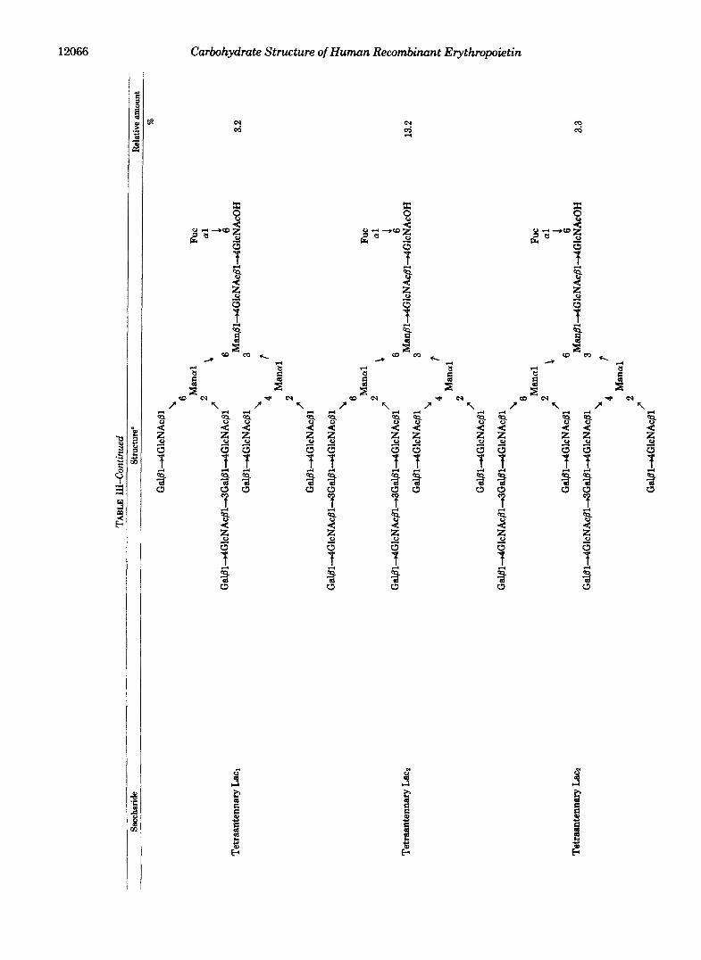
Structure of Asiulo Tetraantennary Saccharides with One N-Acetyllactosaminyl Repeat (Lac&"ethylation analysis of peak 5 in Fig. 5A provided 1 mol of 2,4-substituted mannose,
2,6-substituted mannose, and 3,6-substituted mannose (Table 11), indicating that peak 5 is composed of tetraantennary saccharides. The presence of 3-substituted galactose (0.8 mol) in this asialo form indicates that the saccharides contain one N-acetyllactosaminyl repeat. FAB-MS of the saccharides after permethylation yielded a signal at m/z 913 for Hex- HexNAc-Hex-HexNAc' (Fig. 4C), which is indicative of one N-acetyllactosaminyl repeat.
The saccharides of peak 5 were sequentially digested by P- galactosidase and P-N-acetylglucosaminidase and subjected to HPLC. As shown in Fig. 623, the major product eluted at 15 min, which corresponds to Gal. GlcNAc' Man3 I GlcNAc. (+Fuc)GlcNAcOH. The methylation analysis of this product provided 0.1 mol of 2-substituted mannose, 0.1 mol of 4- substituted mannose, 0.8 mol of 6-substituted mannose, and 1 mol each of terminal mannose and 3,6-substituted mannose as mannose derivatives. These results indicate that 80% of the N-acetyllactosaminyl repeat is attached to C-6 of 2,6- substituted mannose, 10% is attached to C-4 of 2,4-substituted mannose, and 10% is attached to C-2 of either 2,6- or 2,4- substituted mannose (Table 111).
Structure of Tetraantennary Saccharides with Two N-Ace- tyllactosaminyl Repeats (Lac&"ethylation analysis of peak 6 in Fig. 5A provided 1.7 mol of 3-substituted galactose, 4 mol of terminal galactose, and 1 mol each of 2,4-substituted, 2,6- substituted, and 3,6-substituted mannose, in addition to other derivatives (Table 11). The results indicate that this sacchar- ide fraction consists of tetraantennary saccharides with two N-acetyllactosaminyl repeats. FAB-MS of this oligosaccha- ride fraction supported the above conclusion since a fragment ion at m/z 913 corresponding to Hex-HexNAc-Hex- HexNAc' was detected (Fig. 40).
The saccharides were digested sequentially by P-galacto- sidase and P-N-acetylglucosaminidase to yield a major peak at 19 min (peak 2 in Fig. 6C), which corresponds to (Gal. GlcNAc)2. Mans. GlcNAc- (+Fuc)GlcNAcOH, confirm- ing that the starting sample contains tetraantennary sacchar- ides with two N-acetyllactosaminyl repeats. Methylation analysis of this product provided 1 mol of terminal mannose, 0.8 mol of 2,6-substituted mannose, and 0.2 mol each of 6- and 4-substituted mannose, in addition to 1 mol each of 3,6- substituted mannose, terminal galactose, and reducing ter- minal N-acetylglucosamine and 2 mol of 4-substituted N- acetylglucosamine. However, 2,4-substituted mannose or 3- substituted galactose was not detected. These results indicate that each side chain arising from 2,6-substituted mannose was elongated by one N-acetyllactosaminyl repeat in 80% of the saccharides. In addition, 20% of the molecules have N- acetyllactosaminyl repeats in side chains elongating from C- 6 and C-4 (Table 111).
Structure of Tetraantennary Saccharides with Three N- Acetyllactosaminyl Repeats (Lac&-The saccharides in peak 7 eluted at the position where saccharides containing seven N- acetyllactosaminyl units are expected to elute. This is because the difference of the elution time between peaks 7 and 6 is the same as that between peaks 6 and 5. The saccharides were sequentially digested with P-galactosidase and P-N-acetylglu- cosaminidase, and the products were analyzed by HPLC. As shown in Fig. 60, a major peak eluted at 24 min, which corresponds to (Gal.Gl~NAc)~.Man~.GlcNAc(+Fuc)Glc- NAcOH. However, about 35% of the products eluted at the position corresponding to (Gal-GlcNAc)2.Mans-GlcNAc(+ Fuc)GlcNAcOH.
These results suggest that 65% of the peak 7 saccharides are tetraantennary saccharides with three N-acetyllactosa- minyl repeats (Lac3), whereas 35% of the saccharides are

Carbohydrate Structure of Human Recombinant Erythropoietin 12069
tetraantennary saccharides with two N-acetyllactosaminyl repeats (Lac2). These results were confirmed by methylation analysis, as shown in Table 11. Methylation analysis of the saccharides provided 2.4 mol of 3-substituted galactose, 4 mol of terminal galactose, and 1 mol each of 2,4-, 2,6-, and 3,6- substituted mannose. The same analysis indicates that about 45% of the reducing terminal N-acetylglucosamine was un- reduced judging from the amount of 3-O-methyl-N-acetylglu- cosamine. This compound is produced when the reducing terminal N-acetylglucosamine is incompletely reduced before methylation.
In order to elucidate which side chains were elongated to form the N-acetyllactosaminyl repeat, the saccharides which eluted at 24 min (peak 3 in Fig. 6D) were methylated. The saccharides provided 0.6 mol of 2,6-substituted mannose, 0.4 mol of 6-substituted mannose, 0.3 mol of 4-substituted man- nose, 0.3 mol of terminal mannose, 0.4 mol of 2-substituted mannose, and 1 mol of 3,6-substituted mannose as mannose derivatives. These results indicate that each side chain arising from 2,6-substituted mannose and the side chains arising from C-2 or C-4 of 2,4-substituted mannose were elongated by N- acetyllactosaminyl units.
Since FAB-MS of the starting materials afforded a frag- ment ion at m/z 1723 for NeuNAc-+Hex+HexNAc+Hex+ HexNAc-+Hex-+HexNAc' (see Fig. 3A), it is likely that Lacs saccharides contain three N-acetyllactosaminyl units in one of the side chains which are attached to C-2 or C-6 of 2,6- substituted mannose or C-4 of 2,4-substituted mannose. This was confirmed by the fact that peak 3 in Fig. 6D provided a small amount (0.1 mol) of 3-substituted galactose on meth- ylation analysis.
Structures of Asialo N-Linked Saccharides from Recombi- nant Erythropoietin-The results obtained above are sum- marized in Table 111. By measuring radioactivity in each fraction, the relative yields of saccharides were calculated. In some cases, it was necessary to obtain the ratio after exogly- cosidase digestion. For example, the ratio of triantennary with one N-acetyllactosaminyl repeat and tetraantennary in peak 4 was obtained in Fig. 6A.
Fractionation of Intact N-Linked Saccharides by TSK- DEAE Ion-exchange Chromatography-In order to determine how these saccharides are sialylated, intact N-linked sacchar- ides were fractionated by HPLC employing a TSK-DEAE column. As shown in Fig. 7A, sialylated saccharides were essentially separated into four fractions: monosialosyl (frac- tion I) (7% of the total saccharide), disialosyl (fraction 11) (41%), trisialosyl (fraction 111) (48%), and tetrasialosyl (frac- tion IV) (4%) saccharides. No detectable amount of carbo- hydrate was present in other fractions.
After desialylation, monosialosyl saccharides (fraction I) were found to contain biantennary (12% of the total mono- sialosyl saccharides), triantennary (17%), tetraantennary (plus triantennary with one N-acetyllactosamine repeat) (28%), tetraantennary with one N-acetyllactosamine repeat (29%), tetraantennary with two N-acetyllactosamine repeats (1 l%), and tetraantennary with three N-acetyllactosamine repeats (3%) (Fig. 5C). Disialosyl saccharides (fraction 11) were found to consist of triantennary (7% of the total disialo- syl saccharides), tetraantennary (plus triantennary with one N-acetyllactosamine repeat) (43%), and tetraantennary sac- charides with one (36%), two (12%), or three (2%) N-acetyl- lactosamine repeats (Fig. 50). Trisialosyl saccharides (frac- tion 111), however, contain only tetraantennary (47% of the total tetrasialylated saccharides) and tetraantennary sacchar- ides with one (39%), two (12%), or three (2%) N-acetyhcto- saminyl repeats (Fig. 5E). The amount of tetrasialylated
saccharides was significantly low (4.2% of the total). Those saccharides were found to contain tetraantennary saccharides and tetraantennary saccharides with one or two N-acetyllac- tosaminyl repeats (data not shown).
These results indicate that 1) the biantennary saccharide is almost exclusively in a monosialylated form; 2) the trian- tennary saccharides are in monosialylated or disialylated forms; 3) the tetraantennary saccharides are mostly in disi- alylated or trisialylated forms; and 4) the tetraantennary saccharides with one, two, or three N-acetyllactosamine re- peats are mostly in disialylated or trisialylated forms. These results suggest that one of the side chains in the saccharides is almost alway terminated without a sialic acid residue (see below).
In order to know whether any difference exists among different batches of recombinant erythropoietin, two addi- tional batches (Batches 3 and 4 in Table I) of recombinant erythropoietin were subjected to analysis. Interestingly, these samples contained more highly sialylated saccharides: the disialosyl form is 18-21% of the total saccharides; the trisi- alosyl form is 64-67%, the tetrasialosyl form is 10-13%; and the monosialosyl form is less than 6%. However, the relative ratios of asialo biantennary, triantennary, tetraantennary, and tetraantennary saccharides with one, two, or three N- acetyllactosaminyl repeats were almost identical among dif- ferent samples. These results indicate that sialylation may vary among different batches of recombinant erythropoietin but their backbone structures are the same.
Separation of Saccharides with Different Backbone Struc- ture but with the Same Number of Sialic Residues in Side Chains-The results of Fig. 7A suggested to us that each peak in the monosialosyl, disialosyl, or trisialosyl fraction may represent saccharides with different backbone structures but with the same number of sialic acid residues. In order to test this possibility, another 5 mg of recombinant erythropoietin (Batch 2 in Table I) was treated to yield N-linked saccharides, and these saccharides were fractionated by TSK-DEAE ion- exchange chromatography. This sample provided an elution profile almost identical to that in Fig. 7A. Saccharides were divided into four (fraction 11) or three (fraction 111) fractions, desialylated, and subjected to another HPLC employing a Lichrosorb column. Fraction 11-1, which eluted earliest in TSK-DEAE chromatography, provided tetraantennary sac- charides with two or three N-acetyllactosaminyl repeats (Fig. 8A, see Miniprint), whereas the last peak (fraction 11-4) mainly consists of triantennary and tetraantennary sacchar- ides (Fig. 80). Similarly, the earliest peak in the trisialosyl fraction (fraction 111-1) provided Lac2 and a small amount of Lacl and Lac3 (Fig. 8E), whereas the last peak (fraction III- 3) provided almost exclusively tetraantennary saccharides (Fig. 8G). These results indicate that the saccharides with higher numbers of N-acetyllactosamine units elute earlier than those with smaller numbers of N-acetyllactosamine units in TSK-DEAE ion-exchange chromatography.
Localization of a24-Linked Sialic Acid in the Side Chains-In order to know which side chains are preferentially sialylated, fractions 11-2, 11-3 and 111-3 were digested exten- sively with a mixture of P-galactosidase and P-N-acetylglu- cosaminidase, and the products were purified by Sephadex G- 50 gel filtration followed by TSK-DEAE chromatography. The purified products were then subjected to methylation analysis, and the results are summarized as follows.
Disialosyl Tetraantennary Saccharides with One or Two N- Acetyllactosaminyl Repeats (Fraction II-2)"Methylation analysis on the exoglycosidase product of fraction 11-2 pro- vided the following mannose derivatives: 0.9 mol each of 2,6-

12070 Carbohydrate Structure of Human Recombinant Erythropoietin
( A r ~-r,+rn*
FIG. 7. Ion-exchange m L C of N- linked saccharides obtained from recombinant erythropoietin (A) and urinary erythropoietin (€3). N- Linked oligosaccharide fractions ob- tained after Sephadex G-50 gel filtration (fractions 29-41 in Fig. 2B) were applied to a column of TSK-DEAE SW2 equili- brated with 25 mM potassium phosphate buffer, pH 5.0. After washing with the same buffer for 10 min, the column was eluted with a linear gradient from the same buffer to 400 mM potassium phos- phate buffer, pH 5.0. A portion of the fractions indicated by the horizontal ar- rows were pooled and subjected to HPLC with a Lichrosorb column, as shown in Figs. 5 (C-E) and 8.
FIG. 11. Proposed structures of tetraantennary saccharides (A) and tetraantennary saccharides with N- acetyllactosaminyl repeata ( B ) ob- tained from recombinant erythro- poietin. A, sialylation takes place pref- erentially at the side chain arising from C-6 and then from the side chain at C-2 of 2,d-substituted mannose and C-4 of 2,4-substituted mannose. B. the sacchar- ides with one N-acetyllactosaminyl re- peat(m=l ,n=O,ando=O,inW%of the molecules, and o = 1 and m = n = 0 in 10% of the molecules); the saccharides with two N-acetyllactosaminyl repeats (m = n = 1 and o = 0 in 60% of the molecules, and m = o = 1 and n = 0 in 40% of the molecules); and the sacchar- ides with three N-acetyllactosaminyl re- peats (m + n + o = 3).
l6 t
A
0 20 40 100 120 140
Fraction Nunbec (0.3ml I Fraction)
- +NeuNAca2+3Gal81+4Gl cNAc6l ‘6
pllanal\ +NeuNAcaZ+3Ga161+461cNAc81~ 6
- +Fuc a1
6 I -
Man614Gl cNAc614G1 cNAcdsn - +NeuNAca2+3GalB14Gl cNAc6l
‘4 ; Hanal
2 Gal 61461 cNAc61’
B +NeuNAca2+3(Gal614G1cNAc61+3),-Gal61461cNAc61 k +Fuc Manal, -
a1
+NeuNAca2+3( Gal614461 cNAc61+3),*Gal614G1 cNAc6l’ 4
- 6
- +NeuNAca2-.3(Ga161+461cNAc61+3);Ga1614G1cNAc61 3 Man81+4461cNAc614G1cNAc+Asn
‘4 I
substituted and terminal mannose, 0.1 mol of 4- and 6- substituted mannose, and 1 mol of 3,6-substituted mannose. No other mannose derivatives, including 2,4-substituted man- nose, were detected. The results indicate that the 2 sialosyl residues are almost exclusively linked to the side chains arising from C-2 and C-6 of 2,6-substituted mannose (see Fig. 11).
Disialosyl Tetraantennary Saccharide (Fraction II-3)- Methylation analysis of the exoglycosidase product of fraction 11-3 provided the following mannose derivatives: 0.6 mol of terminal mannose, 0.3 mol of 2-substituted mannose, 0.4 mol of 4-substituted mannose, 0.1 mol of 6-substituted mannose, 0.6 mol of 2,6-substituted mannose, and 1 mol of 3,6-substi-
‘Manal’ 2
Gal 814GlcNAc61’
tuted mannose. The results indicate that 60% of the molecules are sialylated in both chains elongating from C-2 and C-6 of 2,6-substituted mannose, and 40% of the molecules are sia- lylated in the side chain arising from C-4 of 2,4-substituted mannose and that from C-6 or C-2 of 2,6-substituted mannose.
Trisialosyl Tetraantennary Saccharide (Fraction III-3)- Methylation analysis after exoglycosidase digestion of frac- tion 111-3 provided the following mannose derivatives: 1 mol each of 2,6- and 3,6-substituted mannose and 0.5 mol each of 2,4- and 4-substituted mannose. However, no other mannose derivative was detected. The results indicate that the nonsi- alylated chain almost exclusively arises from C-2 of 2,4- substituted a-mannose.

Carbohydrate Structure of Human Recombinant Erythropoietin 12071
Structure of Sialylated Tetraantennary Saccharides with or without N-Acetyllactosamine Repeats-Based on the results described above, the structure of intact tetraantennary sac- charides and those with N-acetyllactosaminyl repeats, which represent 85% of the total saccharides, can be proposed as shown in Fig. 11. The tetraantennary saccharides are mainly present as disialosyl or trisialosyl forms, and 2+3-linked sialic acid is attached to the side chains arising from C-6 and C-2 of 2,6-substituted mannose and C-4 of 2,4-substituted man- nose. In the tetraantennary saccharides with N-acetyllacto- saminyl repeats, sialic acid residues are always present in the side chain which contains N-acetyllactosaminyl repeats. This conclusion was supported by FAB-MS analysis. As shown in Fig. 3A, all of the fragment ions containing polylactosaminyl units are sialylated.
In order to delineate this further, trisialylated saccharides (fraction 111) were extensively digested by P-galactosidase and @-N-acetylglucosaminidase. The methylation analysis of this product showed that more than 90% of the side chain attached to C-2 of 2,4-substituted mannose was terminated without sialic acid. These combined results support the proposed structures shown in Fig. 11.
Structure of Carbohydrate Units of Urinary Erythropoie- tin-Since only a limited amount of urinary erythropoietin was available, the following experiments were carried out to analyze carbohydrate units of urinary erythropoietin. Glyco- peptides, prepared by Pronase digestion of urinary erythro- poietin, were subjected to alkaline borohydride treatment. The alkaline borohydride-treated sample was then applied to Bio-Gel P-4 gel filtration. Urinary erythropoietin saccharides provided almost the same elution profile as Fig. 2A. The glycopeptides containing N-linked saccharides (fractions 24- 29) were digested by N-glycanase, and the digest was subjected to Sephadex G-50 gel filtration. Again, the elution profile of N-linked saccharides from urinary erythropoietin was almost identical to that from recombinant erythropoietin (see Fig. 2B).
Methylation analysis of N-linked saccharides (fractions 29- 41 after Sephadex (3-50 gel filtration) provided partially 0- methylated monosaccharide derivatives, which are almost identical to those produced from highly sialylated Batch 3 of recombinant erythropoietin (Table 11). FAB-MS of permeth- ylated N-linked saccharides provided fragment ions for NeuNAc' (m/z 376 and 344), NeuNAc+Hex+Hex NAc+ (m/ z 825), NeuNAc+Hex+HexNAc-+Hex+HexNAc+ (m/z 1274), and NeuNAc+Hex+HexNAc+Hex+HexNAc+ Hex+HexNAc+ (m/z 1723) (Fig. 3B). These results are es- sentially the same as those obtained on recombinant eryth- ropoietin (compare Fig. 3, A and B).
N-Linked saccharides were then desialylated and subjected to HPLC employing a Lichrosorb-NHz column. As shown in Fig. 5B, urinary erythropoietin saccharides provided trianten- nary, tetraantennary, and tetraantennary saccharides with one, two, or three N-acetyllactosaminyl units. The relative proportion among these saccharides is almost identical to that obtained on recombinant erythropoietin except that urinary erythropoietin apparently lacks biantennary saccharides. In order to determine the relative amounts of sialylated N-linked saccharides, intact N-linked saccharides were subjected to TSK-DEAE ion-exchange chromatography. Fig. 7B shows that N-linked saccharides from urinary erythropoietin con- tain disialosyl(27% of the total saccharides), trisialosyl(56%), and tetrasialosyl (17%) saccharides. These results indicate that urinary erythropoietin and recombinant erythropoietin have an almost identical set of N-linked saccharide units but
with slightly different sialylation depending upon the batches of recombinant erythropoietin (see above and Table I).
DISCUSSION
This paper reports the detailed structures of the carbohy- drate moiety of human erythropoietin produced by recombi- nant DNA. The protein analyzed was produced in Chinese hamster ovary cells which were transfected with human eryth- ropoietin cDNA (7). As far as we are aware, this is the first report on the detailed carbohydrate structure of a glycoprotein produced by recombinant DNA in comparison with the gly- coprotein of natural origin. Although Mutsaers et al. (28) reported the carbohydrate structure of human y-interferon produced in Chinese hamster ovary cells, their studies did not investigate those of naturally occurring human y-interferon. The carbohydrate composition (Table I) showed that eryth- ropoietin contains three N-linked saccharides and one 0- linked saccharide, and these conclusions are consistent with the recent report on the amino acid sequence of human urinary erythropoietin (29).
The present study revealed that a large proportion of the carbohydrate moiety of recombinant erythropoietin is com- posed of tetraantennary saccharides with one (32.1% of the total saccharides), two (16.5%), and three (4.7%) N-acetyllac- tosaminyl repeats. The localization of these polylactosaminyl units was elucidated by sequential exoglycosidase digestion followed by methylation analysis, and the results are sum- marized as follows (see also Table 111).
When the saccharides contain one N-acetyllactosaminyl repeat, more than 70% of this repeat is preferentially attached to the side chain arising from C-6 of 2,6-substituted mannose, and 19% of the repeat is attached to that from C-4 of 2,4- substituted mannose. When the saccharides contain two N- acetyllactosaminyl repeats, these repeats are attached to C-2 and C-6 of 2,6-substituted mannose in 80% of the molecules. The rest of the molecule contains N-acetyllactosaminyl re- peats in the side chains arising from C-6 of 2,6-substituted mannose and C-4 of 2,4-substituted mannose. These results indicate that N-acetyllactosaminyl repeats are most prefer- entially added to C-6 of 2,6-substituted mannose and then to C-2 of 2,6-substituted mannose. These conclusions are con- sistent with previous reports on several cellular glycoproteins. For example, Cummings and Kornfeld (30) reported that the mouse lymphoma BW5147 cell line expressed a signX1cant amount of polylactosaminoglycan, whereas its mutant, which lacks the side chain arising from C-6 of 2,6-substituted man- nose, expresses a minimum amount of polylactosaminoglycan. Li et al. (31) isolated polylactosaminoglycan from Chinese hamster ovary cells in which polylactosaminyl units are at- tached to C-2 and C-6 of 2,6-substituted mannose. Similarly, polylactosaminyl units were found in triantennary and te- traantennary saccharides of various origins (24,32-34). These results appear to establish that N-acetyllactosaminyl repeats are preferentially added to C-6 of 2,6-substituted mannose and then to C-2 of 2,6-substituted mannose. These 2,6-sub- stituted mannose residues are usually linked to the C-6 side of @-mannose. In human erythrocytes, polylactosaminyl elon- gation can be found in the side chain arising from C-2 of (Y-
mannose which is linked to C-6 of P-mannose (23, 35). It is likely that human erythroid cells contain very little activity of the N-acetylglucosaminyltransferase which forms a GlcNAcpl4Man branch. As a result, N-acetyllactosamine repeats are formed on the secondary preferable side chain, which is attached to C-2 of a-mannose linked to C-6 of 8- mannose, in these erythroid cells.
This study showed that human erythropoietin exclusively

12072 Carbohydrate Structure of Human Recombinant Erythropoietin
contains a2-3-linked sialic acid. This fact allowed us to elucidate the locaiization of 2+3-linked sialic acid residues among different side chains. This was achieved by extensive digestion of intact saccharides with P-galactosidase and P-N- acetylglucosaminidase followed by methylation analysis of the products. These results can be summarized as follows. 1) When typical triantennary or tetraantennary saccharides con- tain 2 sialic acid residues, they are attached to C-2 and C-6 of 2,6-substituted mannose or C-6 of 2,6-substituted mannose and C-4 of 2,4-substituted mannose. 2) When saccharides with N-acetyllactosaminyl repeats contain 2 sialic acid resi- dues, they are attached almost exclusively to C-2 and C-6 of 2,6-substituted mannose. This localization is essentially iden- tical to that of N-acetyllactosamine repeats. Thus, it is ap- parent that polylactosamine is preferably sialylated through an aB-+S-linkage.
These results are consistent with our previous results ob- tained on polylactosaminoglycans from chronic myelogenous leukemia cells; a2-3-linked sialic acid is present on side chains arising from C-6 and C-2 of 2,6-substituted mannose and C-4 of 2,4-substituted mannose, and those side chains are longer than that terminating with 24-l inked sialic acid (24). Similar results were obtained in human erythrocyte Band 3 polylactosaminoglycans; polylactosaminyl side chains arising from the C-6 side of p-mannose are sialylated through a 2-+ 3-linkage, whereas the shorter chain arising from the C-3 side is sialylated through a 24-linkage (23, 35). Similarly, Ya- mashita et al. (33) and Markle and Cummings (36) found that longer polylactosaminyl side chains are almost exclusively sialylated through a 2+3-linkage. Interestingly, short poly- lactosamine chains in thyroid cell glycoprotein Gp-1 (37) and BW5147 (36) are terminated with 24- l inked sialic acid. Our results also showed that almost no 2-3-linked sialic acid is attached to the side chain arising from C-2 of 2,4-substituted mannose. It is noteworthy that this side chain was found to be exclusively sialylated through a 24-linkage in many glycoproteins including normal and leukemic granulocyte po- lylactosaminoglycans (19-21, 24, 34, 38-41).
By using a bovine colostrum a2-&-sialyltransferase, Jo- ziasse et al. (42) have shown that preferential sialylation takes place first on C-2 of 2,4-substituted mannose and then on C- 4 of 2,4-substituted mannose. This branch (or side chain) specificity appears to be opposite to the distribution of 2-3- linked sialic acid. Thus, it is likely that a2+=3-~ialyltransfer- ase and a24-sialyltransferase have complementary specific- ity toward different side chains. Furthermore, our results raise the possibility that the side chains containing polylactosami- nyl units would be preferable sites for 2+3-linked sialylation.
This study demonstrated that the carbohydrate moiety of human erythropoietin isolated from human urine is indistin- guishable from that of recombinant erythropoietin except for a difference in degree of sialylation. Urinary erythropoietin has a similar degree of sialylation as the highly sialylated batch of recombinant erythropoietin (Tables I and 11). The recombinant erythropoietin was produced in Chinese hamster ovary cells, and urinary erythropoietin is presumably derived from human kidney cells. The results therefore suggest two possibilities. 1) Chinese hamster ovary and human kidney cells contain similar glycosyltransferases. 2) The protein ac- ceptor itself influences glycosylation even when a similar set of glycosyltransferases are not present in two cell types. It will be interesting to see if the carbohydrate moiety of eryth- ropoietin produced in other mammalian cells is similar to those elucidated in this study. This study also demonstrated that the major carbohydrate units of erythropoietin are te- traantennary saccharides with or without N-acetyllactosa-
mine repeats. It has been shown that rat liver cells uptake the asialo form of glycoproteins which contain tri- or tetraanten- nary saccharides (43). It is therefore reasonable that the asialo form of erythropoietin is taken up by liver cells through a galactose-binding protein (15). Our preliminary studies showed that a portion of intact erythropoietin of both recom- binant and urinary origins is taken up by rat liver cells, presumably because of the incomplete sialylation. It will be interesting to test if sialylation by a24-sialyltransferase elongates the serum concentration of erythropoietin and sus- tains in vivo activity longer than the starting erythropoietin.
Acknowledgments-We thank Dr. Tsutomu Kawaguchi (Chugai Pharmaceutical Co., Ltd.) for initiating this joint project, Dr. Fried- rich Piller for useful discussion and Candy Farmer for secretarial assistance.
1.
2.
3. 4.
5.
6.
7.
8.
9.
10.
11.
12.
13.
14.
15.
16.
17.
18.
19.
20.
21.
22.
23.
24.
5;5.
26.
REFERENCES Goldwasser, E., and Kung, C. K. H. (1968) Ann. N. Y. Acad. Sci.
Jacobson, L. O., Goldwasser, E., Fried, W., and Plzak, L. F. (1957)
Fried, W . (1972) Blood 40,671-677 Zanjani, E. D., Ascensao, J. L., McGlave, P. B., Banisadre, M.,
and Ash, R. C. (1981) J. Clin. Invest. 6 7 , 1183-1188 Adamson, J. W . , Eschback, J. W . , and Finch, C. A. (1968) Am. J.
Med. 44,725-733 Miyake, T., Kung, C. K.-H., and Goldwasser, E. (1977) J. Bwl.
Chem. 262,5558-5564 Jacobs, K., Shoemaker, C., Rudersdorf, R., Neill, S. D., Kaufman,
R. M., Mufson, A., Seehra, J., Jones, S. S., Hewick, R., Fitch, E. F., Kawakita, M., Shimizu, T., and Miyake, T. (1985) Nature
Lin, F. K., Suggs, S., Lin, C. H., Browne, J. K., Smalling, R., Egrie, J. C., Chen, K. D., Fox, G. M., Martin, F., Stabinsky, Z., Badrawi, S. M., Lai, P. M., and Goldwasser, E. (1985) Proc. Natl. Acad. Sci. U. S. A. 8 3 , 7580-7584
Powell, J. S., Berkner, K. L., Lebo, R. V., and Adamson, J. W . (1986) Proc. Natl. Acad. Sci. U. S. A. 8 3 , 6465-6469
Winearls, C. G., Oliver, D. O., Pippard, M. J., Reid, C., Downing, M. R., and Cotes, P. M. (1986) Lancet ii, 1175-1178
Eschbach, J. W., Egrie, J. C., Dowwing, M. R., Browne, J. K., and Adamson, J. W . (1987) N. Engl. J. Med. 316,73-78
Rambach, W . A., Shaw, R. A., Cooper, J. A. D., and Apt, H. L. (1958) Proc. Soc. Exp. Biol. Med. 99,482-483
Lowy, P. H., Keighley, G., and Borsook, H. (1960) Nature 186,
Lukowsky, W. A., and Painter, R. H. (1972) Can. J. Biochem.
Goldwasser, E., Kung, C. K.-H., and Eliason, J. (1974) J. Biol. Chem. 249,4202-4206
Simnonsen, C. C., and Levinson, A. D. (1983) Proc. Natl. Acad. Sci. U. S. A. 8 0 , 2495-2499
Fukuda, M., Carlsson, S. R., Klock, J. C., and Dell, A. (1986) J. Bwl. Chem. 261,12796-12806
Plummer, T. H., Jr., Elder, J. H., Alexander, S., Phelan, A. W., and Tarentino, A. L. (1984) J. Biol. Chem. 2 5 9 , 10700-10704
Nilsson, B., Nordkn, N. E., and Svensson, S. (1979) J. Bwl. Chem. 264,4545-4553
Baenziger, J. U., and Fiete, D. (1979) J. Biol. Chem. 254, 789- 795
Endo, M., Suzuki, K., Schmid, K., Fournet, B., Karamanos, Y., Montreuil, J., Dorland, L., Van Halbeek, H., and Vliegenthart, J. F. G. (1982) J. Biol. Chem. 267,8755-8760
Yoshima, H., Matsumoto, A., Mizuochi, T., Kawasaki, T., and Kobata, A. (1981) J. Bwl. Chem. 256,8476-8484
Fukuda, M., Dell, A., and Fukuda, M. N. (1984) J. Bwl. Chem.
Fukuda, M., Bothner, B., Ramsamooj, P., Dell, A., Tiller, P. R., Varki, A., and Klock, J. C. (1985) J. Biol. Chern. 260, 12957- 12967
Jourdian, G. W . , Dean, L., and Roseman, S. (1971) J. Biol. Chem.
Greenwood, F. C., Hunter, W . M., and Glove, J. S. (1963)
149,49-53
Nature 179,633-634
313,806-810
102-103
60,909-917
269,4782-4791
246,430-435
Biochem. J. 89, 114-123
Carbohydrate Structure of Human Recombinant Erythropoietin 12073
27. Laemmli, U. K. (1970) Nature 227,680-685 28. Mutsaers, J. H. G. M., Kamerling, J. P., Devos, R., Guisez, Y.,
Fiers, W., and Vliegenthart, F. G. (1986) Eur. J. Biochem. 166,
29. Lai, P.-H., Everett, R., Wang, F.-F., Arakawa, T., and Goldwas-
30. Cummings, R. D., and Kornfeld, S. (1984) J. Biol. Chem. 269,
31. Li, E., Gibson, R., and Kornfeld, S. (1980) Arch. Biochem. Bio-
32. Eckhardt, A. E., and Goldstein, I. J. (1983) Biochemistry 22,
33. Yamashita, K., Ohkura, T., Tachibana, Y., Takasaki, S., and
34. Fukuda, M., Spooncer, E., Oates, J. E., Dell, A., and Klock, J. C.
35. Fukuda, M., Dell, A., Oates, J. E., and Fukuda, M. N. (1984) J.
651-654
ser, E. (1986) J. Biol. Chem. 261,3116-3121
6253-6260
phys. 199,393-399
5290-5297
Kobata, A. (1984) J. Biol. C h m . 269 , 10834-10840
(1984) J. Biol. Chem. 269,10925-10935
SUPPLEMENTAL MATERIAL TO
CARBOHYDRATE STRUCTURE OF ER'lTHROPOlETlN EXPRESSEO I N CHINESE HMSTER OVARY CELLS B Y A HUMAN ERYTHROPOIETIN COW
BY
Hlroshi Saraki . Br ian Bothner. Anne Oell and Minaru FUkVda
ol igosdcc ar er *ere separ*te T m - n c g ycopep l l o l d t i o n O f 0-l inked DligOsaCChmides frm n c n b i n a n t e r y t h r o p o i e t i n - The 0- l inked
I s r h a n 'in ' f i g . 2 A . The f r d t T o n s :>d :o"td35 ( d ~ ~ ~ & ~ ~ d B ~ - i ) and those frm 36-40 o-Gel P-4 g e l f i l t r a t i o n
(designated 0 - i f ) were separately pooled and sub jec ted to TSK-OEAE ?on exchange c h m a t o - graphy as I h w n i n F i g . 9
The 0-1 f rac t ion p lov ided d is la losy l o l iqOsaCChmides and t v l s ia losv l o l i so raccha- r ides , whereas t h e 0 - i i f m t i o n p r o v i d d m i n i y m o n o r i a l o r y l o l i g o s a c c h a 6 d e s . d;s judged by FAB-MS. The f T a C t i O n l c a r r e r p a n d i n g t o m n o ~ i a l o s y l - , d l r i a l o r y l - . and t r l l i a l o s y l - sacchwidss f rom the 0-1 and (I-H f rac t ions were pooled and subjected t o m e t h y l a t i o n ana lys is and FAR-MS maly611.
molecule (The d e t a i l e d mechaniimr fo r B~c leavage and 6 - e l i m i n a t i o n w i l l be der;ribed elraher;.21. The 01igoSdCChalide was r e g u c n t i a l l y d i g e s t e d by c l o s t r i d i a l s i a l i d a s e and
mthylation-iiiilysis (Table 1V) i n d i c a t e t h a t t h e d l 1 i m l o l y l o l i q o s a c c h a r i d e 11 NeuNAeo- Esher i ch ia co l i 6 -ga lac t0 l i dd le as descr ibed previously (17). These r e w l t s . t o g e t h e r w i t h
ol igosaccharides f m ur ina ry evy th ropo ic t i n were recovered i n f r a c t i o n 30-40 i n Blo-Gel S t r u c t w e s of 0- l inked Ol igosaCChdridel frm u r i n a r y e r y t h r o p o i e t i n - The 0- l inked
P-4 g e l f i l t r a t i o n !5*e Fig. ZA). I n i t i a l L t t m p t l t o f w t h e r f rac t ionate 0 - l inked Ol igosacchar ides fa i led and no s i g n i f i c a n t amount Of 0 - l i n k e d o l i g m a c c h a r i d e l w e ~ e de- t e c t e d a f t e r TSK-DEAE ion exchange c h m d t ~ g r a p h y . T h i s was probably due t o a low quant i ty o f t h e 0 - l i n k e d o l i g o s a ~ ~ h a r i d e s fm urinary ery thropoiet in . Therefore, f ract ions frm 30-40 were poled and d i r e c t l y analyzed by methy la t ion ana lys is and FAB-US analysis.
AI shwn i n Ftg. 100. FAB-MS O f the .aligssACCharldel provided a p o l e c v l a r Ion o f m/z 1234 f o p NeuNLc .H~x.HcxNAc In sdd i t ion . f rament inns fo r NeuNAc 1176 and 344) were detected. A f r igment ion a!'m/z 825 cor respond ing to NeuNAc.Hex.HexNAc may be produced fm N-linked saccharides which were present as contaminants i n th is O l igosdcchar ide f rac t ion . There resu l ts . together w i th methy la t ion ana lys is , ind imtec tha t the major 0- l inked o l igosacchar ide f m u r i n a r y e r y t h r o p o i e t i n i s t h e d i r i a l o r y l s a c c h a r i d e . YhiCh i s pmbab ly NeuNAc~-3Gal61*3~NeuNA~~~)GalNAcOH.
Biol. Chem. 269,8260-8273 36. Merkle, R. K., and Cummings, R. D. (1987) J,. Biol. Chem. 262 ,
37. Edge, A. S. B., and Spiro, R. G. (1985) J. Biol. Chem. 260,15332-
38. Mega, T., Lujan, E., and Yoshida, A. (1980) J. Biol. Chern. 256,
39. Paulson, J. C., Weistein, J., Dorland, L., Van Halbeek, H., and Vliegenthart, J. F. G. (1982) J. Biol. Chem. 267 , 12734-12738
40. Spik, G., Debruyne, V., Montreuil, J., Van Halbeek, H., and Vliegenthart, J. F. G. (1985) FEBS Lett. 183,65-69
41. Yamamoto, K., Tsuji, T., Irimura, T., and Osawa, T. (1981) Biochem. J. 196,701-713
42. Joziasse, D. H., Schiporst, W. E. C. M., Van den Eijenden, D. H., Van Kuik, J. A., Van Halbeek, H., and Vliegenthart, J. F. G. (1987) J. Biol. Chem. 262 , 2025-2033
8179-8189
15338
4057-4061
43. Baenziger, J. U., and Fiete, D. (1980) CeU 22,611-620
TABLE 11. Re la t i ve p ropor t ions Of methylated s w a m obtained frm N-l inked saccha- r i d e s O f recmbinant and ur ina ry e ry th ropo ie t i ns .
Methylated Sugars Recrmbindnt Urinary 3 4 5 6 7 N-saccharides
batch 1 batch 3
F u d t o l -T;5;4Itri-O-Wthyl 0.9 0.9 0.9 0.9 0.95 0.9 0.9 0.9
Ga laCt i to l 2.3.4.6-retra-O-nethyl 1.34 0.17 0.38 3.0 3.90 4.0 4.0 4.0 2.4,6-tr i-O-mthyl 3.52 4.30 4.11 0 0.11 0.8 1.7 2.4
Mami to1 m r i - 0 - m e t h y l 0.16 0.12 0.26 1.00 0.11 0 0 0
3.6.-di-O-nethyl 0.78 0.81 0.76 0.35 1.00 1.0 1.0 0.9 3,4-di-O-nethyl 1.05 1.07 0.98 0.65 0.90 1.0 1.0 1.1 2.1-dl-0-methyl 1.00 1.00 1.00 1.00 1.00 1.0 1.0 1.0
2-N-methylacetamido- 2-deoxyqlucitol 1 .3 ,5 .6 - te t ra -O-~ thy l 0.10 0.12 0.14 t roce 0.05 t race 0.05 0.10
3.6-di-O-mthyl 1,3.5-tr i-O-rethyl 0.80 0.81 0.81 0.9 0.9 0.9 0.85 0 .55
3-0-methyl 5.82 5 .76 5.54 4 .0 5 .0 6.0 6.7 7.4 0.10 0.07 0.05 0.1 0.05 0.05 0.10 0.35
TmLE IY. Re la t i ve p ropor t i ons of methylatea sugar obtained frm 0-l inked saccherfdts o f recmb inan t and u r i n a r y e r y t h r o p i o e t i n r .
Recmbinant
Mo".ali.lolyl O i r i a l o l y l T l l l i . l O l Y l u r i n a r y
G a l d c t i t o l a
2 .4 .6 - tn -O-wthy l 1.0 1.0 1.0 1.0
2-N-rethylacetamido- Z - d e ~ x y q a l a c t i t ~ l
1,4,5,6-tetra-O-methyl 0.8 trace 0 0.2
1,4,5-tn-O-methyl 0.1 1.0 0.9 0.7
4-mono-O-mthylb 0 . 1 0 0.1 0.1
'A m a l l amount O f m a n n m e der iva t ives ( less than 0.1 mole) was detected i n a l l o f t h e
fP1Et iO" l .
b l h i r d e r i v a t i v e was probably produced because Of InCMpletF reduct ion.

12074 Carbohydrate Structure of Human Recombinant Erythropoietin
I ll I
"10
913
D
913
b i n a n t e r y t h r o p o i e t i n . N- l inked sacchar ides, separa ted by HPLC w i t h a column o f L i c r o - F igu re 6. HPLC O f exog1yCI)Sidase d i q e r t r Of N- l i nked saccharides d e r i v e d f rom pecan-
SOP -NH as 5 own i n i g . 5A were d i g e s t e d s e q u e n t i a l l y b y 8 - g a l a C t o S l d a l e and E-N-acetyl- g l u k a 6 i n i d a ; e a n d L r u b j e l t e d t o t h e same HPLC. The chromatograph ic cond i t ions are t he sane I O i n Fig. 5 except that the chromatagrams were m o n i t o r e d b y m e a s u r i n g r a d i o a c t i v i t y .
A . The d l g e s t o f s a c c h a r i d e 4.
B. The d iges t o f sacchar ide 5, Lac1.
C. The d i g e s t o f saccharide 6, Lac . 0. The d i g e s t of racchar lde 7, Lac3.
recomb inan t e ry th ropo ie t i n . The p o s i t i v e s p e c t r a were taken on t h e p e m e t h y l a t e d r a m p l e r . F igu re 1. Fas t atom bombardment-mars spec t ra Of a s i i l o N - l i n k e d s a c c h a r i d e l f r o m
and 2219) were produced by a cleavage between fw N-adtylglucoramin;';er;d;e; ' in - th;
w e ~ e de tec ted I t m/z 2001 for Hex5.HexNAc4 , and 1552 far H e x i HexNAc3 , r e s p e c t i v e l y . c h i t o b i o s y l core. e-c leavage ions der ive$ f10m t r i - a n t e m m y and Dl-pntennary Iacchar ides
B . O e s i a l y l a t e d t e t r a - a n t e n n a l a n d t r i - a n t e n n a r w i t h one N - a c e t l l a c t o r a m i n e
racchar lde . except for t he f o l l ow ing . The A - t y p e i o n s d e r i v e d from b i -an tennary or tri- Isacc6ay ide 4 i n Fig. SA). Almost 6 1 f ragment ions d l tec ted i n A were fyund i n t h i s
antennavy saccharides ( m l z 2219 and 17701 were absent or b a r e l v d e t e c t e d . A l t h o u o h t h e f r a g m e n t i o n a t m/z 913 was p r o m i n e n t , t h e i o n a t 1362 f o r Hex3.H&NAcgf i a s not de tbc ted . .
cha r iae 5 i n F i g . SA). C. O e r i d l y l a t e d t e t r a - a n t e n n a r y w i t h one N-acety l lac tosaminy l repeat , Lac. (sac-
r i d e 3 i n F ia . SA) . There racchar ider were contaminated by o ther componegt r - inc lud ing 0. O e r i a l y l a t e d t e t r a - a n t e n n a r y w i t h t w o N - a c e t y l l a c t o r a m i n y l r e p e a t s . Lac, (raccha-
ae te rgen t . However, r l g n / f i c a n t s i g n a l s a t m/z 913 f o r Hex*HexNAcfHex*HexNA~ (bo th and 0) and 464 f o r Hex-HexNAC (2) were de tec ted .

Carbohydrate Structure of Human Recombinant Erythropoietin
110
90
70
50
30
1 0
110
90
70
50
30
1 0
110
90
70
50
30
10
0 4 12 20 28 36 44 52 Elution Tlme (mmutes)
N-l inked racchariaer from r e c m b i n a n t e r y t h r o p o i e t i n wele separated by TSK-DEAE i o n ex- Figure 8. HPLC o f each peak obtained by TSK-OEAE i o n exchange chromatowauhy.
change ChPanatography as shown i n F i g . 7 4 . Each peak obtained was d e l i a l y l a t e d and sub- j e c t e d t o HPLC M i t h a Lichrerorb-NH2 column I S the ram Cond i t ion of Fig. 5 .
A . 11-1 ( f r a c t i o n s 7 0 - 7 7 , i n Fig. la) .
B. 11-2 ( f r a c t i o n s 78-81 i n F i g . l A i ) .
C. 11-3 ( f r a c t i o n s 82-87 i n Fig . 7A1.
0. 11-4 ( f r a c t i o n s 8 8 - 9 1 i n F i g . 7 A l .
E . 111-1 ( f m c t l o n l 94-98 i n F i g . l a ) .
F. 111-2 ( f r l c t i o n l 99-102 i n F i g . 7Ai ) .
6 . 111-3 ( f r a c t i o n s 103-108 i n F i g . 7 A ) .
0 10 20 30 40 50 Fraction Number
erythropoiet in . OTgoracchar idcr re leased wi th a lka l ine borohydr ide t reatnent were i s o l a t e d F i g w e 9. Ion-exchange HPLC of 0- l inked o l igosacchar ides obta ined f rom recanbinant
chromatography, a3 descr ibed in "Exper imental Procedures" except for the fo l lowing. The by Bio-Gel P-4 g e l f i l t r a t i o n and those saccharides were separated by TSK-DEAE i on exchange
column was e o u i l i b r a t e d w i t h 25 nW Dotarsiurn Dhowhate buffer. DH 5.0. After warh ino wi th the same b u f f e r f o r 5 .in, t h e & l w w was e l u t e d w i t h a l j&w gradient f rom th; same
ml lmin and each f rac t i on con ta ined 0.5 ml, The hor izon ta l arrows i nd i ca te t he f rac t i ons b u f f e r t o 400 nW potassium phosphate buffer. pH 5.0 over 40 m i " . The f low ra te was 1
pooled. No carbohydrate was de tec ted i n o the r f rac t i on$ .
12075
4. 0 - 1 ( f m c t i o n s 30-35 i n F i g . 2 A ) .
8 . 0 - i i ( f r a c t i o n r 36-40 i n F i g . 2 A )

12076 Carbohydrate Structure of Human Recombinant Erythropoietin
859 I
631
I 105 6 9
i 1 1