chapter 17 blood - north seattle collegefacweb.northseattle.edu/jdahms/214win08/lect/chapter 17...
TRANSCRIPT
1
Chapter 17: Blood
Overview• Blood functions• Compostion of whole blood• Plasma• RBCs – structure, function, and
development• Blood types• WBCs• Platelets • Hemostasis
The Cardiovascular System
• A circulating transport system composed of:– a pump (the heart)– a conducting system (blood vessels)– a fluid medium (blood)
• Functions to transport:– oxygen and carbon dioxide– nutrients– hormones– immune system components – waste products
Blood – tissue type?
General Characteristics of Blood
• Blood is a sticky, opaque fluid with a metallic taste
• Color varies from scarlet to dark red High viscosity (due to cells)
• Temperature is 38°C• Normal pH range = 7.35–7.45• Blood volume (liters) = 7% of body weight
(kilograms):– adult male: 5 to 6 liters– adult female: 4 to 5 liters
Blood - General Functions• Transport of dissolved gases, nutrients,
hormones, and metabolic wastes• Regulation of pH, body temperature, ion
composition of interstitial fluids• Restriction of fluid loss at the injury site• Defense against toxins and pathogens

2
Whole Blood
Whole Blood
• Plasma: Fluid component– Water (90%)– Dissolved plasma proteins– Other solutes
• Formed elements: Cells and fragments– RBCs (carry Oxygen)– WBCs (immunity)– Platelets (cell fragments involved in clotting)
Figure 19–1b
Plasma Plasma
• Makes up 50–60% of blood volume• More than 90% of plasma is water• Other constiuents:
– Plasma proteins– Lactic acid, urea, creatinine– Organic nutrients – glucose, carbohydrates, amino
acids– Electrolytes – sodium, potassium, calcium, chloride,
bicarbonate – Respiratory gases – oxygen and carbon dioxide
Body Fluids
Extracellular Fluid (ECF) = Interstitial fluid (IF) and plasma plus a few other body fluids such as CSF
• Plasma and IF exchange water, ions, & small solutes across capillary walls
Intracellular Fluid (ICF)=fluid inside cellsECF and ICF differ in their levels of:• O2 and CO2
• Dissolved proteins: plasma proteins do not pass through capillary walls (too large)
Plasma proteins• Albumins (60%): major component of osmotic
pressure of plasma– Transport proteins for fatty acids, thyroid
hormones, steroid hormones• Globulins (35%): antibodies
(immunoglobulins) and transport proteins:– hormone-binding proteins– metalloproteins– apolipoproteins (lipoproteins)– steroid-binding proteins
• Fibrinogens (4%)-functions in blood clotting (form fibrin)
• Others (1%) including hormones

3
Origins of Plasma Proteins
• 90% made in liver• Others not made in the liver include:
– Antibodies made by plasma cells (a special type of WBC)
– Peptide hormones made by endocrine organs
Serum
• Liquid part of a blood sample in which dissolved fibrinogen has converted to solid fibrin
• Often, this term refers to plasma that has had the clotting proteins removed
Formed Elements
Formed Elements
• These are the cells (and quasi-cellular) constituents of blood
• Red blood cells (RBCs) make up 99.9% of blood’s formed elements
• White blood cells and platelets make up the rest
Components of Whole Blood
Figure 17.2
Measuring RBCs
• Red blood cell count: reports the number of RBCs in 1 microliter whole blood – Male: 4.5–6.3 million– female: 4.2–5.5 million
• Hematocrit (packed cell volume, PCV): percentage of RBCs in centrifuged whole blood– male: 40–54 (avg = 46)– female: 37–47 (avg = 42)
RBCs make up about 1/3 of all cells in the body!
4
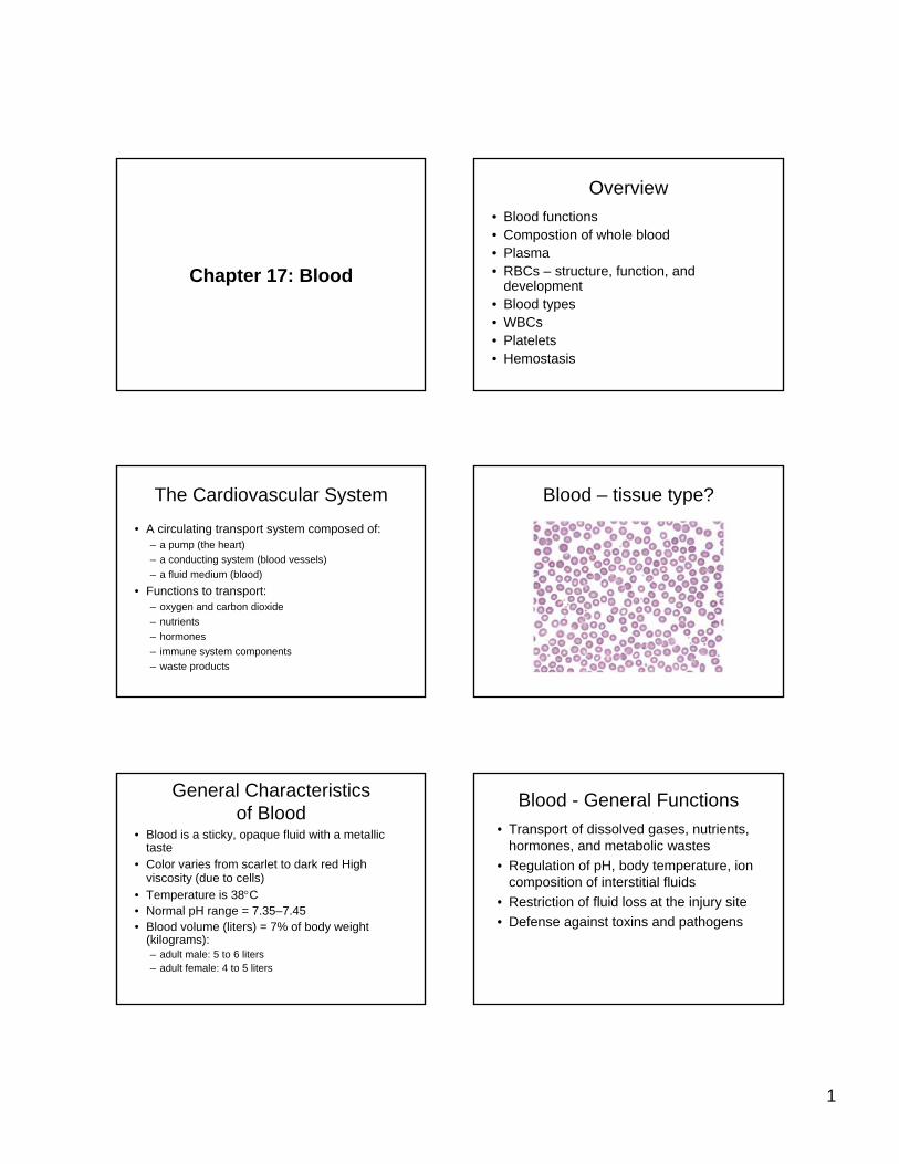
Why do RBCs look hollow?
No nucleus
Biconcave structure
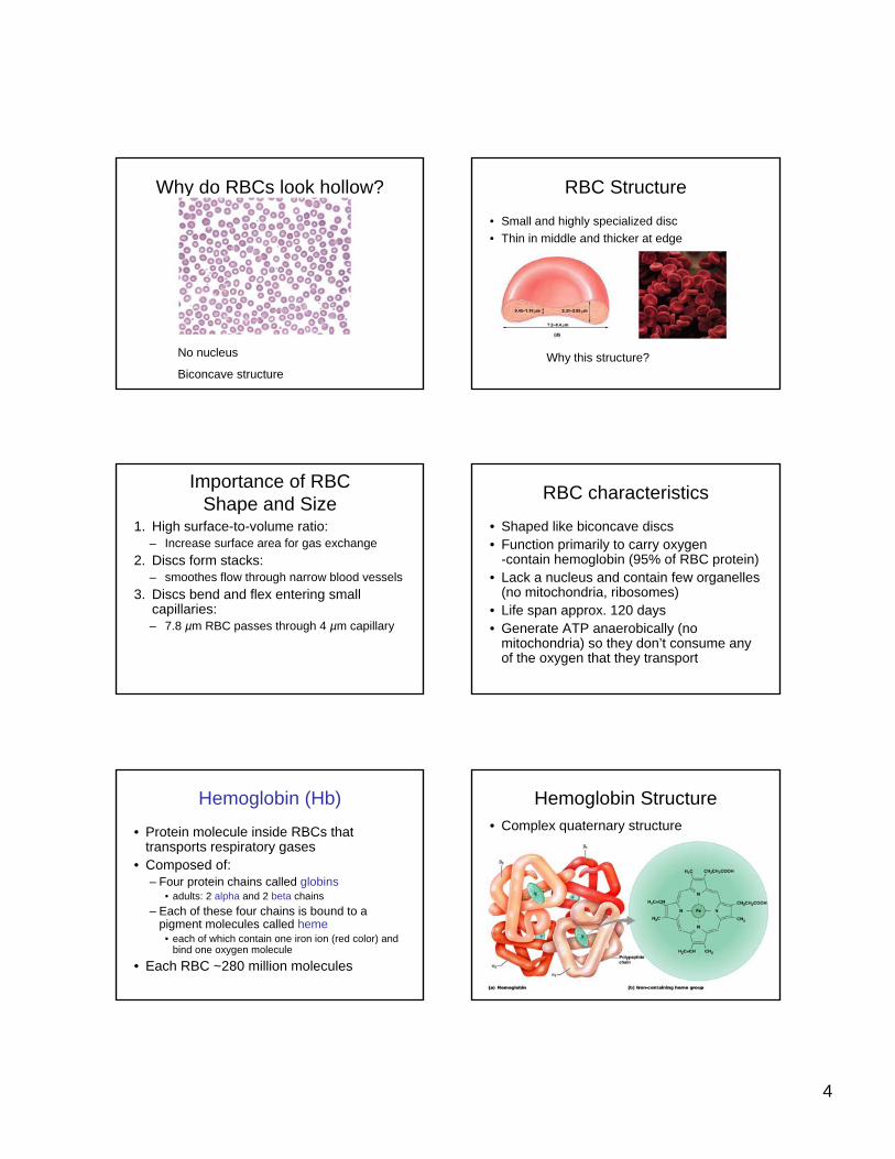
RBC Structure
• Small and highly specialized disc• Thin in middle and thicker at edge
Figure 19–2d
Why this structure?
Importance of RBC Shape and Size
1. High surface-to-volume ratio:– Increase surface area for gas exchange
2. Discs form stacks:– smoothes flow through narrow blood vessels
3. Discs bend and flex entering small capillaries:– 7.8 µm RBC passes through 4 µm capillary
RBC characteristics
• Shaped like biconcave discs • Function primarily to carry oxygen
-contain hemoglobin (95% of RBC protein)• Lack a nucleus and contain few organelles
(no mitochondria, ribosomes)• Life span approx. 120 days• Generate ATP anaerobically (no
mitochondria) so they don’t consume any of the oxygen that they transport
Hemoglobin (Hb)
• Protein molecule inside RBCs that transports respiratory gases
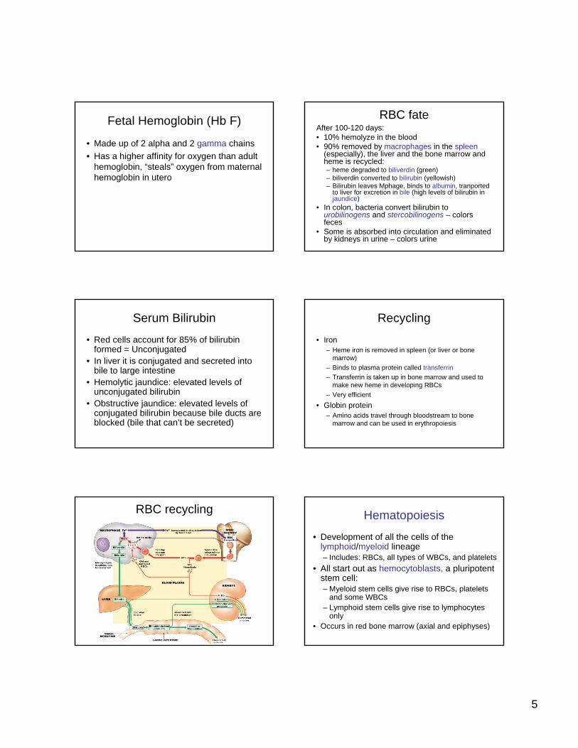
• Composed of:– Four protein chains called globins
• adults: 2 alpha and 2 beta chains– Each of these four chains is bound to a
pigment molecules called heme• each of which contain one iron ion (red color) and
bind one oxygen molecule
• Each RBC ~280 million molecules
Hemoglobin Structure• Complex quaternary structure
Figure 19–3
5
Fetal Hemoglobin (Hb F)
• Made up of 2 alpha and 2 gamma chains• Has a higher affinity for oxygen than adult
hemoglobin, “steals” oxygen from maternal hemoglobin in utero
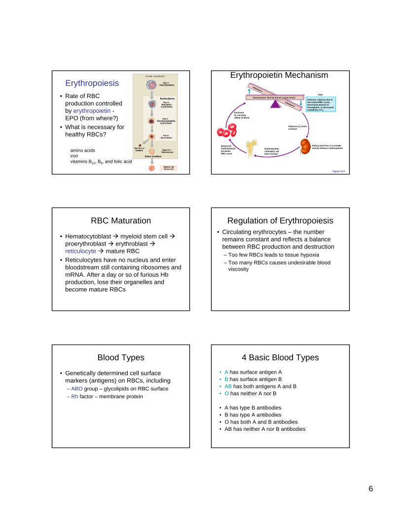
RBC fateAfter 100-120 days:• 10% hemolyze in the blood• 90% removed by macrophages in the spleen
(especially), the liver and the bone marrow and heme is recycled:– heme degraded to biliverdin (green)– biliverdin converted to bilirubin (yellowish)– Bilirubin leaves Mphage, binds to albumin, tranported
to liver for excretion in bile (high levels of bilirubin injaundice)
• In colon, bacteria convert bilirubin to urobilinogens and stercobilinogens – colors feces
• Some is absorbed into circulation and eliminated by kidneys in urine – colors urine
Serum Bilirubin
• Red cells account for 85% of bilirubinformed = Unconjugated
• In liver it is conjugated and secreted into bile to large intestine
• Hemolytic jaundice: elevated levels of unconjugated bilirubin
• Obstructive jaundice: elevated levels of conjugated bilirubin because bile ducts are blocked (bile that can’t be secreted)
Recycling
• Iron– Heme iron is removed in spleen (or liver or bone
marrow)– Binds to plasma protein called transferrin– Transferrin is taken up in bone marrow and used to
make new heme in developing RBCs– Very efficient
• Globin protein– Amino acids travel through bloodstream to bone
marrow and can be used in erythropoiesis
RBC recycling Hematopoiesis
• Development of all the cells of the lymphoid/myeloid lineage– Includes: RBCs, all types of WBCs, and platelets
• All start out as hemocytoblasts, a pluripotentstem cell:– Myeloid stem cells give rise to RBCs, platelets
and some WBCs– Lymphoid stem cells give rise to lymphocytes
only• Occurs in red bone marrow (axial and epiphyses)
6
Erythropoiesis• Rate of RBC
production controlled by erythropoietin -EPO (from where?)
• What is necessary for healthy RBCs?
amino acidsironvitamins B12, B6, and folic acid
Homeostasis: Normal blood oxygen levels
IncreasesO2-carryingability of blood
Erythropoietinstimulates redbone marrow
Reduces O2 levelsin blood
Kidney (and liver to a smallerextent) releases erythropoietin
Enhancederythropoiesisincreases RBC count
Stimulus: Hypoxia due todecreased RBC count,decreased amount of hemoglobin, or decreased availability of O2
Start
Imbalance
Imbalance
Erythropoietin Mechanism
Figure 17.6
Figure 19–5
RBC Maturation
• Hematocytoblast myeloid stem cellproerythroblast erythroblastreticulocyte mature RBC
• Reticulocytes have no nucleus and enter bloodstream still containing ribosomes and mRNA. After a day or so of furious Hbproduction, lose their organelles and become mature RBCs
Regulation of Erythropoiesis• Circulating erythrocytes – the number
remains constant and reflects a balance between RBC production and destruction– Too few RBCs leads to tissue hypoxia– Too many RBCs causes undesirable blood
viscosity
Blood Types
• Genetically determined cell surface markers (antigens) on RBCs, including– ABO group – glycolipids on RBC surface– Rh factor – membrane protein
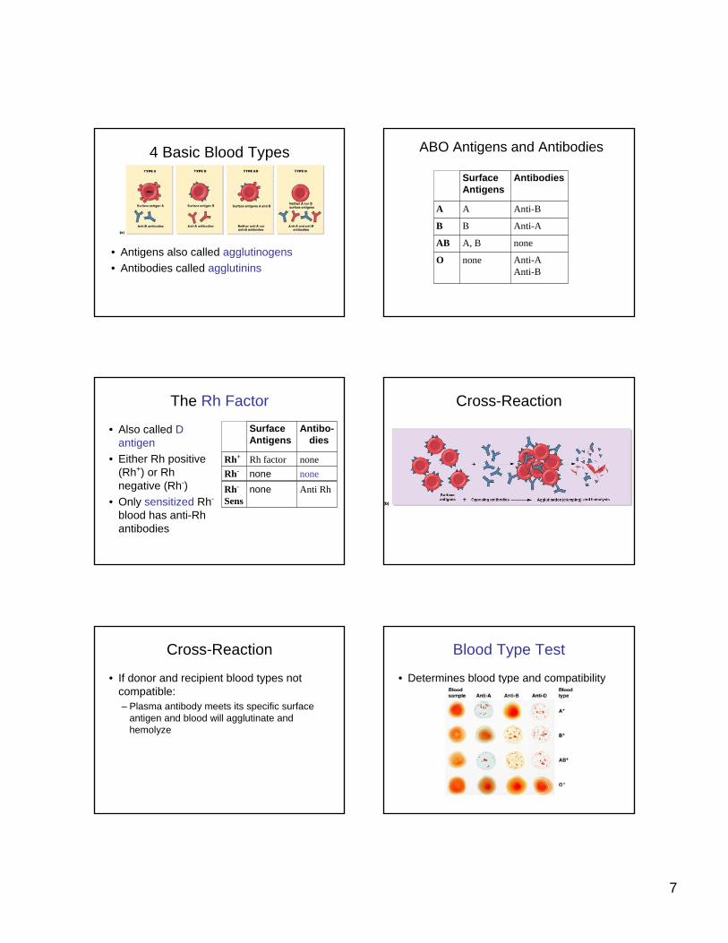
4 Basic Blood Types• A has surface antigen A• B has surface antigen B• AB has both antigens A and B• O has neither A nor B
• A has type B antibodies• B has type A antibodies• O has both A and B antibodies• AB has neither A nor B antibodies
7
Figure 19–6a
4 Basic Blood Types
• Antigens also called agglutinogens• Antibodies called agglutinins
Anti-AAnti-B
noneO
noneA, BAB
Anti-ABB
Anti-BAA
AntibodiesSurfaceAntigens
ABO Antigens and Antibodies
The Rh Factor
• Also called D antigen
• Either Rh positive (Rh+) or Rhnegative (Rh-)
• Only sensitized Rh-
blood has anti-Rhantibodies
nonenoneRh-noneRh factorRh+
Antibo-dies
SurfaceAntigens
Anti RhnoneRh-
Sens
Figure 19–6b
Cross-Reaction
Cross-Reaction
• If donor and recipient blood types not compatible:– Plasma antibody meets its specific surface
antigen and blood will agglutinate and hemolyze
Blood Type Test
• Determines blood type and compatibility
Figure 19–7
8
Cross-Match Test
• Performed on donor and recipient blood for compatibility to blood surface antigens other than ABO and Rh
Blood type questions
• Which blood type is the best in emergency settings (hint: which type can be given to anyone?)
• Which blood type is the lucky one that can receive blood from any donor?
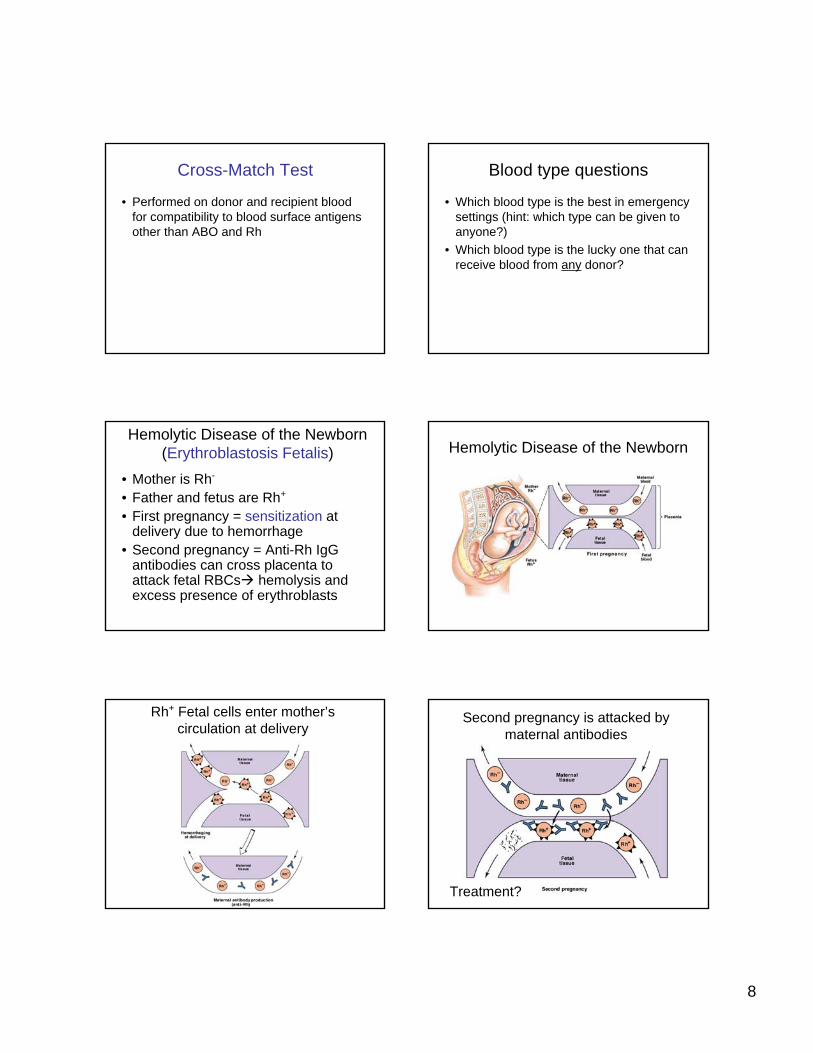
• Mother is Rh-
• Father and fetus are Rh+
• First pregnancy = sensitization at delivery due to hemorrhage
• Second pregnancy = Anti-Rh IgGantibodies can cross placenta to attack fetal RBCs hemolysis and excess presence of erythroblasts
Hemolytic Disease of the Newborn (Erythroblastosis Fetalis) Hemolytic Disease of the Newborn
Rh+ Fetal cells enter mother’s circulation at delivery
Second pregnancy is attacked by maternal antibodies
Treatment?

9
Transfusions
• Unit whole blood = 500ml• About half of this is plasma which contains
antibodies. There is a slight risk of graft versus host (GVH) reactions, but since the volume in one unit is only about 10% of total plasma volume, usually gets diluted out
• If RBCs are needed, can use packed RBCs instead of whole blood
White Blood Cells (WBCs)
• Leukocytes: the only blood components that are complete cells; have nuclei and other organelles, not involved in oxygen transport.
• Functions:– Defend against pathogens– Remove toxins and wastes– Attack abnormal cells
WBC in blood vs. tissue
• Very small numbers in blood:– 6000 to 9000 per microliter– Outnumbered 1000:1 by RBCs– But only 1% of WBC are in blood
• Most WBCs are not found in blood but instead in connective tissue proper and in lymphatic system organs
• Can leave capillaries via diapedesis
Circulating WBCs
• WBCs can migrate out of capillaries into tissues via diapedesis
• Have amoeboid movement (using actin)• Attracted to chemical stimuli (positive
chemotaxis)• Some are phagocytic: neutrophils,
eosinophils, and monocytes
5 Types of WBCs
1. Neutrophils2. Lymphocytes3. Monocytes4. Eosinophils5. Basophils
“Never Let Monkeys Eat Bananas”Figure 19–9
Types of WBCs
10
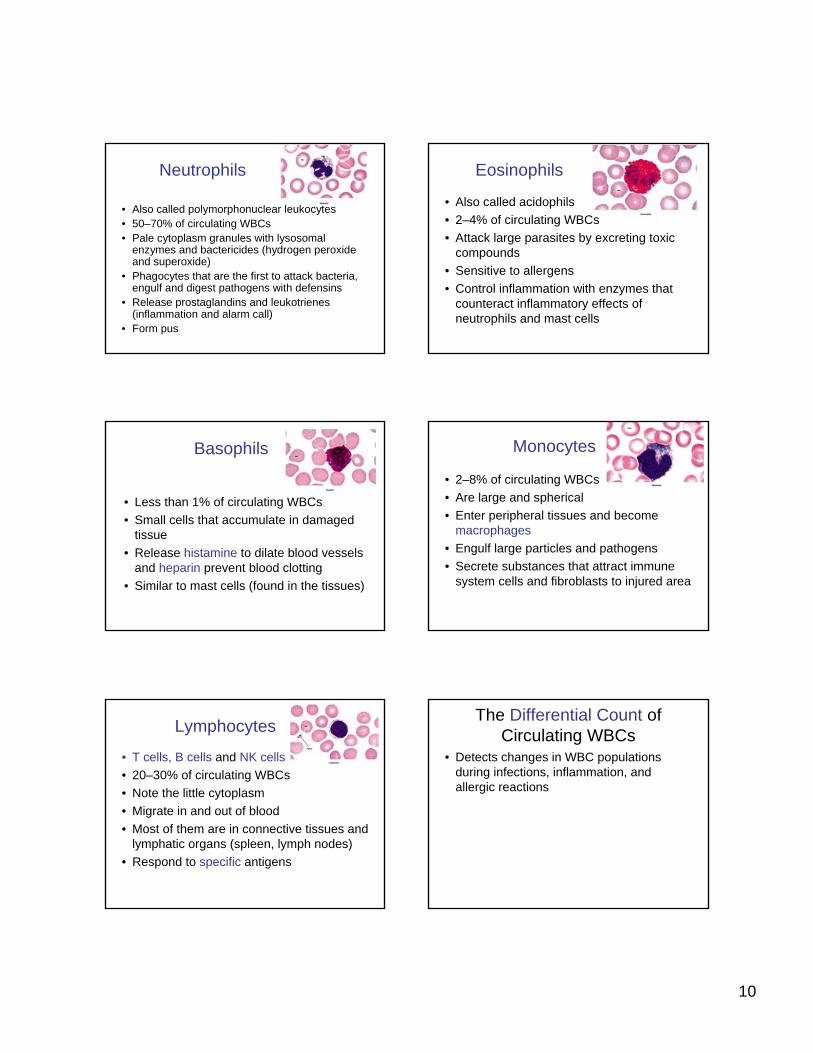
Neutrophils
• Also called polymorphonuclear leukocytes • 50–70% of circulating WBCs• Pale cytoplasm granules with lysosomal
enzymes and bactericides (hydrogen peroxide and superoxide)
• Phagocytes that are the first to attack bacteria, engulf and digest pathogens with defensins
• Release prostaglandins and leukotrienes(inflammation and alarm call)
• Form pus
Eosinophils
• Also called acidophils• 2–4% of circulating WBCs• Attack large parasites by excreting toxic
compounds• Sensitive to allergens • Control inflammation with enzymes that
counteract inflammatory effects of neutrophils and mast cells
Basophils
• Less than 1% of circulating WBCs• Small cells that accumulate in damaged
tissue• Release histamine to dilate blood vessels
and heparin prevent blood clotting• Similar to mast cells (found in the tissues)
Monocytes
• 2–8% of circulating WBCs• Are large and spherical• Enter peripheral tissues and become
macrophages• Engulf large particles and pathogens• Secrete substances that attract immune
system cells and fibroblasts to injured area
Lymphocytes
• T cells, B cells and NK cells• 20–30% of circulating WBCs• Note the little cytoplasm• Migrate in and out of blood• Most of them are in connective tissues and
lymphatic organs (spleen, lymph nodes)• Respond to specific antigens
The Differential Count of Circulating WBCs
• Detects changes in WBC populations during infections, inflammation, and allergic reactions

11
WBC Disorders
• Leukopenia:– abnormally low WBC count
• Leukocytosis:– high WBC count (normal response to
infection)• Leukemia:
– extremely high WBC count
Blood disease nomenclature
• -penia (poverty): too little of a cell type in the blood
• -cytosis: too much of a cell type in the blood
• -emia: refering to the presence of something (anything) in the blood
Figure 19–10
Hematopoiesis: WBCs WBC classes• Granulocytes – neutrophils, eosinophils, and
basophils– Contain cytoplasmic granules that stain specifically
(acidic, basic, or both) with Wright’s stain– Are larger and usually shorter-lived than RBCs– Have lobed nuclei– Are all phagocytic cells
• Agranulocytes – lymphocytes and monocytes:– Lack visible cytoplasmic granules– Have spherical (lymphocytes) or kidney-shaped
(monocytes) nuclei
WBC Production• Like RBCs, WBCs originate from
hemocytoblasts in the bone marrow• Hemocytoblasts differentiate into myeloid stem
cells and lymphoid stem cells• Myeloid stem cells become myeloblasts, which
give rise to neurophils, basophils, and eosinophils (granulocytes), OR monoblasts, which become monocytes.
• Lymphoid stem cells become lymphoblasts, and give rise to lymphocytes (B, T, and NK cells)
• All complete their development in the bone marrow except T cells, which mature in the thymus
4 Colony-Stimulating Factors (CSFs)
• Hormones that regulate blood cell populations:M-CSF:
• stimulates monocyte productionG-CSF:
• stimulates granulocyte production (neutrophils, eosinophils, and basophils)
GM-CSF:• stimulates granulocyte and monocyte production
Multi-CSF:• accelerates production of granulocytes, monocytes,
platelets, and RBCs (all blood except lymphocytes)
12
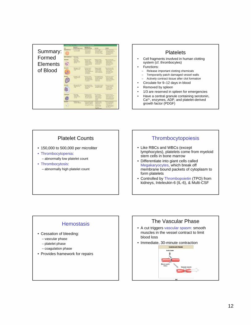
Summary: Formed Elements of Blood
Table 19–3
Platelets• Cell fragments involved in human clotting
system (cf. thrombocytes)• Functions:
– Release important clotting chemicals– Temporarily patch damaged vessel walls– Actively contract tissue after clot formation
• Circulate for 9–12 days in blood• Removed by spleen• 1/3 are reserved in spleen for emergencies• Have a central granule containing serotonin,
Ca2+, enzymes, ADP, and platelet-derived growth factor (PDGF)
Platelet Counts
• 150,000 to 500,000 per microliter• Thrombocytopenia:
– abnormally low platelet count• Thrombocytosis:
– abnormally high platelet count
Thrombocytopoiesis
• Like RBCs and WBCs (except lymphocytes), platelets come from myeloid stem cells in bone marrow
• Differentiate into giant cells called Megakaryocytes, which break off membrane bound packets of cytoplasm to form platelets
• Controlled by Thrombopoietin (TPO) from kidneys, Inteleukin-6 (IL-6), & Multi-CSF
Hemostasis
• Cessation of bleeding:– vascular phase– platelet phase– coagulation phase
• Provides framework for repairs
Figure 19–11a
The Vascular Phase• A cut triggers vascular spasm: smooth
muscles in the vessel contract to limit blood loss
• Immediate, 30-minute contraction
13
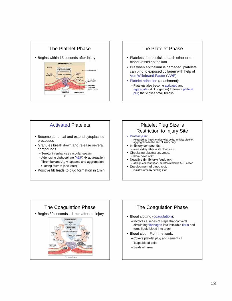
The Platelet Phase
• Begins within 15 seconds after injury
Figure 19–11b
The Platelet Phase
• Platelets do not stick to each other or to blood vessel epithelium
• But when epithelium is damaged, platelets can bind to exposed collagen with help ofVon Willebrand Factor (VWF)
• Platelet adhesion (attachment):– Platelets also become activated and
aggregate (stick together) to form a platelet plug that closes small breaks
Activated Platelets
• Become spherical and extend cytoplasmicprocesses
• Granules break down and release several compounds – Serotonin enhances vascular spasm– Adenosine diphosphate (ADP) aggregation– Thromboxane A2 spasms and aggregation– Clotting factors (see later)
• Positive f/b leads to plug formation in 1min
Platelet Plug Size isRestriction to Injury Site
• Prostacyclin:– released by intact endothelial cells, inhibits platelet
aggregation to the site of injury only• Inhibitory compounds:
– released by other white blood cells• Circulating plasma enzymes:
– break down ADP• Negative (inhibitory) feedback:
– at high concentration, serotonin blocks ADP action• Development of blood clot:
– isolates area by sealing it off
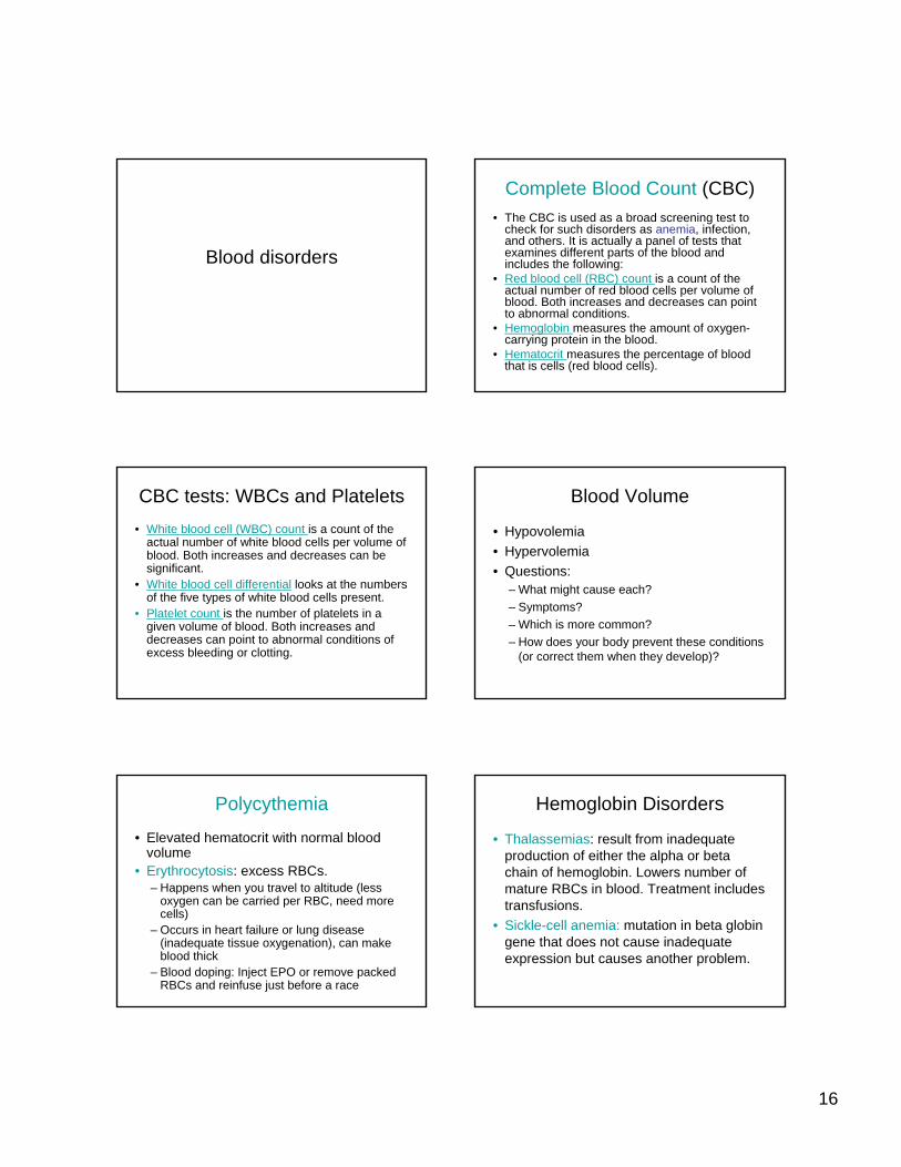
The Coagulation Phase• Begins 30 seconds – 1 min after the injury
Figure 19–12a
The Coagulation Phase
• Blood clotting (coagulation):– Involves a series of steps that converts
circulating fibrinogen into insoluble fibrin and turns liquid blood into a gel
• Blood clot = Fibrin network: – Covers platelet plug and cements it– Traps blood cells– Seals off area

14
Coagulation
Figure 17.13a
Clotting Factors
• Proteins or ions in plasma required for normal clotting– 11 major proteins– Calcium ions
3 Coagulation Pathways
• Extrinsic pathway:– begins in the vessel wall outside bloodstream
• Intrinsic pathway:– begins with circulating proenzymes within
bloodstreamNormally, both are activated• Common pathway:
– where intrinsic and extrinsic pathways converge
The Extrinsic Pathway
• Damaged cells release tissue factor (TF)also called factor III
• TF + other compounds including Calcium = enzyme complex
• Activates Factor X (ten)Shorter, faster pathway that bypasses several steps in the intrinsic pathway
The Intrinsic Pathway
• Activation of proenzymes by exposed collagen
• Combines with PF–3 from platelets• Series of reactions involving calcium result
in factors VIII and IX combining to activate Factor X
Slower, more productive pathwayHappens in vitro (acivated by glass
surfaces)
The Common Pathway
• Activated Factor X leads to enzyme prothrombinase (prothrombin activator)
• This converts prothrombin to thrombin• Thrombin converts fibrinogen (a ubiquitous
plasma protein) to fibrin• Fibrin polymer covers the platelet plug

15
Thrombin
• Stimulates formation of tissue factor, which stimulates release of PF-3 by platelets
• This positive feedback loop involves both intrinsic and extrinsic pathways and accelerates clotting
Clotting Area is Restricted
1. Anticoagulants (plasma proteins):– antithrombin-III– Fibrin itself binds thrombin and prevents it
from exerting positive feedback2. Heparin from endothelium3. Prostacyclin from endothelium4. Protein C (activated by thrombomodulin)
activates plasmin
Other Factors
• Calcium ions (Ca2+) and vitamin K (from diet and colon bacteria) are both essential to the clotting process
Clot Retraction• After clot has formed, platelets contract and pull
torn area together, squeezing out serum• Stabilizes injury site, facilitates repair• Takes 30–60 minutes • Repair
– Platelet-derived growth factor (PDGF) stimulates rebuilding of blood vessel wall
– Fibroblasts form a connective tissue patch– Stimulated by vascular endothelial growth factor
(VEGF), endothelial cells multiply and restore the endothelial lining
Fibrinolysis
• Slow process of dissolving clot• thrombin and tissue plasminogen activator
(t-PA): activate plasminogen– Note that this is the same thrombin that
helped activate the fibrin in the first place• Plasminogen produces plasmin, which
digests fibrin strands
Summary• Blood functions• Compostion of whole blood• Plasma• RBCs – structure, function, and
development• Blood types• WBCs• Platelets • Hemostasis
16
Blood disorders
Complete Blood Count (CBC)• The CBC is used as a broad screening test to
check for such disorders as anemia, infection, and others. It is actually a panel of tests that examines different parts of the blood and includes the following:
• Red blood cell (RBC) count is a count of the actual number of red blood cells per volume of blood. Both increases and decreases can point to abnormal conditions.
• Hemoglobin measures the amount of oxygen-carrying protein in the blood.
• Hematocrit measures the percentage of blood that is cells (red blood cells).
CBC tests: WBCs and Platelets• White blood cell (WBC) count is a count of the
actual number of white blood cells per volume of blood. Both increases and decreases can be significant.
• White blood cell differential looks at the numbers of the five types of white blood cells present.
• Platelet count is the number of platelets in a given volume of blood. Both increases and decreases can point to abnormal conditions of excess bleeding or clotting.
Blood Volume
• Hypovolemia• Hypervolemia• Questions:
– What might cause each?– Symptoms?– Which is more common?– How does your body prevent these conditions
(or correct them when they develop)?
Polycythemia
• Elevated hematocrit with normal blood volume
• Erythrocytosis: excess RBCs. – Happens when you travel to altitude (less
oxygen can be carried per RBC, need more cells)
– Occurs in heart failure or lung disease (inadequate tissue oxygenation), can make blood thick
– Blood doping: Inject EPO or remove packed RBCs and reinfuse just before a race
Hemoglobin Disorders
• Thalassemias: result from inadequate production of either the alpha or beta chain of hemoglobin. Lowers number of mature RBCs in blood. Treatment includes transfusions.
• Sickle-cell anemia: mutation in beta globingene that does not cause inadequate expression but causes another problem.
17
Thalassemias• Alpha-thalassemia
– We have four copies of alpha globin gene– 3 good/1bad: carrier– 2good/2bad: alpha-thalassemia trait– 1good/3bad: microcytic anemia– 4bad: die before birth
• Beta-thalassemia– We have only two copies of beta globin gene– No good copies: beta-thalassemia major:
• Severe microcytic anemia• Low hematocrit (below 20)
– One good copy: beta-thalassemia trait• Few clinical symptoms
Anemias
• Hematocrit or hemoglobin levels are below normal, caused by several conditions
• Characterized by a decrease in the oxygen carrying capacity of the blood (due to the problems with RBCs or with hemoglobin)
• Can be macrocytic (big RBCs) or microcytic
Sickle-Cell Anemia
• Mutation in beta globin gene resulting in production of HbS
• At low oxygen, cells with HbS become rigid and adopt a “sickle” shape: makes them fragile and can become stuck in small capillaries (last 10 days in blood)
• One bad copy: sickling trait• Two bad copies: SCA• Treatments?
Transfusions, hydroxyurea, butyrate
Pernicious Anemia
• Low RBC production due to lack of vitamin B12
• Vitamin B12 absorption requires Intrinsic factor (IF) from cells in the stomach. No IF, no B12.
Iron Deficiency Anemia
• Caused by low dietary iron or blood loss• RBCs made without enough functional
hemoglobin: microcytic• Low hematocrit• 12% of menstruating women may have it• Treatment?
Changes in blood parameters
• Macrocytic anemia caused by vitamin B12 deficiency.
• Microcytic anemia is seen in iron deficiency anemia or thalassemias.
18
Iron Loading
• Excess iron intake, gets depostied in peripheral tissues notably heart valves
• Very dangerous, leads to heart failure• Can develop as a result of repeated
transfusions of whole blood given to severely anemic patients – they need the functional RBCs, but the RBCs keep getting broken down and the iron is retained
Leukemia
• Blood cancer – no solid tumor (cf. lymphoma)
• Myeloid or lymphoid • Lymphoid more common in children• Myeloid more common in adults• Treatment?
Clotting Disorders: Excessive Clotting
• Embolus• Thrombus• Anticoagulant therapies:
– Heparin: activates antithrombin III– Coumadin: blocks Vitamin K action– t-PA: activates plasmin– Streptokinase/urokinase: also activate plasmin– Asprin: inactivates platelet enzymes and prostacyclin
production– EDTA – Calcium chelator
Clotting Disorders: Inadequate Clotting
• Hemophilia A: Gene for factor VIII is on X chromosome (sex-linked) and so this type of hemophilia is almost exclusively in males
• DIC – disseminated intravascularcoagulation: small fibrin clots form throughout the blood, leads to shortage of fibrin when it is needed