chapter 20, 21. lymphatic and immune systemsfacweb.northseattle.edu/jdahms/214win08/lect/chapters...
TRANSCRIPT
1
Chapter 20, 21. Lymphatic and Immune Systems
Part I. Tissues and Nonspecific Immunity
Overview
• Lymphatic system functions• Lymph vessel anatomy• Lymphocytes• Lymphatic tissues (nodules)• Lymphatic organs (nodes, thymus, spleen)• Nonspecific defenses
The Lymphatic System• Consists of two semi-independent parts:
– A network of lymphatic vessels– Lymphoid tissues and organs scattered throughout the body
• Returns interstitial fluid and leaked plasma proteins back to the blood
• Lymph = interstitial fluid once it has entered lymphatic vessels• Produces, maintains, and distributes lymphocytes• Protects us against pathogens: microscopic organisms that cause
disease:– viruses– bacteria– fungi– parasites
• Each attacks in a specific way
Parts of the Lymphatic System• Lymph:
– a fluid derived from plasma/interstitial fluid– does not have plasma proteins
• Lymphatic vessels (lymphatics): – network that carries lymph from peripheral tissues
to the venous system• Lymphoid tissues and lymphoid organs:
– found throughout the body• Lymphocytes, phagocytes, and other immune
system cells
The Immune System
• Includes all body cells and tissues involved in production of immunity, not just lymphatic system
• What else? Integumentary, digestive, cardiovascular, respiratory, others all contribute to the immune system
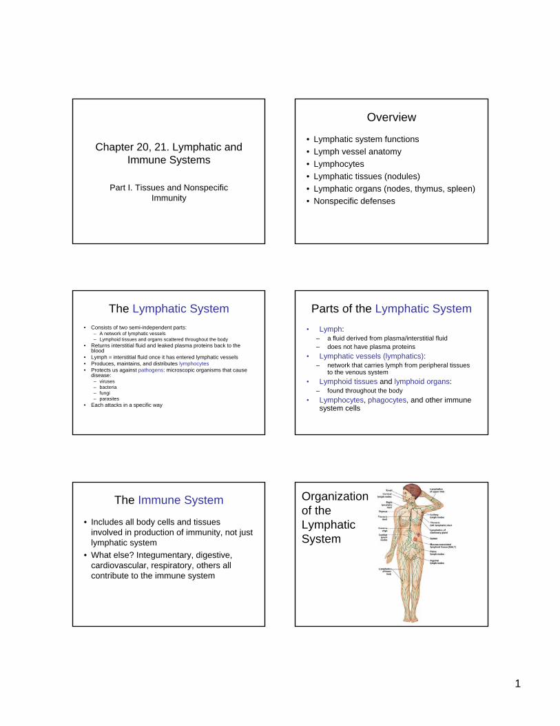
Organization of the Lymphatic System
Figure 22–1
2
Lymphocyte Circulation• From blood plasma to interstitial fluid
(through capillary exchange) to lymph and back to the venous system
Lymphatic Vessels
• One-way system of vessels that carry lymph toward the heart
• Lymphatic system begins with smallest vessels called lymphatic capillaries (terminal lymphatics)
• Lymphatic caps differ from blood caps in 4 ways:– start as dead end pockets rather than tubes– larger diameters– thinner walls– flat or irregular in section
Figure 22–2
Lymphatic Capillaries Lymphatic Capillaries
• Endothelial cells loosely bound together with overlap which acts as one-way valve:– allows fluids, solutes, viruses, and bacteria to enter
(very permeable)– prevents return to intercellular space
• During inflammation, lymph capillaries can absorb:– Cell debris– Pathogens– Cancer cells
• Cells in the lymph nodes cleanse and “examine”this debris
• Lacteals = special lymphatic capillaries in small intestine, transport lipids from digestive tract
Lymph Flow
• From lymphatic capillaries to larger lymphatic collecting vessels containing larger one-way valves
• Valves make them appear beaded• Structure similar to veins• Lymphatic vessels travel with veins
Figure 22–3
Lymphatic Vessels and Valves
3
Superficial and Deep Lymphatic Vessels
• Superficial lymphatic vessels are located in:– skin– mucous membranes– serous membranes lining body cavities
• Deep lymphatic vessels are larger vessels that accompany deep arteries and veins
• Superficial and deep join to form large lymphatic trunks, which empty into 2 major collecting vessels: – thoracic duct – right lymphatic duct
Figure 22–4
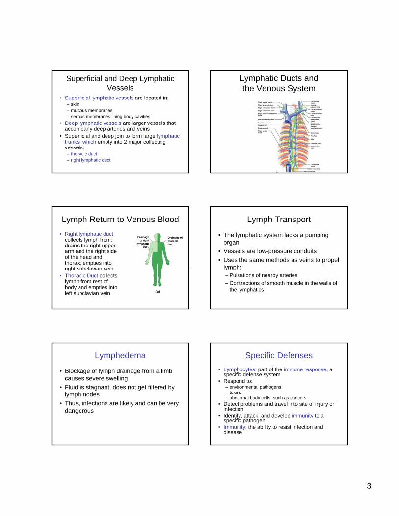
Lymphatic Ducts and the Venous System
Lymph Return to Venous Blood• Right lymphatic duct
collects lymph from: drains the right upper arm and the right side of the head and thorax; empties into right subclavian vein
• Thoracic Duct collects lymph from rest of body and empties into left subclavian vein
Lymph Transport
• The lymphatic system lacks a pumping organ
• Vessels are low-pressure conduits• Uses the same methods as veins to propel
lymph:– Pulsations of nearby arteries– Contractions of smooth muscle in the walls of
the lymphatics
Lymphedema
• Blockage of lymph drainage from a limb causes severe swelling
• Fluid is stagnant, does not get filtered by lymph nodes
• Thus, infections are likely and can be very dangerous
Specific Defenses• Lymphocytes: part of the immune response, a
specific defense system• Respond to:
– environmental pathogens– toxins– abnormal body cells, such as cancers
• Detect problems and travel into site of injury or infection
• Identify, attack, and develop immunity to a specific pathogen
• Immunity: the ability to resist infection and disease
4
Lymphocyte Production
• Lymphocytes are produced in:– lymphoid tissues (e.g., tonsils)– lymphoid organs (e.g., spleen, thymus)– red bone marrow
• Make up 20–30% of circulating leukocytes• Majority are stored in lymphoid organs, not
circulating (remember that only about 1% of your WBCs are in the blood)
Classes of Circulating Lymphocytes
• T cells: thymus-dependent– Make up 80% of circulating lymphocytes
• B cells: bone–marrow derived– Make up 10–15% of circulating lymphocytes
• NK cells: natural killer cells– Make up 5–10% of circulating lymphocytes
Types of T Cells1. Cytotoxic T cells
– Attack cells infected by viruses– Produce cell-mediated immunity
2. Helper T cells– Stimulate function of T cells and B cells
3. Suppressor T cells– Inhibit function of T cells and B cells
4. Inflammatory T cells • The last two are called regulatory T Cells
because they control the sensitivity of immune response
B Cells
• Differentiate into plasma cells, which produce and secrete antibodies(immunoglobin proteins)
• Antibodies bind to their specific target antigen and initiate antibody-mediated immunity
Antigens
• Antigen – anything the body perceives as foreign– Bacteria and their toxins; viruses– Mismatched RBCs or cancer cells
Natural Killer (NK) Cells• Also called large granular lymphocytes • Responsible for immunological
surveillance• Attack:
– foreign cells– virus-infected cells– cancer cells
5
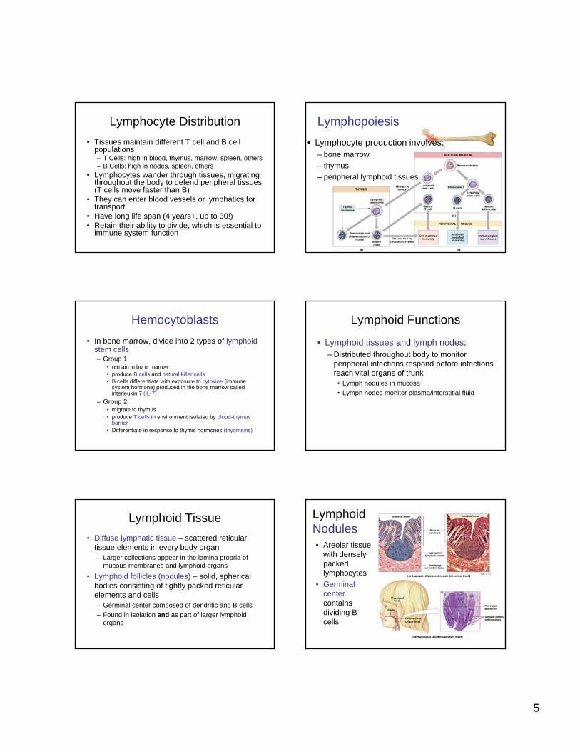
Lymphocyte Distribution• Tissues maintain different T cell and B cell
populations– T Cells: high in blood, thymus, marrow, spleen, others– B Cells: high in nodes, spleen, others
• Lymphocytes wander through tissues, migrating throughout the body to defend peripheral tissues (T cells move faster than B)
• They can enter blood vessels or lymphatics for transport
• Have long life span (4 years+, up to 30!)• Retain their ability to divide, which is essential to
immune system functionFigure 22–5
Lymphopoiesis• Lymphocyte production involves:
– bone marrow– thymus– peripheral lymphoid tissues
Hemocytoblasts• In bone marrow, divide into 2 types of lymphoid
stem cells – Group 1:
• remain in bone marrow• produce B cells and natural killer cells • B cells differentiate with exposure to cytokine (immune
system hormone) produced in the bone marrow called interleukin 7 (IL-7)
– Group 2:• migrate to thymus• produce T cells in environment isolated by blood-thymus
barrier • Differentiate in response to thymic hormones (thyomsins)
Lymphoid Functions
• Lymphoid tissues and lymph nodes:– Distributed throughout body to monitor
peripheral infections respond before infections reach vital organs of trunk
• Lymph nodules in mucosa• Lymph nodes monitor plasma/interstitial fluid
Lymphoid Tissue• Diffuse lymphatic tissue – scattered reticular
tissue elements in every body organ– Larger collections appear in the lamina propria of
mucous membranes and lymphoid organs• Lymphoid follicles (nodules) – solid, spherical
bodies consisting of tightly packed reticular elements and cells– Germinal center composed of dendritic and B cells– Found in isolation and as part of larger lymphoid
organs
Figure 22–6
Lymphoid Nodules• Areolar tissue
with densely packed lymphocytes
• Germinal centercontains dividing B cells
6
Distribution of Lymphoid Nodules
• Respiratory tract (tonsils)• Along digestive tract (MALT = Mucosa
Associated Lymphoid Tissue e.g. Peyer’spatches, appendix)
• Urinary tract• Found within some lymphoid organs
(Lymph nodes, spleen)
Lymphoid Organs
• Are separated from surrounding tissues by a fibrous connective-tissue capsule = encapsulated.
• Include:– Lymph nodes– Thymus – Spleen
Figure 22–7
Lymph Nodes
Diameter = 1-25 mm
Lymph Node• Principal lymphoid organs of the body• Embedded in connective tissue and clustered
along lymphatic vessels• Aggregations of these nodes occur near the
body surface in inguinal, axillary, and cervical regions of the body (Lymph Glands)
• Functions– Act as filters, purifying lymph before returning it to
venous circulation, removes debris, pathogens, 99% of antigens
– monitor for antigens and mount an attack against them (activate the immune system)
Lymph Node - Structure• Surrounded by fibrous capsule• Trabeculae: fibrous partitions made of collagen
fibers that extend from capsule into interior of lymph node
• Hilus: shallow indentation where blood vessels and nerves reach the lymph node
• Afferent Lymphatic Vessels: carry lymph from peripheral tissues to lymph node
• Efferent Lymphatic Vessels: leave lymph node at hilus, carry lymph to venous circulation
Lymph Flow• Flows from afferent lymphatics through lymph
node in a network of sinuses:• Enters subcapsular sinus:
– contains macrophages and dendritic cells• Through outer cortex:
– contains B cells within germinal centers• Through deep cortex:
– dominated by T cells• Through the core (medulla):
– contains B cells and plasma cells– organized into medullary cords
• Into hilus and efferent lymphatics (less of theses than afferent)
7
Antigen Presentation
• First step in immune response• Extracted antigens are “presented” to
lymphocytes by macrophages, dendriticcells (in lymph nodes, these are in the subcapsular area)
Lymphoid Organs
Figure 20.5
• The spleen, thymus gland, and tonsils
• Peyer’s patches and bits of lymphatic tissue scattered in connective tissue
• All are composed of reticular connective tissue
• All help protect the body• Only lymph nodes filter
lymph
Figure 22–8
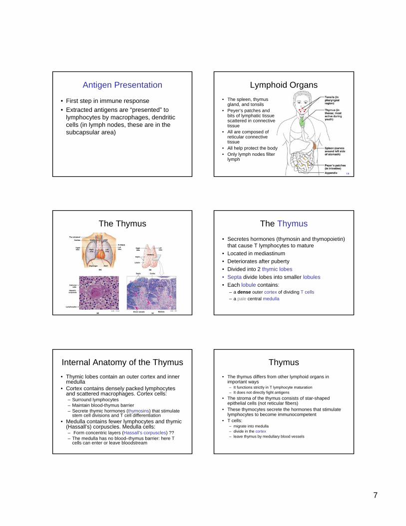
The Thymus The Thymus
• Secretes hormones (thymosin and thymopoietin) that cause T lymphocytes to mature
• Located in mediastinum• Deteriorates after puberty• Divided into 2 thymic lobes• Septa divide lobes into smaller lobules• Each lobule contains:
– a dense outer cortex of dividing T cells– a pale central medulla
Internal Anatomy of the Thymus• Thymic lobes contain an outer cortex and inner
medulla• Cortex contains densely packed lymphocytes
and scattered macrophages. Cortex cells:– Surround lymphocytes– Maintain blood-thymus barrier– Secrete thymic hormones (thymosins) that stimulate
stem cell divisions and T cell differentiation• Medulla contains fewer lymphocytes and thymic
(Hassall’s) corpuscles. Medulla cells:– Form concentric layers (Hassall’s corpuscles) ??– The medulla has no blood–thymus barrier: here T
cells can enter or leave bloodstream
Thymus• The thymus differs from other lymphoid organs in
important ways– It functions strictly in T lymphocyte maturation– It does not directly fight antigens
• The stroma of the thymus consists of star-shaped epithelial cells (not reticular fibers)
• These thymocytes secrete the hormones that stimulate lymphocytes to become immunocompetent
• T cells:– migrate into medulla– divide in the cortex– leave thymus by medullary blood vessels
8
Figure 22–9
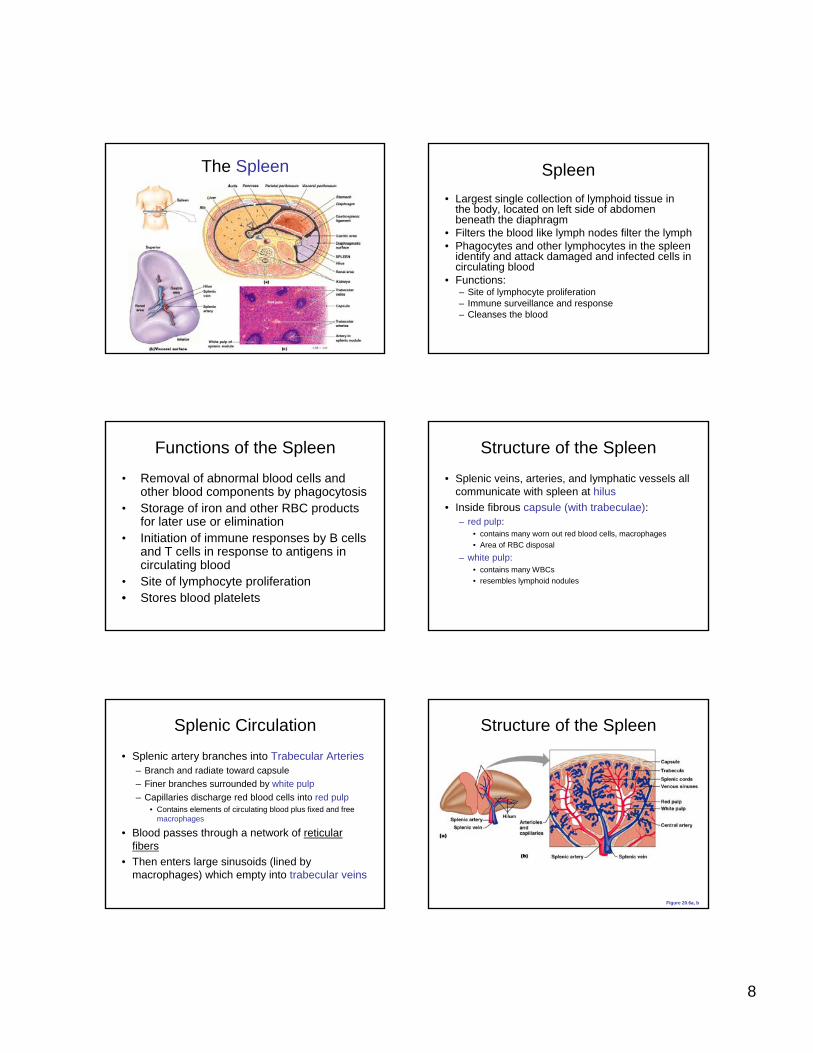
The Spleen Spleen• Largest single collection of lymphoid tissue in
the body, located on left side of abdomen beneath the diaphragm
• Filters the blood like lymph nodes filter the lymph• Phagocytes and other lymphocytes in the spleen
identify and attack damaged and infected cells in circulating blood
• Functions:– Site of lymphocyte proliferation– Immune surveillance and response– Cleanses the blood
Functions of the Spleen
• Removal of abnormal blood cells and other blood components by phagocytosis
• Storage of iron and other RBC products for later use or elimination
• Initiation of immune responses by B cells and T cells in response to antigens in circulating blood
• Site of lymphocyte proliferation• Stores blood platelets
Structure of the Spleen
• Splenic veins, arteries, and lymphatic vessels all communicate with spleen at hilus
• Inside fibrous capsule (with trabeculae):– red pulp:
• contains many worn out red blood cells, macrophages• Area of RBC disposal
– white pulp:• contains many WBCs• resembles lymphoid nodules
Splenic Circulation
• Splenic artery branches into Trabecular Arteries– Branch and radiate toward capsule– Finer branches surrounded by white pulp– Capillaries discharge red blood cells into red pulp
• Contains elements of circulating blood plus fixed and free macrophages
• Blood passes through a network of reticular fibers
• Then enters large sinusoids (lined by macrophages) which empty into trabecular veins
Structure of the Spleen
Figure 20.6a, b
9
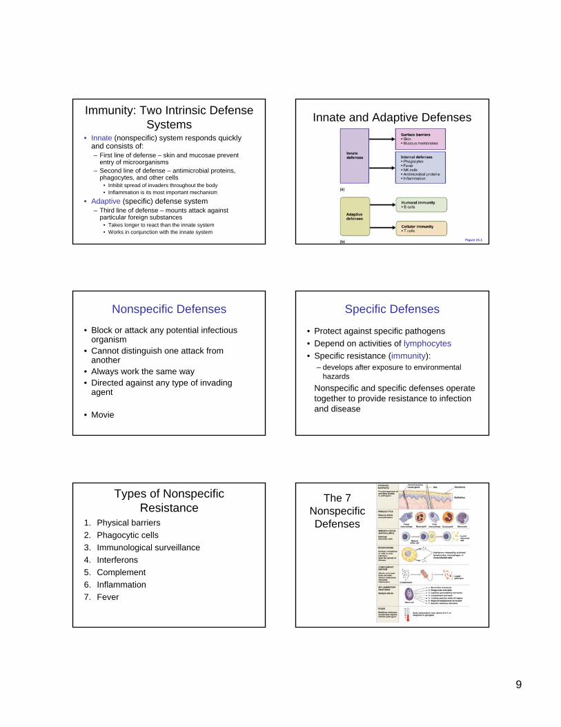
Immunity: Two Intrinsic Defense Systems
• Innate (nonspecific) system responds quickly and consists of:– First line of defense – skin and mucosae prevent
entry of microorganisms– Second line of defense – antimicrobial proteins,
phagocytes, and other cells • Inhibit spread of invaders throughout the body• Inflammation is its most important mechanism
• Adaptive (specific) defense system– Third line of defense – mounts attack against
particular foreign substances• Takes longer to react than the innate system• Works in conjunction with the innate system
Innate and Adaptive Defenses
Figure 21.1
Nonspecific Defenses
• Block or attack any potential infectious organism
• Cannot distinguish one attack from another
• Always work the same way • Directed against any type of invading
agent
• Movie
Specific Defenses
• Protect against specific pathogens• Depend on activities of lymphocytes• Specific resistance (immunity):
– develops after exposure to environmental hazards
Nonspecific and specific defenses operate together to provide resistance to infection and disease
Types of Nonspecific Resistance
1. Physical barriers2. Phagocytic cells3. Immunological surveillance4. Interferons5. Complement6. Inflammation7. Fever
Figure 22–10
The 7 Nonspecific Defenses
10
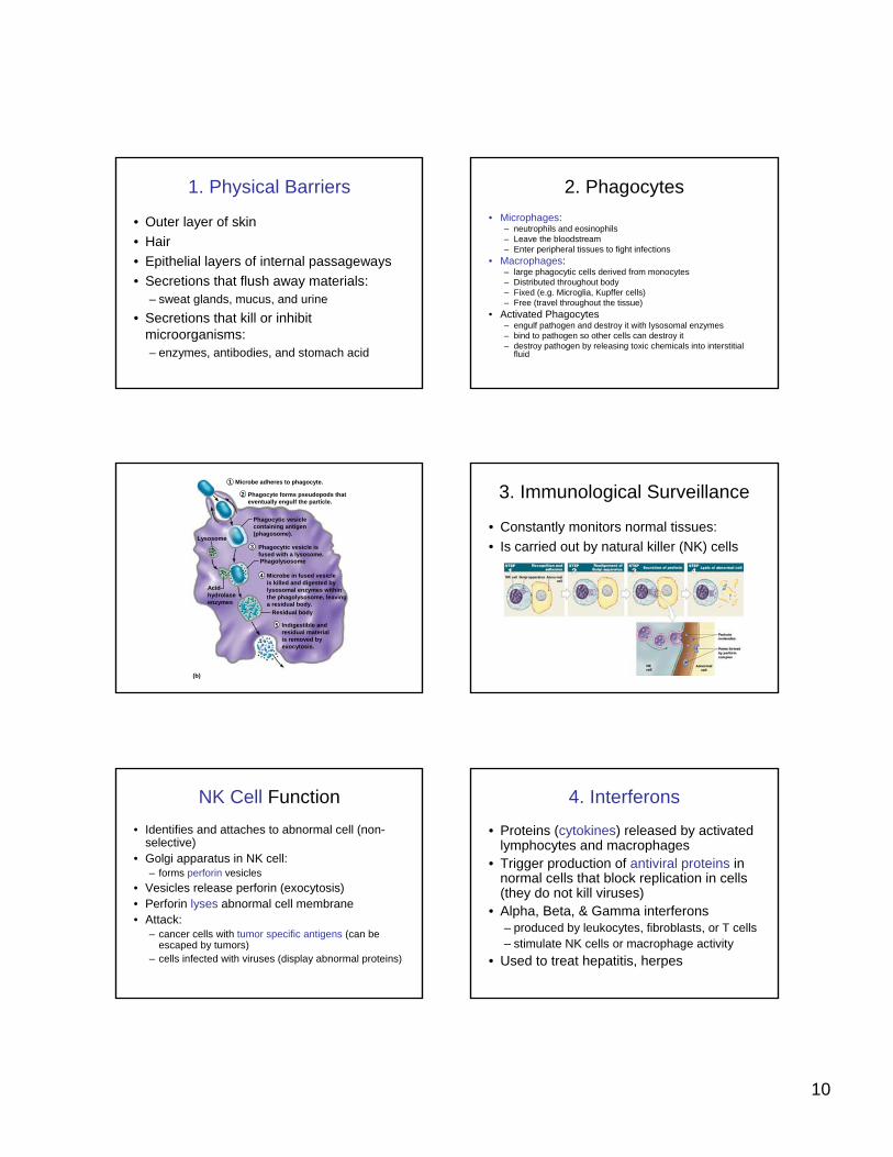
1. Physical Barriers
• Outer layer of skin• Hair• Epithelial layers of internal passageways• Secretions that flush away materials:
– sweat glands, mucus, and urine• Secretions that kill or inhibit
microorganisms:– enzymes, antibodies, and stomach acid
2. Phagocytes• Microphages:
– neutrophils and eosinophils– Leave the bloodstream– Enter peripheral tissues to fight infections
• Macrophages:– large phagocytic cells derived from monocytes– Distributed throughout body– Fixed (e.g. Microglia, Kupffer cells)– Free (travel throughout the tissue)
• Activated Phagocytes – engulf pathogen and destroy it with lysosomal enzymes– bind to pathogen so other cells can destroy it– destroy pathogen by releasing toxic chemicals into interstitial
fluid
(b)
Lysosome
Microbe adheres to phagocyte.
Phagocyte forms pseudopods thateventually engulf the particle.
Phagocytic vesicle isfused with a lysosome.
Microbe in fused vesicleis killed and digested bylysosomal enzymes withinthe phagolysosome, leavinga residual body.
Indigestible andresidual materialis removed byexocytosis.
Phagocytic vesiclecontaining antigen(phagosome).
Residual body
Acidhydrolaseenzymes
Phagolysosome
4
3
2
1
5
3. Immunological Surveillance
• Constantly monitors normal tissues:• Is carried out by natural killer (NK) cells
NK Cell Function• Identifies and attaches to abnormal cell (non-
selective)• Golgi apparatus in NK cell:
– forms perforin vesicles• Vesicles release perforin (exocytosis)• Perforin lyses abnormal cell membrane• Attack:
– cancer cells with tumor specific antigens (can be escaped by tumors)
– cells infected with viruses (display abnormal proteins)
4. Interferons
• Proteins (cytokines) released by activated lymphocytes and macrophages
• Trigger production of antiviral proteins in normal cells that block replication in cells (they do not kill viruses)
• Alpha, Beta, & Gamma interferons– produced by leukocytes, fibroblasts, or T cells – stimulate NK cells or macrophage activity
• Used to treat hepatitis, herpes
11
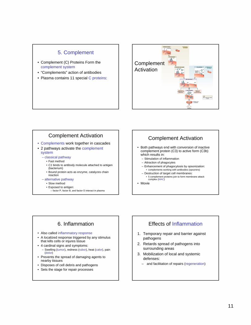
5. Complement
• Complement (C) Proteins Form the complement system
• “Complements” action of antibodies• Plasma contains 11 special C proteins:
Figure 22–12
Complement Activation
Complement Activation• Complements work together in cascades• 2 pathways activate the complement
system– classical pathway
• Fast method• C1 binds to antibody molecule attached to antigen
(bacterium)• Bound protein acts as enzyme, catalyzes chain
reaction– alternative pathway
• Slow method• Exposed to antigen:
– factor P, factor B, and factor D interact in plasma
Complement Activation• Both pathways end with conversion of inactive
complement protein (C3) to active form (C3b) which results in:– Stimulation of inflammation– Attraction of phagocytes– Enhancement of phagocytosis by opsonization:
• complements working with antibodies (opsonins)– Destruction of target cell membranes:
• 5 complement proteins join to form membrane attack complex (MAC)
• Movie
6. Inflammation• Also called inflammatory response• A localized response triggered by any stimulus
that kills cells or injures tissue• 4 cardinal signs and symptoms:
– Swelling (tumor), redness (rubor), heat (calor), pain (dolor)
• Prevents the spread of damaging agents to nearby tissues
• Disposes of cell debris and pathogens• Sets the stage for repair processes
Effects of Inflammation
1. Temporary repair and barrier against pathogens
2. Retards spread of pathogens into surrounding areas
3. Mobilization of local and systemic defenses:
– and facilitation of repairs (regeneration)
12
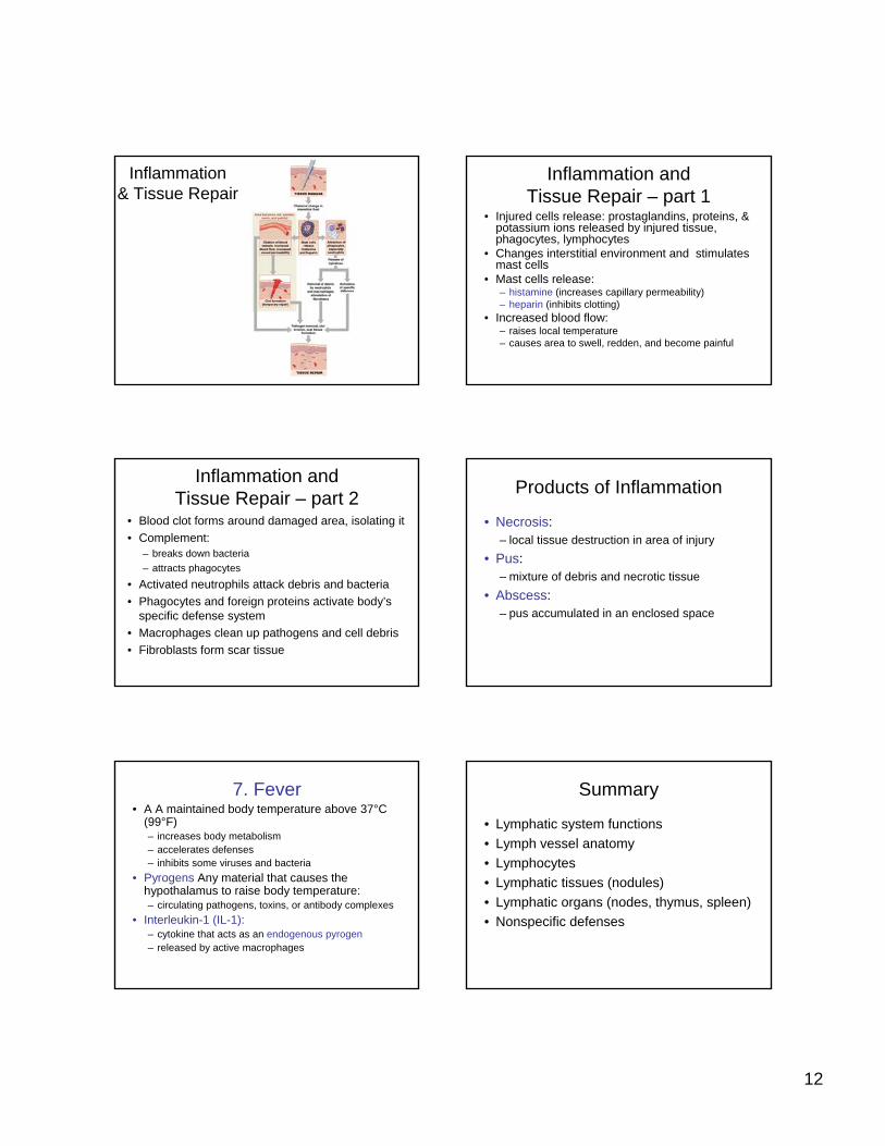
Figure 22–13
Inflammation& Tissue Repair
Inflammation and Tissue Repair – part 1
• Injured cells release: prostaglandins, proteins, & potassium ions released by injured tissue, phagocytes, lymphocytes
• Changes interstitial environment and stimulates mast cells
• Mast cells release:– histamine (increases capillary permeability)– heparin (inhibits clotting)
• Increased blood flow:– raises local temperature– causes area to swell, redden, and become painful
Inflammation and Tissue Repair – part 2
• Blood clot forms around damaged area, isolating it• Complement:
– breaks down bacteria– attracts phagocytes
• Activated neutrophils attack debris and bacteria• Phagocytes and foreign proteins activate body’s
specific defense system• Macrophages clean up pathogens and cell debris• Fibroblasts form scar tissue
Products of Inflammation
• Necrosis:– local tissue destruction in area of injury
• Pus:– mixture of debris and necrotic tissue
• Abscess:– pus accumulated in an enclosed space
7. Fever• A A maintained body temperature above 37°C
(99°F)– increases body metabolism– accelerates defenses– inhibits some viruses and bacteria
• Pyrogens Any material that causes the hypothalamus to raise body temperature:– circulating pathogens, toxins, or antibody complexes
• Interleukin-1 (IL-1):– cytokine that acts as an endogenous pyrogen– released by active macrophages
Summary
• Lymphatic system functions• Lymph vessel anatomy• Lymphocytes• Lymphatic tissues (nodules)• Lymphatic organs (nodes, thymus, spleen)• Nonspecific defenses