changing patterns in cytoskeletal mrnaexpressionandprotein
TRANSCRIPT

Proc. Natl. Acad. Sci. USAVol. 89, pp. 5749-5753, July 1992Developmental Biology
Changing patterns in cytoskeletal mRNA expression and proteinsynthesis during murine erythropoiesis in vivoLUANNE L. PETERS*t, ROBERT A. WHITES, CONNIE S. BIRKENMEIER*, MICHAEL L. BLOOM*,SAMUEL E. Luxt, AND JANE E. BARKER*§*The Jackson Laboratory, Bar Harbor, ME 04609; and *Children's Hospital and Dana-Farber Cancer Institute, Harvard University, Boston, MA 02115
Communicated by Elizabeth S. Russell, March 26, 1992
ABSTRACT The major cytoskeletal proteins a-spectrin,(3-spectrin, and ankyrin are synthesized and assembled into asupportive membrane skeleton during erythroid differentia-tion. Information on the temporal appearance of mRNA andprotein species is essential for understanding both the cyto-skeletal assembly process and the function of various isoforms.We have isolated highly enriched populations of fetal erythroidcells at various stages of maturation. mRNAs for erythroidankyrin, a-spectrin, and fi-spectrin were expressed at allstages but there were differences in transcript types and levels.The ratio of 9-kilobase (kb) to 7.5-kb erythroid ankyrintranscripts decreased markedly during differentiation, butthere was no change in the ratio of the 10.1-kb and 9.3-kberythroid fl-spectrin transcripts. The relative amounts ofankyrin, a-spectrin, and fl-spectrin mRNA increased duringyolk sac cell differentiation, whereas only a-spectrin mRNAincreased during differentiation of the fetal liver cells. Theamounts of fl-spectrin mRNA exceeded the amounts of a-spec-trin mRNA in the early precursors from both yolk sac and fetalliver; protein synthetic levels showed the same pattern. The16-day fetal peripheral reticulocytes, on the other hand, hadthe adult mRNA and protein synthetic ratios with a/f > 1.The data indicate that at least two mechanisms exist to meetchanging erythroid membrane cytoskeletal requirements dur-ing development in utero: (t) stage-specific processing of themRNA for the major cytoskeletal linker protein ankyrin and(it) developmentally regulated a/fl-spectrin protein syntheticrates.
The erythrocyte membrane skeleton is composed primarilyof tetramers of a (240 kDa) and f3 (220 kDa) spectrin joinedtogether by junctional complexes consisting of short actinfilaments and protein 4.1 to form a two-dimensional network(1, 2). Adducin, protein 4.9, myosin, and tropomyosin arealso present at the junctional complexes (1, 2). The spectrinlattice is attached to the erythrocyte plasma membranethrough at least two binding sites. One of these is at thejunctional complexes and is mediated by protein 4.1-glycophorin linkages (3, 4). The major plasma membraneattachment site is provided by ankyrin (protein 2.1), whichcontains high-affinity binding sites for both fl-spectrin and thetransmembrane anion exchanger (protein 3) (5). Binding ofthe membrane protein 4.2 to the ankyrin complex is thoughtto stabilize the interaction (6, 7). The integrity of the mem-brane cytoskeleton is essential to the survival ofthe red bloodcell, since heritable defects in spectrin and/or ankyrin resultin severe hemolytic anemia in humans and mice (8-13).Numerous studies have investigated the synthesis and
assembly of cytoskeletal proteins into the red-cell membraneskeleton (14-25). As it is difficult to obtain enriched popu-lations of stage-specific cells in vivo from adult animals, moststudies have been limited primarily to virus-transformed cell
lines (murine erythroleukemia) or spleen cells induced todifferentiate with dimethyl sulfoxide or erythropoietin, re-spectively (14-20, 22-24). Although the results have beeninformative, there are inconsistencies among the publishedobservations, especially related to the a/,S-spectrin proteinsynthetic ratios (17, 18, 20, 22). Furthermore, studies of thea/83 synthetic ratio by in vitro labeling of freshly isolatedchicken embryonic cells (26) and phenylhydrazine-treated rat(21) and mouse (27) cells are in agreement with one anotherbut the ratios are different from those noted in the majorityof the transformed cells.As an alternative to studying mixed populations from adult
mice or cell lines, we have utilized fetal mouse cells. Murineerythroid differentiation in utero occurs in two waves: (i)nucleated yolk sac-derived cells progress from pre-type 1 totype 3 erythroblasts during days 10-14 and (ii) fetal livercells, primarily pro- and basophilic erythroblasts at 12-14days, generate predominantly reticulocytes in the peripheralblood by 16 days (28). The transcripts and protein prQductsfrom erythroid a and fi spectrins were investigated simulta-neously. Stage-related shifts in mRNA levels and in a/,8-spectrin protein synthetic ratios did occur during normalerythroid maturation in utero. We suggest that the differencesreflect changing requirements of the erythrocyte membraneduring the extensive remodeling that accompanies erythro-cyte differentiation.
MATERIALS AND METHODSAnimals. Erythroid cells were obtained from normal fe-
tuses by timed matings of C57BL/6J (B6)-+/+ breedingpairs. The appearance of a vaginal plug was designated day0 of pregnancy. Adult spleen cells and reticulocytes wereobtained from B6-+/+ mice treated with phenylhydrazine(P7126; Sigma) as described (29). Phenylhydrazine treatmentresulted in pronounced reticulocytosis (.95%) and splenicerythroid hyperplasia.
Isolation of Fetal Cells. Females were killed by cervicaldislocation on day 10, 12, 13, 14, or 16 of pregnancy. Fetuseswere removed and placed in sterile phosphate-buffered saline(PBS, pH 7.4; 310-4200AG; GIBCO). Fetal livers were dis-sected in fresh cold (40C) sterile PBS and dissociated byrepeated aspiration through a sterile Pasteur pipette. Periph-eral blood from normal fetuses was obtained by terminalbleeding into sterile PBS. Samples from each day (13, 14, or16) were pooled and placed on ice. Normal yolk sac cells werecollected from 10- and 12-day fetuses by removing Reichert'smembrane and dissociating the yolk sac in fresh PBS. Cellswere collected by centrifugation (40C) at 1000 X g for 5 min.
Differential Cell Counts. Slides of dissociated fetal livercells, yolk sac/blood cells, circulating fetal blood cells, anddissociated spleen cells from phenylhydrazine-treated nor-
tPresent address: Children's Hospital and Dana-Farber CancerInstitute, Harvard Medical School, Boston, MA 02115.§To whom reprint requests should be addressed.
5749
The publication costs of this article were defrayed in part by page chargepayment. This article must therefore be hereby marked "advertisement"in accordance with 18 U.S.C. §1734 solely to indicate this fact.
Dow
nloa
ded
by g
uest
on
Dec
embe
r 19
, 202
1

5750 Developmental Biology: Peters et al.
mal adult mice were prepared (Shandon Cytospin 2) andstained with Wright/Giemsa reagents with an automatedHemastainer (Miles, Dallas). At least 300 cells per slide wereclassified by conventional morphological criteria (28) and theclassification system of Kelemen et al. (30). Reticulocyteswere identified by the new methylene blue method (31).
Hybridization Probes. Transcripts of erythroid ankyrin(Ank-J) were detected with a 4.6-kilobase (kb) murine eryth-roid ankyrin cDNA (mAnk-1; ref. 32). mAnk-1 was specificfor the erythroid ankyrin as demonstrated by tests in whichmRNAs from the ankyrin-deficient nb/nb and +/+ reticu-locytes were analyzed on Northern blots. The ankyrin cDNAused showed normal levels of ankyrin mRNA in reticulocytesfrom normal mice but greatly reduced levels in nb/nb retic-ulocytes (33). Erythroid spectrin transcripts were detectedwith a 700-base-pair (bp) murine P-spectrin cDNA (pB58; ref.34) and a 750-bp murine a-spectrin cDNA (pB129; ref. 35).The specificity ofboth the a- and the ,-spectrin cDNA for theerythroid transcript was demonstrated by Northern analysisof the a-spectrin-deficient sph'J/sph'J and f3-spectrin-deficient ja/ja reticulocyte mRNA, respectively (10, 11).Northern Blots. Total RNA from fetal erythroid cells and
from phenylhydrazine-treated adult spleen cells was pre-pared by the acid guanidinium thiocyanate/phenol/chloroform method (36). Poly(A)+ RNA was isolated byoligo(dT)-cellulose chromatography (37). RNA was electro-phoresed in denaturing agarose/formaldehyde gels and trans-ferred to nylon filters (38). Probes were labeled with 32P bythe random hexamer method (39). Hybridizations were per-formed as specified by Zetabind nylon filters (AMF Cuno)except that 1% SDS was used. After hybridization, filterswere washed and exposed to Kodak XAR-5 film at -70°Cwith an intensifying screen. RNA sizes were determinedrelative to 18S and 28S RNA.
Immunoprecipitation and SDS/PAGE ofNewly Synthesizeda- and f3-Spectrin. Cells were washed at 37°C in minimalessential medium (MEM) without methionine (16-222-49;Flow Laboratories). An aliquot containing either 108 16-dayfetal reticulocytes or 107 12-day fetal liver cells was incubatedfor 90 min at 37°C in 300 ,ul ofMEM containing 200 ,uCi (7.4MBq) of [35S]methionine (1186 Ci/mmol; NEG-009A; NewEngland Nuclear) and 10% fetal bovine serum in a 5%C02/95% air atmosphere. The cells were washed extensivelyin medium, lysed in 1% Nonidet P-40, and centrifuged at 780x g to remove nuclei. The supernatant, containing mem-
branes and cytoplasmic material, was added to 300 ,1u ofPBS
containing 0.03 mM phenylmethylsulfonyl fluoride (P-7626;Sigma) and 0.05% SDS and incubated with 30 ,ul of antibodyfor 1 hr at room temperature with gentle agitation. Thespecificity of the spectrin antibody for the erythroid spectrinand quantitation of the precipitated spectrin monomers havebeen described (27). The antibody recognized equivalentamounts of a- and f3-spectrin monomers and the concentra-tion was adjusted for complete precipitation ofthe solubilizedspectrins. The conjugate was precipitated by incubation withprotein A-Sepharose CL-4B (Pharmacia; 100 mg/ml in PBS)for 1 hr at room temperature. The precipitate was washedfour times in PBS and the final pellet was dissolved in 50 ,u1of SDS sample buffer, boiled for 2 min, and centrifuged topellet the Sepharose. The supernatant was loaded on a
denaturing polyacrylamide gel.SDS/PAGE (40) was done with a 5% stacking gel and a
10%1 running gel. The gels were soaked in En3Hance, washed,dried, and exposed to Kodak XAR-5 film at -70°C. Autora-diographs were scanned with a Bio-Rad 620 video densitom-eter and the areas under the a- and P-spectrin peaks weredetermined with a Bio-Rad 3392A integrator. The a/l syn-thetic ratio was calculated from these areas.
RESULTS
Erythroid Precursors in Fetal Erythrogenerative Tissue.During murine ontogeny, the yolk sac is the initial site oferythropoiesis and produces large nucleated cells (28). Cellsofyolk sac origin are present in the circulation from days 8-14but their frequency decreases as smaller nonnucleated cellsproduced in the fetal liver are released into the circulation.The liver is an active site of erythropoiesis from 11 days ofgestation to birth but its contribution to the peripheral bloodis limited until after day 12. Maternally derived erythrocytesin the fetus are small, enucleated cells and are easily dis-cernible from yolk sac cells but not from liver-derived cells.For our purposes it was necessary to identify and quantify
the erythroid precursors. Fetal erythroblasts were stainedand differential counts were performed. The results aresummarized in Table 1 and cells characteristic of the devel-oping fetuses are shown in Fig. 1. Primitive nucleated eryth-roblasts derived from the yolk sac were present from day 10to day 14. At day 10, >95% of the nucleated cells werepre-type 1 erythroblasts, and by day 13, the cells matured totype 3 cells. There were -z15% maternal cells detected from10 to 13 days of gestation but, since these cells were not
Table 1. Types of cells present in erythroid compartmentsNucleated cells Nonnucleated cells
Compartment % Classification % Classification
Yolk sac10 days 85 >95% pre-type 1 erythroblasts 15 Maternal red cells12 days 85 >95% types 1, 2 erythroblasts 15 Maternal red cells
Blood13 days 86 >95% type 3 erythroblasts 14 Maternal red cells16 days 1 Not classified 99 Fetal reticulocytes
Liver12-14 days 82 809% pro- and basophilic erythroblasts, 18 Fetal reticulocytes
20%o polychromatophilic andorthochromic erythroblasts
SpleenAdult* 75 70%o pro- and basophilic erythroblasts, 25 Adult reticulocytes
9%o polychromatophilic andorthochromic erythroblasts, 21%leucocytes
At least 300 cells from each of three to five animals were classified. Results are expressed as thepercent of the total number of cells present. Reticulocytes were enumerated after exposure to newmethylene blue stain.*Phenylhydrazine-treated.
Proc. Natl. Acad. Sci. USA 89 (1992)
Dow
nloa
ded
by g
uest
on
Dec
embe
r 19
, 202
1
Proc. Natl. Acad. Sci. USA 89 (1992) 5751
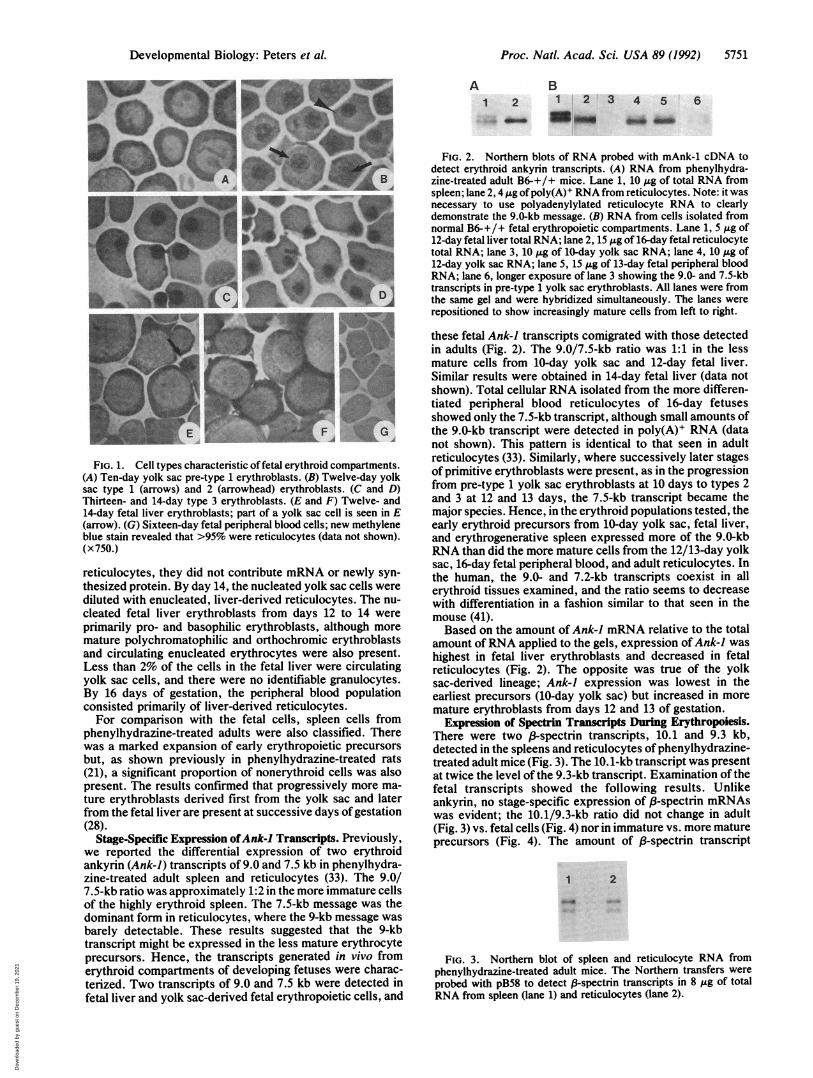
A B1 2 1 2 3 4 5 6
m .. . MO _e b
FIG. 1. Cell types characteristic offetal erythroid compartments.(A) Ten-day yolk sac pre-type 1 erythroblasts. (B) Twelve-day yolksac type 1 (arrows) and 2 (arrowhead) erythroblasts. (C and D)Thirteen- and 14-day type 3 erythroblasts. (E and F) Twelve- and14-day fetal liver erythroblasts; part of a yolk sac cell is seen in E(arrow). (G) Sixteen-day fetal peripheral blood cells; new methyleneblue stain revealed that >95% were reticulocytes (data not shown).(x750.)
reticulocytes, they did not contribute mRNA or newly syn-
thesized protein. By day 14, the nucleated yolk sac cells werediluted with enucleated, liver-derived reticulocytes. The nu-
cleated fetal liver erythroblasts from days 12 to 14 were
primarily pro- and basophilic erythroblasts, although more
mature polychromatophilic and orthochromic erythroblastsand circulating enucleated erythrocytes were also present.Less than 2% of the cells in the fetal liver were circulatingyolk sac cells, and there were no identifiable granulocytes.By 16 days of gestation, the peripheral blood populationconsisted primarily of liver-derived reticulocytes.For comparison with the fetal cells, spleen cells from
phenylhydrazine-treated adults were also classified. Therewas a marked expansion of early erythropoietic precursors
but, as shown previously in phenylhydrazine-treated rats(21), a significant proportion of nonerythroid cells was alsopresent. The results confirmed that progressively more ma-
ture erythroblasts derived first from the yolk sac and laterfrom the fetal liver are present at successive days of gestation(28).
Stage-Specific Expression ofAnk-l Transcripts. Previously,we reported the differential expression of two erythroidankyrin (Ank-J) transcripts of 9.0 and 7.5 kb in phenylhydra-zine-treated adult spleen and reticulocytes (33). The 9.0/7.5-kb ratio was approximately 1:2 in the more immature cellsof the highly erythroid spleen. The 7.5-kb message was thedominant form in reticulocytes, where the 9-kb message was
barely detectable. These results suggested that the 9-kbtranscript might be expressed in the less mature erythrocyteprecursors. Hence, the transcripts generated in vivo fromerythroid compartments of developing fetuses were charac-terized. Two transcripts of 9.0 and 7.5 kb were detected infetal liver and yolk sac-derived fetal erythropoietic cells, and
FIG. 2. Northern blots of RNA probed with mAnk-1 cDNA todetect erythroid ankyrin transcripts. (A) RNA from phenylhydra-zine-treated adult B6-+/+ mice. Lane 1, 10 1Ag of total RNA fromspleen; lane 2, 4 ,ug ofpoly(A)+ RNA from reticulocytes. Note: it wasnecessary to use polyadenylylated reticulocyte RNA to clearlydemonstrate the 9.0-kb message. (B) RNA from cells isolated fromnormal B6-+/+ fetal erythropoietic compartments. Lane 1, 5 ,ug of12-day fetal liver total RNA; lane 2, 15 Ag of 16-day fetal reticulocytetotal RNA; lane 3, 10 ,ug of 10-day yolk sac RNA; lane 4, 10 Ag of12-day yolk sac RNA; lane 5, 15 1&g of 13-day fetal peripheral bloodRNA; lane 6, longer exposure of lane 3 showing the 9.0- and 7.5-kbtranscripts in pre-type 1 yolk sac erythroblasts. All lanes were fromthe same gel and were hybridized simultaneously. The lanes wererepositioned to show increasingly mature cells from left to right.
these fetal Ank-J transcripts comigrated with those detectedin adults (Fig. 2). The 9.0/7.5-kb ratio was 1:1 in the lessmature cells from 10-day yolk sac and 12-day fetal liver.Similar results were obtained in 14-day fetal liver (data notshown). Total cellular RNA isolated from the more differen-tiated peripheral blood reticulocytes of 16-day fetusesshowed only the 7.5-kb transcript, although small amounts ofthe 9.0-kb transcript were detected in poly(A)+ RNA (datanot shown). This pattern is identical to that seen in adultreticulocytes (33). Similarly, where successively later stagesof primitive erythroblasts were present, as in the progressionfrom pre-type 1 yolk sac erythroblasts at 10 days to types 2and 3 at 12 and 13 days, the 7.5-kb transcript became themajor species. Hence, in the erythroid populations tested, theearly erythroid precursors from 10-day yolk sac, fetal liver,and erythrogenerative spleen expressed more of the 9.0-kbRNA than did the more mature cells from the 12/13-day yolksac, 16-day fetal peripheral blood, and adult reticulocytes. Inthe human, the 9.0- and 7.2-kb transcripts coexist in allerythroid tissues examined, and the ratio seems to decreasewith differentiation in a fashion similar to that seen in themouse (41).Based on the amount of Ank-1 mRNA relative to the total
amount ofRNA applied to the gels, expression ofAnk-J washighest in fetal liver erythroblasts and decreased in fetalreticulocytes (Fig. 2). The opposite was true of the yolksac-derived lineage; Ank-) expression was lowest in theearliest precursors (10-day yolk sac) but increased in moremature erythroblasts from days 12 and 13 of gestation.
Expression of Spectrin Transcripts During Erythropoiesis.There were two 8-spectrin transcripts, 10.1 and 9.3 kb,detected in the spleens and reticulocytes of phenylhydrazine-treated adult mice (Fig. 3). The 10.1-kb transcript was presentat twice the level of the 9.3-kb transcript. Examination of thefetal transcripts showed the following results. Unlikeankyrin, no stage-specific expression of f3-spectrin mRNAswas evident; the 10.1/9.3-kb ratio did not change in adult(Fig. 3) vs. fetal cells (Fig. 4) nor in immature vs. more matureprecursors (Fig. 4). The amount of 8-spectrin transcript
1 2
FIG. 3. Northern blot of spleen and reticulocyte RNA fromphenylhydrazine-treated adult mice. The Northern transfers were
probed with pB58 to detect 3-spectrin transcripts in 8 ,g of totalRNA from spleen (lane 1) and reticulocytes (lane 2).
Developmental Biology: Peters et al.
..;k.
.k
"I
FL. A -. A
Dow
nloa
ded
by g
uest
on
Dec
embe
r 19
, 202
1

5752 Developmental Biology: Peters et al.
1 2 3 4
A
am
B
FIG. 4. Northern blot of total RNA isolated from B6-+/+ fetalerythroid compartments. A single blot was hybridized first withpB129 to demonstrate a-spectrin transcripts (B), then stripped andsubsequently probed with pB58 to demonstrate /3-spectrin tran-scripts (A). All hybridization and wash conditions were identical. Thea-spectrin filter was exposed for 4 days, 83-spectrin for 3 days. Lane1, 10 ,ug of 12-day fetal liver; lane 2, 15 ug of 10-day yolk sac; lane3, 15 ,ug of 12-day yolk sac; lane 4, 20 ,ug of 16-day reticulocytes.
relative to the total RNA concentration increased during yolksac differentiation but decreased during differentiation of theliver precursors to reticulocytes in a pattern similar to Ank-JmRNA.An a-spectrin transcript of 10.2 kb was detected in phe-
nylhydrazine-treated adult spleen and reticulocytes (data notshown) and was also present in all fetal cells, except the10-day yolk sac cells (Fig. 4). The relative amount of thea-spectrin transcript increased during differentiation of boththe yolk sac- and fetal liver-derived erythroblasts. Interest-ingly, the amount of a-spectrin transcript relative to the totalRNA was significantly lower than the amount of ,B-spectrintranscript in the early erythropoietic precursors from bothyolk sac and fetal liver. This was not due to differences inexperimental conditions, since the a-spectrin and,-spectrincDNA probes were similar in size (750 bp and 700 bp,respectively) and were labeled to the same specific activities(108 cpm/,g of DNA), and the hybridization and washconditions were identical. The same Northern transfer filterwas exposed for 4 days to show a-spectrin transcripts and 3days to show A3-spectrin transcripts. This pattern of increased,B-spectrin mRNA expression over a-spectrin mRNA in earlyerythropoietic precursor cells was consistent with the a < (3spectrin protein synthesis observed in erythropoietin-stimulated Friend virus-infected murine spleen cells (24) butunlike the protein synthetic profile seen in reticulocytes fromphenylhydrazine-treated adult mice (27) and rats (21).
Immunoprecipitation of Newly Synthesized a- and (8-Spec-trin in Fetal Erythroid Cells. To determine whether themRNA levels were indicative of protein synthetic patterns,total newly synthesized a- and (3-spectrin were quantified in12-day fetal liver erythroblasts and 16-day fetal reticulocytes.Cells were incubated in the presence of [35S]methionine,lysed, and precipitated with an antibody that recognizesequivalent amounts of a- and (3-spectrin on Western blots(27). In 12-day fetal liver, total ,8-spectrin synthesis was 3-foldgreater than that of a-spectrin (a/(3 synthetic ratio = 0.33)(Fig. 5). In 16-day fetal reticulocytes, however, total a-spec-trin synthesis was 2-fold greater than ,B-spectrin synthesis(a/(3 synthetic ratio = 2.0). In this regard, 16-day fetalreticulocytes were similar to phenylhydrazine-treated mousereticulocytes (27). Hence, total a- and (3-spectrin synthesiscorrelates with the Northern blot data.
DISCUSSIONThe relevant observations from this study are that (i) the7.5-kb ankyrin mRNA, while present in the less matureerythroblasts from all erythogenerative tissues tested, be-comes the major transcript in the more mature erythroblasts;
1 2 W3~~FIG. 5. Immunoprecipitation of total newly synthesized spectrin.
Cells were metabolically labeled with [35S]methionine and immuno-precipitated with spectrin antibody. Lanes 1 and 2, 12-day fetal livercells (107); lane 3, 16-day fetal reticulocytes (106). The shift in the a/,8synthetic ratio from 12-day fetal liver erythroblasts to 16-day fetalreticulocytes is apparent.
(ii) there is more 3- than a-spectrin mRNA in yolk sac-derived nucleated cells and in the fetal liver erythroblasts, butthe converse occurs in the more mature fetal liver-derivedand adult reticulocytes; and (iii) the relative amount ofmRNA for each cytoskeletal protein is dependent not only onthe stage of differentiation but also on the source of theerythroblasts. The observed mRNA changes are summarizedin Table 2 and, since we have shown that the amount ofa- andf3-spectrin synthesized is consistent with mRNA levels, alsopredict the protein synthetic ratios.
Regardless of cell origin (fetal yolk sac, fetal liver, adultspleen), our results indicate that ankyrin mRNA is differen-tially processed depending on the stage of erythroid matura-tion. We find nearly equivalent levels of 9.0- and 7.5-kbmRNA in the early cells, and predominance of the 7.5-kbmRNA in later stages of erythroid cell differentiation. Al-though these results support our contention (33) that the 9-kbmRNA generates a product essential for structural integrityof the early erythroid cell membrane skeleton, the situationis probably more complex. Structural analyses ofankyrin andprotein 4.1 cDNAs indicate that alternative splicing of mul-tiple small exons can produce many transcripts from a singlegene (41-46) that are structurally different but comigrateduring electrophoretic separation. Therefore, the apparent9.0- to 7.5-kb switch of ankyrin mRNA during erythroiddevelopment could involve several 9.0- and/or 7.5-kb tran-scripts designed to produce proteins with minor structuralchanges but significant changes in binding properties.Ankyrin isoforms display different f-spectrin and anionexchanger binding affinities (47) and bind alternative proteinsin the erythroid membrane (48, 49). It is possible that a closerexamination of transcripts from the erythroid a- and P3-spec-trin genes may reveal similarly complex patterns of splicing.As extensive remodeling of the erythrocyte membrane oc-curs during erythropoiesis (50, 51), it is reasonable to proposethat stage-specific mRNA splicing produces isoforms of the
Table 2. Summary of transcript characteristicsTranscriptst
Cell Major Ankyrin Spectrinsource cell type* 9/7.5 kb Amount f3 a
Yolk sac Pre-1 1 + + ND1, 2 <0.1 ++ ++ +3 <0.1 +++ +++ ++
Fetal liver Pro, Baso 1 +++ +++ +Retic <0.1 + + ++
Spleen Pro, Baso 0.5Retic <0. 1
*Pro, proerythroblast; Baso, basophilic erythroblast; Retic, reticu-locyte. Refer to Table 1 for time of appearance of cell types.tFor /-spectrin, the ratio of the 10.1- and 9.3-kb transcripts was 2 inall samples. For a-spectrin, only a 10.2-kb mRNA was seen. ND,not detectable; -, no data.
Proc. Natl. Acad. Sci. USA 89 (1992)
Dow
nloa
ded
by g
uest
on
Dec
embe
r 19
, 202
1

Proc. Natl. Acad. Sci. USA 89 (1992) 5753
spectrin-binding proteins, ankyrin and 4.1, that function tomeet membrane requirements unique to each developmentalstage.Heterodimers of a- and (3-spectrin are incorporated stoi-
chiometrically (1:1) into the membrane despite the fact thatunequal amounts of the two proteins are synthesized (21, 27).Expression of a- and f-spectrin transcripts and the a/(3protein synthetic ratio can vary significantly depending uponphysiological conditions. For example, in patients withankyrin-deficient hemolytic anemia (8), in some dimethylsulfoxide-induced murine erythroleukemia cells (18, 22), andin erythropoietin-stimulated, Friend virus-infected murinespleen cells (24), f3-spectrin expression and synthesis areincreased over those of a-spectrin (a/,8 < 1). In adultreticulocytes, the a/l, synthetic ratio is usually 2-3:1. Thealtered a/,3 synthetic ratio produced by erythropoietin stim-ulation increases P3-spectrin mRNA expression and synthesiswithout affecting that of a-spectrin. This phenomenon resultsin increased membrane spectrin incorporation (24). In thepresent study, higher levels ofP-spectrin mRNA than a-spec-trin mRNA and increased 83-spectrin synthesis relative toa-spectrin synthesis were found in early erythroid precursorsof the fetal liver. The cells showing increased /8-spectrinmRNA expression and protein synthesis in this study areknown to be erythropoietin-sensitive (pro- and basophilicerythroblasts) (23). The increased expression and synthesisof/-spectrin seen in these early erythroid precursors suggestincreased membrane demand for spectrin tetramers, mostlikely reflecting the dynamic nature of the differentiatingerythrocyte membrane. Physiological control of the a//3synthetic ratio may provide an important mechanism formaintaining the erythroid cell membrane during erythropoie-sis.The erythrocyte in vivo possesses two putative mecha-
nisms to meet varying membrane binding and/or mechanicalproperties during differentiation: (i) generation of stage-specific ankyrin and protein 4.1 isoforms by alternativemRNA processing and (ii) the ability to alter a/,3-spectrinsynthetic ratios. In the future, more detailed analyses mustinclude the use of region-specific antibodies (52) and oligo-nucleotide probes (53). Only when unique cytoskeletal prod-ucts are identified and their interactions with other proteinsare established will we begin to understand the process oferythroid cell differentiation.
We thank Dottie Hamblen and Nancy Hamblen for maintaining themice. We thank Drs. John Eppig and Laurie Rund, who provideduseful suggestions for improving the manuscript. This work wassupported by Grant HL29305 (J.E.B.) from the National Institutes ofHealth.
1. Bennett, V. (1990) Physiol. Rev. 70, 1029-1065.2. Bennett, V. (1989) Biochim. Biophys. Acta 988, 107-121.3. Reid, M., Chasis, J. & Mohandas, N. (1987) Blood 69, 1068-1072.4. Conboy, J., Mohandas, N., Tchernia, G. & Kan, Y. W. (1986) N.
Engl. J. Med. 15, 680-685.5. Bennett, V. & Stenbuck, P. J. (1980) J. Biol. Chem. 255, 6424-6432.6. Korsgren, C. & Cohen, C. M. (1988) J. Biol. Chem. 263, 10212-
10218.7. Rybicki, A. C., Heath, R., Wolf, J., Lubin, B. & Schwartz, R. S.
(1988) J. Clin. Invest. 81, 893-901.8. Hanspal, M., Yoon, S.-H., Yu, H., Hanspal, J. S., Lambert, S.,
Palek, J. & Prchal, J. T. (1991) Blood 77, 165-173.9. Lux, S. E., Tse, W. T., Menninger, J. C., John, K. M., Harris, P.,
Shalev, O., Chilcote, R. R., Marchesi, S. L., Watkins, P. C.,Bennett, V., McIntosh, F. S., Collins, F. S., Francke, U., Ward,D. C. & Forget, B. G. (1990) Nature (London) 345, 736-739.
10. Bodine, D. M., IV, Birkenemier, C. S. & Barker, J. E. (1984) Cell37, 721-729.
11. Birkenmeier, C. S., McFarland-Starr, E. C. & Barker, J. E. (1988)Proc. Natl. Acad. Sc.USA 85, 8121-8125.
12. White, R. A., Birkenmeier, C. S., Lux, S. E. & Barker, J. E. (1990)Proc. Natl. Acad. Sci. USA 87, 3117-3121.
13. Davies, K. A. & Lux, S. E. (1989) Trends Genet. 5, 222-227.14. Eisen, H., Bach, R. & Emery, R. (1977) Proc. Natl. Acad. Sci. USA
74, 3898-3902.15. Rossi, G. R., Aducci, P., Gambari, R., Minetti, M. & Vernole, P.
(1978) J. Cell. Physiol. 97, 293-304.16. Marshall, L. M. & Hunt, R. C. (1982) J. Cell Sci. 54, 98-113.17. Glenney, J. & Glenney, P. (1984) Exp. Cell Res. 152, 15-21.18. Pfeffer, S. R., Huima, T. & Redman, C. M. (1986)J. CellBiol. 103,
103-113.19. Koury, M. J., Bondurant, M. C. & Mueller, T. J. (1986) J. Cell.
Physiol. 126, 259-265.20. Koury, M. J., Bondurant, M. C. & Rana, S. S. (1987) J. Cell.
Physiol. 133, 438-448.21. Hanspal, M. & Palek, J. (1987) J. Cell Biol. 105, 1417-1424.22. Lehnert, M. E. & Lodish, H. F. (1988) J. Cell Biol. 107, 413-426.23. Koury, M. J. & Bondurant, M. C. (1988) J. Cell. Physiol. 137,
65-74.24. Hanspal, M., Kalraija, R., Hanspal, J., Sahr, K. E. & Palek, J.
(1991) J. Biol. Chem. 266, 15626-15630.25. Chang, H., Langer, P. J. & Lodish, H. F. (1976) Proc. Natl. Acad.
Sci. USA 73, 3206-3210.26. Lazarides, E. (1987) Cell 51, 345-356.27. Barker, J. E., Bodine, D. M. & Birkenmeier, C. S. (1986) in Mem-
brane Skeletons and Cytoskeletal-Membrane Associations, eds.Bennett, V., Cohen, C. M., Lux, S. E. & Palek, J. (Liss, NewYork), pp. 313-324.
28. Russell, E. S. & Bernstein, S. E. (1968) in Biology of the Labora-tory Mouse, ed. Green, E. L. (McGraw Hill, New York), 2nd Ed.,pp. 351-360.
29. Chui, D. H., Patterson, K. M. & Bayley, S. T. (1980) Br. J.Haematol. 44, 431-439.
30. Kelemen, E., Calvo, W. & Fliedner, T. M. (1979) Atlas ofHumanHemopoietic Development (Springer, New York).
31. Brown, B. (1984) Principles and Procedures ofHematology (Lea &Febiger, Philadelphia), 4th Ed., pp. 54-59.
32. White, R. A., Birkenmeier, C. S., Peters, L. L., Barker, J. E. &Lux, S. E., Mamm. Genome, in press.
33. Peters, L. L., Birkenmeier, C. S., Bronson, R. T., White, R. A.,Lux, S. E., Otto, E., Bennett, V., Higgins, A. & Barker, J. E.(1991) J. Cell Biol. 114, 1233-1241.
34. Cioe, L., Laurila, P., Meo, P., Krebs, K., Goodman, S. & Curtis,P. J. (1987) Blood 70, 915-920.
35. Cioe, L. & Curtis, P. J. (1985) Proc. Natl. Acad. Sci. USA 82,1367-1371.
36. Chomczynski, P. & Sacchi, N. (1987) Anal. Biochem. 162, 156-159.37. Aviv, H. & Leder, P. (1972) Proc. Natl. Acad. Sci. USA 69,
1408-1412.38. Sambrook, J., Fritsch, E. F. & Maniatis, T. (1989) Molecular
Cloning: A Laboratory Manual (Cold Spring Harbor Lab., ColdSpring Harbor, NY), 2nd Ed.
39. Feinberg, A. P. & Vogelstein, B. (1983) Anal. Biochem. 132, 6-13.40. Laemmli, U. K. (1970) Nature (London) 227, 680-685.41. Lambert, S., Yu, H., Prchal, J. T., Lawler, J., Ruff, P., Speicher,
D., Cheung, M. C., Kan, Y. W. & Palek, J. (1990) Proc. Natl. Acad.Sci. USA 87, 1730-1734.
42. Lux, S. E., John, K. M. & Bennett, V. (1990) Nature (London) 344,36-40.
43. Peters, L. L., Birkenmeier, C. S., Lux, S. E., White, R. A. &Barker, J. E. (1989) Blood 74, 59 (abstr.).
44. Birkenmeier, C. S., Peters, L. L. & Barker, J. E. (1991) Blood 78,364 (abstr.).
45. Conboy, J. G., Chan, J., Mohandas, N. & Kan, Y. W. (1988) Proc.Natl. Acad. Sci. USA 85, 9062-9065.
46. Tang, T. K., Leto, T. L., Correas, I., Alonso, M. A., Marchesi,V. T. & Benz, E. J., Jr. (1988) Proc. Natl. Acad. Sci. USA 85,3713-3717.
47. Hall, T. G. & Bennett, V. (1987) J. Biol. Chem. 262, 10537-10545.48. Georgatos, S. D. & Marchesi, V. T. (1985) J. Cell Biol. 100,
1955-1961.49. Inaba, M. & Maede, Y. (1988) J. Biol. Chem. 263, 17763-17771.50. Chasis, J. A., Prenant, M., Leung, A. & Mohandas, N. (1989) Blood
74, 1112-1120.51. Geiduschek, J. B. & Singer, S. J. (1979) Cell 16, 149-163.52. Tang, T. K., Leto, T., Marchesi, V. T. & Benz, E. J. (1990) J. Cell
Biol. 110, 617-624.53. Conboy, J. G., Chan, J. Y., Chasis, J. A., Kan, Y. W. & Mohan-
das, N. (1991) J. Biol. Chem. 266, 8273-8280.
Developmental Biology: Peters et al.
Dow
nloa
ded
by g
uest
on
Dec
embe
r 19
, 202
1