challenges in the medical management of patients with ... · objectives •describe the...
TRANSCRIPT

Challenges in the Medical Management of Patients with Aortic
Stenosis
November 4, 2014
Theresa Cary, RN, MSN, ACNS-BC, CCRN, CHFN
Judy Pearce, RN, BSN, CCRN

The authors have no conflicts of interest to disclose
Objectives
• Describe the pathophysiology of AS
• Identify clinical manifestations of AS
• Discuss medical and nursing management of nonsurgical patients

Introduction
• Aortic stenosis (AS)
- Narrowing of aortic valve orifice
- → Obstruction of left ventricular outflow
• Calcific aortic valve disease (CAVD)
- Affects > 4% North Americans and Europeans
- Increasing in prevalence

Normal Valve Function
• Essential to cardiovascular and cardiopulmonary physiology
• Heart valves ensure forward progression of blood through the heart
• Open & close in response to pressure changes during systole and diastole

Normal Heart & Valve Function
Open tricuspid &
mitral valves in
early/mid diastole
Closed tricuspid
& mitral valves in
early systole
Open pulmonic
and aortic valves
in mid systole
Closed pulmonic
and aortic valves
In late systole
© Cleveland Clinic

Systole
Lub © Cleveland Clinic
Diastole
Dub © Cleveland Clinic

Systolic Contraction & Twist

©Cleveland Clinic
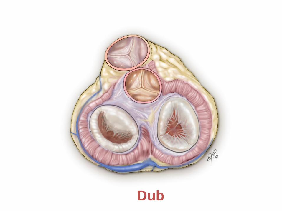
Aortic Valve A & P
• 3 cup-shaped leaflets
• Top edge (free margin)
• Base
• Annulus connects aortic valve to the fibrous skeleton of the heart
©Cleveland Clinic

Fibrous Skeleton
• Dense fibrous connective tissue
©Cleveland Clinic

Aortic Valve Anatomy & Physiology
• Commissures join leaflets edge to edge
• Penetrate aortic wall
• Absorb stress of systole and diastole

Aortic Valve A & P
• Sinus of Valsalva
• Aortic wall bulges outward
• Creates space behind leaflets
©Cleveland Clinic

3 Sinuses of Valsalva
• Bulge creates space
- Prevent obstruction to coronary arteries during systole
- Provide space for blood to pool during diastole for coronary artery filling
©Cleveland Clinic

Sinus of Valsalva
• Blood flow outlet narrows
• Blood forced between the Sinus of Valsalva and open cusps, filling coronary arteries
• Backward curling
• Free margins meet
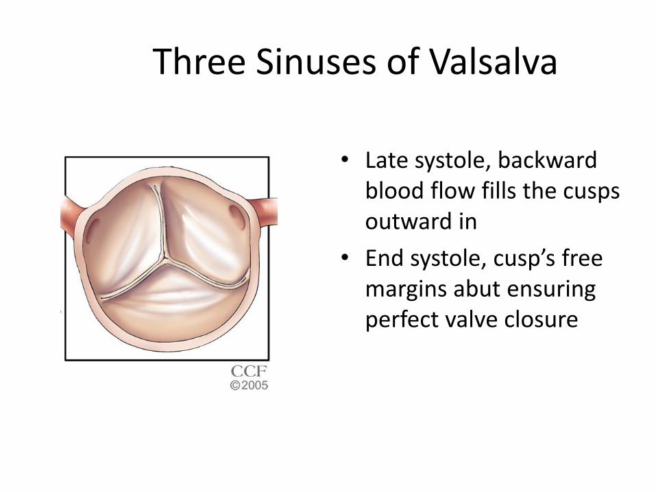
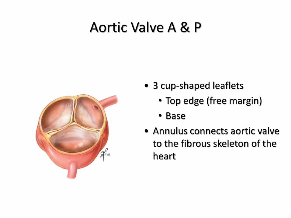
Three Sinuses of Valsalva
• Late systole, backward blood flow fills the cusps outward in
• End systole, cusp’s free margins abut ensuring perfect valve closure

Layers of Leaflet
• Fibrosa
• Ventricularis
• Spongiosa
© Cleveland Clinic

Aortic valve leaflet A&P
• Fibrosa – collagen: distributes pressure load
• Spongiosa - glycosaminoglycans, proteoglycans: cushion, minimize friction
• Ventricularis - elastic fibers: maintains shape

Aortic Valve Leaflet A&P
• Valvular interstitial cells (VICS):
- Maintain structure & function
- Inhibit angiogenesis
- Repair cellular damage
• Valvular extracellular matrix (VECM):
- Collagen fibers
- Elastin fibers
- Glycosaminoglycans, proteoglycans

Aortic Stenosis
• Progression from sclerosis to stenosis
- Sclerosis – mild valve thickening and/or calcification without obstruction
- Stenosis – increasing obstruction of blood flow and progression of:
• Leaflet thickening
• Calcium nodule formation
• Angiogenesis
– 10% advance from sclerosis to stenosis

Causes of Aortic Stenosis
• Valve calcification of:
- Tri-leaflet AV
• Most common cause of AS

Bicuspid Aortic Valve
• 1-2% of adults More likely to develop AS
• Stenosis occurs earlier
- 50s to 60s bicuspid
- 70s to 80s tricuspid
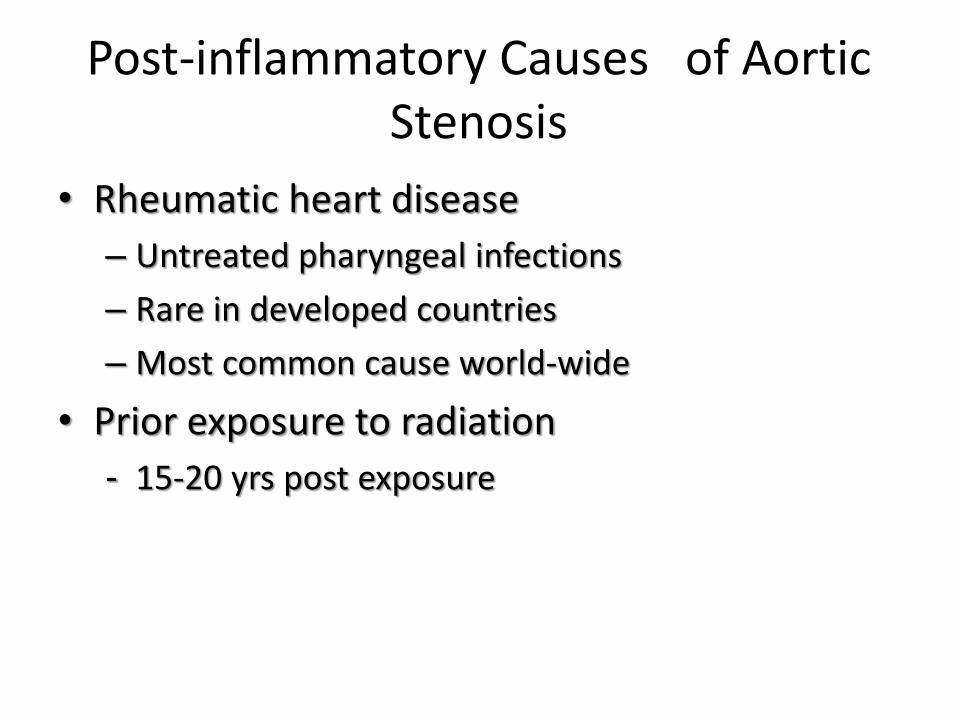
Post-inflammatory Causes of Aortic Stenosis
• Rheumatic heart disease
– Untreated pharyngeal infections
– Rare in developed countries
– Most common cause world-wide
• Prior exposure to radiation
- 15-20 yrs post exposure

Aortic Valve Calcification
• CAVD is an active cellular biological process
• Not an inevitable consequence of aging
• VICS no longer repair injuries to VECM
– Alterations of cells within the layers of the AV
• Exact cause is still unclear

Pathophysiology of AS
• Chronic resistance to LV ejection → concentric LV hypertrophy and myocardial fibrosis
• Stronger LV systolic contraction needed to
- Maintain adequate stroke volume and cardiac output
- EF is maintained
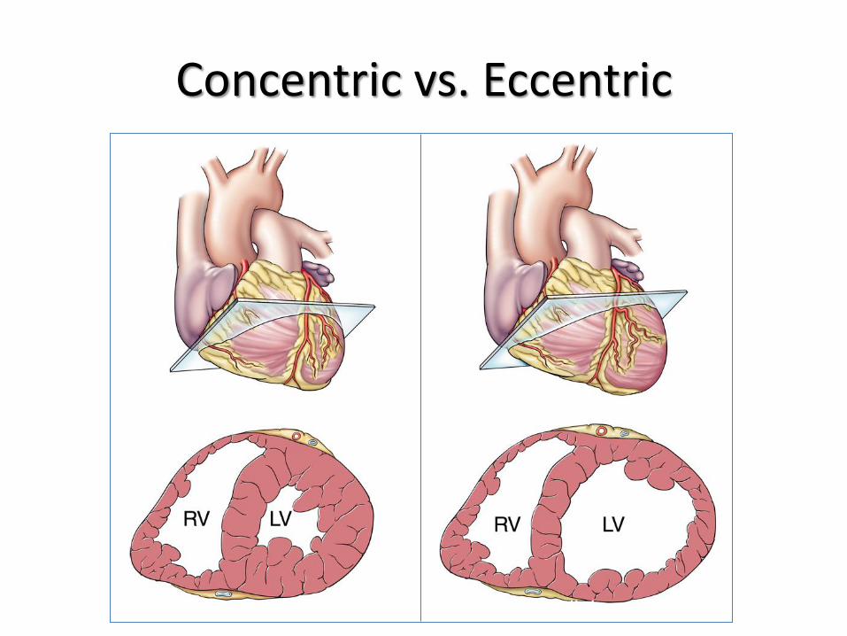
Concentric vs. Eccentric
© Cleveland Clinic

Consequences of Concentric Hypertrophy
• Decreased
- Myocardial elasticity
- Coronary blood flow
• Increased
- Myocardial workload
- Myocardial oxygen consumption
- Mortality

Consequences of Concentric Hypertrophy
• Increased
- Diastolic pressure
- Delayed LV untwisting (relaxation)
• Optimal stroke volume and cardiac output increasingly dependent upon a forceful atrial contraction (atrial kick)

Consequences of Concentric Hypertrophy
• Mitral valve regurgitation
- Increased LV pressure puts a strain on the mitral valve
- Increase pressure in the lungs
• Pulmonary venous hypertension
• Reactive vasoconstriction of the pulmonary vasculature

2014 AHA/ACC Guideline for the Management of Patients with
Valvular Heart Disease
• Stages A-D
Grading of AS
• Aortic jet velocity
• Mean aortic valve pressure gradient
• Aortic valve area

Pressure Gradient
• Peak LV and aortic pressure tracings
• LV pressure higher than aortic pressure
• Grading table uses mean pressure gradient
Wikipedia, Aortic Valve Stenosis
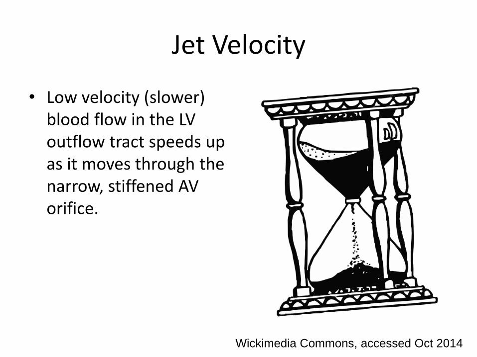
Jet Velocity
• Low velocity (slower) blood flow in the LV outflow tract speeds up as it moves through the narrow, stiffened AV orifice.
Wickimedia Commons, accessed Oct 2014

Clinical Manifestations of AS
• Initial manifestations - Decreased exercise tolerance
• Dyspnea on exertion
• Exertional dizziness
• Lightheadedness with exertion
• Late manifestations - Angina
- Syncope
- Heart failure

Clinical Manifestations of AS
• Jugular vein distension
• Pulmonary rales
• Carotid pulse abnormalities
• Systolic ejection murmur

Carotid Pulse
• Gentle, careful palpation
• Pulsus tardus
- Slowly increasing carotid upstroke
- Takes longer to reach peak pressure
• Weaker pulse amplitude
Carotid Pulse
• Indication of:
- Resistance to AV opening
- Subsequent delay in LV ejection
- Decreasing volume
• Indications may be masked in the elderly d/t age related changes in arterial compliance and stiffness

Systolic Ejection Murmur
• Crescendo-decrescendo
- 2nd intercostal space right sternal boarder with bell of stethoscope
• May radiate to carotids
• In elderly, may radiate to the apex

Medical Management of Asymptomatic Patients
• Decrease cardiovascular risk factors
• Monitoring and education
• Medication therapy
Decrease Cardiovascular Risk Factors
• HTN
• Diabetes
• Smoking tobacco
• High cholesterol
• Overweight
• Lack of exercise
Monitoring and Education
• Monitoring
- F/U visits, echocardiograms
• Education
- Disease progression
- Change in exercise tolerance
- Physical activity
Medication Therapy
• There is currently no known medical therapy to
– Prevent CAVD
– Delay the progression of AS

Medication Therapy
• Prophylactic antibiotics
– Rheumatic AS only
• Treat HTN according to standard GDMT
– B-blockers historically considered unsafe
• Statins??
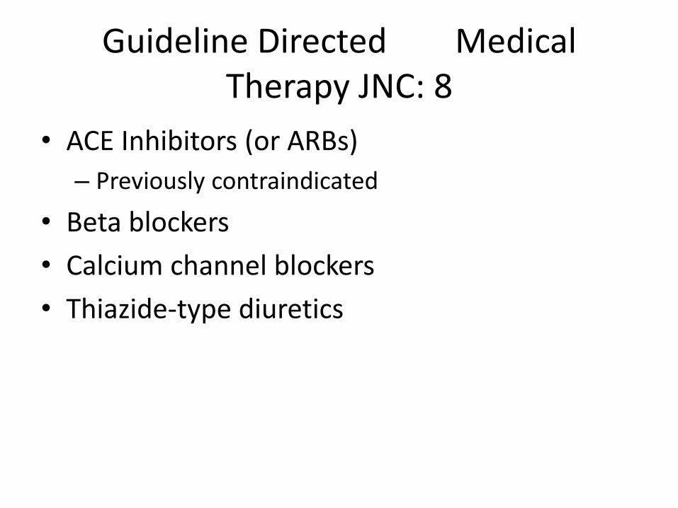
Guideline Directed Medical Therapy JNC: 8
• ACE Inhibitors (or ARBs)
– Previously contraindicated
• Beta blockers
• Calcium channel blockers
• Thiazide-type diuretics
James, et al. JAMA, 2014.
Nadir et al. J Am Coll Cardiol, 2011.
Pai et al. Ann Thorac Surg, 2006.
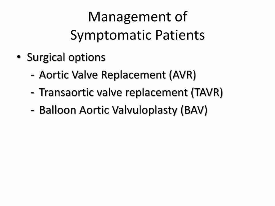
Management of
Symptomatic Patients • Surgical options
- Aortic Valve Replacement (AVR)
- Transaortic valve replacement (TAVR)
- Balloon Aortic Valvuloplasty (BAV)
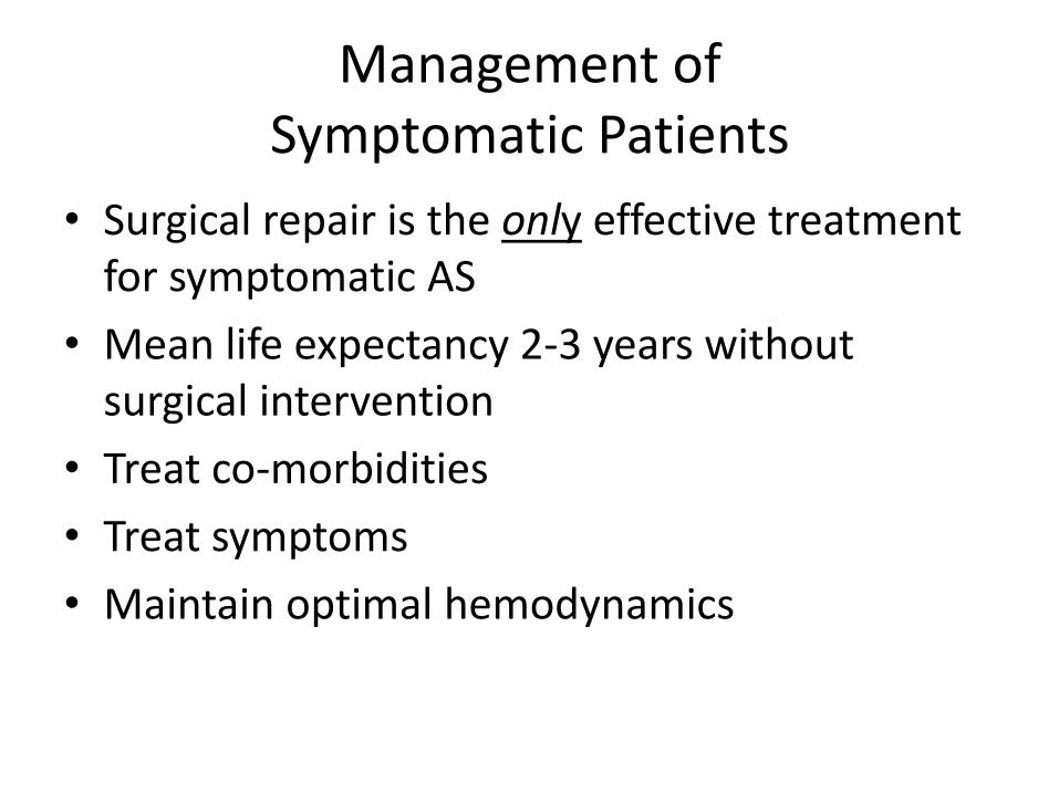
Management of Symptomatic Patients
• Surgical repair is the only effective treatment for symptomatic AS
• Mean life expectancy 2-3 years without surgical intervention
• Treat co-morbidities
• Treat symptoms
• Maintain optimal hemodynamics

Management of
Symptomatic Patients
• Angina
• Syncope
• Pulmonary congestion
• Acute pulmonary edema
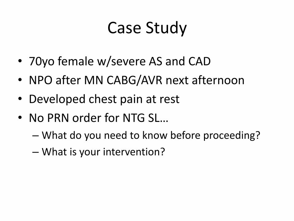
Case Study
• 70yo female w/severe AS and CAD
• NPO after MN CABG/AVR next afternoon
• Developed chest pain at rest
• No PRN order for NTG SL…
– What do you need to know before proceeding?
– What is your intervention?
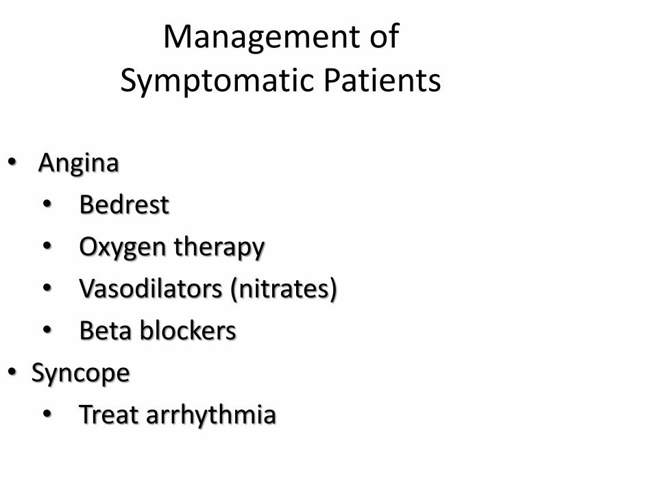
Management of
Symptomatic Patients
• Angina
• Bedrest
• Oxygen therapy
• Vasodilators (nitrates)
• Beta blockers
• Syncope
• Treat arrhythmia

Case Study, continued
• B/P 108/65, usual B/P 130/70
– Chest pain unrelieved w/rest and O2
• IV Nitroglycerin 10mcg/min
– B/P ↓ 90/60
– Chest pain unrelieved
• Normal Saline 150cc/hr
• Transferred to ICU
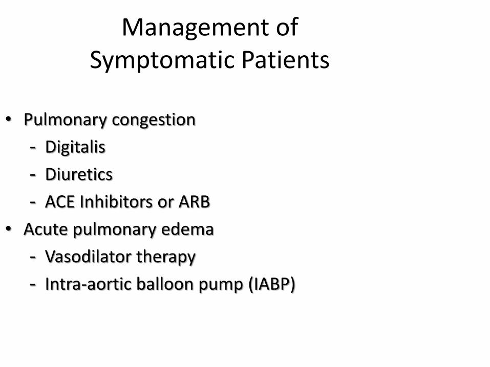
Management of
Symptomatic Patients
• Pulmonary congestion
- Digitalis
- Diuretics
- ACE Inhibitors or ARB
• Acute pulmonary edema
- Vasodilator therapy
- Intra-aortic balloon pump (IABP)

Nursing Considerations
• Thorough grasp of the tenuous balance between the narrow range of preload & afterload necessary to maintain forward blood flow & adequate CO
• Extremely sensitive to changes in preload – High preload → pulmonary congestion
– Low preload → low output failure
• Dependent on a strong atrial contraction

Nursing Considerations
• Consider hemodynamics as they relate to signs & symptoms of AS
• Consider hemodynamic effects of medications, treatments, plan of care
• Avoid systemic hypotension – Myocardial ischemia
– Reduced contractility
– Worsening hypotension
– Worsening coronary perfusion

Goals in Daily Plan of Care
• Balance rest & activity
– Maintain HR, B/P, temperature and fluid volume status
• Monitor for indicators of decompensation
– Hypoxia, arrhythmias, B/P changes, SOB, chest pain, prolonged NPO status
• Identify decompensation early to prevent deterioration in clinical status
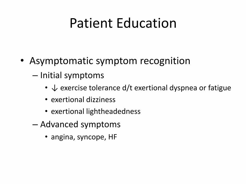
Patient Education
• Asymptomatic symptom recognition
– Initial symptoms
• ↓ exercise tolerance d/t exertional dyspnea or fatigue
• exertional dizziness
• exertional lightheadedness
– Advanced symptoms
• angina, syncope, HF

Patient Education
• Symptomatic symptom recognition
– worsening signs and symptoms with prompt reporting
– balance rest/activity to avoid symptoms
• Impact of medication adherence on cardiac function
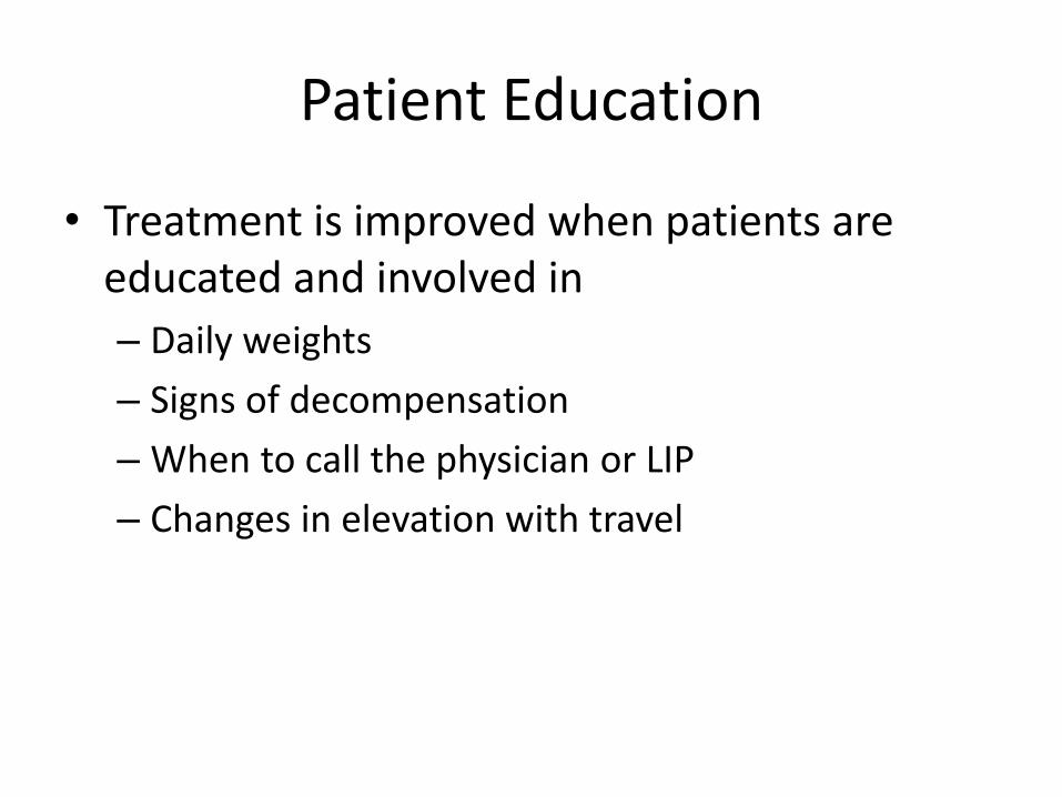
Patient Education
• Treatment is improved when patients are educated and involved in
– Daily weights
– Signs of decompensation
– When to call the physician or LIP
– Changes in elevation with travel

Conclusion
• Symptomatic AS cannot be corrected without surgery
• Medical management of patients with AS, particularly severe symptomatic AS, is challenging
• Nurses must be astute about the tenuous hemodynamic balance of patients with severe AS
