cell biology for clinical pharmacy students md102 module iii: cell division (lecture # 11) dr. ahmed...
TRANSCRIPT

Cell Biology for Clinical Pharmacy StudentsMD102Module III: Cell Division (Lecture # 11)
Dr. Ahmed Sherif [email protected]://sites.google.com/site/ahmedsattia/

Objectives
By the end of this lecture you should be familiar with:
• Different stages of Meiosis
• Gametogenesis
• Differences and similarities between Meiosis and Mitosis
• Types of cell death
• Apoptosis roles in development, Cancer, organ transplant and AIDS
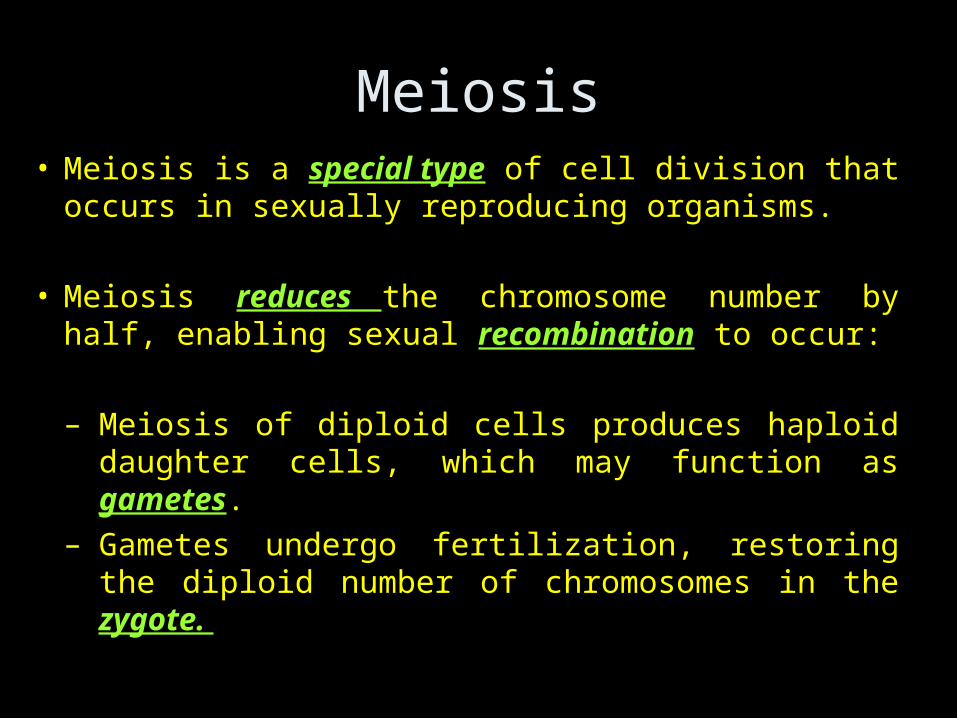
Meiosis• Meiosis is a special type of cell division that
occurs in sexually reproducing organisms.
• Meiosis reduces the chromosome number by
half, enabling sexual recombination to occur:
– Meiosis of diploid cells produces haploid daughter cells, which may function as gametes.
– Gametes undergo fertilization, restoring the diploid number of chromosomes in the zygote.

• Most cells in the human body are produced by mitosis. These are the somatic (or vegetative) line cells.
• Cells that become gametes are referred to as germ line cells.
• The vast majority of cell divisions in the human body are mitotic, with meiosis being restricted to the gonads.
Stages of Meiosis
• The stages of meiosis can be broken down into two main stages, Meiosis I and Meiosis II:
• Meiosis I can be broken down into four sub
stages: Prophase I, Metaphase I, Anaphase I and Telophase I.
• Meiosis II can be broken down into four sub
stages: Prophase II, Metaphase II, Anaphase II and Telophase II.
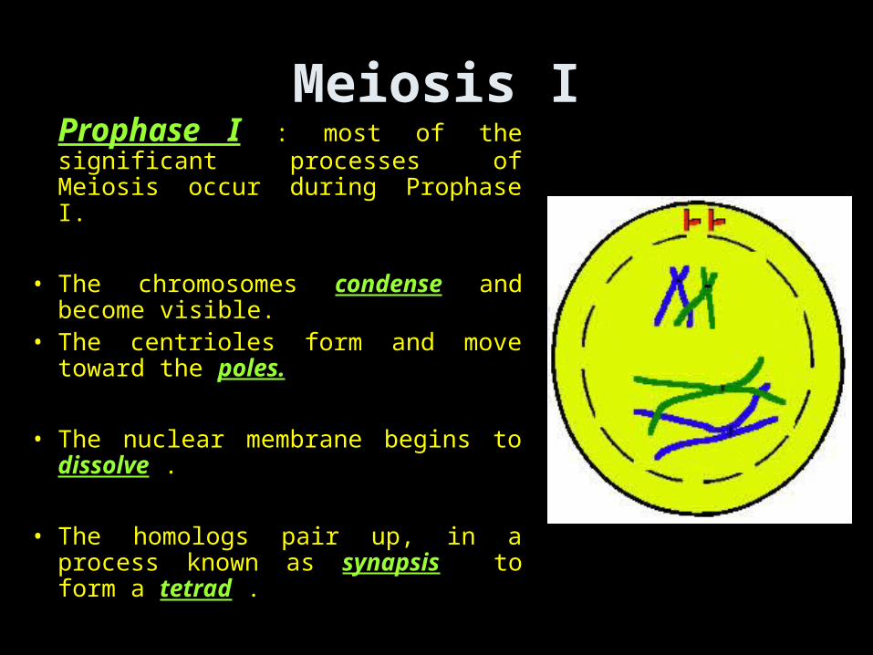
Meiosis IProphase I : most of the significant processes of Meiosis occur during Prophase I.
• The chromosomes condense and become visible.
• The centrioles form and move toward the poles.
• The nuclear membrane begins to dissolve .
• The homologs pair up, in a process known as synapsis to form a tetrad .

Each tetrad is comprised of four chromatids - the two homologous, each with their sister chromatid.

Crossing over• Genetic material from the
homologous chromosomes is randomly swapped.
• This creates four unique chromatids, which increases the overall genetic diversity of the gametes.
Chiasma: The site where the exchange of chromosome segments between homologous chromosomes takes place (pl= chiasmata).

Metaphase I
• Microtubules grow from the centrioles and attach to one side of the centromeres.
• The tetrads line up along the cell equator.
• Each homologous pair
can orient in either of two ways at the plane of cell division.

Anaphase I
• The centromeres break and homologous chromosomes separate.
• Note that the sister chromatids are still attached.
• Cytokinesis begins.

Telophase I
• The chromosomes may decondense (depends on species).
• Cytokinesis reaches completion, creating two haploid daughter cells.

Meiosis II
Prophase II
• Centrioles form and move toward the poles.
• The nuclear membrane dissolves.

Metaphase II
• Microtubules grow from the centrioles and attach to the centromeres.
• The sister
chromatids line up along the cell equator.
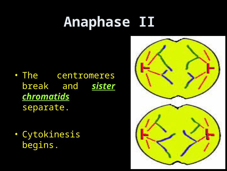
Anaphase II
• The centromeres break and sister chromatids separate.
• Cytokinesis begins.

Telophase II
• The chromosomes may decondense (depends on species).
• Cytokinesis reaches completion, creating four haploid daughter cells.

Gametogenesis
• Gametogenesis is the process of forming gametes (by definition haploid, n) from diploid cells of the germ line.
• Spermatogenesis is the process of forming sperm cells by meiosis (in animals) in specialized organs known as gonads (in males these are termed testes).
• After division the cells undergo differentiation to become sperm cells.

Gametogenesis• Oogenesis is the process of forming an ovum (egg)
by meiosis (in animals) in specialized gonads known as ovaries.
• Whereas in spermatogenesis all 4 meiotic products develop into gametes, oogenesis places most of the cytoplasm into the large egg. The other cells, the polar bodies, do not develop. This all the cytoplasm and organelles go into the egg.
• Human males produce 200,000,000 sperm per day, while the female produces one egg (usually) each menstrual cycle.

Meiosis and Genetic Variation • Meiosis and fertilization introduce genetic
variation in three ways:
• 1-Crossing over between homologous chromosomes at prophase I.
• 2-Independent assortment of homologous pairs at metaphase I: – Each homologous pair can orient in either of two ways
at the plane of cell division. – The total number of possible outcomes = 2n (n =
number of haploid chromosomes).
• 3- Random chance fertilization between any one female gamete with any other male gamete.

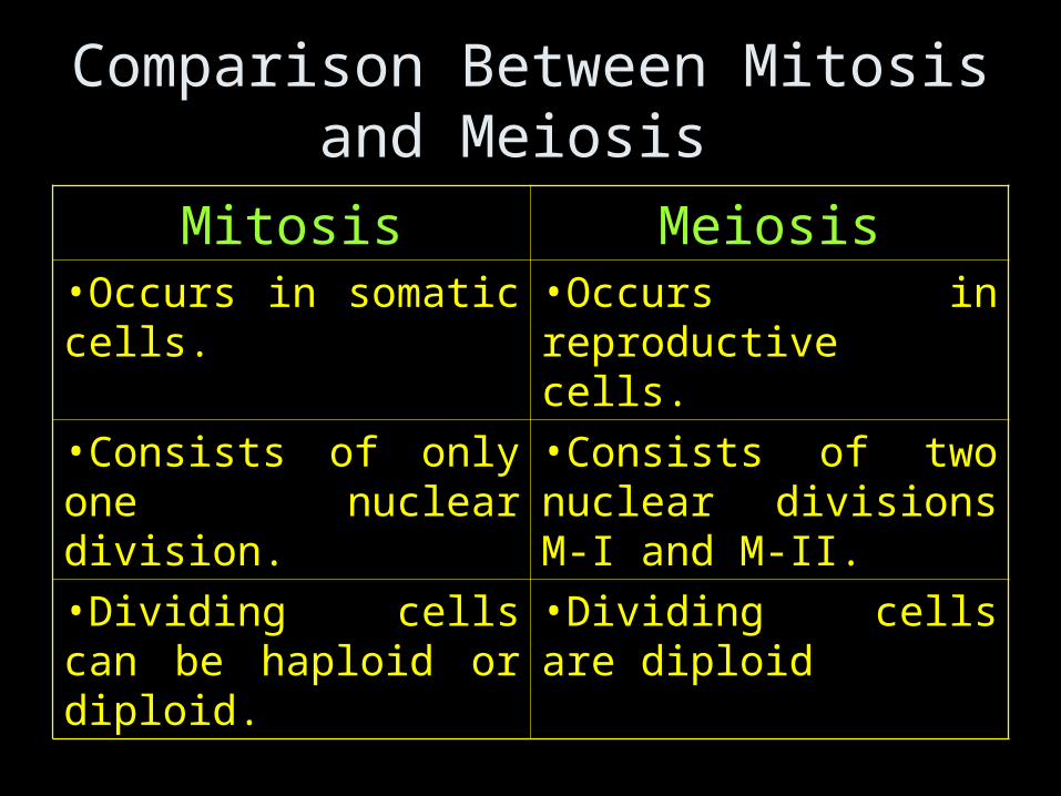
Comparison Between Mitosis and Meiosis
Mitosis Meiosis•Occurs in somatic cells. •Occurs in reproductive
cells.
•Consists of only one nuclear division.
•Consists of two nuclear divisions M-I and M-II.
•Dividing cells can be haploid or diploid.
•Dividing cells are diploid

Mitosis Meiosis•Involves division of chromosomes.
•Involves separation of homologous chromosomes in M-I and division of chromosomes in M-II.
•Does not involve either pairing of homologous chromosomes or crossing over.
•Pairing of homologous chromosomes and crossing over occur during Prophase-I.
•Two daughter cells are formed.
•Four daughter cells are formed.

Mitosis Meiosis•Number of chromosomes present in the mother cell is maintained in both the daughter cells. Therefore it is an equational division.
•Diploid number of chromosomes is reduced to haploid in each daughter cell. Therefore it is a reduction division
•Original characters of the chromosomes are maintained in the daughter cells.
•Chromosomal characters are altered due to "crossing over" causing recombination of genes

Mitosis Meiosis
Daughter cells are similar to each other and also to the original mother cell.
Daughter cells differ from each other as well as from the original mother cell.
Helps in growth and body repairs.
Helps in the sexual reproduction and regulation of chromosome number in the life cycle of sexually reproducing organism

Life Spans• There are about 210 cell types.• Each cell during its life span :
1- Born2- Differentiae3- Function4- Die
Neutrophil: 6-7 hours circulating to few days in tissue
RBCs : 120 daysBrain neuron: 50 to 100 years

AgingAging is generally characterized by:
• The declining ability to respond to stress.
• Increasing homeostatic imbalance.
• Increased risk of aging-associated diseases.
Death is the ultimate consequence of aging.

Two distinct modes of cell death
Apoptosis vs. Necrosis
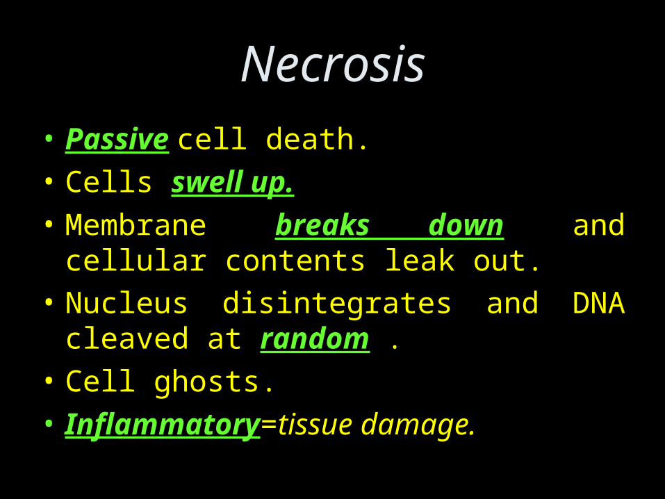
Necrosis• Passive cell death.
• Cells swell up.
• Membrane breaks down and cellular contents leak out.
• Nucleus disintegrates and DNA cleaved at random .
• Cell ghosts.
• Inflammatory=tissue damage.
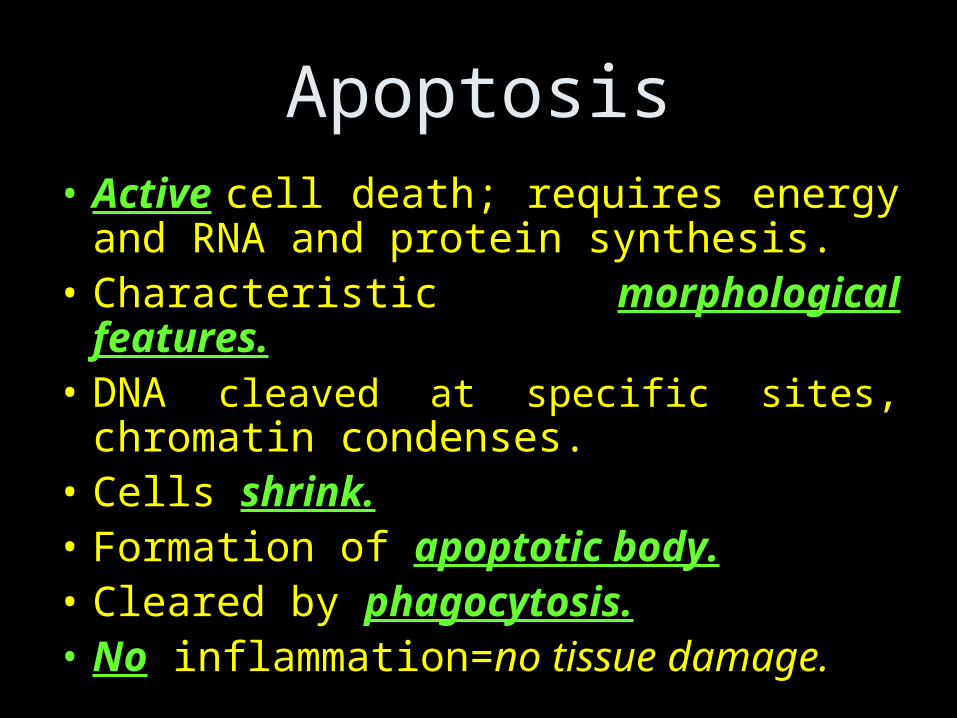
Apoptosis• Active cell death; requires energy and
RNA and protein synthesis.• Characteristic morphological features.• DNA cleaved at specific sites, chromatin
condenses.• Cells shrink.• Formation of apoptotic body.• Cleared by phagocytosis.• No inflammation=no tissue damage.

CaspasesProteases that are central for the apoptosis process.
2 functional categories:• Initiator caspases (trigger onset of apoptosis by
activating the caspase cascade)
Caspase 8,9,10 & 12.
• Effector caspases (undertake the actual work of destroying critical components of the cell)
Caspase 3,6,& 7.
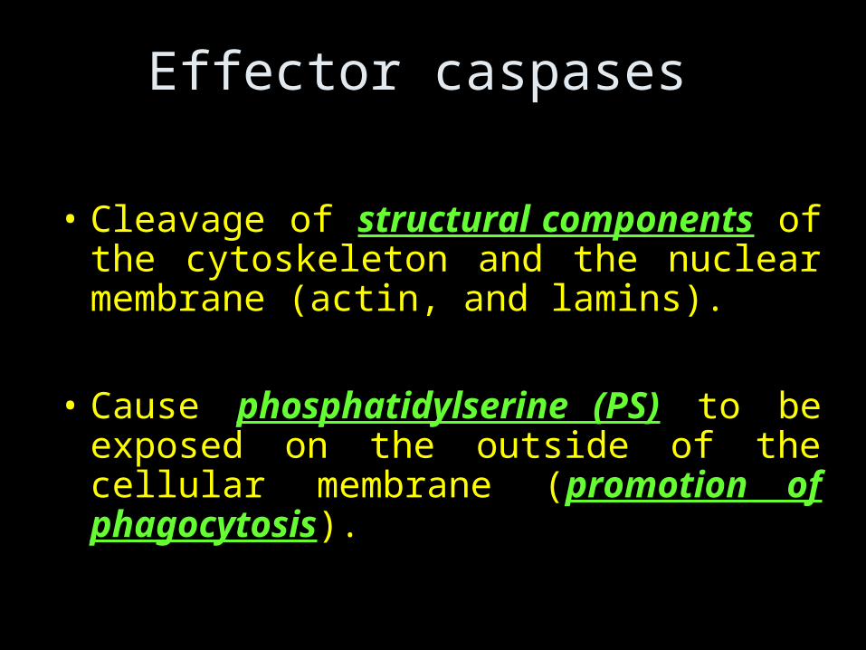
Effector caspases
• Cleavage of structural components of the cytoskeleton and the nuclear membrane (actin, and lamins).
• Cause phosphatidylserine (PS) to be exposed on the outside of the cellular membrane (promotion of phagocytosis).

Effector caspases
• Inhibit genes that regulate DNA repair during cell cycle.
• Inactivate enzymes responsible for stability, integrity and repair of DNA.
• Cleave ICAD (inhibitor of the caspase-activated DNase).
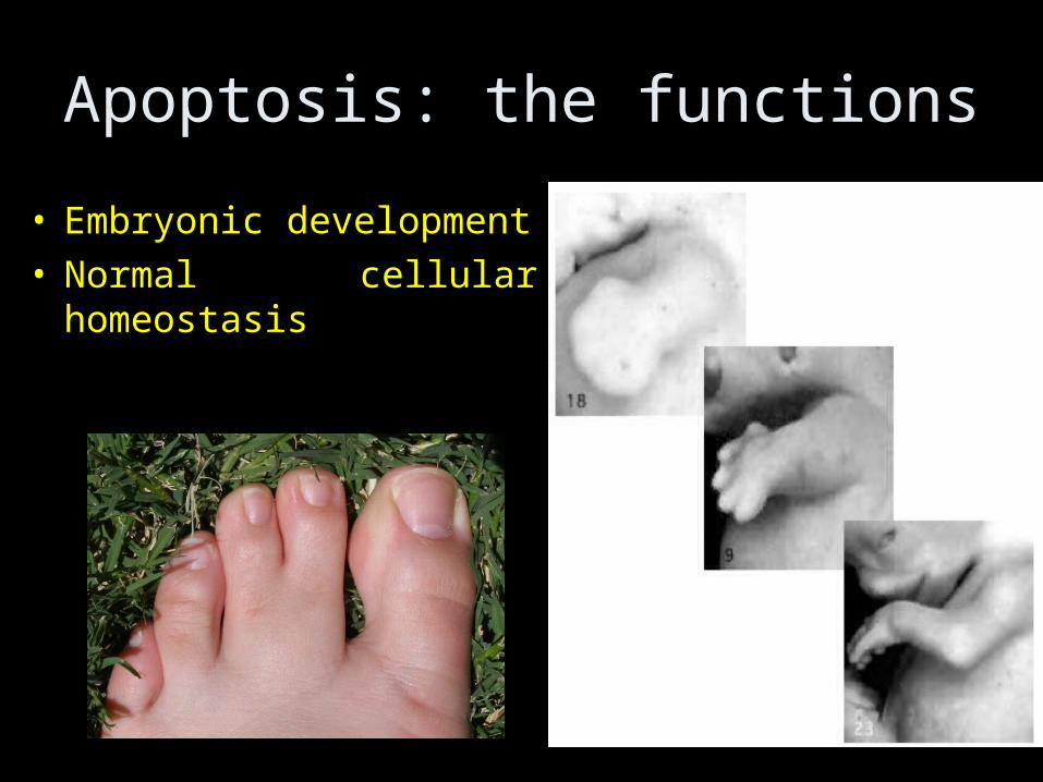
Apoptosis: the functions
• Embryonic development• Normal cellular
homeostasis

Apoptosis in normal physiology
• Intestinal epithelial cells every 4-5 days are substituted by new ones and this process involves apoptosis.
• Precursors of red cells are eliminated by apoptosis. When the levels of red cells lower there is increase in Erythropoietin (EPO) which inhibits the apoptosis of these precursors.

Apoptosis in normal physiology
• Regression of the cells of mammary gland after weaning of offspring.
• More or less the 90% of epithelial cells accumulated during pregnancy in the mammary gland undergo apoptosis.
• This phenomenon is called involution.

Deregulation of apoptosis
Insufficient apoptosis found in:
• Cancer
• Autoimmunity
Accelerated cell death is found in:
• Degenerative diseases
• Immunodeficiency

Apoptosis and Organ Transplants
• Transplant rejection is mainly due to attack of the new organs by the host immune system cells.
• New strategies for evading this (instead of using immunosuppressive drugs) is making at least some of the transplanted cells express apoptosis ligands.
• So they kill the immune cells that might attack the transplanted cells.

Apoptosis and AIDS
• The progression of the human immunodeficiency virus (HIV) to AIDS is primarily due to the depletion of CD4+ T-helper lymphocytes.
• One of the mechanisms by which T-helper cells are depleted is apoptosis.
• HIV enzymes inactivate anti-apoptotic proteins and simultaneously activate pro-apoptotic procaspase-8.
• Infected cells may also die as a direct consequence of the viral infection.

Summary
• Different stages of Meiosis
• Gametogenesis
• Differences and similarities between Meiosis and Mitosis
• Types of cell death
• Apoptosis roles in development, Cancer, organ transplant and AIDS