ccr8 expression identifies cd4 memory t cells · pdf fileccr8 expression identifies cd4...
TRANSCRIPT

of May 17, 2018.This information is current as
Th2 Effector Lymphocytes Regulatory and+Cells Enriched for FOXP3
CCR8 Expression Identifies CD4 Memory T
Hodge and Roland KolbeckSunita Badola, Eric Fedyk, Anthony J. Coyle, Martin R.Wang, Javier Cote-Sierra, Mark Ryan, Alice McDonald, Dulce Soler, Tobias R. Chapman, Louis R. Poisson, Lin
http://www.jimmunol.org/content/177/10/6940doi: 10.4049/jimmunol.177.10.6940
2006; 177:6940-6951; ;J Immunol
Referenceshttp://www.jimmunol.org/content/177/10/6940.full#ref-list-1
, 29 of which you can access for free at: cites 68 articlesThis article
average*
4 weeks from acceptance to publicationFast Publication! •
Every submission reviewed by practicing scientistsNo Triage! •
from submission to initial decisionRapid Reviews! 30 days* •
Submit online. ?The JIWhy
Subscriptionhttp://jimmunol.org/subscription
is online at: The Journal of ImmunologyInformation about subscribing to
Permissionshttp://www.aai.org/About/Publications/JI/copyright.htmlSubmit copyright permission requests at:
Email Alertshttp://jimmunol.org/alertsReceive free email-alerts when new articles cite this article. Sign up at:
Print ISSN: 0022-1767 Online ISSN: 1550-6606. Immunologists All rights reserved.Copyright © 2006 by The American Association of1451 Rockville Pike, Suite 650, Rockville, MD 20852The American Association of Immunologists, Inc.,
is published twice each month byThe Journal of Immunology
by guest on May 17, 2018
http://ww
w.jim
munol.org/
Dow
nloaded from
by guest on May 17, 2018
http://ww
w.jim
munol.org/
Dow
nloaded from
CCR8 Expression Identifies CD4 Memory T Cells Enrichedfor FOXP3� Regulatory and Th2 Effector Lymphocytes
Dulce Soler,1 Tobias R. Chapman, Louis R. Poisson, Lin Wang, Javier Cote-Sierra, Mark Ryan,Alice McDonald, Sunita Badola, Eric Fedyk, Anthony J. Coyle,2 Martin R. Hodge,and Roland Kolbeck3
CD4� Th2 cells are important regulators of allergic inflammation. CCR8 is thought to play a role in Th2-mediated responses,however, expression of CCR8 in peripheral blood has not been fully characterized. Using a fluorescent form of the ligand selectivefor CCR8 (F-CCL1), we identified the leukocytes expressing CCR8 in human, monkey, and mouse peripheral blood. CCR8expression is primarily restricted to a subset of human CD4 memory T lymphocytes (15%). Approximately 40% of CCR8�CD4�
T cells express Th2 cytokines IL-4 or IL-13 while 13% express the Th1 cytokine IFN-�. In fact, 50% of all Th2, but only 5% ofTh1, cells express CCR8. Upon anti-CD3/anti-CD28 mAb-mediated activation, CCR8�CD4� T cells secrete 3- to 7-fold higherlevels of IL-4, IL-5, IL-9, and IL-13 and 10- to 20-fold lower levels of IFN-� or IL-17, compared with CCR8�CD4� memory Tcells. Two-thirds of CCR8�CD4 T cells express cutaneous lymphocyte-associated Ag while the majority lack gut-homing receptors.CCR8�CD4� cells express CCR7 and CD62L and are present in spleen and lymph nodes of mice. Approximately 25% ofCCR8�CD4 T cells express CD25high while 20% of CCR8�CD4� express the T regulatory cell transcription factor FOXP3accounting for 60% of all FOXP3-expressing CD4� T cells. In conclusion, CCR8 marks a diverse subset of CD4 memory T cellsenriched for T regulatory and Th2 cells which have the potential for recruitment into sites of allergic inflammation where theycould participate in the induction and regulation of the allergic response. The Journal of Immunology, 2006, 177: 6940–6951.
T he T lymphocyte pool consists of naive T cells and Ag-experienced memory T cells which can be further dividedinto nonpolarized (TNPM),4 also referred to as central
memory (TCM), and effector memory (TEM) T cells. TNPM expandand acquire T-effector functions in response to Ag re-encounterwhile TEM represent a circulating pool of polarized T cells capableof producing immediate effector functions upon restimulation.CD4� TEM cells include Th1 and Th2 cells which migrate to in-flamed peripheral tissues where they secrete effector cytokines(IFN-� by Th1 and IL-4, IL-5, IL-9, and IL-13 by Th2 cells, re-spectively) involved in amplifying the immune response (1, 2). Inrecent years, other populations of memory T cells that have thecapacity to down-regulate immune responses through either cell-to-cell contact or the release of soluble mediators such as IL-10and TGF-�, so-called regulatory T (TREG) cells, have gained in-creased attention (3). A subpopulation of TREG cells, known asnaturally occurring TREG, are generated in the thymus during Tcell development and are best characterized by high expressionlevels of the IL-2R �-chain (CD25) and by the expression of the
forkhead family transcription factor FOXP3 which is necessary fortheir development and function (4, 5).
Migration of diverse T cell subsets during homeostasis and in-flammation is controlled by the concerted expression of adhesionmolecules, such as integrins and selectins, and chemokine recep-tors. For example, coexpression of L-selectin (CD62L) and thechemokine receptor CCR7 by T cells is a prerequisite for homingto lymphoid tissues (6 – 8) while expression of the integrin �4�7
or cutaneous lymphocyte-associated Ag (CLA) are required forlymphocyte migration to the gastrointestinal tract or the skin,respectively (9 –11).
Chemokine receptors belong to the class of seven transmem-brane G protein-coupled receptors and have been shown to medi-ate a variety of biological processes upon chemokine binding,including angiogenesis, leukocyte activation, and chemokine-induced transendothelial migration through integrin activation andsubsequent transmigration (12). Chemokines are small secretedproteins (�8 kDa) which can be divided into four subfamiliesbased on the spacing of two conserved cysteine residues (12). Theycan also be distinguished by their pattern of regulation: 1) lym-phoid chemokines such as CCL19, CCL21, and CXCL13 are con-stitutively expressed in lymphoid tissues and mediate the migra-tion of leukocytes into and within lymphoid tissues by engagementof their respective receptors CCR7 and CXCR5 (6, 13, 14), 2)chemokines constitutively expressed in nonlymphoid tissues, forexample, CCL1 in the skin which attracts CCR8� T cells (15), and3) inducible chemokines such as CCL11 (16), CCL17 (17, 18), andCXCL10 (19) attract effector cells into inflamed tissues throughengagement of CCR3, CCR4, and CXCR3, respectively.
We and others have recently reported the expression of the che-mokine CCL1 by IgE-activated mast cells in vitro and in vivo,implicating CCL1 in the recruitment of inflammatory cell typesinvolved in allergic inflammation (Refs. 20–22 and J. A. Gonzalo,Y. Qiu, J. M. Lora, A. Al-Garawi, J. L. Villeval, J. Boyce, C.
Inflammation, Millennium Pharmaceuticals, Cambridge, MA 02139
Received for publication February 10, 2006. Accepted for publication August28, 2006.
The costs of publication of this article were defrayed in part by the payment of pagecharges. This article must therefore be hereby marked advertisement in accordancewith 18 U.S.C. Section 1734 solely to indicate this fact.1 Address correspondence and reprint requests to Dr. Dulce Soler, Millennium Phar-maceuticals, 35 Landsdowne Street, Cambridge, MA 02139. E-mail address:[email protected] Current address: MedImmune, One MedImmune Way, Gaithersburg, MD 20878.3 Current address: Peptimmune, 64 Sidney Street, Cambridge, MA 02139.4 Abbreviations used in this paper: TNPM, nonpolarized memory T cell; TCM, centralmemory T cell; TEM, effector memory T cell; TREG, regulatory T cell; CLA, cuta-neous lymphoid-associated Ag; F-CCL1, fluorescently labeled CCL-1; DAPI,4�,6�-diamidino-2-phenylindole.
The Journal of Immunology
Copyright © 2006 by The American Association of Immunologists, Inc. 0022-1767/06/$02.00
by guest on May 17, 2018
http://ww
w.jim
munol.org/
Dow
nloaded from

Martinez, G. Marquez, I. Goya, Q. Hamid, et al., submitted forpublication). Indeed, CCR8, the only identified receptor for CCL1,belongs to a small group of chemokine receptors, including CCR3and CCR4 that have been shown to be preferentially associatedwith Th2 effector cells (23–25). Th2 cells are recruited to sites ofallergic mucosal inflammation where they secrete the Th2 cyto-kines IL-4, IL-5, IL-9, and IL-13 and orchestrate the hallmarks ofallergic lung inflammation such us IgE class switching, mast celland eosinophil activation, mucus hypersecretion, and airway hy-perresponsiveness. The functional involvement of the CCR8/CCL1 axis in the recruitment of Th2 effector cells in vivo is sup-ported by an increase in CCR8� CD4 T cell numbers in allergicasthma (Ref. 26 and J. A. Gonzalo, Y. Qiu, J. M. Lora, A. Al-Garawi, J. L. Villeval, J. Boyce, C. Martinez, G. Marquez, I. Goya,Q. Hamid, et al., submitted for publication) and by a recent studydemonstrating CCL1 and CCR8 up-regulation in atopic dermatitis(21). There are also reports suggesting CCR8 expression byCD4�CD25� TREG cells, skin-homing CLA� T cells, monocytes,NK cells, and dendritic cells (21, 27–32). Recently, the majority ofCD4� and CD8� T cells isolated from normal human skin, as wellas small subpopulations of CD4� and CD8� peripheral blood Tcells were reported to express CCR8 as well as both, CD45RA�
and CD45RO� (15). The authors proposed a mechanism by whichCCR8� T cells home to and reside in healthy skin tissue and playa role in immune surveillance.
Intrigued by the increased numbers of CCR8� CD4 T cells inallergic asthma and atopic dermatitis and by the substantial num-bers of CCR8-expressing T cells isolated from healthy human skin,we characterized the cell types expressing CCR8 in peripheralblood. The identification and phenotypic characterization of pe-ripheral blood-derived leukocytes expressing the chemokine re-ceptor CCR8 has been hampered by the limited availability ofspecific and suitable mAbs. Therefore, to identify cells expressingfunctional CCR8, we used fluorescently labeled CCL1 (F-CCL1),the ligand specific for CCR8. We discovered that CCR8 expressionis absent from naive CD4� T cells in the periphery and that it islargely restricted to a small but diverse subset of CD4� TNPM andTEM that preferentially produce Th2 cytokines. The CCR8�CD4�
T cell compartment includes cells capable of homing to secondarylymphoid tissues and to peripheral tissues such as the skin and thelung, but not to the gut. It also contains a small subset of TREG
cells as indicated by the expression of FOXP3. CCR8-expressingcells functionally respond to CCL1-dependent activation with di-rectional migration in vitro. Our data support a functional role forthe CCR8/CCL1 axis in Th2 effector cell recruitment into sites ofallergic mucosal inflammation and suggest that CCR8 participatesin the induction, amplification, and/or the resolution phases of in-flammatory responses.
Materials and MethodsProteins and Abs
Recombinant human CCL1 was obtained from R&D Systems and recom-binant human CCL1 labeled with Alexa Fluor 647 (F-CCL1) was obtainedfrom Dictagene. Fluorochrome-conjugated Abs to human proteins werepurchased from the following sources: CD3 (UCHT1), CD4 (L200), CD8(RPA-T8), CD19 (HIB19), CD27 (M-T271), CD45R0 (UCHL1), CD56(B159), CD62L (Dreg56), CD94 (HP-3D9), CD16 (3G8), CD29 (MAR4),CRTH2 (BM16), and CLA (HECA-452) were obtained from BD Bio-sciences; CD25 (4E3) from Miltenyi Biotec; ICOS from eBioscience;CD123 (9F5) from StemCell Technologies; and CD49d from ChemiconInternational. Unconjugated Abs to CCR3 (7B11), CCR4 (1G1), CCR5(2D7), CCR7 (7H12), CCR9 (3C3), CCR10 (1B5), CXCR3 (1C6), CXCR6(7F3), and �4�7 (act-1) were available in-house and were detected usingrelevant fluorochrome-conjugated secondary Abs. Fluorochrome-conju-gated Abs to cynomolgus monkey CD4 (L200), CD8 (RPA-T8), CD56(B159), CD16 (3G8), CD20 (2H7), CD25 (M-A251), and CD45RA (5H9)
were obtained from BD Biosciences. Fluorochrome-conjugated Abs tomouse CD4 (RM4-5), CD44 (IM7), CD8 (53-6.7), CD3 (145-2C11), andNK-1.1 (PK136) were obtained from BD Biosciences. For immunofluo-rescence microscopy, a goat polyclonal anti-human FOXP3 Ab and anFITC-conjugated donkey polyclonal to goat IgG H and L chains wereobtained from Abcam.
Staining of whole blood, lymph nodes, and spleen with F-CCL1
Wild-type and CCR8�/�C57BL6 mice were maintained in pathogen-freeconditions and used between 6 and 8 wk. CCR8�/� mice were obtainedfrom the laboratory of Dr. C. Martinez (Centro Nacional de BiotecnologiaUniversidad Autonoma, Madrid, Spain). Lymph nodes and spleens fromthese animals were taken and recovered cells were filtered through a70-�m cell strainer to prepare a single-cell suspension. Blood was obtainedfrom cynomolgus monkeys (Charles River Laboratories) and healthy hu-man volunteers after obtaining informed consent.
A total of 200 �l of whole blood, or 100 �l of a 5 � 106/ml cellsuspension were stained at 37°C (5% CO2/95% O2) for 30 min in a hu-midified incubator with 5 nM F-CCL1. Staining with F-CCL1 at 4°C or37°C yielded similar results but fluorescence intensity was greater at 37°C,most likely due to internalization of receptor/ligand complexes. Timecourse of binding experiments showed that maximum binding wasachieved in 30 min at 4°C and in �3 h at 37°C. To assess CCR8 expressionin various cell subsets and to examine costaining with other cellular mark-ers, samples were then placed on ice and stained with relevant mAbs (seeProteins and Abs) for 30 min at 4°C. RBC were lysed with ammoniumchloride lysis solution (StemCell Technologies) and samples were ana-lyzed by flow cytometry (BD FACSCalibur; BD Biosciences) usingCellQuest software. To verify specificity and selectivity, all our phenotypicexperiments included control samples with excess unlabeled CCL1 andsmall molecule antagonists selective for CCR8 (T. Jenkins, B. Guan, M.Dai, G. Li, T. Lightburn, S. Huang, S. Freeze, D. Burdi, S. Jacutin-Porte,R. Bennett, et al., submitted for publication).
Isolation of CCR8� and CCR8�CD4� populations
Complete medium consisted of RPMI 1640, 2 mM L-glutamine, 100 �Mnonessential amino acids, 1 mM sodium pyruvate, 10 �g/ml penicillin/streptomycin (all obtained from Invitrogen Life Technologies), and 10%United States-defined FBS (HyClone).
The isolation of CCR8� and CCR8�CD4� memory T cells from humanblood was performed as follows. Human PBMCs were isolated from freshbuffy coats (Oklahoma Blood Institute) by centrifugation on Ficoll-PaquePLUS (Amersham Biosciences). Pure (�98%) CD4� memory T cells wereobtained by negative selection using a CD4 memory� T cell isolation kit(Miltenyi Biotec) according to the manufacturer’s instructions. These cellswere subsequently stained with 1 nM F-CCL1 overnight in complete me-dium at 37°C and sorted the following day by FACS into CCR8� andCCR8� subsets using a MOFLO (DakoCytomation) cell sorter. CCR8�/�
populations were typically of �97% purity. To examine the coexpressionof CD25, CCR8, and FOXP3, CD4� memory T cells were also sorted intoCD25� and CD25� subpopulations before CCR8 sorting. Briefly, PBMCwere isolated as above, and CD4� T cells isolated using a CD4 T cellisolation kit II (Miltenyi Biotec). From these cells, CD25� T cells wereisolated using a CD4�CD25� regulatory T cell isolation kit (Miltenyi Bio-tec) and put to one side. Naive CD4� cells and CD45RO� memory T cellswere separated from the remaining CD25�CD4� cells using CD45RO�
microbeads (Miltenyi Biotec). Both CD4 memory� populations werestained with F-CCL1 (1 nM overnight) and anti-CD45RO-FITC and sortedbased on CCR8/CD45RO expression by FACS as above.
Chemotaxis assays
After CD4� memory T cells had been sorted into CCR8� and CCR8�
subsets by FACS, they were incubated for 24 h at 37°C in complete me-dium to allow for washout of F-CCL1 labeling and receptor recycling tothe cell surface. Cell migration was subsequently assessed using 3-�m porepolycarbonate membranes in a 96-well multiscreen-MIC plate format (Mil-lipore). Before performing the experiment, the upper surface of the che-motaxis membrane was precoated with a confluent monolayer of ECV-304cells. CCL1 was diluted in chemotaxis buffer (CTXB: HBSS with 10 mMHEPES and 0.1% fatty acid-free BSA) to the indicated concentrations andplaced in the lower wells of the chemotaxis plate. A total of 100 �l of cellsuspension (2 � 105 cells) was added to the upper wells and the plate wasincubated at 37°C for 4 h in a humidified 5% CO2 incubator. After incu-bating, the chemotaxis membranes were removed and the migrated cells inthe lower chamber were loaded with Calcein AM dye (Invitrogen LifeTechnologies) before reading with the Discovery-1 cellular imaging plat-form. The resulting data were analyzed using MetaMorph software (both
6941The Journal of Immunology
by guest on May 17, 2018
http://ww
w.jim
munol.org/
Dow
nloaded from

Molecular Devices) and expressed as the mean chemotactic index (averagenumber of migrated cells exposed to chemokine divided by the number ofcells exposed to CTXB alone) from triplicate samples � SD.
Intracellular cytokine staining
For intracellular staining, immediately after sorting, cells were stimulatedwith 10 ng/ml PMA and 1 �g/ml ionomycin (both Sigma-Aldrich) for 6 h,the last 5 h in the presence of 10 �g/ml brefeldin A (BD FastImmune; BDBiosciences) in complete medium at 37°C. Cells were washed with PBSand fixed with 4% paraformaldehyde, before being permeabilized with0.5% saponin in PBA (5% BSA in PBS; no Ca2�/Mg2�). Cells werestained with combinations of the following Abs for 30 min at room tem-perature: mouse Abs to IL-4-PE (3007) and IL-13-PE (32007) (both R&DSystems), a rat Ab to IL-10-PE (JES3-9D7) and a mouse Ab to IFN-�-FITC (25723.11) (both BD Biosciences) and a rat Ab to IL-4-allophyco-cyanin (MP4-25D2) (eBioscience). Samples were subsequently washedone time with saponin buffer, one time with PBA, and analyzed by flowcytometry.
Analysis of cytokine mRNA expression and protein secretion
To examine cytokine expression and secretion, sorted cells were resus-pended in complete medium containing 50 ng/ml IL-2 (R&D Systems) and10 �g/ml anti-CD28 (BD Pharmingen) and plated at 2 � 105/well in a96-well EIA high protein-binding plates (Costar) that had been precoatedwith anti-CD3 (10 �g/ml; BD Pharmingen). Cells were incubated for 6 and24 h and supernatants were removed and stored at �80°C. Supernatantswere sent to Pierce Endogen for Multiplexed Searchlight ELISA (Pierce)analysis of the following cytokines: IL-4, IL-5, IL-8, IL-9, IL-10, IL-13,IL-17, IFN-�, and TNF-�. The cell pellets after supernatant removal werealso stored at �80°C in preparation for RNA extraction to examine cyto-kine gene expression, described as follows.
Quantitative RT-qPCR
Total RNA was isolated using RNAeasy technology on a Biorobot 8000workstation and DNase treated according to the manufacturer’s protocol(Qiagen). The purity and yield of the RNA were assessed using the Nano-drop ND-1000. Integrity of the RNA was measured with RNA 6000 NanoLabChip on a Agilent 2100 Bioanalyzer (Agilent Technologies) and cal-culated using the RNA Integrity Number algorithm. First-strand cDNAsynthesis was performed with a reverse transcription system (Applied Bio-systems) according to the manufacturer’s protocol, except that botholigo-dT and random hexamers were used for priming. The integrity ofcDNA samples was assessed via qTIAM analysis of 18S and �2-micro-globulin transcripts by real-time PCR in an ABI PRISM 7700 (AppliedBiosystems). Primers and MGB Eclipse probes (Nanogen) for real-timePCR (see below), were designed specifically to U133A&B GeneChipprobe sets (Affymetrix) used in initial high-density microarray analysis.Transcripts of interest were assayed in a multiplexed format, using human�2-microglobulin RNA, as an endogenous control; analysis of MAS 5.0data from corresponding U133A&B GeneChip data verified that levels of�2-microglobulin transcript remained constant across this set of RNA sam-ples. Transcripts were amplified using Taq polymerase (Sigma-Aldrich)and cycling parameters of: an initial 95°C for 2 min, then 40 cycles of 95°Cfor 20 s, 58°C for 20 s, 76°C for 20 s. Data were analyzed using SDS 1.7(Applied Biosystems) software. Data were analyzed using the comparativecycle threshold method, with the amount of transcript of interest normal-ized to �2-microglobulin transcripts (user bulletin no. 2 from Applied Bio-systems). Forward/reverse/probe primers were: IL-4, ACACAACTGAGAAGGAAACCTT/CCTGTCGAGCCGTTTCAGGAAT/TACAGCCACCATGAGA*A; IL-5, CCCACAAGTGCATTGGTG/TCAGAGTCTCATTGGCTATCAGCAG/GCTTTCTACTCA*TCG; IL-9, GAGACTGTCTCAGATGACCA/GACTCTTCAGAAATGTCAGCGCGTT/GCAAACAA*GATACCC; IL-13, TGTGCAGCCCTGGAAT/TGTCTCGGACATGCAAGCTGGAAA/CAGTGCCATCGAGA*A; IL-17, ACTTGGGCTGCATCAAC/GGACCAGGATCTCTTGCTGGAT/GACTACCA*CATGA*AC; TNF-�, GATCATCTTCTCGAACCC/GTTATCTCTCAGCTCCACGCCATT/TAGCAAACCCTCAAGC; IFN-�, CATCCAAGTGATGGCTGAAC/TACTGGGATGCTCTTCGACCTCGAAA/AACAGG*GAAGCGAAAA; �2-microglobulin, GCCTGCCGTGTGAACCATGTGACTTTGTC/CGGCATCTTCAAACCTCCATGA/GTTAAGTGGGATCGAGA; FOXP3,CTGAGTCTGCACAAGTGCTTT/TTGGAACACCTGCTGGGCCTCT/TGGACCGTGGATGAGC. The base immediately preceding the asteriskis a specially modified base that the vendor (Epoch Biosciences (nowNanogen)) provides to increase the stability of the interaction of the primer.The shills are to indicate the separation between the sequences of the threeprimers of each gene.
Examination of FOXP3 expression by immunofluorescencemicroscopy
Sorted cells were resuspended at 1 � 106/ml in complete medium. A totalof 200 �l of cell suspension was loaded into a Shandon single Cytofunnel,and the cells were spun onto microscope slides using a Shandon Cytospin3 (both Thermo Electron). Slides were air-dried overnight, fixed in acetonefor 5 min, and air-dried for 1 h. Slides were blocked with 10% FBS in PBSfor 15 min in a humid chamber, then stained with anti-FOXP3 (1/200dilution in blocking buffer) for 2 h in a humidified chamber. Slides werethen washed for 10 min in PBS with 0.2% gelatin. For detection, slideswere stained with tetramethylrhodamine isothiocyanate-conjugated donkeyanti-goat IgG (1/200 dilution in 5% donkey serum in PBS) for 30 min ina humidified chamber in the dark. Control slides were prepared and incu-bated with secondary Ab alone to control for background staining. Slideswere washed again with PBS containing 0.2% gelatin and mounted using4�,6�-diamidino-2-phenylindole (DAPI) mounting medium to allow for ex-amination of nuclear colocalization. Slides were then viewed with an im-munofluorescence microscope and images captured for analysis.
ResultsCD4 memory T cells are the predominant cell type expressingCCR8 in peripheral blood
We recently found that the number of CCR8-expressing cells inlungs of asthmatic individuals is increased 4-fold when comparedwith healthy controls and that �70% of all lung CD4� T cellsexpress CCR8 (J. A. Gonzalo, Y. Qiu, J. M. Lora, A. Al-Garawi,J. L. Villeval, J. Boyce, C. Martinez, G. Marquez, I. Goya, Q.Hamid, et al., submitted for publication). Thus, it was important tocharacterize CCR8 expression in human peripheral blood leuko-cytes and its potential to orchestrate allergic mucosal inflammationin the lung. CCL1 has been shown to specifically bind to andactivate CCR8. To identify CCR8-expressing cells, we used F-CCL1 which exhibits similar chemotactic potency as unlabeledCCL1 (data not shown). For staining peripheral blood leukocytes,F-CCL1 was used at concentrations that induce maximal cell mi-gration (1–5 nM). These concentrations also achieved maximalstaining. In human whole blood, F-CCL1 consistently stained asubpopulation of CD4�CD45RO� memory T cells (15 � 4%; n �50 different donors), representing 91 � 7% of all labeled cellsidentified (Fig. 1A; Table I). Staining specificity and selectivity forCCR8 was thoroughly verified by 1) competition with excess un-labeled CCL1 (Fig. 1, A and B) or TCA-3 for mouse samples, 2)absence of staining in lymphocytes of CCR8-deficient mice (Fig.1C), and importantly, by 3) competition with several small mole-cule antagonists shown to be selective for CCR8 vs all other che-mokine receptors and a large panel of other G protein-coupledreceptors. In addition, MC148-huFc, an engineered fusion proteincontaining at the N terminus MC148, a selective CCR8 viral che-mokine antagonist (33), also gave the same pattern of staining asF-CCL1, and both chemokines were competitive with each other(data not shown). All non-CD4 expression of CCR8 is confined tosmall subsets of CD8� T lymphocytes (2 � 1.6%; n � 12) andCD56� NK cells (0.7 � 0.46%; n � 12) (Table I). The Th2/Tcytotoxic 2 cell-associated PGD2 receptor, CRTH2 (34), is ex-pressed in a subset of CCR8�CD8� T cells (Fig. 1A). Within theCD56� NK cell compartment, CCR8 expression is associatedpreferentially (16 � 3.8%) with a small subset that expresses highCD56 levels (CD56highCD94highCD16�CD3�) (Fig. 1A) (35, 36).All other subsets of NK cells investigated including NKT cells,CD8�CD56� and CD8�CD56� NK cells, had levels of expres-sion similar to the total CD56� population. CCR8 expression wasnot observed in CD4�CD45RO� or CD4�CD45RA� naive Tcells (Fig. 1A), or CD4�CD45RA�CD45RO� T cells, CD19� Blymphocytes, plasmacytoid (CD123highBDCA-2�) or myeloid(CD123lowBDCA-1� or CD123�BDCA-3�) dendritic cells,CD123� basophils, CD14� monocytes, CD16� neutrophils, or
6942 EXPRESSION OF CCR8 BY PERIPHERAL BLOOD CD4 MEMORY T CELLS
by guest on May 17, 2018
http://ww
w.jim
munol.org/
Dow
nloaded from

CCR3�CD49d� eosinophils (data not shown). Similarly, CD4memory T cells were the predominant cell type expressing CCR8in peripheral blood from cynomolgus monkeys (�10%; Fig. 1B;Table I) and naive mice (�10%; Fig. 1C, Table I), respectively.We also identified low numbers of CCR8�CD8� T cells and NKcells in peripheral blood from cynomolgus monkeys (Table I) andas in humans, the CD56highCD16� NK population was enrichedfor CCR8� cells (data not shown). In mice, CCR8 was not ex-pressed on CD8� T cells but it was expressed by a small number
of NK (NK1.1; 0.4%) cells and NKT cells (2%) (data not shown).Leukocyte types and numbers expressing CCR8 in peripheralblood of human, monkey, and mouse are summarized in Table I.In addition, we identified CCR8�CD4 memory T cells, NK andNKT cell populations in naive mouse spleens (14, 1, and 3%,respectively) and lymph nodes (24, 3, and 4%, respectively) butnot in the corresponding tissues derived from CCR8-deficient mice(data not shown).
In summary, our data clearly indicate that CD4� memory Tcells are the predominant cell type expressing CCR8 in peripheralblood across species (human, monkey, mouse) and that these cellseither have the potential to migrate into secondary lymphoid or-gans and/or acquire a CCR8� phenotype during activation in theselymphoid tissues as indicated by the presence of CCR8�CD4�
memory T cells in naive mouse spleens and lymph nodes.
Activated CCR8�CD4 memory T cells preferentially produceTh2 cytokines in vitro
Th effector cells are best characterized by the types of cytokinesexpressed upon activation: Th1 effector cells produce IFN-�whereas Th2 effector cells produce IL-4, IL-5, IL-9, and IL-13. Toinvestigate the potential of CCR8�CD4 memory T cells to expresseither Th1 or Th2 cytokines, we sorted CD4 memory T cells iso-lated from human peripheral blood into CCR8� and CCR8� pop-ulations (purity �97%) (Fig. 2A) and determined the relative cellnumbers expressing IL-4, IL-10, IL-13, and IFN-� by intracellularstaining following activation in vitro. To ensure that the sortedpopulations were representative of their respective counterparts inblood, we compared their relative phenotypic frequencies with re-spect to expression of CLA, CD25, CCR5, CCR7, CD62L, CD27,and CRTH2 and found no significant differences. As shown in Fig.2, B and C, the percentage of activated cells expressing IL-4 was6-fold higher in the CCR8� than in the CCR8� population(CCR8� 26 � 3%; CCR8� 4 � 1%) and cells expressing IL-13were 10-fold more frequent in the CCR8� than in the CCR8�
population (CCR8� 20 � 2%; CCR8� 2 � 0.6%). Conversely,cells expressing IFN-� were 3-fold more frequent in the CCR8�
population (CCR8� 13 � 4%; CCR8� 42 � 8%). Approximatelyone-third of all CCR8� IL-4- and IL-13-expressing cells coex-pressed both cytokines most likely reflecting a probabilistic distri-bution of expression of each cytokine as recently demonstrated forin vitro-cultured Th2 cells (37). Within the CCR8� population,there was very little coexpression of IL-4 or IL-13 with IFN-�,whereas �50% of IL-4- and IL-13-expressing CCR8� cells coex-pressed IFN-� likely representing still uncommitted Th0 cells.Both cell populations exhibited a similar frequency of IL-10-ex-pressing cells (2%). In summary, �40% of all CCR8� memory Tcells express either IL-4 or IL-13 after activation, consistent withTh2 effector function. When compared with the total number ofCD4 memory T cells the CCR8� population contains �40% of allIL-4, 60% of all IL-13, 15% of all IL-10, but only 5% of allIFN-�-expressing cells (Fig. 2D). Of particular significance is thealmost complete inclusion of all IFN-�-producing Th1 effectorcells in the CCR8� population (95%).
The enrichment of Th2 effector cells within the CCR8�CD4memory T cell population was further supported by the higherlevels of IL-4 (3 � 0.9-fold), IL-5 (5 � 1-fold), IL-9 (2 � 0.5-fold), and IL-13 (3 � 0.8-fold) secreted by CCR8� than byCCR8� CD4 memory T cells 24 h after anti-CD3/anti-CD28 ac-tivation (Fig. 2E). Levels of the Th1 cytokine IFN-� (12 � 6-fold)and IL-17 (8 � 0.1-fold) were greater in the CCR8� population.The differences in IL-9 (6-fold), IL-13 (7-fold), and IL-17 (20-fold) levels were even more pronounced 48 h after activation (data
FIGURE 1. CCR8 expression is mainly restricted to a subset of memoryCD4� T cells in human, monkey, and mouse peripheral blood. A, Flowcytometric analysis of human blood stained with 5 nM F-CCL1 alone(PBS) or with 40 nM unlabeled CCL1 and markers for CD4� memory Tcells (CD4, CD45RO; top panel), CD8 and CRTH2 T cells (bottom panel,left), and CD56� NK cells (bottom panel, right). CCR8 expression wasexamined in the naive CD4� T cell (CD4�CD45RO�), memory CD4� Tcell (CD4�CD45RO�), CD8� T cell, lymphocyte, and CD56high cell gates,and specificity of staining was demonstrated in all cases by competitionwith unlabeled CCL1. The percentage of CCR8-expressing cells is indi-cated in the top right quadrant of each plot. For CCR8 vs CRTH2 expres-sion in the CD8 gate, the frequency of all four possible subsets is indicated.B, Flow cytometric analysis of cynomolgus monkey whole blood stainedwith CCL1, CD4, and CD45RA. CCR8 expression was examined in thenaive CD4� T cell gate (CD4�CD45RA�) and the memory CD4� T cellgate (CD4�CD45RA�) and verified to be specific by competition withunlabeled CCL1. C, Flow cytometric analysis of mouse whole blood, aswell as spleen and lymph node cell suspensions from wild-type and CCR8-deficient mice stained with CCL1, CD4, and CD44high. CCR8 expressionwas examined in the gated memory CD4 cell population (CD4�CD44high).These plots are representative and complete data set are summarized inTable I.
6943The Journal of Immunology
by guest on May 17, 2018
http://ww
w.jim
munol.org/
Dow
nloaded from
not shown). Protein levels correlated with mRNA levels as deter-mined by quantitative real-time PCR 6 or 24 h after anti-CD3/anti-CD28 activation (Fig. 2F). The cytokine expression data clearlyindicate that CCR8 expression selects for a subset of peripheralblood CD4 memory T cells enriched in Th2 effector cells.
Phenotypic characterization of CCR8�CD4 memory T cells
CCR8�CD4� T cells have been proposed to express skin-homingreceptors and TREG markers but a complete phenotypic character-ization in whole blood has not been reported. To further charac-terize blood-derived CCR8�CD4� T cells and their homing po-tential, we investigated their phenotype in detail.
CCR4 and CCR10, as well as CCR8 and CLA, have been pro-posed to be critically involved in skin-specific T cell homing dur-ing homeostasis and inflammation (15, 38–40). Approximatelytwo-thirds of CCR8�CD4 memory T cells express the skin hom-ing receptor CLA (66 � 12%; n � 20) the majority of which (86 �12%; n � 10) also express the chemokine receptor CCR10 (Fig.3B, Table II). The CCR8�CD4 memory T cell subset contains�36 � 5% of all CLA� and 65 � 7% of all CCR10�CD4 memoryT cells. In addition, while CCR8� CD4 memory T cells contain�22 � 6% of all CCR4�CD4 memory T cells (Table II), CCR4 isexpressed by nearly the entire CCR8� population (97 � 3%) (Fig.3B; Table II). It is therefore possible that homing of theCCR8�CLA� cells is restricted to the skin under conditions whereone or a combination of appropriate chemokines are expressed inthe skin.
Interestingly, we observed that within the CCR8� population,IL-4- and IL-13-producing cells were enriched 2-fold in the non-skin homing (CLA�CCR8�) as compared with the skin-homing(CLA�CCR8�) subset (30 vs 15% and 21 vs 16% for IL-4 andIL-13, respectively). Conversely, IFN-�-producing cells were2-fold enriched in the CLA�CCR8� population when comparedwith the CLA�CCR8� subset (12 vs 6%). However, in theCCR8� subsets, the frequency of IL-4, IL-13, or IFN-�-producingcells appeared to be independent of CLA expression. Therefore,Th2 cytokine-producing effector cells are enriched in theCCR8�CLA� population, presumably a subset capable of homingto the lung.
The integrin �4�1 (CD49dCD29) has been implicated in lunghoming by virtue of its interaction with VCAM-1 (41). AllCCR8�CD4� T cells express the � subunit CD29, however, wefound that only half of CCR8�CD4� T cells express �4 althoughthe vast majority of CCR8� CD4� T cells express this � subunit.Interestingly, the majority of CLA�CCR8� cells express high lev-els of �4 while the skin-homing cells were mostly negative orintermediate/low for �4 (Fig. 3D) and expressed �5 (data notshown). It is thus possible that the CLA�CCR8��4
high�1� subset,
enriched in Th2 cells (see above), might preferentially migrate tothe lung and participate in allergic inflammation of the airway.
The integrin �4�7 and chemokine receptor CCR9 are involvedin gut homing of T cells (10, 11, 42, 43). Only 1.5 � 0.44% ofCCR8�CD4 T cells are �4�7
high–accounting for only 0.6% of all�4�7
high CD4 memory cells. Even less coexpress CCR9 and�4�7
high (0.7 � 0.05% representing only 1.5% of all double-pos-itive cells in the CD4 memory population) (Fig. 3C). Therefore,97–99% of CD4 memory cells expressing gut-homing receptorsreside in the CCR8� compartment and peripheral CCR8�CD4 Tcells most likely do not home to the gut. The majority of peripheralblood CCR8�CD4 memory T cells express chemokine receptorCCR7 (90%) and CD62L (75%), which colocalize on �70% of thecells (Fig. 3D, Table II), indicating the ability of these cells tomigrate to secondary lymphoid tissues.
High expression levels of the IL-2R �-chain (CD25) have beenassociated with cell activation and TREG cells. About half (47 � 6;n � 18 donors) of all CD25highCD4 memory T cells express CCR8accounting for a quarter (25 � 6%; n � 22 donors) of all CCR8-expressing CD4 T cells. CD25high cells are enriched 3-fold in theCCR8� subset as compared with the entire CD4 memory T cellpopulation (Fig. 3E, Table II). Interestingly, about two-thirds of allCCR8�CD25high T cells belong to the subset of potentially skin-homing T cells (Fig. 3E; Table II). This CCR8�CLA�CD25high
subset accounts for one-quarter of all skin-homing CCR8�CD4memory T cells. CD25high is also expressed in one-quarter of theCLA�CCR8� subset, which may be defined as systemic homing,due to the lack of skin and gut-homing properties.
The CD28-related molecule ICOS functions to regulate clonalexpansion and more recently has been associated with regulatoryfunctions (44, 45). ICOS is expressed in a quarter of CCR8�CD4�
cells, representing 40% of all ICOS-expressing CD4 memory Tcells (Fig. 3F). Similarly, lack of expression of the TNFR homologCD27 has been associated with effector-type T cells and with cellsrecently activated by Ag (46, 47). Approximately one-third of allCD27�CD4 memory T cells express CCR8. Both ICOS expres-sion and absence of CD27 are enriched 2-fold in the CCR8� pop-ulation (Table II) indicating an accumulation of TEM cells.
We also identified small subpopulations of CCR8� cells ex-pressing the Th2-associated receptors CRTH2 (9 � 4%; n � 14)and CCR3 (10 � 6%) (Table II). In both cases, these cells repre-sent about one-third of all CRTH2� and CCR3�CD4 memory Tcells and they are enriched 2-fold in the CCR8 compartment ascompared with the total CD4 memory population (Fig. 3F, TableII). CCR4 which is also associated with Th2 function is expressedin virtually all CCR8�CD4� cells (Fig. 3B). At the same time,chemokine receptors which are enriched in Th1 effector cells suchas CXCR3, CCR2, CCR5, and CXCR6 (25) were expressed by 30,27, 24, and 11% of the CCR8� cells, respectively. These frequen-cies are either the same or lower compared with the total CD4memory population and account for minor fractions of all CD4memory cells expressing these receptors (Fig. 3G, Table II).
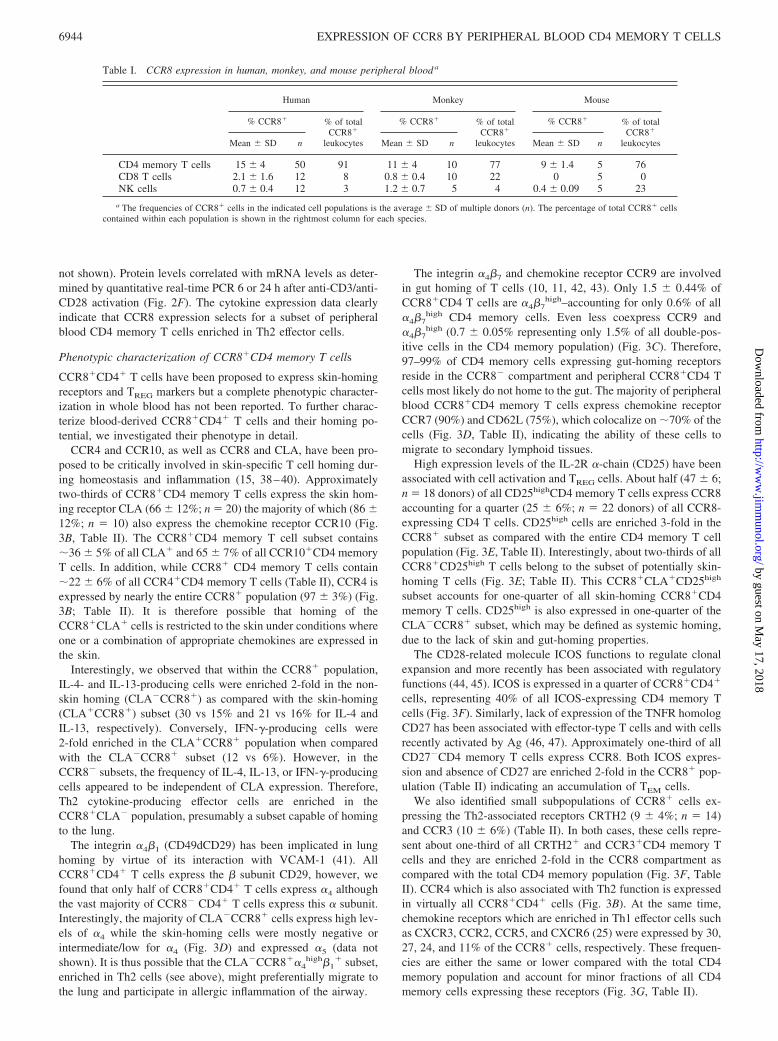
Table I. CCR8 expression in human, monkey, and mouse peripheral blood a
Human Monkey Mouse
% CCR8� % of totalCCR8�
leukocytes
% CCR8� % of totalCCR8�
leukocytes
% CCR8� % of totalCCR8�
leukocytesMean � SD n Mean � SD n Mean � SD n
CD4 memory T cells 15 � 4 50 91 11 � 4 10 77 9 � 1.4 5 76CD8 T cells 2.1 � 1.6 12 8 0.8 � 0.4 10 22 0 5 0NK cells 0.7 � 0.4 12 3 1.2 � 0.7 5 4 0.4 � 0.09 5 23
a The frequencies of CCR8� cells in the indicated cell populations is the average � SD of multiple donors (n). The percentage of total CCR8� cellscontained within each population is shown in the rightmost column for each species.
6944 EXPRESSION OF CCR8 BY PERIPHERAL BLOOD CD4 MEMORY T CELLS
by guest on May 17, 2018
http://ww
w.jim
munol.org/
Dow
nloaded from

FOXP3 expression by CCR8�CD4 memory T cells
The forkhead transcription factor family member FOXP3 is themost specific marker for thymic-derived TREG cells and is requiredfor TREG cell development and function (4). CCR8 has recently
been shown to be expressed by CD4�CD25� TREG cells and tomediate CCL1-dependent migration of those cells in vitro (27–29).It is however unclear to what degree CCR8 expression is confinedto TREG cells. To address this issue, we used a FOXP3-specific Ab
FIGURE 2. CCR8� memory CD4� T cells prefer-entially produce Th2 cytokines. A, Freshly isolatedmemory CD4� T cells were labeled with F-CCL1 andsorted into the CCR8� and CCR8� subsets. Flow cy-tometric analysis demonstrating CCR8 expression inisolated memory CD4�cells before sorting (left panel)and in sorted CCR8� and CCR8� memory CD4 cells.Staining specificity was monitored using excess unla-beled CCL1 (second from left). B, A representativeexample of CCR8� and CCR8�CD4 memory T cellsactivated for 6 h with ionomycin/PMA, stained with anti-cytokine Abs, and examined by flow cytometry forintracellular cytokine expression. Plots show the rela-tive frequencies of all four possible cell subsets indi-cated in the right top corner. C, Percentage of cytokine-producing cells in the CCR8� (f) and CCR8� (�)memory CD4� T cell subsets (average � SD of threeindependent experiments/donors). D, Distribution of allmemory CD4 T cells expressing a given cytokine be-tween the CCR8� (f) and CCR8� (�) memory CD4 Tcell populations (average � SD of three independentexperiments). E and F, Levels of (E) secreted cytokineand (F) cytokine mRNA following activation of CCR8�
and CCR8� memory CD4 T cells with anti-CD3/anti-CD28 for 24 h. Shown is a representative experiment ofthree independent experiments. Numbers indicate folddifferences between the two populations.
6945The Journal of Immunology
by guest on May 17, 2018
http://ww
w.jim
munol.org/
Dow
nloaded from

and enumerated CCR8� and CCR8�CD4 memory T cells whosenuclei were positive for FOXP3, as determined by costaining withthe nuclear dye DAPI. FOXP3 protein localized to the nuclei of�21% of all CCR8� cells and only 3% of CCR8� cells (Fig. 4, Aand B), accounting for �60 and 40% of all FOXP3� CD4 memoryT cells, respectively (Fig. 4C). All CCR8�FOXP3� cells expressCD25 and represent 50–70% of the CCR8�CD25� population(Fig. 4D). No FOXP3� nuclei could be identified in CD4 naive Tcells or in the absence of the FOXP3 Ab indicating staining spec-
ificity (data not shown). Interestingly, cells with high-intensityFOXP3 staining exclusively localized to the CCR8� population.The distribution of FOXP3� cells across the different cell popu-lations investigated was supported by FOXP3 mRNA expressiondata, showing the highest FOXP3 mRNA levels in CD25�CCR8�
and CD25�CCR8� memory T cells (Fig. 4E). In addition, ac-tivation with anti-CD3/anti-CD28 for 6 h resulted in increasedFOXP3 mRNA levels (3- to 5-fold) in the CD25�CCR8� andCD25�CCR8� subsets with no effect on the CD25� subsets (data
FIGURE 3. Phenotypic analysis of human peripheralblood memory CD4 T cells expressing CCR8. A, Flowcytometry plots indicating the gates used in B–G. Thefrequencies of all four possible subsets in each plot areindicated in the top right quadrant. B, CCR8 coexpres-sion with the skin-homing cell surface markers CLA,CCR10, and CCR4. C, CCR8 expression analysis inCD4 memory T cells stained with the gut-homing re-ceptor �4�7 (left plot); analysis of CCR9 vs �4�7 ex-pression in CCR8�CD4� T cells (middle plot) and inCCR8�CD4� T cells (right plot). The quadrants wereestablished to distinguish the �4�7
high and �4�7int pop-
ulations rather than according to the isotype control.D, Analysis of CLA vs CD49d (�4) expression inCCR8�CD4 T cells (left plot); CCR8 expression inCD4 memory T cells stained with CD62L (middle plot)and coexpression of lymphoid-homing receptors CCR7and CD62L in CCR8�CD4� T cells (right plot).E, CCR8 and CD25 coexpression in CD4 memory T cells(left plot); expression of CLA vs CD25 in CCR8�CD4T cells (middle plot) (quadrants were established to dis-tinguish the CD25high from CD25int/low) and coexpres-sion of CCR8 with CD27 in CD4 memory T cells(right). F, Coexpression of CCR8 with CRTH2, CCR3,and ICOS in memory CD4 T cells. G, Coexpression ofCCR8 with CCR5, CXCR3, and CXCR6. Shown arerepresentative examples from multiple donors.
6946 EXPRESSION OF CCR8 BY PERIPHERAL BLOOD CD4 MEMORY T CELLS
by guest on May 17, 2018
http://ww
w.jim
munol.org/
Dow
nloaded from

not shown). The high overlap of FOXP3, CD25high, CLA, andCCR10 expression (see above and Table II) indicates the existenceof CCR8� TREG cells with skin-homing potential. Nevertheless,our data indicate that TREG cells, as identified by FOXP3 expres-sion, comprise a minority of the entire CCR8� CD4 memory T cellpopulation (�20%) (Fig. 4B).
CCR8�CD4 memory T cells migrate in response to CCL1
The main proposed biological function mediated by CCR8 is li-gand-induced cell migration. We compared the responsiveness ofsorted CCR8� and CCR8� memory T cells to CCL1 in transwellchemotaxis assays to address functional expression of CCR8 onCD4 memory T cells. The CCR8� cells specifically migrated toCCL1, which induced a maximum response at 0.3 nM (Fig. 5)whereas the CCR8� cells were unresponsive. Both populationsresponded similarly well to the CCR7 ligand CCL21 (data notshown).
DiscussionCCR8 has been proposed to play a role in Th2-mediated responses,in TREG function, and in skin immunosurveillance but the precisephenotype of CCR8-expressing cells in the periphery is poorlycharacterized. We describe the phenotype and functional proper-ties of CCR8� cells in peripheral whole blood.
The selectivity, specificity, and high affinity of CCL1 forCCR8 presented the possibility of preparing a fluorescently la-beled variant of CCL1 that could be used as a probe for theidentification of leukocytes expressing CCR8. A subpopulationof CD4�CD45RO� memory T cells (15%) is the most abundantleukocyte type expressing CCR8 in human (�90%), monkey(77%), and mouse (76%) peripheral blood. CCR8 is also expressedby small populations of CD8� T cells and CD56� NK cells. Mostintriguingly, we identified preferred association of CCR8 withCD56highCD94highCD16�CD3� NK cells in some of the donorsinvestigated. These cells might represent a unique population ofactivated NK cells in vivo (35, 36), as supported by a study re-porting CCR8 expression by adherent and IL-2-activated, but notnonactivated, NK cells in vitro (31).
Very limited expression of CCR8 on T cells in peripheral bloodhas been reported recently by Schaerli et al. (15) using a newlydeveloped anti-CCR8 polyclonal Ab raised against the N-terminalpeptide. However, in contrast to their report, we found that allCCR8�CD4 T cells did not coexpress CD45RO and CD45RA butexclusively express high levels of CD45RO, indicative of memoryT cells. The reasons for this discrepancy are unclear although post-translational modifications in the N-terminal peptide of chemokinereceptors are known to occur and could affect cross-reactivity ofthe Ab to primary cells. In addition, specificity of the polyclonalanti-CCR8 Ab to primary cells was not demonstrated as conclu-sively as we demonstrated specificity and selectivity of F-CCL1binding. The same report suggests that 30–40% of peripheralblood-derived CCR8� T cell clones cultured in vitro expressIFN-�, and only 10% express IL-4. This is in contrast to our resultsindicating that �40% of all CD4�CCR8� T cells are Th2 cellswhile 13% are Th1 cells. This difference could be due to the use ofT cell clones as compared with freshly isolated peripheral blood Tcells. Although �13% of CCR8� CD4 memory T cells expressedIFN-�, this population only represents �5% of all IFN-�-produc-ing CD4 memory T cells, whereas CCR8� Th2 cells represent�50% of all Th2 cells. These results are in agreement with reportsindicating expression of CCR8 on in vitro differentiated and acti-vated Th2 cells (23, 24, 48). We also found that levels of secretedIL-17 are far greater in the CCR8� population. IL-17 has recentlybeen suggested to be a crucial mediator of autoimmune responsesthought to be Th1 in nature (49). Most recently, IL-17-producingCD4� memory Th cells (Th17) have been proposed to represent aseparate differentiation lineage than Th1 or Th2 cells (50).
The presence of a small population of CCR8� Th1 effector cellswas also supported by CCR8 coexpression with the Th1-associatedchemokine receptors CXCR3 (30%), CCR2 (27%), CCR5 (24%),and CXCR6 (11%). However, while CD4 memory T cell subsetsexpressing these receptors are enriched in Th1 effectors, they alsocontain a significant frequency of nonpolarized cells and Th2 ef-fectors and thus their expression is not restricted to Th1 cells (25).More interestingly, although �40% of CCR8�CD4 cells are Th2effectors, only 9% expressed CRTH2 which is considered the most
Table II. CCR8� CD4 memory T cells are enriched in cells expressing CLA, CCR10, CCR4, CD25high, ICOS, CRTH2, CCR3, and CD27�
as compared to the total CD4 memory T cell populationa
Marker
CD4 Memory CCR8 CD4 Memory% of Marker� CD4Memory Expressing
CCR8 n% expression % expressionFold
enrichment
Skin homing CLA 24 � 4 66 � 12 2.8 36 � 5 18CCR10 12 � 2 65 � 8 5.4 65 � 7 15CCR4 62 � 12 97 � 3 1.6 22 � 6 5
Gut homing CCR9 7 � 3 2 � 1 0.3 4 � 2 9�4�7
high 26 � 6 1.5 � 0.4 0.06 0.6 10CCR9/�4�7
high 5 � 1 0.7 � 0.05 0.14 1.5 4Lymphoid homing CCR7 87 � 9 90 � 9 1.1 15 � 3 6
CD62L 82 � 8 75 � 10 1.1 14 � 2 8Treg and/or activation CD25high 8 � 2 25 � 6 3.1 47 � 6 22
CLA�CD25high 5 � 0.2 18 � 6 3.6 36 � 13 4CCR10�CD25high 3 � 1.4 19 � 2 6 59 � 9 4ICOS 12 � 3 26 � 6 2 34 � 5 6CD27� 10 � 5 20 � 6 2 35 � 18 7
Th2 CRTH2 4 � 2 9 � 2 2.2 33 � 8 14CCR3 4 � 2 10 � 6 2.5 35 � 11 5
Th1 CCR5 25 � 5 24 � 5 1.0 13 � 5 5CXCR3 59 � 12 30 � 12 0.5 7 � 2 5CXCR6 13 � 4 11 � 5 0.8 12 � 5 5
a Phenotypic characterization of CCR8� and total CD4 memory T cell populations was performed by flow cytometry. Numbers represent average expression frequencies �SD of the indicated markers in either population for the number of donors investigated (n). Fold enrichment refers to the ratio between the frequency in the CCR8� subset overthat of the total memory CD4� population. The second column from the right shows the percentage of total CD4 memory T cells expressing any given marker that also express CCR8.
6947The Journal of Immunology
by guest on May 17, 2018
http://ww
w.jim
munol.org/
Dow
nloaded from

reliable marker for peripheral Th2 effector cells (34, 51, 52). Al-though CRTH2 expression in CD4 T cells is restricted to Th2effectors, our data clearly indicate that CRTH2 is expressed onlyon a fraction of all peripheral blood Th2 cells in agreement withthe findings of Iwasaki et al. (52) who found CRTH2 expression inabout half of Th2 cells in a broad panel of subjects. Other che-moattractant receptors associated with Th2 cells, including CCR8(Refs. 23 and 24 and data presented herein), CCR3 (53), and CCR4(23, 54), might be involved in mediating migration of these cellsinto inflamed tissues. CCR8 is the chemokine receptor most highlyenriched in Th2 cells. In fact, we and others have reported in-creased numbers of CCR8� T cells in asthmatic lung biopsies(Ref. 26 and J. A. Gonzalo, Y. Qiu, J. M. Lora, A. Al-Garawi, J.L. Villeval, J. Boyce, C. Martinez, G. Marquez, I. Goya, Q.Hamid, et al., submitted for publication) and atopic skin lesions
(21). In addition, we observed a correlation between mRNA levelsof CCR8 with those of IL-13, IL13-R�2, IL-5, eosinophil cationicprotein, and mast cell tryptase in human asthmatic lung biopsies.CCR8 mRNA was also detected in cells isolated by laser capturemicroscopy from bronchial alveolar-associated lymphoid tissuefrom normal (nonasthmatic) subjects (E. Fedyk, unpublished ob-servation). The presence of CCR8� Th2 effector cells in peripheralblood (data herein), their accumulation at sites of allergic inflam-mation, and the concomitant identification of activated mast cellsas a major source producing CCL1 in vivo indicates an importantrole for the CCL1-CCR8 axis in the orchestration of allergic mu-cosal inflammation. In support of this notion is the phenotype ofCCR8-deficient mice in a mast cell-dependent model of allergic air-way inflammation, including reduced lung inflammation, Th2 cyto-kine levels, airway hyperresponsiveness, and mucus hypersecretion
FIGURE 4. FOXP3 expression by 20% ofCCR8�CD4 memory T cells. A, Freshly isolatedCCR8� and CCR8�CD4 memory T cells were stainedwith an Ab specific for FOXP3 and the nuclear dyeDAPI. The merged images are shown. B, Percentage ofCCR8� (f) and CCR8� (�) cells expressing nuclearFOXP3 (average � SD of three independent experi-ments). C, Percentage of total FOXP3� CD4 memory Tcells which are CCR8� (f) or CCR8� (�) (average �SD of three independent experiments). D, Representa-tive flow cytometry analysis of sorted CD4 memory Tcells based on CCR8 and CD25 expression (upperpanel). Representative images of nuclear FOXP3 stain-ing of the sorted populations (lower panel). E, Real-time PCR analysis of FOXP3 mRNA expression in thefour subsets shown in D and in naive CD4 T cells.
6948 EXPRESSION OF CCR8 BY PERIPHERAL BLOOD CD4 MEMORY T CELLS
by guest on May 17, 2018
http://ww
w.jim
munol.org/
Dow
nloaded from

(J. A. Gonzalo, Y. Qiu, J. M. Lora, A. Al-Garawi, J. L. Villeval,J. Boyce, C. Martinez, G. Marquez, I. Goya, Q. Hamid, et al.,submitted for publication).
Importantly, although CCR8 expression enriches for Th2 effec-tor cells in peripheral blood, its expression is not exclusive to thiseffector subset. Actually, �50% of CCR8�CD4� memory T cellsdo not express either IFN-�, IL-4, or IL-13. We prefer to call thesecells TNPM rather than TCM because we find no correlation be-tween CCR7 expression and effector phenotype, or lack thereof, inagreement with other investigators (25, 55). In fact, 90% ofCCR8�CD4� T cells express CCR7 with no bias toward TNPM orTEM subsets. Although TEM cells were initially described as mem-ory T cells with effector function and lack of CCR7 expression(56), it is now generally accepted that only a small fraction ofcirculating TEM have lost CCR7 expression and that rather, theterm effector memory is more accurately ascribed to cells capableof rapidly producing cytokines upon Ag encounter regardless oftheir lymphoid tissue-homing capacity. Indeed, this notion is alsoconsistent with our observation that 80% of CRTH2 CD4 T cells,a well-recognized terminally differentiated Th2 subset, expressCCR7. It remains to be determined whether the differences inCCR7 expression are due to the properties of the Abs used in thevarious studies. Effector-dependent modifications of CCR7 maydifferentially affect Ab reactivity and/or CCR7 function.
We found coexpression of CD62L and CCR7 in �70% ofCCR8� CD4 cells and for the first time we show the presence ofCCR8�CD4�CD44high memory T cells in naive mouse lymphnodes and spleens, further supporting the ability of CCR8� T cellsto home to secondary lymphoid tissues. Whether these cells rep-resent previous peripheral blood CCR8� T cells or cells thatup-regulate CCR8 while in lymphoid tissue remains to beinvestigated.
Similarly to CCR7/CD62-L coexpression being required for Tcell homing to lymphoid tissues, expression of the integrin �4�7
or CLA is a prerequisite for T cell homing to the gut or theskin, respectively. We found that �4�7 was not expressed byCCR8�CD4 memory T cells in agreement with a report indicatingthe absence of CCR8� cells among T cells isolated from normalsmall intestine or colon (15). However, two-thirds of theCCR8�CD4� memory T cells express the skin homing-associatedreceptor CLA, suggesting a functional role for CCR8 in mediatingskin-specific homing. Consistent with these data, CD4 memory� Tcells selected via chemotaxis to CCL1 are enriched in CLA ex-pression (27). In addition, constitutive expression of CCL1 in nor-
mal skin as well as functional expression of CCR8 on CD4� andCD8� T cells isolated from normal skin have been reported (15),suggesting a functional role of the CCL1-CCR8 axis in skin-spe-cific immunosurveillance. In another study, increased levels ofCCL1 in atopic dermatitis lesions correlated with increased num-bers of CCR8� T cells suggesting an involvement of CCR8 inmediating T cell migration to inflamed skin (21). Nevertheless,functional involvement of CCR8 in skin-specific T cell homingduring homeostasis and/or acute inflammation remains to be dem-onstrated. We report here coexpression of the chemokine receptorsCCR10 and CCR4 on CCR8�CLA� CD4 memory T cells. Bothreceptors have been shown to mediate T cell recruitment to in-flamed skin (39, 57). The relative contribution of CCR4, CCR8,and CCR10 or their synergistic cooperation in mediating skin-spe-cific T cell migration, as demonstrated for CCR4 and CCR10 (39),still requires more detailed investigation using gene-deficient miceor function blocking Abs. Tissue microlocalization of functionallydistinct T cell subsets may involve the sequential action of severalchemokine receptors with one receptor mediating migration fromblood while others may mediate migration within the tissue.
The expression of CCR8 by thymus-derived TREG cells is sup-ported by several studies showing functional CCR8 expression(cell migration) on human thymic CD4�CD25� (29) and periph-eral blood-derived CD4�CD25� TREG (28). High expression ofthe IL-2R� subunit CD25 is considered the most reliable cell sur-face marker for naturally occurring TREG cells, although CD25 isalso up-regulated on activated T cells (5). TREG cells have beenshown to play an important role in controlling autoimmunity andin the regulation of pathological and physiological immune re-sponses (58). Our data indicate that �25% of the CCR8�CD4�
memory T cells express high CD25 levels and that 50% of totalCD4�CD25high cells are contained within the CCR8 compartment.These results are in good agreement with the fact that �20% of theCCR8� T cells and 3% of the CCR8� T cells express FOXP3(with all FOXP3 staining confined to the CD25� sorted subsets).The CCR8� cells expressing FOXP3 represent �60% of allFOXP3�CD4� memory T cells. Of note is the observation thatFOXP3 staining intensity was highest in CCR8� T cells, whichmight correlate with a stronger suppressor phenotype. Virtually allCD4�CD25� cells in the thymus express CCR8 (29) suggestingthat CCR8 expression might be lost in the periphery by a subset oforiginally CCR8�FOXP3� cells, although we cannot completelyexclude the possibility of contamination of the CCR8� subset witha small fraction of CCR8� cells. However, the concordance ofCD25high staining in whole blood among the CCR8� and CCR8�
subsets with the FOXP3 data in the sorted populations, makes thispossibility unlikely. CCL1 is expressed in the thymus and CCR8/CCL1 interactions may play a role in TREG cell development byeither directing the localization of these cells to specific thymicstructures or by rescuing them from negative selection upon TCR/MHC activation given that CCR8 signaling has been shown tohave antiapoptotic effects (59–62). However, it should be notedthat CCR8-deficient mice do not exhibit any of the characteristicsof the severe autoimmune and lymphoproliferative disorder result-ing from FOXP3 deficiency (63, 64). Therefore, either the role ofCCR8 in TREG development and function is not essential or che-mokine receptor redundancy (e.g., CCR4) compensates for the ab-sence of CCR8 function. This is supported by a recent study thatdemonstrates involvement of CCR4 in TREG migration into cardiacallografts (65). We conclude that CCR8 expression is not restrictedto CD4 TREG cells, but that the CCR8�CD4 T cell population isenriched in FOXP3� TREG cells.
FIGURE 5. CCL1 induces cell migration of sorted CCR8� but notCCR8� CD4 memory T cells. Freshly isolated cells were incubated over-night to allow for CCR8 re-expression before chemotaxis assays. The che-motactic index was calculated as the fold difference in migrated cellnumbers between the indicated chemokine concentration and the con-trol without chemokine. Average of triplicate determinations � SD arerepresented.
6949The Journal of Immunology
by guest on May 17, 2018
http://ww
w.jim
munol.org/
Dow
nloaded from

In summary, we provide data indicating that the chemokine re-ceptor CCR8 is expressed by a small and heterogeneous popula-tion of peripheral blood CD4 memory T cells enriched in Th2effector and TREG cells. CCR8�CD4� memory T cells comprisetwo major subsets according to their tissue-homing specificity:skin-homing cells, which account for approximately two-thirds ofCCR8�CD4� cells and nonskin, nongut homing cells which ac-count for about one-third of CCR8�CD4� cells. Within each hom-ing subset, the relative frequencies of TNPM, TEM, and CD25high
cells are very similar as in the total CCR8 population. The majordifference between these two homing subsets is the relative fre-quencies of Th1 and Th2 cells. In the skin-homing subset, thefrequencies of Th1 and Th2 effectors are similar while in the sys-temic homing subset, the frequency of Th2 effectors is three to fivetimes higher. Altogether, CCR8 may participate in the inductionand amplification phase of inflammatory responses to pathogen orallergen by recruiting TEM cells. At the same time it may alsoparticipate in the recruitment of TREG cells contributing to theestablishment of the appropriate balance of effector/regulatorycells which will influence the final outcome of the inflammatoryresponse. TREG cells would provide the regulatory function re-quired to contain collateral damage that may be caused by an ex-aggerated immune response and/or also participate in the resolu-tion phase of an ongoing inflammatory response. CCL1/CCR8interactions may also be important for intratissue migration ofTREG to target cells producing CCL1, including dendritic or Tcells. In this regard, APCs and Th1 cells have been reported toproduce CCL1 (66, 67). CCR8� TREG attraction by Th1 cellsmight further shift the balance toward a Th2 response. Whereas thefunctional involvement of CCR8 in skin-specific T cell recruitmentstill needs to be validated, data in mouse models of allergic airwayinflammation (Ref. 68 and J. A. Gonzalo, Y. Qiu, J. M. Lora, A.Al-Garawi, J. L. Villeval, J. Boyce, C. Martinez, G. Marquez, I.Goya, Q. Hamid, et al., submitted for publication) indicate thefunctional importance of the CCL1-CCR8 axis in Th2 responses.Thus, functional inhibition of the chemokine receptor CCR8 or itsligand CCL1 might prove a valuable strategy for the treatment ofallergic diseases such as asthma or atopic dermatitis.
DisclosuresThe authors have no financial conflict of interest.
References1. Sallusto, F., J. Geginat, and A. Lanzavecchia. 2004. Central memory and effector
memory T cell subsets: function, generation, and maintenance. Annu. Rev. Im-munol. 22: 745–763.
2. Seder, R. A., and R. Ahmed. 2003. Similarities and differences in CD4� andCD8� effector and memory T cell generation. Nat. Immunol. 4: 835–842.
3. Shevach, E. M. 2000. Regulatory T cells in autoimmunity. Annu. Rev. Immunol.18: 423–449.
4. Fontenot, J. D., M. A. Gavin, and A. Y. Rudensky. 2003. Foxp3 programs thedevelopment and function of CD4�CD25� regulatory T cells. Nat. Immunol. 4:330–336.
5. Sakaguchi, S. 2005. Naturally arising Foxp3-expressing CD25�CD4� regulatoryT cells in immunological tolerance to self and non-self. Nat. Immunol 6:345–352.
6. Cyster, J. G. 1999. Chemokines and cell migration in secondary lymphoid organs.Science 286: 2098–2102.
7. Forster, R., A. Schubel, D. Breitfeld, E. Kremmer, I. Renner-Muller, E. Wolf, andM. Lipp. 1999. CCR7 coordinates the primary immune response by establishingfunctional microenvironments in secondary lymphoid organs. Cell 99: 23–33.
8. von Andrian, U. H., and C. R. Mackay. 2000. T-cell function and migration: twosides of the same coin. N. Engl. J. Med. 343: 1020–1034.
9. Fuhlbrigge, R. C., J. D. Kieffer, D. Armerding, and T. S. Kupper. 1997. Cuta-neous lymphocyte antigen is a specialized form of PSGL-1 expressed on skin-homing T cells. Nature 389: 978–981.
10. Berlin, C., E. L. Berg, M. J. Briskin, D. P. Andrew, P. J. Kilshaw, B. Holzmann,I. L. Weissman, A. Hamann, and E. C. Butcher. 1993. �4�7 integrin mediateslymphocyte binding to the mucosal vascular addressin MAdCAM-1. Cell 74:185–195.
11. Butcher, E. C., M. Williams, K. Youngman, L. Rott, and M. Briskin. 1999.Lymphocyte trafficking and regional immunity. Adv. Immunol. 72: 209–253.
12. Mackay, C. R. 2001. Chemokines: immunology’s high impact factors. Nat. Im-munol. 2: 95–101.
13. Kim, C. H., and H. E. Broxmeyer. 1999. Chemokines: signal lamps for traffickingof T and B cells for development and effector function. J. Leukocyte Biol.65: 6–15.
14. Jung, S., and D. R. Littman. 1999. Chemokine receptors in lymphoid organ ho-meostasis. Curr. Opin. Immunol. 11: 319–325.
15. Schaerli, P., L. Ebert, K. Willimann, A. Blaser, R. S. Roos, P. Loetscher, andB. Moser. 2004. A skin-selective homing mechanism for human immune sur-veillance T cells. J. Exp. Med. 199: 1265–1275.
16. Ying, S., D. S. Robinson, Q. Meng, J. Rottman, R. Kennedy, D. J. Ringler,C. R. Mackay, B. L. Daugherty, M. S. Springer, S. R. Durham, et al. 1997.Enhanced expression of eotaxin and CCR3 mRNA and protein in atopic asthma:association with airway hyperresponsiveness and predominant co-localization ofeotaxin mRNA to bronchial epithelial and endothelial cells. Eur. J. Immunol. 27:3507–3516.
17. Terada, N., T. Nomura, W. J. Kim, Y. Otsuka, R. Takahashi, H. Kishi, T.Yamashita, N. Sugawara, S. Fukuda, T. Ikeda-Ito, and A. Konno. 2001. Ex-pression of C-C chemokine TARC in human nasal mucosa and its regulationby cytokines. Clin. Exp. Allergy 31: 1923–1931.
18. Berin, M. C., L. Eckmann, D. H. Broide, and M. F. Kagnoff. 2001. Regulatedproduction of the T helper 2-type T-cell chemoattractant TARC by human bron-chial epithelial cells in vitro and in human lung xenografts. Am. J. Respir. CellMol. Biol. 24: 382–389.
19. Balashov, K. E., J. B. Rottman, H. L. Weiner, and W. W. Hancock. 1999. CCR5�
and CXCR3� T cells are increased in multiple sclerosis and their ligands MIP-1�and IP-10 are expressed in demyelinating brain lesions. Proc. Natl. Acad. Sci.USA 96: 6873–6878.
20. Gilchrest, H., B. Cheewatrakoolpong, M. Billah, R. W. Egan, J. C. Anthes, andS. Greenfeder. 2003. Human cord blood-derived mast cells synthesize and releaseI-309 in response to IgE. Life Sci. 73: 2571–2581.
21. Gombert, M., M. C. Dieu-Nosjean, F. Winterberg, E. Bunemann, R. C. Kubitza,L. Da Cunha, A. Haahtela, S. Lehtimaki, A. Muller, J. Rieker, et al. 2005. CCL1-CCR8 interactions: an axis mediating the recruitment of T cells and Langerhans-type dendritic cells to sites of atopic skin inflammation. J. Immunol. 174:5082–5091.
22. Nakajima, T., N. Inagaki, H. Tanaka, A. Tanaka, M. Yoshikawa, M. Tamari,K. Hasegawa, K. Matsumoto, H. Tachimoto, M. Ebisawa, et al. 2002. Markedincrease in CC chemokine gene expression in both human and mouse mast celltranscriptomes following Fc� receptor I cross-linking: an interspecies compari-son. Blood 100: 3861–3868.
23. D’Ambrosio, D., A. Iellem, R. Bonecchi, D. Mazzeo, S. Sozzani, A. Mantovani,and F. Sinigaglia. 1998. Selective up-regulation of chemokine receptors CCR4and CCR8 upon activation of polarized human type 2 Th cells. J. Immunol. 161:5111–5115.
24. Zingoni, A., H. Soto, J. A. Hedrick, A. Stoppacciaro, C. T. Storlazzi,F. Sinigaglia, D. D’Ambrosio, A. O’Garra, D. Robinson, M. Rocchi, et al. 1998.The chemokine receptor CCR8 is preferentially expressed in Th2 but not Th1cells. J. Immunol. 161: 547–551.
25. Kim, C. H., L. Rott, E. J. Kunkel, M. C. Genovese, D. P. Andrew, L. Wu, andE. C. Butcher. 2001. Rules of chemokine receptor association with T cell polar-ization in vivo. J. Clin. Invest. 108: 1331–1339.
26. Panina-Bordignon, P., A. Papi, M. Mariani, P. Di Lucia, G. Casoni, C. Bellettato,C. Buonsanti, D. Miotto, C. Mapp, A. Villa, et al. 2001. The C-C chemokinereceptors CCR4 and CCR8 identify airway T cells of allergen-challenged atopicasthmatics. J. Clin. Invest. 107: 1357–1364.
27. Colantonio, L., A. Iellem, F. Sinigaglia, and D. D’Ambrosio. 2002. Skin-homingCLA� T cells and regulatory CD25� T cells represent major subsets of humanperipheral blood memory T cells migrating in response to CCL1/I-309. Eur.J. Immunol. 32: 3506–3514.
28. Iellem, A., M. Mariani, R. Lang, H. Recalde, P. Panina-Bordignon, F. Sinigaglia,and D. D’Ambrosio. 2001. Unique chemotactic response profile and specificexpression of chemokine receptors CCR4 and CCR8 by CD4�CD25� regulatoryT cells. J. Exp. Med. 194: 847–853.
29. Annunziato, F., L. Cosmi, F. Liotta, E. Lazzeri, R. Manetti, V. Vanini, P.Romagnani, E. Maggi, and S. Romagnani. 2002. Phenotype, localization, andmechanism of suppression of CD4�CD25� human thymocytes. J. Exp. Med.196: 379 –387.
30. Tiffany, H. L., L. L. Lautens, J. L. Gao, J. Pease, M. Locati, C. Combadiere,W. Modi, T. I. Bonner, and P. M. Murphy. 1997. Identification of CCR8: ahuman monocyte and thymus receptor for the CC chemokine I-309. J. Exp. Med.186: 165–170.
31. Inngjerdingen, M., B. Damaj, and A. A. Maghazachi. 2000. Human NK cellsexpress CC chemokine receptors 4 and 8 and respond to thymus and activation-regulated chemokine, macrophage-derived chemokine, and I-309. J. Immunol.164: 4048–4054.
32. Qu, C., E. W. Edwards, F. Tacke, V. Angeli, J. Llodra, G. Sanchez-Schmitz,A. Garin, N. S. Haque, W. Peters, N. van Rooijen, et al. 2004. Role of CCR8 andother chemokine pathways in the migration of monocyte-derived dendritic cellsto lymph nodes. J. Exp. Med. 200: 1231–1241.
33. Luttichau, H. R., J. Stine, T. P. Boesen, A. H. Johnsen, D. Chantry, J. Gerstoft,and T. W. Schwartz. 2000. A highly selective CC chemokine receptor (CCR)8antagonist encoded by the poxvirus molluscum contagiosum. J. Exp. Med. 191:171–180.
34. Cosmi, L., F. Annunziato, M. I. G. Galli, R. M. E. Maggi, K. Nagata, andS. Romagnani. 2000. CRTH2 is the most reliable marker for the detection of
6950 EXPRESSION OF CCR8 BY PERIPHERAL BLOOD CD4 MEMORY T CELLS
by guest on May 17, 2018
http://ww
w.jim
munol.org/
Dow
nloaded from
circulating human type 2 Th and type 2 T cytotoxic cells in health and disease.Eur. J. Immunol. 30: 2972–2979.
35. Campbell, J. J., S. Qin, D. Unutmaz, D. Soler, K. E. Murphy, M. R. Hodge,L. Wu, and E. C. Butcher. 2001. Unique subpopulations of CD56� NK and NK-Tperipheral blood lymphocytes identified by chemokine receptor expression rep-ertoire. J. Immunol. 166: 6477–6482.
36. Ottaviani, C., F. Nasorri, C. Bedini, O. de Pita, G. Girolomoni, and A. Cavani.2006. CD56brightCD16� NK cells accumulate in psoriatic skin in response toCXCL10 and CCL5 and exacerbate skin inflammation. Eur. J. Immunol. 36:118–128.
37. Guo, L., J. Hu-Li, and W. E. Paul. 2005. Probabilistic regulation in TH2 cellsaccounts for monoallelic expression of IL-4 and IL-13. Immunity 23: 89–99.
38. Soler, D., T. L. Humphreys, S. M. Spinola, and J. J. Campbell. 2003. CCR4versus CCR10 in human cutaneous TH lymphocyte trafficking. Blood 101:1677–1682.
39. Reiss, Y., A. E. Proudfoot, C. A. Power, J. J. Campbell, and E. C. Butcher. 2001.CC chemokine receptor (CCR)4 and the CCR10 ligand cutaneous T cell-attract-ing chemokine (CTACK) in lymphocyte trafficking to inflamed skin. J. Exp. Med.194: 1541–1547.
40. Hudak, S., M. Hagen, Y. Liu, D. Catron, E. Oldham, L. M. McEvoy, andE. P. Bowman. 2002. Immune surveillance and effector functions of CCR10�
skin homing T cells. J. Immunol. 169: 1189–1196.41. Wardlaw, A. J., C. Guillen, and A. Morgan. 2005. Mechanisms of T cell migra-
tion to the lung. Clin. Exp. Allergy 35: 4–7.42. Zabel, B. A., W. W. Agace, J. J. Campbell, H. M. Heath, D. Parent, A. I. Roberts,
E. C. Ebert, N. Kassam, S. Qin, M. Zovko, et al. 1999. Human G protein-coupledreceptor GPR-9–6/CC chemokine receptor 9 is selectively expressed on intesti-nal homing T lymphocytes, mucosal lymphocytes, and thymocytes and is re-quired for thymus-expressed chemokine-mediated chemotaxis. J. Exp. Med. 190:1241–1256.
43. Kunkel, E. J., J. J. Campbell, G. Haraldsen, J. Pan, J. Boisvert, A. I. Roberts,E. C. Ebert, M. A. Vierra, S. B. Goodman, M. C. Genovese, et al. 2000. Lym-phocyte CC chemokine receptor 9 and epithelial thymus-expressed chemokine(TECK) expression distinguish the small intestinal immune compartment: epi-thelial expression of tissue-specific chemokines as an organizing principle inregional immunity. J. Exp. Med. 192: 761–768.
44. Miyamoto, K., C. I. Kingsley, X. Zhang, C. Jabs, L. Izikson, R. A. Sobel,H. L. Weiner, V. K. Kuchroo, and A. H. Sharpe. 2005. The ICOS molecule playsa crucial role in the development of mucosal tolerance. J. Immunol. 175:7341–7347.
45. Warnatz, K., L. Bossaller, U. Salzer, A. Skrabl-Baumgartner, W. Schwinger,M. van der Burg, J. J. van Dongen, M. Orlowska-Volk, R. Knoth, A. Durandy,et al. 2005. Human ICOS-deficiency abrogates the germinal center reaction andprovides a monogenic model for common variable immunodeficiency. Blood107: 3045–3052.
46. De Jong, R., M. Brouwer, B. Hooibrink, T. Van der Pouw-Kraan, F. Miedema,and R. A. Van Lier. 1992. The CD27� subset of peripheral blood memory CD4�
lymphocytes contains functionally differentiated T lymphocytes that develop bypersistent antigenic stimulation in vivo. Eur. J. Immunol. 22: 993–999.
47. Hintzen, R. Q., R. de Jong, S. M. Lens, M. Brouwer, P. Baars, and R. A. van Lier.1993. Regulation of CD27 expression on subsets of mature T-lymphocytes. J. Im-munol. 151: 2426–2435.
48. Colantonio, L., H. Recalde, F. Sinigaglia, and D. D’Ambrosio. 2002. Modulationof chemokine receptor expression and chemotactic responsiveness during differ-entiation of human naive T cells into Th1 or Th2 cells. Eur. J. Immunol. 32:1264–1273.
49. Kolls, J. K., and A. Linden. 2004. Interleukin-17 family members and inflam-mation. Immunity 21: 467–476.
50. Harrington, L. E., R. D. Hatton, P. R. Mangan, H. Turner, T. L. Murphy,K. M. Murphy, and C. T. Weaver. 2005. Interleukin 17-producing CD4� effectorT cells develop via a lineage distinct from the T helper type 1 and 2 lineages. Nat.Immunol. 6: 1123–1132.
51. Cosmi, L., F. Annunziato, G. Galli, R. Manetti, E. Maggi, and S. Romagnani.2001. CRTH2: marker for the detection of human Th2 and Tc2 cells. Adv. Exp.Med. Biol. 495: 25–29.
52. Iwasaki, M., K. Nagata, S. Takano, K. Takahashi, N. Ishii, and Z. Ikezawa. 2002.Association of a new-type prostaglandin D2 receptor CRTH2 with circulating Thelper 2 cells in patients with atopic dermatitis. J. Invest. Dermatol. 119:609–616.
53. Sallusto, F., C. R. Mackay, and A. Lanzavecchia. 1997. Selective expression ofthe eotaxin receptor CCR3 by human T helper 2 cells. Science 277: 2005–2007.
54. Imai, T., M. Nagira, S. Takagi, M. Kakizaki, M. Nishimura, J. Wang, P. W. Gray,K. Matsushima, and O. Yoshie. 1999. Selective recruitment of CCR4-bearingTh2 cells toward antigen-presenting cells by the CC chemokines thymus andactivation-regulated chemokine and macrophage-derived chemokine. Int. Immu-nol. 11: 81–88.
55. Campbell, J. J., K. E. Murphy, E. J. Kunkel, C. E. Brightling, D. Soler, Z. Shen,J. Boisvert, H. B. Greenberg, M. A. Vierra, S. B. Goodman, et al. 2001. CCR7expression and memory T cell diversity in humans. J. Immunol. 166: 877–884.
56. Sallusto, F., D. Lenig, R. Forster, M. Lipp, and A. Lanzavecchia. 1999. Twosubsets of memory T lymphocytes with distinct homing potentials and effectorfunctions. Nature 401: 708–712.
57. Homey, B., H. Alenius, A. Muller, H. Soto, E. P. Bowman, W. Yuan, L. McEvoy,A. I. Lauerma, T. Assmann, E. Bunemann, et al. 2002. CCL27-CCR10 interac-tions regulate T cell-mediated skin inflammation. Nat. Med. 8: 157–165.
58. Fehervari, Z., and S. Sakaguchi. 2004. CD4� Tregs and immune control. J. Clin.Invest. 114: 1209–1217.
59. Van Snick, J., F. Houssiau, P. Proost, J. Van Damme, and J. C. Renauld. 1996.I-309/T cell activation gene-3 chemokine protects murine T cell lymphomasagainst dexamethasone-induced apoptosis. J. Immunol. 157: 2570–2576.
60. Ruckes, T., D. Saul, J. Van Snick, O. Hermine, and R. Grassmann. 2001. Auto-crine antiapoptotic stimulation of cultured adult T-cell leukemia cells by over-expression of the chemokine I-309. Blood 98: 1150–1159.
61. Spinetti, G., G. Bernardini, G. Camarda, A. Mangoni, A. Santoni, M. C.Capogrossi, and M. Napolitano. 2003. The chemokine receptor CCR8 mediatesrescue from dexamethasone-induced apoptosis via an ERK-dependent pathway.J. Leukocyte Biol. 73: 201–207.
62. Louahed, J., S. Struyf, J. B. Demoulin, M. Parmentier, J. Van Snick, J. VanDamme, and J. C. Renauld. 2003. CCR8-dependent activation of the RAS/MAPKpathway mediates anti-apoptotic activity of I-309/ CCL1 and vMIP-I. Eur. J. Im-munol. 33: 494–501.
63. Goya, I., R. Villares, A. Zaballos, J. Gutierrez, L. Kremer, J. A. Gonzalo,R. Varona, L. Carramolino, A. Serrano, P. Pallares, et al. 2003. Absence of CCR8does not impair the response to ovalbumin-induced allergic airway disease. J. Im-munol. 170: 2138–2146.
64. Brunkow, M. E., E. W. Jeffery, K. A. Hjerrild, B. Paeper, L. B. Clark,S. A. Yasayko, J. E. Wilkinson, D. Galas, S. F. Ziegler, and F. Ramsdell. 2001.Disruption of a new forkhead/winged-helix protein, scurfin, results in the fatallymphoproliferative disorder of the scurfy mouse. Nat. Genet. 27: 68–73.
65. Lee, I., L. Wang, A. D. Wells, M. E. Dorf, E. Ozkaynak, and W. W. Hancock.2005. Recruitment of Foxp3� T regulatory cells mediating allograft tolerancedepends on the CCR4 chemokine receptor. J. Exp. Med. 201: 1037–1044.
66. Schaerli, P., and B. Moser. 2005. Chemokines: control of primary and memoryT-cell traffic. Immunol. Res. 31: 57–74.
67. Iellem, A., L. Colantonio, S. Bhakta, S. Sozzani, A. Mantovani, F. Sinigaglia, andD. D’Ambrosio. 2000. Inhibition by IL-12 and IFN-� of I-309 and macrophage-derived chemokine production upon TCR triggering of human Th1 cells. Eur.J. Immunol. 30: 1030–1039.
68. Freeman, C. M., B. C. Chiu, V. R. Stolberg, J. Hu, K. Zeibecoglou, N. W.Lukacs, S. A. Lira, S. L. Kunkel, and S. W. Chensue. 2005. CCR8 is expressedby antigen-elicited, IL-10-producing CD4�CD25� T cells, which regulateTh2-mediated granuloma formation in mice. J. Immunol. 174: 1962–1970.
6951The Journal of Immunology
by guest on May 17, 2018
http://ww
w.jim
munol.org/
Dow
nloaded from