cav3 t-type calcium channel isoforms differentially
TRANSCRIPT

CaV3 T-type calcium channel isoforms differentiallydistribute to somatic and dendritic compartments in ratcentral neurons
Bruce E. McKay,1 John E. McRory,1 Michael L. Molineux,1 Jawed Hamid,1 Terrance P. Snutch,2 Gerald W. Zamponi1
and Ray W. Turner1
1Hotchkiss Brain Institute, University of Calgary, 3330 Hospital Dr, N.W., Calgary, Alberta, T2N 4N1, Canada2Michael Smith Laboratories, University of British Columbia, Vancouver, British Columbia, Canada
Keywords: Cav3.1, Cav3.2, Cav3.3, cerebellum, cortex, hippocampus, thalamus
Abstract
Spike output in many neuronal cell types is affected by low-voltage-activated T-type calcium currents arising from the Cav3.1, Cav3.2and Cav3.3 channel subtypes and their splice isoforms. The contributions of T-type current to cell output is often proposed to reflect adifferential distribution of channels to somatic and dendritic compartments, but the subcellular distribution of the various rat T-typechannel isoforms has not been fully determined. We used subtype-specific Cav3 polyclonal antibodies to determine their distributionin key regions of adult Sprague–Dawley rat brain thought to exhibit T-type channel expression, and in particular, dendritic low-voltage-activated responses. We found a selective subcellular distribution of Cav3 channel proteins in cell types of the neocortex andhippocampus, thalamus, and cerebellar input and output neurons. In general, the Cav3.1 T-type channel immunolabel is prominent inthe soma ⁄ proximal dendritic region and Cav3.2 immunolabel in the soma and proximal-mid dendrites. Cav3.3 channels are distinct indistributing to the soma and over extended lengths of the dendritic arbor of particular cell types. Cav3 distribution overlaps with celltypes previously established to exhibit rebound burst discharge as well as those not recognized for this activity. Additionalimmunolabel in the region of the nucleus in particular cell types was verified as corresponding to Cav3 antigen through analysis ofisolated protein fractions. These results provide evidence that different Cav3 channel isoforms may contribute to low-voltage-activated calcium-dependent responses at the somatic and dendritic level, and the potential for T-type calcium channels to contributeto multiple aspects of neuronal activity.
Introduction
Low-voltage-activated (LVA) calcium currents provide an importantcontribution to spike output patterns of neurons. T-type channels arerecognized as key determinants of LVA calcium-dependent responses,including low-threshold calcium spikes (LTS), bistable behaviour,rebound depolarizations and augmentation of synaptic responses(Huguenard, 1996; Perez-Reyes, 2003). As such, T-type channels areimportant to cell and circuit functions that range from sensory and paintransmission through thalamocortical sleep-wake cycles.
Molecular analyses have identified three isoforms of the T-typecalcium channel, a1G ⁄ Cav3.1, a1H ⁄ Cav3.2, and a1I ⁄ Cav3.3 (Lee et al.,1999; McRory et al., 2001; Perez-Reyes, 2003). Expression studiesindicate that these isoforms can differ in their voltage-dependent andkinetic properties (McRory et al., 2001; Chemin et al., 2002; Perez-Reyes, 2003), suggesting the potential to differentially affect spikeoutput. In situ hybridization has established a widespread cellularexpression of the three isoforms, with distinct differences in mRNAsignal intensity between neighbouring but functionally distinct brainnuclei (Craig et al., 1999; Kase et al., 1999; Talley et al., 1999). Bycomparison, the distribution of Cav3 channel proteins has been
described over only a limited subset of nuclei and focused on theCav3.1 and Cav3.3 T-type channels (Craig et al., 1999; Yunker &McEnery, 2003; Molineux et al., 2006).It is important to identify the subcellular distribution of all three Cav3
calcium channel isoforms, as the contribution of T-type current to celloutput has been attributed in many cases to its activation over specificregions of the soma-dendritic axis (Huguenard, 1996). In hippocampalpyramidal cells single channel T-type activity has been recorded atsomatic and dendritic levels, where LVA calcium spikes can be initiatedin response to synaptic activation (Magee et al., 1995; Golding et al.,1999). In thalamus, differences in the distribution and kinetic propertiesof T-type currents have been shown capable of influencing the nature ofoscillatory output of principal cells and inhibitory interneuronsinvolved in the sleep-wake cycle (Huguenard et al., 1993; Papeet al., 2004; Joksovic et al., 2006). These differences can even extendto kinetic properties of inactivation between the somatic and dendriticcompartments, suggesting a selective distribution or modulation ofCav3 channel isoforms over discrete regions of the cell axis (Joksovicet al., 2005a). In cerebellar Purkinje cells T-type current has beendetected in somatic and dendritic regions and contributes to both burstand interburst depolarizations (Cavelier & Bossu, 2003; Swensen &Bean, 2003). In each case the distribution of Cav3 channel isoformsover the soma-dendritic axis may be a primary determinant of LVAresponses, but their pattern of distribution has not been established.
Correspondence: Dr Ray W. Turner, as above.E-mail: [email protected]
Received 30 June 2006, revised 17 August 2006, accepted 23 August 2006
European Journal of Neuroscience, Vol. 24, pp. 2581–2594, 2006 doi:10.1111/j.1460-9568.2006.05136.x
ª The Authors (2006). Journal Compilation ª Federation of European Neuroscience Societies and Blackwell Publishing Ltd

In the present study we compare the distribution of immunolabel forCav3 channel isoforms in four regions reported to exhibit LVA calciumcurrent or spike responses, i.e. cortex, hippocampus, thalamus andcerebellum. We report a widespread and distinct expression of Cav3isoforms that may underlie LVA responses in somatic and dendriticregions of particular cell types. We further identify Cav3 expression innuclear protein fractions from the hippocampus, revealing a previouslyunrecognized potential for these channels to contribute to nuclearactivity.
Materials and methods
Animal care
Male Sprague–Dawley rats (n ¼ 38; Charles River, Canada) weremaintained and prepared for histology according to guidelinesapproved by the Canadian Council for Animal Care.
Immunocytochemistry
All chemicals were obtained from Sigma (St. Louis, MO) unlessotherwise indicated. General procedures for Cav3.x immunocyto-chemistry followed the procedures detailed in Molineux et al. (2006).Briefly, male rats (P18–P40) were anaesthetized with sodium pento-barbital (65 mg ⁄ kg; MTC Pharmaceuticals, Cambridge, ON, Canada)and perfused intracardially with 250 mL of 0.1 m phosphate-buffer(PB, pH 7.4), followed by 100 mL of 4% paraformaldehyde (Para,pH 7.4) at room temperature. Brains were postfixed in 4% Para atroom temperature for 1 h and overnight at 4 �C. Brains used forglutamic acid decarboxylase (GAD) 67 staining were fixed with 4%paraformaldehyde and 0.3% gluteraldehyde, postfixed 1 h on ice, andovernight at 4 �C. All tissue was cut as free-floating 30–40 lmparasagittal or coronal sections by vibratome in ice-cold PB, andreacted in a working solution of 3% normal donkey, horse or goatserum (Jackson Immuno-Research, West Grove, PA), 0.1% Tween,and 1% dimethylsulphoxide (DMSO) in PB, with gentle agitationthroughout all reactions. Primary antibodies were added to theworking solution and reacted at 4 �C for 24–48 h and washed3 · 15 min in working solution at room temperature. Secondaryantibodies were incubated for at least 4 h at room temperature andwashed in PB. Sections were then mounted on gel-coated slides andcover-slipped with antifade medium (90% glycerol ⁄ PBS ⁄ 0.1%p-phenylenediamine; pH 10), sealed with nail polish and stored at)20 �C. Controls consisted of omitting the primary antibodies andpreabsorbing primary antibodies with an excess of purified protein.Antibodies directed to the Cav3.1 calcium channel were produced
using the Cav3.1 I–II linker sequence (ELRKSLLPPLIIHTAATPMS),which corresponds to amino acids #1010–1027 (accession number# AF290212). The channel peptide was cross-linked to KLH, injectedinto rabbits, and the serum purified for use. The epitopes used for theCav3.2 and 3.3 antibodies were produced using the Caulobactorexpression system (Invitrogen) and corresponded to amino acids1195–1273 (accession number # AF290213) and 1013–1115 (acces-sion # AF290214) for the Cav3.2 and Cav3.3 calcium channel II–IIIlinkers, respectively. Purified fusion proteins were injected into rabbitsand the polyclonal antibodies were IgG purified before use. Thespecificity of each of the Cav3 antibodies has been thoroughlyestablished (Molineux et al., 2006). All reactions were carried outusing flourophore-conjugated secondary antibodies to avoid spuriouslabelling of endogenous biotin (McKay et al., 2004). Cav3.xantibodies were used at concentrations of Cav3.1 (1 : 250–1000),Cav3.2 (1 : 1000–10 000), Cav3.3 (1 : 2000–10 000) and revealed
using a secondary Cy3-donkey anti-rabbit IgG (1 : 1000; MolecularProbes, Eugene, OR). Monoclonal or polyclonal antibodies used tocounterlabel cell specific proteins were localized using secondary(1 : 1000) Alexa Fluor 488-goat anti-mouse IgG or -donkey anti-mouse IgG (Molecular Probes), or Alexa Fluor 555-goat anti-rabbitIgG or -goat anti-chicken IgG. Primary antibodies used for colabellingstudies were monoclonal anti-microtubule-associated protein (MAP-2,1 : 500), calbindin-28DK (1 : 1000, Swant, Bellinzona, CH), parval-bumin (1 : 25 000), or GAD67 (1 : 1000), Chemicon, Temecula CA),and polyclonal chicken anti-lamin A (1 : 1000, Cedarlane, HornbyON Canada) and rabbit anti-annexin A3 (1 : 500, Abcam, CambridgeUK). Immunolabel was imaged using an Olympus FV300 BX50confocal microscope (Carsen Group, Markham ON) equipped forisolating Cy3 and FITC-like fluorolabels. All image adjustments wereconfined to brightness ⁄ contrast and intensity levels using Photoshop 7and Illustrator 9.
Western blotting
Male Sprague–Dawley rats were anaesthetized with sodium pento-barbital and the hippocampus removed and placed on ice. Cellcompartment components from the hippocampus were isolated as tothe protocol detailed in the Qiagen Qproteome Cell Compartment Kit(Qiagen, Mississauga ON). For Western blots 5 lg of whole proteinfrom each cell compartment sample was prepared in 2· SDS loadingbuffer enriched with 0.1 m DTT, separated by a 10% SDS-PAGE gel,and transferred to PVDF membrane (Millipore, Billerica, MA).Membranes were blocked in PBS, 0.1% Tween 20 and 5% (w ⁄ v)powdered milk prior to overnight incubation with polyclonalantibodies against annexin A3 (1 : 1000), lamin (1 : 1000), Cav3.1(1 : 5000), Cav3.2 (1 : 15 000) or Cav3.3 (1 : 15 000). Secondaryantibodies used were horseradish peroxidase (HRP)-conjugated rabbitanti-chicken (1 : 5000, Abcam, Cambridge UK) or goat anti-rabbitIgG (1 : 5000) and detected using the Amersham’s HCL detection kitas to the manufacturer’s instructions.
Results
Subcellular distribution of Cav3.1, Cav3.2 and Cav3.3 T-typecalcium channels
The current study examined Cav3 T-type calcium channel isoformdistribution in key regions of rat brain in which T-type currentshave been suggested to have important roles in modulating celloutput. We were particularly interested in Cav3 channel distributionbetween the soma and dendritic structures of individual cell types inneurons of the cortex, hippocampus, thalamus, and cerebellar inputand output cells.The polyclonal antibodies used to localize Cav3 channel isoforms
have previously been shown to be specific to each of the rat Cav3isoforms, and brain sections are negative if preabsorbed with purifiedCav3 fusion protein or if the primary antibody is omitted (Molineuxet al., 2006). We expect the antibodies to label all known alternativelyspliced isoforms of Cav3.1, 3.2 and 3.3 given the conserved regionsused to generate the antibodies. We observed consistent penetration ofCav3 antibodies to �10–15-lm depth under our conditions, andalways compared the pattern of Cav3 label to a counterlabel againstinternal or structural proteins to improve identification of positive ornegative cell structures. The use of cell-specific immunolabels (i.e.parvalbumin, calbindin-28K, GAD) allowed both analysis of Cav3expression in identified cell classes and the detailed examination ofdendritic structures (MAP-2).
2582 B. E. McKay et al.
ª The Authors (2006). Journal Compilation ª Federation of European Neuroscience Societies and Blackwell Publishing LtdEuropean Journal of Neuroscience, 24, 2581–2594
Interpretation of immunolabel distributionsThe localization of immunofluorescent signals in relation to functionalion channels in situ requires careful consideration of the limitationsinherent to resolving low-level signals in light microscopy. Forinstance, identifying plasma membrane labelling of channels inneurons is more demanding than that encountered in expressionsystems, where overexpression of an ion channel can readily supportvisual identification of membrane-associated label. As often found inlocalization studies of native channels, a distinct membrane-associatedCav3 label that can be interpreted as channel insertion in the plasmamembrane was detected in only specific cell types. It is important torecognize that a lack of apparent membrane labelling does not rule outinsertion of these channels, as LVA T-type calcium currents arerecorded in many cell types that did not present with distinct Cav3immunofluorescence at the membrane level. Rather, in most cells Cav3immunolabel was seen as a diffuse label in the cell interior, a patternexpected for channels localized to at least the Golgi complex or in theprocess of being transported to the membrane (referred to here as‘cytoplasmic’, which includes organelles). In some cell types Cav3immunolabel could also be detected in the region of the nucleus, apattern not typically expected but readily visualized in particular celltypes (i.e. Cav3.3 in dentate gyrus granule cells, Fig. 3J). Wepreviously performed exhaustive control studies to establish that theseantibodies are specific to Cav3 proteins in rat CNS, ruling out non-specific labelling (Molineux et al., 2006). There is ample precedencefor the expression of ion channels in the nuclear envelope, includingvoltage-gated potassium, chloride and cation channels, and calcium-activated potassium channels, with additional expression of Na-KATPase (Maruyama et al., 1995; Draguhn et al., 1997; Valenzuelaet al., 1997; Franco-Obregon et al., 2000; Mazzanti et al., 2001;Garner, 2002; Marchenko et al., 2005). Moreover, a restingtransmembrane potential can be recorded across the nuclear envelopeat a level more negative than the plasma membrane potential, andextensive systems exist to regulate calcium concentration in thenucleoplasm (Draguhn et al., 1997; Mazzanti et al., 2001; Marchenkoet al., 2005; Marchenko & Thomas, 2006). Therefore, to understandbetter the source of Cav3 channel immunolabel we first carried out aseries of protein separation ⁄ Western blot and colocalization studies.
To test for the subcellular distribution of T-type calcium channelisoforms we separated plasma membrane, cytoplasmic, nuclear andcytoskeletal proteins from hippocampus using a commercially avail-able protein separation kit. We first verified the specificity of theextraction by establishing that an antibody against annexin A3, amember of an extensive family of membrane and cytoplasmicproteins, labelled the appropriate fractions on a Western blot(Fig. 1A; Gerke et al., 2005). Conversely, the nuclear-specific proteinlamin was detected on Western blots only in the nuclear proteinfraction (Fig. 1A; Gruenbaum et al., 2005). A nuclear localization oflamin but exclusion of annexin A3 from the nucleus was verifiedfurther through double-label immunocytochemistry of CA3 hippo-campal pyramidal cells (Fig. 1B). Finally, Western blot analysis ofisolated protein fractions from hippocampus established that antibod-ies to the three Cav3 channel isoforms detected bands of the correctmolecular weight in all three fractions of plasma membrane,cytoplasmic and nuclear proteins, but not cytoskeletal proteins(Fig. 1C). These data indicate that each of the Cav3 calcium channelisoforms are expressed at the plasma membrane at levels at least ashigh as that found in the cytoplasm. We cannot fully distinguishwhether labelling in the nuclear region corresponds to channelexpression in the nuclear envelope, internal nuclear membranes oreven potential inclusion of membranes of the smooth ER, which iscontinuous with the outer membrane of the nuclear envelope.
However, these data confirm that labelling in the nuclear region,when observed, is specific to Cav3 calcium channel isoforms.It also became clear that Cav3 immunolabel could differ in terms
of relative fluorescent intensity between nuclei or in its distributionover the soma-dendritic axis of individual cells. Differences in theintensity of immunolabel were also apparent between Cav3 isoformlabels processed in parallel from the same animal. The extent towhich this could reflect actual differences in membrane channelexpression or density of membrane-inserted calcium channels isunknown. We did not attempt to quantify these differences.Ultimately the level of channel expression in the membrane needsto be verified with direct electrophysiological measurement. Yet,relative differences in the perceived fluorescent intensity that wereapparent between Cav3 isoforms when compared at the sameconfocal laser intensity were prominent enough to warrant adescriptive comparison (Table 1). Comparisons between Cav3intensity levels made here are thus necessarily subjective andexpressed in terms of relative intensity levels, with no impliedrelation to protein level or density of membrane-associated andfunctional channel isoforms.
Cav3 neuronal expression
Cav3 channel isoforms were widely expressed, with most cell typeslabelling for one or more isoforms. As a general rule, Cav3.1 subtypeimmunolabel was primarily restricted to the somatic and ⁄ or proximaldendritic region and exhibited the lightest labelling of all threeisoforms. Cav3.2 channel immunolabel was often more intense thanCav3.1, with a distribution at the soma and over at least the proximalto mid portions of dendrites of a number of cell classes. Cav3.3channels typically exhibited the most intense immunolabel, and couldbe expressed at the soma as well as extended regions of dendrites. It isimportant to note that this represents only a general pattern to whichseveral exceptions could be found. The extent of Cav3 isoformdistribution between somata and dendrites was also highly cell-specific and is detailed for each of the regions indicated below. In eachcase we first summarize the available physiological data implicatingT-type channels in cell output to help place the immunolabel results incontext with known electrophysiological patterns.
Cortex
Pyramidal neurons within layer Vof the neocortex express robust LVAcalcium currents (Sayer et al., 1990; de la Pena & Geijo-Barrientos,1996). Synaptic or intracellular stimulation triggers a low thresholdcalcium spike (LTS) that drives a burst of somatic Na+ spikes(Markram & Sakmann, 1994; Larkum & Zhu, 2002). Patch-clamprecordings have identified a dendritic origin for LTS that is greatest inthe region of the distal dendritic tuft (Larkum & Zhu, 2002). LTSbehaviour has been identified further in layer V interneurons where itenables dendritic-initiated signalling to activate the soma (Goldberget al., 2004). Cav3.1 immunolabel has been reported in corticalpyramidal cell bodies and proximal apical dendrites (Craig et al.,1999).We observed immunolabel for Cav3 channel isoforms in all cortical
layers and regions, with the most prominent cell type corresponding tolarge diameter pyramidal cells. Colabelling with MAP-2 revealedlabelling of most cells for Cav3 channels, although cells negative for agiven Cav3 isoform could be detected in all cases. A distinct layer-specific banding pattern previously reported for mRNA expression(Kase et al., 1999; Talley et al., 1999) was not apparent in
Cav3 channel soma-dendritic distribution 2583
ª The Authors (2006). Journal Compilation ª Federation of European Neuroscience Societies and Blackwell Publishing LtdEuropean Journal of Neuroscience, 24, 2581–2594

Table 1. Soma-dendritic distribution and relative immunofluorescent intensity of Cav3 immunolabelling in specific rat brain cell types
Cell class and Cav3 isoform Soma Proximal dendrites Mid dendrites Distal dendrites
CortexLayer V pyramidalCav3.1 ++ + – –Cav3.2 ++ ++ – –Cav3.3 +++ +++ +++ ++
Layers 1–4 pyramidalCav3.1 ++ + – –Cav3.2 +++ ++ – –Cav3.3 +++ ++ – –
Hippocampal formationCA3 pyramidalCav3.1 + ⁄ ++ + – –Cav3.2 ++ ++ + ⁄ ++ –Cav3.3 +++ +++ +++ ++
CA1 pyramidalCav3.1 ++ + – –Cav3.2 ++ ++ + ⁄ ++ –Cav3.3 +++ +++ ++ – ⁄ +
Subicular pyramidalCav3.1 ++ + – –Cav3.2 ++ ++ + ⁄ ++ –Cav3.3 +++ +++ +++ +++
Dentate gyrus granule cellsCav3.1 + – – –Cav3.2 ++ – – –Cav3.3 + – – –
Midline thalamic nucleiMHbCav3.1 + ⁄ ++ – – ndCav3.2 + – – ndCav3.3 + – – nd
LHbCav3.1 +++ – – ndCav3.2 ++ – ⁄ ++ – ndCav3.3 + – – nd
PVCav3.1 +++ – – ndCav3.2 ++ – – ndCav3.3 + – – nd
ThalamusRelay nucleiCav3.1 ++ – ⁄ ++ – ndCav3.2 ++ – ⁄ ++ – ⁄ + ndCav3.3 + – ⁄ + – ⁄ ++ nd
nRTCav3.1 ++ – ⁄ ++ – ⁄ ++ ndCav3.2 ++ – – ndCav3.3 – ⁄ + – ⁄ + – ⁄ ++ nd
Local circuit cellsCav3.1 + – ⁄ + – ⁄ + ndCav3.2 ++ – ⁄ + – ndCav3.3 ++ – – nd
CerebellumPurkinjeCav3.1 + – ⁄ + – ⁄ + –Cav3.2 ++ – ⁄ + – ⁄ + –Cav3.3 +++ +++ +++ +++
Deep cerebellumCav3.1 – ⁄ +++ – ⁄ +++ – ⁄ +++ – ⁄ +++Cav3.2 – ⁄ + – – –Cav3.3 – ⁄ +++ – ⁄ +++ – ⁄ +++ – ⁄ +++
Inferior oliveCav3.1 ++ – – ndCav3.2 – – – ndCav3.3 + – – nd
Ratings for intensity refer to that most commonly observed in relation to other regions within a tissue section or between isoforms at a set laser intensity. Definitions:proximal dendrites, <30 lm; mid-dendrites, 30–100 lm; distal dendrites, >100 lm; +, light; ++, moderate; +++, intense; –, below threshold for light microscopicdetection ()/++ and )/+++ refer to cells that were either negative or positive for a given isoform); nd, not determined. Label distribution for cortical and hippocampalpyramidal cells refers only to apical dendrites, and in the DCN to the large diameter cell class.
2584 B. E. McKay et al.
ª The Authors (2006). Journal Compilation ª Federation of European Neuroscience Societies and Blackwell Publishing LtdEuropean Journal of Neuroscience, 24, 2581–2594

immunostaining, with a fairly homogeneous intensity across layers foreach of the Cav3 isoforms. The greatest distinction between isoformlabelling was in the extent of Cav3 distribution over the soma-dendriticaxis of individual cells. Cav3.1 T-type channel immunolabel waslargely restricted to the soma and proximal regions of pyramidal cellsas a cytoplasmic label that was sufficient to delineate the boundaries ofthe plasma membrane (Fig. 2A). Cav3.2 T-type immunolabel wasslightly less intense than Cav3.1, but included the soma as well asmore extended (<200 lm) regions of the apical dendrites (Fig. 2B).Cav3.3 T-type channel immunolabel stood out in labelling somaticregions, and in particular, extended lengths of pyramidal cell apicaldendrites that emanated primarily from cells in layer V (Fig. 2C). Inmany cases, Cav3.3 immunolabel highlighted elongated regions ofindividual apical dendrites that could be tracked visually over 600 lmfrom cells in layer V (Fig. 2C). The distal tuft of neocortical pyramidalcell apical dendrites also expressed immunolabel but often with lessapparent intensity than the primary shaft, with the thin branchingdendrites readily distinguished only for Cav3.3 immunolabel(Fig. 2C). By comparison, virtually no label for any of the T-typechannel isoforms was found in basilar dendrites. An additional aspectof Cav3.1 distribution was a distinctive label associated with some, butnot all, local GAD-positive interneurons. A higher power image inFig. 2D reveals that many neurons colabelled for GAD and Cav3.1 atthe soma were positioned adjacent to GAD-negative and presumedpyramidal cell bodies. However, Cav3.1 label did not extendnoticeably to interneuron dendritic membranes or the synapticterminal boutons that surround pyramidal cell somata, indicating aspecific subcellular distribution for this T-type calcium channelisoform in local interneurons. As found in many brain regions, Cav3labelling in the nuclear region was primarily evident for Cav3.3immunolabel in either pyramidal cells or interneurons (Fig. 2C).
A comparison between the cortical expression pattern of Cav3channels to previous physiological studies indicate that at least one ormore of the Cav3 isoforms are expressed in cell types exhibiting LVAcalcium responses. From this pattern we can infer that LVA spikes inthe somatic region of pyramidal neurons may involve all threeisoforms, while dendritic T-type activity in layer V pyramidal cellslikely incorporates Cav3.2 and particularly Cav3.3 in mid-distaldendritic regions.
Hippocampus
Patch-clamp recordings have shown that single T-type channels arepresent in the somata and apical dendrites of hippocampal pyramidalcells, with a potential increase in channel density in dendritic regions(Fisher et al., 1990; Magee & Johnston, 1995; Kavalali et al., 1997).LVA calcium currents have also been reported to contribute to burstdischarge in both hippocampal and subicular pyramidal cells (Junget al., 2001; Metz et al., 2005). Dentate gyrus granule cells expressLVA calcium channels and generate LVA spike responses (Fisheret al., 1990; Zhang et al., 1993). Cav3.1 protein has further beenreported in the cell body region of dentate granule cells (Craig et al.,1999). The hippocampal and dentate regions contain a multitudeof interneuron subtypes (Freund & Buszaki, 1996). Although
Fig. 2. Differential subcellular distribution of Cav3.x isoforms in neocortex.Red labels indicate Cav3 channel isoforms (A–D) and green labels MAP-2-labelled structures in (A and B) and GAD-positive cells in (D). (A) Cav3.1expression is restricted primarily to the soma and most proximal extent ofapical dendrites of cortical pyramidal cells. (B) Cav3.2 labels pyramidal cellsomata but extends further into apical dendrites (filled arrows). A putativeinterneuron is indicated by an open arrow. (C) A montage of all layers inneocortex reveals Cav3.3 label in cell bodies, extended lengths of apicaldendrites (column of arrows), and in some branches of the distal tuft ofpyramidal cell dendrites (top 4 arrows). Cav3.3-positive cell bodies are apparentin all layers of cortex. (D) Cav3.1 labels the cell bodies of GAD-positiveneurons in cortex (asterisks), but not the GAD-positive terminal boutons (smallfilled arrows) that outline nearby pyramidal cell bodies (open arrows). Shownin (D) are the superimposed labels for Cav3.1 and GAD (left panel) and GADalone (right panel). Scale bars, 20 lm.
Fig. 1. T-type calcium channel isoforms are localized to multiple cellcompartments. (A) Western blots establish the ability to isolate select proteinfractions from the adult rat hippocampus, as shown by labelling with anantibody against the nuclear-specific protein lamin, and a cytoplasmic andmembrane expression for annexin A3. Molecular weights are indicated on theright. (B) Double-label immunocytochemistry of CA3 pyramidal cells toconfirm the exclusion of annexin A3 immunolabel from the nuclear region (leftpanel) and restricted nuclear immunolabel for lamin (centre panel). (C) Bandsat the correct molecular weight are detected by each of the Cav3 isoformantibodies in aWestern blot of hippocampal cytoplasmic, membrane and nuclearprotein fractions, but not in the cytoskeletal fraction.
Cav3 channel soma-dendritic distribution 2585
ª The Authors (2006). Journal Compilation ª Federation of European Neuroscience Societies and Blackwell Publishing LtdEuropean Journal of Neuroscience, 24, 2581–2594

interneurons typically exhibit a fast spiking phenotype comparedto the burst discharge normally associated with calcium channelexpression, recordings have shown calcium conductances in dendrites(Goldberg et al., 2004).We observed immunolabel for all three T-type calcium channel
isoforms in pyramidal cells, with a gradient of relative intensity for allisoforms that could be seen in single tissue slices to increase from alow level in the CA3 field to a higher intensity in CA1, and highest inthe subiculum. The general subcellular distribution for the three Cav3isoforms was apparent in pyramidal cells, with a restricted expressionof Cav3.1 in the soma ⁄ proximal dendritic region, more extendedlabelling by Cav3.2 in soma and apical dendrites, and finally the somaand extended lengths of apical dendrites for Cav3.3 (Fig. 3A–G). Themost prominent distribution of Cav3 immunolabel was found forCav3.3 in the soma and over the apical dendritic axis of CA1pyramidal cells, and in subicular pyramidal cells reached an intensityand degree of dendritic extension similar to that in neocortex (Fig. 3Cand D). The pattern of expression in single cells was generally adiffuse internal label, with no clearly definable puncta at the lightmicroscopic level. Apical dendritic labelling of pyramidal cells in CAand subicular regions could only be discerned for the main dendritic
shafts and not the oblique dendrites in stratum radiatum, whilebranches in the distal tuft of apical dendrites in stratum lacunosum-moleculare were less evident than in cortical pyramidal cells.Similarly, we could not detect a noticeable change in Cav3.3immunolabel between mid- and distal-apical dendritic regions, andno distinct label was detected in pyramidal cell basilar dendrites inhippocampal or subicular regions. In the dentate gyrus we observed arelatively weak expression of Cav3.1 but stronger staining for Cav3.2and Cav3.3 at the cell body level of granule cells (Fig. 3H–J). Wecould detect no label for any of the Cav3 channel isoforms in granulecell dendrites despite resolving these structures with a MAP-2counterlabel (Fig. 3H–J). When present, the more prominent labellingin the nuclear region was for Cav3.3, particularly in CA1 and dentateregions (Fig. 3C and J).We identified GAD-labelled interneurons or cells with a morphol-
ogy and location consistent with interneurons that were either positiveor negative for different Cav3 channel immunolabels (Fig. 4). Mostinterneurons were positive for Cav3.3 or in some cases Cav3.2 orCav3.1. Figure 4A indicates a positive label for Cav3.3 at the soma andat least proximal regions of horizontally projecting processes of aparvalbumin-positive stratum oriens-alvear interneuron. GAD-positive
Fig. 3. Differential expression and distribution of Cav3.x isoforms in hippocampus. Columns indicate immunolabel for Cav3.x isoforms (red) with correspondingMAP-2-labelled (green) cell structures. Rows correspond to regions of labelling. A low level of expression for Cav3.1 is detected primarily in the somatic region ofCA1 (A) and CA3 pyramidal cells (E) and dentate gyrus granule cells (H). Cav3.2 immunolabel extends at a light density over at least the proximal regionsof pyramidal cell apical dendrites (B and F) but only the soma of dentate granule cells (I). Cav3.3 immunolabel is expressed at higher levels in all regions ofhippocampus, with clear labelling of CA1 and CA3 pyramidal cell apical dendrites (C and G), but only somatic labelling of dentate granule cells (J). The highestintensity and greatest extent of apical dendritic label is present in pyramidal cells of the subiculum (D). (H–J) A class of interneuron positioned in the molecularlayer of dentate gyrus is positive for all three Cav3 isoforms at the soma (open arrows), with additional dendritic label for Cav3.1 (H; solid arrows). GC, granule celllayer; mol, molecular layer; SP, stratum pyramidale; SR, stratum radiatum. Scale bars, 20 lm.
2586 B. E. McKay et al.
ª The Authors (2006). Journal Compilation ª Federation of European Neuroscience Societies and Blackwell Publishing LtdEuropean Journal of Neuroscience, 24, 2581–2594

interneurons colabelled for Cav3.3 were located within or adjacent tostratum pyramidale of the CA1 region (Fig. 4B), while manypresumed multipolar and fusiform interneurons in the hilar regionwere positive for Cav3.3 (Fig. 4C) or Cav3.2 (Fig. 4D). PotentialMOPP or VIP-containing basket cell interneurons positioned in theinner molecular layer of the dentate gyrus were positive for all threeCav3 isoforms at the soma (Fig. 3H–J; Freund & Buszaki, 1996). Anadditional clear extension of Cav3.1 into the dendrites of these cellsthat project into the molecular layer was apparent (Fig. 3H).
Cav3 isoforms can thus be predicted to contribute to LVA calcium-dependent responses in somatic membrane of pyramidal cells, and forat least Cav3.3 in apical dendritic regions of CA1 and particularlysubicular pyramidal cells. The dendritic LVA responses reported indentate granule cells may derive from other channel subtypes, givenan apparent restriction of Cav3 channel distribution to the somatic andnuclear region. The expression of Cav3 channel isoforms in interneu-rons in both the hippocampal and dentate gyrus regions was also moreprominent than for interneurons in neocortical regions.
Thalamus
The thalamic nuclei represent one of the most extensively studiedregions for the expression and functional output of T-type calciumchannels. Previous recordings provide a detailed account of theelectrical properties of LVA calcium currents and their role ingenerating rebound discharge and membrane potential oscillations(Pape et al., 2004). Most studies have focused on three primary celltypes: (i) relay cells; (ii) local GABAergic circuit neurons withinprincipal thalamic nuclei, and (iii) neurons of the nucleus reticularis(nRT). Similar work has been carried out in the midline habenularnuclei and dorsal paraventricular (PV) thalamic nuclei (Huguenardet al., 1993; Kim & Chang, 2005; Richter et al., 2005). Together thesestudies provide a wealth of evidence for LVA-mediated responsesassociated with T-type calcium channel activity, and the importance ofdistributing these channels to dendritic regions in specific cell types(Huguenard, 1996). However, the extent to which the activity ofthalamic subnuclei reflects a differential expression or distribution ofCav3 channel isoforms has not been fully determined. We focusedattention on the regions most examined in previous electrophysiolog-ical studies, including the medial and lateral habenular nuclei (MHb,LHb), PV, dorsal thalamic nuclei [dorsal lateral geniculate (dLGN),lateral posterior (LP), and lateral dorsal (LD)], and ventrobasal (VB)nuclei [ventroposterolateral (VPl), ventroposteromedial (VPm)], andnRT.
Midline thalamic nuclei
A prominent T-type current that underlies a LVA calcium spike andrebound discharge has been distinguished in LHb neurons (Kim &Chang, 2005). T-type currents in dissociated LHb cells can be furtherdistinguished from relay or nRT cells on the basis of kinetic properties(Huguenard et al., 1993). In contrast, cells in the adjacent MHb exhibitonly tonic firing, with no evidence for rebound discharge (Kim &Chang, 2005).
We observed a difference in the intensity and number of cellslabelled for Cav3 isoforms that was sufficient to clearly definehabenular nuclear boundaries. Cav3.1 T-type channel label wasdetected in MHb and LHb nuclei as both a cytosolic and putativemembrane-associated punctate label (Fig. 5A and D). AlthoughCav3.1 label could be detected in the MHb the relative intensity oflabel in individual cells was far greater in LHb neurons. In the LHb
distinct puncta of Cav3.1 label outlined the circumference ofindividual cell somata (Fig. 5D), with little or no evidence forCav3.1 dendritic label in either nucleus despite resolving thesestructures with a MAP-2 counter-label. Cav3.2 channel label waspresent in both MHb and LHb cells as a diffuse or a punctatemembrane-associated label in individual cells (Fig. 5B and E). Wefurther detected some extension of Cav3.2 label into proximaldendritic stumps of cells in LHb but not MHb cells, and an additionaldense Cav3.2 cytosolic label in a less frequently encountered andsmaller diameter cell type in the LHb (Fig. 5E). Unlike other structuresexamined, Cav3.3 label in the MHb and LHb was expressed in a lowerpercentage of cells and with a lower relative intensity (Fig. 5C and F).Similarly, we could detect no evidence for a dendritic extension ofCav3.3 T-type channels in either the MHb or LHb, with all labelrestricted to a punctate somatic label or in some cases an additionalnuclear label.Neurons of the PV were recently shown to generate LVA calcium
spikes that support rebound discharge through T-type calciumchannels (Richter et al., 2005). The boundaries between habenularnuclei and the PV were readily apparent in Cav3.1 immunolabelledtissue, with a decrease in the number of cells in the PV compared toMHb, but with fluorescent intensity as high as Cav3.1 in the LHb.Cav3.1 immunolabel was again apparent as an intense punctate andmembrane-associated signal that surrounded PV cell somata (Fig. 5G).Cav3.2 was detected as a more sparsely distributed but intensepunctate label surrounding PV cell somata (Fig. 5H). Cav3.3 labellingin the PV was light compared to Cav3.1 and 3.2, in a much lowerpercentage of cells and included both somatic and nuclear labelling(Fig. 5I). As found for the MHb and LHb, we could detect no Cav3label in PV cell dendrites even with high power resolution ofsurrounding dendrites with MAP-2.Cav3 labelling in the midline thalamic nuclei is consistent with
previous reports of rebound discharge in LHb and PV neurons ascompared to a relatively lower intensity label for Cav3 protein andonly tonic spike activity in the adjacent MHb (Huguenard et al., 1993;Richter et al., 2005). These structures were also distinct in exhibitingone of the clearest examples of a punctate pattern of label that can beassociated with membrane-associated channels. As compared tocortical and hippocampal neurons, labelling for Cav3.1 in the LHband PV was among the highest intensity of all structures examined butwithout detectable dendritic label.
Thalamic relay and nRT cells
Several experimental approaches have provided evidence for adistribution of Cav3 channels over extended regions of the soma-dendritic axis of thalamic relay, local circuit cells, and nRT cells. Patchrecordings, calcium imaging and modelling of relay cells in the dLGNand ventrobasal (VB) nuclei indicate that T-type calcium channels arelocated at the soma and dendrites, with a higher peak density andactivation in dendrites 18–60 lm from the soma (Munsch et al., 1997;Zhou et al., 1997; Destexhe et al., 1998; Williams & Stuart, 2000).Local GABAergic interneurons in thalamic nuclei exhibit a greateractivation of LVA current in dendrites at 60 lm over that at the soma(Munsch et al., 1997) and less calcium current in dissociated cellswhen dendrites have been removed (Pape et al., 1994; Munsch et al.,1997; Tarasenko et al., 1997; Zhuravleva et al., 1999, 2001). Similarevidence exists for nRT cells, where models and recordings show thatmultiple aspects of LVA calcium-mediated responses depend on thedendritic distribution of T-type channels (Destexhe et al., 1996).Moreover, differences in the rate of inactivation of somatic and
Cav3 channel soma-dendritic distribution 2587
ª The Authors (2006). Journal Compilation ª Federation of European Neuroscience Societies and Blackwell Publishing LtdEuropean Journal of Neuroscience, 24, 2581–2594

dendritic T-type currents have been measured with on-cell patchrecordings in nRT cells (Joksovic et al., 2005a), directly supporting aselective distribution or modulation of Cav3 isoforms in somatic vs.dendritic regions.
Relay cells
We observed an extensive distribution of immunolabel for Cav3isoforms in all thalamic nuclei, with Cav3 immunolabel localized tolarge diameter presumed relay cells, in GABAergic local circuit
cells, and nRT cells (Fig. 6). There were few differences in therelative intensity of fluorescent label for Cav3 isoforms between mainthalamic nuclei and the nRT. Rather, differences were apparent in therelative intensity of label for different isoforms between cells andover the soma-dendritic axis of individual cell classes. In general,Cav3.1 and Cav3.2 immunolabels were present in the soma of relaycells in both dorsal and ventral divisions, and Cav3.3 as a lightersomatic label. All Cav3 isoforms could be distinguished in individualcells to some degree in the somatic region and dendrites £30 lmfrom the soma, although no attempt was made to quantify thepercentage of cells labelled in different nuclei. The most prominentdendritic distribution was apparent for Cav3.3 channels, with labeldetected in relay cell dendritic segments up to 50 lm in length morefrequently than Cav3.1 or Cav3.2 (Fig. 6F and I). For each isoformthe relative intensity and number of cells exhibiting a Cav3 label insoma or dendrites detected by MAP-2 colabelling varied, with someidentified cells being negative for a given Cav3 isoform. In fact,neighbouring relay cells with dendrites of equivalent length could benegative or positive for dendritic Cav3.3 (data not shown). Labellingfor any of the Cav3 isoforms could be distinguished in the nuclearregion for a number of cells, but most frequently for Cav3.3(Fig. 6A–F).
nRT cells
Large diameter nRT neurons and their projections were readilydistinguished using a GAD or parvalbumin counterlabel (Fig. 6G–I).Immunolabel for Cav3.1 and Cav3.2 isoforms was consistently presentat the somatic level of nRT cells (Fig. 6A–F). Cav3.3 label wasgenerally of lighter relative intensity and was present in a lowerpercentage of nRT cells compared to Cav3.1 or Cav3.2 (Fig. 6I),suggesting that expression of this isoform was restricted to a limited
Fig. 5. Differential expression and distribution of Cav3 isoforms in habenularand paraventricular nuclei. Columns indicate immunolabel for Cav3.x isoformsand rows correspond to regions of labelling. (A–C) MHb neurons show weaklabelling for Cav3.2 and Cav3.3 and strongest labelling for Cav3.1. (D–I) LHband PV cells show strong punctate and membrane-associated labelling forCav3.1 that delineates nuclear and plasma membrane boundaries compared toweaker labels for Cav3.2 and Cav3.3. Scale bars, 20 lm.
Fig. 4. Cav3 channel expression in hippocampal interneurons. Left panelsshow Cav3 immunolabel and right panels the associated cell structures revealedusing parvalbumin (A), GAD (B) or MAP-2 (C and D) immunolabel. Openarrows indicate cell somas and small filled arrows dendritic or axonalprocesses. (A and B) Immunolabel for Cav3.3 is found in the soma andproximal dendrites of a class of horizontal stratum-oriens alvear interneuron(A), and in most GAD-labelled interneurons within and next to the CA1pyramidal cell layer (B). (C and D) The soma and MAP-2 labelled processes ofmany hilar interneurons are positive for Cav3.3 (C) or Cav3.2 (D). GC, gran-ule cell layer; SO, stratum oriens; SP, stratum pyramidale; SR, stratumradiatum. Scale bars, 20 lm.
2588 B. E. McKay et al.
ª The Authors (2006). Journal Compilation ª Federation of European Neuroscience Societies and Blackwell Publishing LtdEuropean Journal of Neuroscience, 24, 2581–2594

number of cells. In some cases long sections of nRT cell dendrites (upto 100 lm) could be found in tissue reacted for Cav3.1 or Cav3.3(Fig. 6G and I).
GABAergic local circuit neurons
Immunolabel for GAD revealed extensive axonal arborizationsthroughout all thalamic nuclei that are expected to arise from bothnRT afferents and local circuit neurons (Harris & Hendrickson,1987). A light and relatively diffuse label for Cav3 channels oftenfound in thalamic nuclei could well correspond to labelling of thisdense axonal network, but Cav3 immunolabel was not conclusivelyidentified in axons or terminals in GAD colabelling studies. Aspreviously reported, GABAergic cells were infrequently encounteredin ventral thalamic nuclei, but were easily distinguished in the LP,lateral dorsal (LD), dLGN and medial geniculate nuclei (MGN)using a GAD counterlabel. Local circuit cells displayed a relativelylight level of somatic labelling for Cav3.1, but label could beidentified in some cases in dendritic regions (Fig. 7A). However,other GAD-positive processes of the same cell or nearby cells wereunlabelled by Cav3.1 in the same section (Fig. 7A). Light orvariable labelling for Cav3.2 could be found on somata of GAD-positive cells (Fig. 7B). Cav3.3 label was most intense in this cellclass but was restricted to the nuclear and somatic region (Fig. 7C).The labelling reported here indicates that thalamic cells exhibit adistribution of Cav3 channels consistent with an important role fordendritic LVA calcium channels in determining cell output. Agreater variability between MAP-2 labelled dendrites and Cav3immunolabel compared to cortical or hippocampal regions alsosuggests a greater heterogeneity of Cav3 channel distribution overthe soma-dendritic axis. Thus, relay cells exhibit a distribution ofany of the three isoforms in at least the proximal stump ofdendrites, but the extent of this distribution that could be detected at
Fig. 6. Cav3 channel expression in thalamic relay nuclei and the nRT. Columns indicate immunolabel for Cav3.x isoforms (left panels) with corresponding cellstructures labelled with MAP-2 (A–F) or parvalbumin (G–I) (right panels). Rows correspond to region of labelling in the dorsal (LP) and ventral (VPL) thalamicnuclei and the nRT. (A–C) Relay cells in both LP and VPL nuclei label at the soma for Cav3 isoforms, with immunolabel in some cells at the somatic and ⁄ orproximal dendritic level (arrows). (G–I) Cells of the nRT label for all three isoforms, but with more prominent somatic label for Cav3.1 and Cav3.2. Note that somesomata are negative for Cav3.3 (I) (open arrows). Extended lengths of nRT cell dendrites are positive for either Cav3.1 (G) or Cav3.3 (I) (arrows). LP, lateralposterior nucleus; nRT, nucleus reticularis; VPL, ventroposterolateral nucleus. Scale bar, 20 lm.
Fig. 7. Cav3 channel expression in local circuit thalamic interneurons. Cav3label is shown on the left and GAD67 labelling on the right. (A) Local circuitneurons in the medial geniculate nuclei (MGN) positive for GAD label lightlyat the soma for Cav3.1 (open arrows). Labelling of extended lengths ofdendrites of MGN local circuit cells for Cav3.1 could be found for some (filledarrows), but not all cells (see cell indicated by open arrow in lower left of A).(B) GAD-positive cells in the LP exhibit varying levels of Cav3.2 immuno-label (open arrow) with little extension into dendrites. (C) Cav3.3 labels GAD-positive cells but is concentrated in the nuclear region. Scale bars, 20 lm.
Cav3 channel soma-dendritic distribution 2589
ª The Authors (2006). Journal Compilation ª Federation of European Neuroscience Societies and Blackwell Publishing LtdEuropean Journal of Neuroscience, 24, 2581–2594

the light microscopic level varies between cells. Local circuitinterneurons are somewhat unique in distributing Cav3.1 channels toextended regions of dendrites and prominent labelling for Cav3.3 inthe nuclear region.
Cerebellum and inferior olive
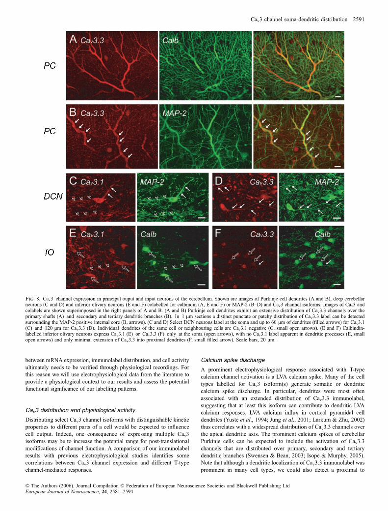
Electrophysiology and calcium imaging studies have established thatPurkinje cells and neurons of the deep cerebellar nuclei (DCN)generate LVA-mediated responses attributed to T-type calcium current.In Purkinje cells T-type single channels can be recorded in dendriticregions (Mouginot et al., 1997) and contribute to both the burst andinterburst depolarizations (Swensen & Bean, 2003; Isope & Murphy,2005). In DCN cells Cav3 channels contribute to a rebound burstdischarge following Purkinje cell inhibition (Gauck et al., 2001;Molineux et al., 2006). Inferior olivary neurons provide climbing fibreinput to both DCN and Purkinje cells and were the first cell typeidentified as generating LVA calcium spikes (Llinas & Yarom, 1981).Unlike other major cells with prominent IT, the LTS in inferior olivaryneurons was proposed to be initiated at the soma rather than dendrites(Llinas & Yarom, 1981).The distribution of Cav3 channel proteins in cerebellar neurons was
recently described, revealing a widespread expression of at least oneor more Cav3 isoforms in the main cerebellar cell types, and a strikingdistribution of Cav3.3 to at least Purkinje cell dendrites (Molineuxet al., 2006). Here we focus on the extent of dendritic Cav3 channeldistribution in the three major input and output cells of cerebellum:Purkinje, DCN and inferior olivary neurons.
Purkinje cells
Cav3 channels were distributed in Purkinje cells with the same generalpattern we detected in other brain regions: primarily Cav3.1 andCav3.2 at the soma or proximal dendrites, and Cav3.3 at the soma andover extensive regions of the dendritic tree of most Purkinje cells(Molineux et al., 2006). Craig et al. (1999) found that Cav3.1 labelcould also extend over the dendrites of at least some Purkinje cells ofmouse cerebellum. In our hands, Cav3.1 and Cav3.2 could be detectedin some rat Purkinje cell dendrites, but only in a very small fraction ofcells.Figure 8A and B provides magnified images of the distribution of
Cav3.3 channels in Purkinje cell dendrites. We found that Cav3.3immunolabel was distributed as either a diffuse cytosolic label(Fig. 8A) or as a distinct punctate or patchy distribution over theprimary dendritic shafts across the entire extent of the molecularlayer (Fig. 8B). A lower intensity of Cav3.3 immunolabel extendedinto the smaller diameter secondary and tertiary dendritic branches(Fig. 8B), but no label of dendritic spines could be detected even at100· magnification. In thin confocal sections the patchy distributionof Cav3.3 immunolabel could be visualized further as beingpositioned on the circumference of the dendritic shaft, as highlightedby an internal cytoplasmic core of MAP-2 counterlabel (Fig. 8B;arrows). Labelling in the region of the nucleus was also distin-guished for stellate cells in the molecular layer for Cav3.3 (Fig. 8Aand B).
Deep cerebellar nucleus
We have previously shown that large diameter DCN neuronsdifferentially label for the three Cav3 channel isoforms, with oneclass of bursting DCN neuron consistently labelling for Cav3.1 at thesoma and a non-bursting neuron class for Cav3.3 (Molineux et al.,
2006). The Cav3.1 labelled cells correspond to both excitatory andGAD-positive cells, while Cav3.3 was located only on excitatoryneurons. As shown in Fig. 8C, large diameter cells occasionallyexhibited Cav3.1 channel distribution up to 60 lm from the soma.More commonly, we detected a Cav3.3 channel dendritic distributionthat could extend over a distance of 120 lm and beyond at least thefirst dendritic branch point (Fig. 8D). Labelling in the nuclear regionwas apparent for both Cav3.1 and Cav3.3.
Inferior olivary neurons
Inferior olivary cells identified through a calbindin counterlabelexpressed detectable levels of only Cav3.1 and Cav3.3 at the soma(Fig. 8E and F) and were negative for Cav3.2 (not shown). The mostintense label was found for Cav3.1 at the soma, with no apparentdendritic label for Cav3.1 (Fig. 8E). Cav3.3 label was comparativelylight and associated with the somatic and nuclear regions (Fig. 8F).
Discussion
The present study establishes the distribution of Cav3.1, Cav3.2 andCav3.3 calcium channel isoforms in key brain regions reported toexpress these channels and ⁄ or generate putative T-type mediatedresponses. Cav3 channels were detected across a large spectrum ofcells, and in many cases individual cell types expressed more than oneCav3 isoform. Furthermore, Cav3 isoforms exhibit a differentialdistribution over somatic and dendritic regions, suggesting that selectCav3 channel isoforms differentially contribute to LVA calcium-dependent responses in distinct subcellular compartments.
Determination of Cav3 channel distribution
The function of T-type calcium channels has been investigated in depththrough physiological recordings, while more recently the pattern ofCav3 mRNA expression has been detailed (Craig et al., 1999; Kaseet al., 1999; Talley et al., 1999). The distribution of Cav3 proteindescribed here agrees well with the cell types previously demonstratedto be capable of generating LVA calcium-dependent responses. TheCav3 immunostaining also largely agrees with that reported for Cav3mRNA. However, it has become recognized that the correspondencebetween detectable mRNA and protein levels obtained through in situhybridization and immunocytochemistry can differ considerably, withneither serving as a faithful predictor of the other (Gygi et al., 1999;Zhang et al., 2002). One example is the finding that �80% of the nRTcell T-type current is blocked by N2O (a specific blocker of Cav3.2channels in expression systems) despite much higher apparent levels ofCav3.3 mRNA (Talley et al., 1999; Joksovic et al., 2005b). Thus, thelocation and relative intensity of immunolabel was similar to thatreported for mRNA in some cell types (i.e. hippocampal pyramidalcells, inferior olive), while others showed less correspondence (i.e.thalamic relay nuclei vs. nRT). We previously found that thedistribution of Cav3 immunolabel in cerebellar cell types was alsodifferent from the reported mRNA expression (Molineux et al., 2006).However, Cav3 protein distribution proved to entirely match the abilityto generate LTS in six different cerebellar cell types when competingoutward potassium currents were blocked. Thus, even when calciumchannels are transported to the membrane, their activation can bemasked by the coexpression of potassium channels (Pape et al., 1994;Molineux et al., 2005, 2006). Work to date then indicates that Cav3immunostaining with these antibodies can be an accurate reflection ofthe cells that express T-type channels. Nevertheless, the relation
2590 B. E. McKay et al.
ª The Authors (2006). Journal Compilation ª Federation of European Neuroscience Societies and Blackwell Publishing LtdEuropean Journal of Neuroscience, 24, 2581–2594
between mRNA expression, immunolabel distribution, and cell activityultimately needs to be verified through physiological recordings. Forthis reason we will use electrophysiological data from the literature toprovide a physiological context to our results and assess the potentialfunctional significance of our labelling patterns.
Cav3 distribution and physiological activity
Distributing select Cav3 channel isoforms with distinguishable kineticproperties to different parts of a cell would be expected to influencecell output. Indeed, one consequence of expressing multiple Cav3isoforms may be to increase the potential range for post-translationalmodifications of channel function. A comparison of our immunolabelresults with previous electrophysiological studies identifies somecorrelations between Cav3 channel expression and different T-typechannel-mediated responses.
Calcium spike discharge
A prominent electrophysiological response associated with T-typecalcium channel activation is a LVA calcium spike. Many of the celltypes labelled for Cav3 isoform(s) generate somatic or dendriticcalcium spike discharge. In particular, dendrites were most oftenassociated with an extended distribution of Cav3.3 immunolabel,suggesting that at least this isoform can contribute to dendritic LVAcalcium responses. LVA calcium influx in cortical pyramidal celldendrites (Yuste et al., 1994; Jung et al., 2001; Larkum & Zhu, 2002)thus correlates with a widespread distribution of Cav3.3 channels overthe apical dendritic axis. The prominent calcium spikes of cerebellarPurkinje cells can be expected to include the activation of Cav3.3channels that are distributed over primary, secondary and tertiarydendritic branches (Swensen & Bean, 2003; Isope & Murphy, 2005).Note that although a dendritic localization of Cav3.3 immunolabel wasprominent in many cell types, we could also detect a proximal to
Fig. 8. Cav3 channel expression in principal ouput and input neurons of the cerebellum. Shown are images of Purkinje cell dendrites (A and B), deep cerebellarneurons (C and D) and inferior olivary neurons (E and F) colabelled for calbindin (A, E and F) or MAP-2 (B–D) and Cav3 channel isoforms. Images of Cav3 andcolabels are shown superimposed in the right panels of A and B. (A and B) Purkinje cell dendrites exhibit an extensive distribution of Cav3.3 channels over theprimary shafts (A) and secondary and tertiary dendritic branches (B). In 1 lm sections a distinct punctate or patchy distribution of Cav3.3 label can be detectedsurrounding the MAP-2 positive internal core (B, arrows). (C and D) Select DCN neurons label at the soma and up to 60 lm of dendrites (filled arrows) for Cav3.1(C) and 120 lm for Cav3.3 (D). Individual dendrites of the same cell or neighbouring cells are Cav3.1 negative (C, small open arrows). (E and F) Calbindin-labelled inferior olivary neurons express Cav3.1 (E) or Cav3.3 (F) only at the soma (open arrows), with no Cav3.1 label apparent in dendritic processes (E, smallopen arrows) and only minimal extension of Cav3.3 into proximal dendrites (F, small filled arrow). Scale bars, 20 lm.
Cav3 channel soma-dendritic distribution 2591
ª The Authors (2006). Journal Compilation ª Federation of European Neuroscience Societies and Blackwell Publishing LtdEuropean Journal of Neuroscience, 24, 2581–2594

mid-dendritic distribution of Cav3.2 channels or even Cav3.1 channels,suggesting the additional involvement of these isoforms. In contrast,LVA calcium currents of inferior olivary neurons appear to derive fromCav3.1 and Cav3.3 isoforms localized to the soma.
Rebound discharge
A second output that results from the activation of T-type calciumchannels is a rebound depolarization following a preceding hyper-polarization. This activity can initiate a high frequency spike outputin response to inhibition or contribute to oscillatory swings inmembrane potential. Recent work has indicated that rebounddischarge in thalamic relay cells is blocked in a Cav3.1 knockoutanimal (Kim et al., 2001). An extensive analysis of mRNA throughRT-PCR also reported a correlation between the expression of Cav3.1mRNA and the ability to generate rebound bursts (Toledo-Rodriguezet al., 2004). We recently established a correlation between Cav3.1channel expression and rebound discharge in large diameter DCNcells (Molineux et al., 2006). Cerebellar Golgi cells and Purkinjecells also label for Cav3.1 and generate rebound depolarizations,while non-bursting DCN cells and stellate cells that lack this isoformdo not. A similar correlation between Cav3.1 expression and reboundburst discharge appears to exist between the habenular nuclei,where intense somatic Cav3.1 immunolabel in LHb cells correlatesto rebound burst capability, whereas MHb cells that exhibit onlylight Cav3.1 label do not exhibit rebound bursts (Kim & Chang,2005).Collectively, the available data suggests that expression of Cav3.1
channels may be sufficient and potentially necessary to generaterebound discharge in a wide variety of neurons. Nevertheless, theextent to which these correlations may also reflect the relative densityof Cav3.1 channel expression, varying kinetic properties of Cav3isoforms through post-translational modulation, or the degree ofinterplay with other ion channels remains to be determined.
Kinetic properties
Kinetic properties of individual Cav3 isoforms have been characterizedprimarily in expression systems at room temperature (McRory et al.,2001; Chemin et al., 2002; Perez-Reyes, 2003). The degree ofcorrespondence between the properties of expressed channels andthose in native neurons is not fully known. As most of the cell typesexamined here express more than one Cav3 channel isoform, it may bedifficult to predict the properties of whole-cell Cav3 current. Differ-ences in the properties of T-type currents have been reported betweenthalamic regions (Huguenard et al., 1993; Joksovic et al., 2006). Ourimmunolabel did not reveal striking differences in the expressionpattern of the Cav3 isoform protein between thalamic nuclei, but thisdoes not rule out the selective translocation of a given isoform to theplasma membrane or post-translational modifications as a source forphysiological differences. It is also difficult to relate differences inburst properties to soma-dendritic Cav3 channel distributions (i.e. relayvs. nRT cells). In general, relay cells are positive for all three Cav3isoforms at the soma and proximal dendrites and shift to a distributionof Cav3.3 label in distal dendrites. Cav3.1 and Cav3.2 label is readilydetected at the soma of nRT cells, but Cav3.1 and ⁄ or Cav3.3immunolabel can be distributed over extensive regions of dendriticmembrane in a given cell. Recent patch recordings reveal that T-typecalcium channels at the soma of nRT cells exhibit a faster rate ofinactivation (s ¼ 28 ms) than those recorded in proximal dendrites(s ¼ 53 ms; Joksovic et al., 2005a). These findings are at least
consistent with a higher density of Cav3.3 channels in dendriticregions that may influence spike output in these cells (Destexhe et al.,1996; Destexhe et al., 1998; Williams & Stuart, 2000).
Synaptic responses
LVA calcium currents have been shown to contribute to synapticallyactivated calcium influx over extensive regions of the dendritic arbor(Yuste et al., 1994; Magee et al., 1995). Our data suggest that all threeT-type calcium channel isoforms may contribute to this activity inproximal dendritic regions of many cell types, but that more distalinvolvement of T-type channels to synaptic input is likely to bemediated primarily by Cav3.3 channels. Another important potentiallocation of T-type channel distribution is dendritic spines, where lowvoltage synaptic responses could be amplified by Cav3 activation.Putative T-type mediated calcium influx has been identified in thespines of Purkinje cell dendrites from younger animals (Isope &Murphy, 2005). We were unable to obtain any evidence here for Cav3channel distribution to dendritic spines, including that of Purkinjecells, where these structures can be readily visualized using acalbindin counterlabel. A distribution to dendritic spines may thenbe restricted to early developmental stages or simply fall belowresolution at the light microscopic level. Alternatively, calcium entryinto dendritic spines may be secondary to calcium influx in theadjacent dendritic shaft or involve other LVA-like channels (Sabatini& Svoboda, 2000).
Nuclear regulation
The most unexpected labelling pattern for Cav3 protein was in thenuclear region of some cells. To our knowledge this is the first reportand test for voltage-dependent calcium channels localized to thenuclear protein fraction, even though several other ion channel typeshave been directly recorded (Mazzanti et al., 2001). The exact locationat which these channels are expressed remains to be determined.Membranes of the smooth ER are continuous with the outer membraneof the nuclear envelope, where a high density of large diameter nuclearpore complexes (NPCs) are expressed. The classical view of the NPCis of a fixed and open structure that allows free transfer of compoundsbetween the cytoplasm and nucleoplasm. This understanding has beensubstantially revised with findings that the NPC is actually semiper-meable, with an additional set of eight smaller diameter pores in theouter ring of the NPC that allows passage of smaller inorganic ions(Pante & Aebi, 1995). Patch-clamp recordings have measuredconductances in nuclear membranes at 800–1000 nS for theunblocked NPC core, as well as smaller conductances of 2–200 nS.The source of these smaller conductances has not been determined, butcould potentially correspond to the small diameter outer pores of theNPC. Altogether, the nuclear envelope acts as a high resistance andsemipermeable membrane that maintains transmembrane gradients forion species (Mazzanti et al., 2001). Nucleocytoplasmic transmem-brane potentials of up to )33 mV below that of the plasma membranepotential have been measured, although this waits to be determined forneuronal nuclei. The nuclear envelope is recognized further for actingas a calcium store for the release of calcium following IP3 orryanodine receptor activation that can react to events at the level of theplasma membrane or cytoplasm (Gerasimenko et al., 1995; Harding-ham et al., 2001; Marchenko & Thomas, 2006; Zhang et al., 2006).Indeed, postsynaptic activity can regulate the extent and time-courseof nuclear calcium increases, providing a means of integratingneuronal firing patterns in terms of the level of gene transcription
2592 B. E. McKay et al.
ª The Authors (2006). Journal Compilation ª Federation of European Neuroscience Societies and Blackwell Publishing LtdEuropean Journal of Neuroscience, 24, 2581–2594

(Hardingham et al., 2001; Power & Sah, 2002). The nucleus thusexhibits the necessary calcium concentration gradient and potentiallyvoltage gradient to employ the LVA properties of Cav3 channels. Assuch, our results suggest the potential for T-type calcium channels tohave previously unrecognized roles in regulating nuclear membraneactivity, and add to a growing list of voltage-activated ion channelsthat can be localized to the nuclear region.
Acknowledgements
We gratefully acknowledge the expert technical assistance of MirnaKruskic. Funded by the CIHR (TPS, GWZ, RWT) and Heart andStroke Foundation (GWZ). RWT is an AHFMR Scientist, GWZ anAHFMR Senior Scholar and Canada Research Chair, and TPS aCanada Research Chair. BEM was funded by the Killam Trust, CIHR-CGS and Steinhauer Doctoral awards.
Abbreviations
DCN, deep cerebellar nuclei; dLGN, dorsal lateral geniculate; GAD, glutamicacid decarboxylase; LTS, low-threshold calcium spikes; LHb, lateral habenula;LP, lateral posterior nucleus; LVA, low-voltage-activated; MAP-2, microtubule-associated protein; MHb, medial habenula; NPC, nuclear pore complexes; nRT,nucleus reticularis; PB, phosphate-buffer; PV, paraventricular nucleus.
References
Cavelier, P. & Bossu, J.L. (2003) Dendritic low-threshold Ca2+ channels in ratcerebellar Purkinje cells: possible physiological implications. Cerebellum, 2,196–205.
Chemin, J., Monteil, A., Perez-Reyes, E., Bourinet, E., Nargeot, J. & Lory, P.(2002) Specific contribution of human T-type calcium channel isotypes(alpha (1G), alpha (1H) and alpha (1I)) to neuronal excitability. J. Physiol.,540, 3–14.
Craig, P.J., Beattie, R.E., Folly, E.A., Banerjee, M.D., Reeves, M.B., Priestley,J.V., Carney, S.L., Sher, E., Perez-Reyes, E. & Volsen, S.G. (1999)Distribution of the voltage-dependent calcium channel alpha1G subunitmRNA and protein throughout the mature rat brain. Eur. J. Neurosci., 11,2949–296.
de la Pena, E. & Geijo-Barrientos, E. (1996) Laminar localization, morphology,and physiological properties of pyramidal neurons that have the low-threshold calcium current in the guinea-pig medial frontal cortex. J.Neurosci., 16, 5301–5311.
Destexhe, A., Contreras, D., Steriade, M., Sejnowski, T.J. & Huguenard, J.R.(1996) In vivo, in vitro, and computational analysis of dendritic calciumcurrents in thalamic reticular neurons. J. Neurosci., 16, 169–185.
Destexhe, A., Neubig, M., Ulrich, D. & Huguenard, J. (1998) Dendritic low-threshold calcium currents in thalamic relay cells. J. Neurosci., 18, 3574–3588.
Draguhn, A., Borner, G., Beckmann, R., Buchner, K., Heinemann, U. &Hucho, F. (1997) Large-conductance cation channels in the envelope ofnuclei from rat cerebral cortex. J. Membr. Biol., 158, 159–166.
Fisher, R.E., Gray, R. & Johnston, D. (1990) Properties and distribution ofsingle voltage-gated calcium channels in adult hippocampal neurons. J.Neurophysiol., 64, 91–104.
Franco-Obregon, A., Wang, H.W. & Clapham, D.E. (2000) Distinct ion channelclasses are expressed on the outer nuclear envelope of T- and B-lymphocytecell lines. Biophys. J., 79, 202–214.
Freund, T.F. & Buszaki, G. (1996) Interneurons of the hippocampus.Hippocampus, 6, 347–470.
Garner, M.H. (2002) Na,K-ATPase in the nuclear envelope regulates Na+:K+
gradients in hepatocyte nuclei. J. Membr. Biol., 187, 97–115.Gauck, V., Thomann, M., Jaeger, D. & Borst, A. (2001) Spatial distribution of
low- and high-voltage-activated calcium currents in neurons of the deepcerebellar nuclei. J. Neurosci., 21, RC158.
Gerasimenko, O.V., Gerasimenko, J.V., Tepikin, A.V. & Petersen, O.H. (1995)ATP-dependent accumulation and inositol trisphosphate- or cyclic ADP-ribose-mediated release of Ca2+ from the nuclear envelope. Cell, 80, 439–444.
Gerke, V., Creutz, C.E. & Moss, S.E. (2005) Annexins: linking Ca2+ signallingto membrane dynamics. Nature Rev. Mol. Cell Biol., 6, 449–461.
Goldberg, J.H., Lacefield, C.O. & Yuste, R. (2004) Global dendritic calciumspikes in mouse layer 5 low threshold spiking interneurones: implications forcontrol of pyramidal cell bursting. J. Physiol., 558, 465–478.
Golding, N.L., Jung, H.Y., Mickus, T. & Spruston, N. (1999) Dendritic calciumspike initiation and repolarization are controlled by distinct potassiumchannel subtypes in CA1 pyramidal neurons. J. Neurosci., 19, 8789–8798.
Gruenbaum, Y., Margalit, A., Goldman, R.D., Shumaker, D.K. & Wilson, K.L.(2005) The nuclear lamina comes of age. Nature Rev. Mol. Cell Biol., 6, 21–31.
Gygi, S.P., Rochon, Y., Franza, B.R. & Aebersold, R. (1999) Correlationbetween protein and mRNA abundance in yeast. Mol. Cell. Biol., 19, 1720–1730.
Hardingham, G.E., Arnold, F.J. & Bading, H. (2001) Nuclear calcium signalingcontrols CREB-mediated gene expression triggered by synaptic activity.Nature Neurosci., 4, 261–267.
Harris, R.M. & Hendrickson, A.E. (1987) Local circuit neurons in the ratventrobasal thalamus – a GABA immunocytochemical study. Neuroscience,21, 229–236.
Huguenard, J.R. (1996) Low-threshold calcium currents in central nervoussystem neurons. Annu. Rev. Physiol., 58, 329–348.
Huguenard, J.R., Gutnick, M.J. & Prince, D.A. (1993) Transient Ca2+ currentsin neurons isolated from rat lateral habenula. J. Neurophysiol., 70, 158–166.
Isope, P. & Murphy, T.H. (2005) Low threshold calcium currents in ratcerebellar Purkinje cell dendritic spines are mediated by T-type calciumchannels. J. Physiol., 562, 257–269.
Joksovic, P.M., Bayliss, D.A. & Todorovic, S.M. (2005a) Different kineticproperties of two T-type Ca2+ currents of reticular thalamic neurons and theirmodulation by enflurane. J. Physiol., 566, 125–142.
Joksovic, P.M., Brimelow, B.C., Murbartian, J., Perez-Reyes, E. & Todorovic,S.M. (2005b) Contrasting anesthetic sensitivities of T-type Ca2+ channels ofreticular thalamic neurons and recombinant Ca(v)3.3 channels. Br. J.Pharmacol., 144, 59–70.
Joksovic, P., Nelson, M., Jevtovic-Todorovic, V., Patel, M., Perez-Reyes, E.,Campbell, K., Chen, C.C. & Todorovic, S. (2006) CaV3.2 is the majormolecular substrate for redox regulation of T-type Ca2+ channels in the ratand mouse thalamus. J. Physiol., 574, 415–430.
Jung, H.Y., Staff, N.P. & Spruston, N. (2001) Action potential bursting insubicular pyramidal neurons is driven by a calcium tail current. J. Neurosci.,21, 3312–3321.
Kase, M., Kakimoto, S., Sakuma, S., Houtani, T., Ohishi, H., Ueyama, T. &Sugimoto, T. (1999) Distribution of neurons expressing alpha 1G subunitmRNA of T-type voltage-dependent calcium channel in adult rat centralnervous system. Neurosci. Lett., 268, 77–80.
Kavalali, E.T., Zhuo, M., Bito, H. & Tsien, R.W. (1997) Dendritic Ca2+
channels characterized by recordings from isolated hippocampal dendriticsegments. Neuron, 18, 651–663.
Kim, U. & Chang, S.-Y. (2005) Dendritic morphology, local circuitry, andintrinsic electrophysiology of neurons in the rat medial and lateral habenularnuclei of the epithalamus. J. Comp. Neurol., 483, 236–250.
Kim, D., Song, I., Keum, S., Lee, T., Jeong, M.J., Kim, S.S., McEnery, M.W. &Shin, H.S. (2001) Lack of the burst firing of thalamocortical relay neuronsand resistance to absence seizures in mice lacking alpha (1G) T-type Ca(2+)channels. Neuron, 31, 35–45.
Larkum, M.E. & Zhu, J.J. (2002) Signaling of layer 1 and whisker-evoked Ca2+
and Na+ action potentials in distal and terminal dendrites of rat neocorticalpyramidal neurons in vitro and in vivo. J. Neurosci., 22, 6991–7005.
Lee, J.H., Gomora, J.C., Cribbs, L.L. & Perez-Reyes, E. (1999) Nickel block ofthree cloned T-type calcium channels: low concentrations selectively blockalpha1H. Biophys. J., 77, 3034–3042.
Llinas, R. & Yarom, Y. (1981) Properties and distribution of ionic conductancesgenerating electroresponsiveness of mammalian inferior olivary neurones invitro. J. Physiol., 315, 569–584.
Magee, J.C., Christofi, G., Miyakawa, H., Christie, B., Lasser-Ross, N. &Johnston, D. (1995) Subthreshold synaptic activation of voltage-gated Ca2+
channels mediates a localized Ca2+ influx into the dendrites of hippocampalpyramidal neurons. J. Neurophysiol., 74, 1335–1342.
Magee, J.C. & Johnston, D. (1995) Synaptic activation of voltage-gatedchannels in the dendrites of hippocampal pyramidal neurons. Science, 268,301–304.
Marchenko, S.M. & Thomas, R.C. (2006) Nuclear Ca2+ signalling in cerebellarPurkinje neurons. Cerebellum, 5, 36–42.
Marchenko, S.M., Yarotskyy, V.V., Kovalenko, T.N., Kostyuk, P.G. & Thomas,R.C. (2005) Spontaneously active and InsP3-activated ion channels in cell
Cav3 channel soma-dendritic distribution 2593
ª The Authors (2006). Journal Compilation ª Federation of European Neuroscience Societies and Blackwell Publishing LtdEuropean Journal of Neuroscience, 24, 2581–2594

nuclei from rat cerebellar Purkinje and granule neurones. J. Physiol., 565,897–910.
Markram, H. & Sakmann, B. (1994) Calcium transients in dendrites ofneocortical neurons evoked by single subthreshold excitatory postsynapticpotentials via low-voltage-activated calcium channels. Proc. Natl Acad. Sci.USA, 91, 5207–5211.
Maruyama, Y., Shimada, H. & Taniguchi, J. (1995) Ca(2+)-activated K(+)-channels in the nuclear envelope isolated from single pancreatic acinar cells.Pflugers Arch., 430, 148–150.
Mazzanti, M., Bustamante, J.O. & Oberleithner, H. (2001) Electrical dimensionof the nuclear envelope. Physiol. Rev., 81, 1–19.
McKay, B.E., Molineux, M.L. & Turner, R.W. (2004) Biotin is endogenouslyexpressed in select regions of the rat central nervous system. J. Comp.Neurol., 473, 86–96.
McRory, J.E., Santi, C.M., Hamming, K.S., Mezeyova, J., Sutton, K.G., Baillie,D.L., Stea, A. & Snutch, T.P. (2001) Molecular and functional characteriza-tion of a family of rat brain T-type calcium channels. J. Biol. Chem., 276,3999–4011.
Metz, A.E., Jarsky, T., Martina, M. & Spruston, N. (2005) R-type calciumchannels contribute to afterdepolarization and bursting in hippocampal CA1pyramidal neurons. J. Neurosci., 25, 5763–5773.
Molineux, M.L., Fernandez, F.R., Mehaffey, W.H. & Turner, R.W. (2005) A-type and T-type currents interact to produce a novel spike latency-voltagerelationship in cerebellar stellate cells. J. Neurosci., 25, 10863–10873.
Molineux, M.L., McRory, J.E., McKay, B.E., Hamid, J., Mehaffey, W.H.,Rehak, R., Snutch, T.P., Zamponi, G.W. & Turner, R.W. (2006) Specific T-type calcium channel isoforms are associated with distinct burst phenotypesin deep cerebellar nuclear neurons. PNAS, 103, 5555–5560.
Mouginot, D., Bossu, J.L. & Gahwiler, B.H. (1997) Low-threshold Ca2+
currents in dendritic recordings from Purkinje cells in rat cerebellar slicecultures. J. Neurosci., 17, 160–170.
Munsch, T., Budde, T. & Pape, H.C. (1997) Voltage-activated intracellularcalcium transients in thalamic relay cells and interneurons. Neuroreport, 8,2411–2418.
Pante, N. & Aebi, U. (1995) Toward a molecular understanding of the structureand function of the nuclear pore complex. Int. Rev. Cytol., 162B, 225–255.
Pape, H.C., Budde, T., Mager, R. & Kisvarday, Z.F. (1994) Prevention ofCa(2+)-mediated action potentials in GABAergic local circuit neurones of ratthalamus by a transient K+ current. J. Physiol., 478, 403–422.
Pape, H.C., Munsch, T. & Budde, T. (2004) Novel vistas of calcium-mediatedsignalling in thalamus. Pflugers Arch., 448, 131–138.
Perez-Reyes, E. (2003) Molecular physiology of low-voltage-activated T-typecalcium channels. Physiol. Rev., 83, 117–161.
Power, J.M. & Sah, P. (2002) Nuclear calcium signaling evoked by cholinergicstimulation in hippocampal CA1 pyramidal neurons. J. Neurosci., 22, 3454–3462.
Richter, T.A., Kolaj, M. & Renaud, L.P. (2005) Low-voltage-activated Ca2+
channels are coupled to Ca2+-induced release in rat thalamic midlineneurons. J. Neurosci., 25, 8267–8271.
Sabatini, B.L. & Svoboda, K. (2000) Analysis of calcium channels in singlespines using optical fluctuation analysis. Nature, 408, 589–593.
Sayer, R.J., Schwindt, P.C. & Crill, W.E. (1990) High- and low-thresholdcalcium currents in neurons acutely isolated from rat sensorimotor cortex.Neurosci. Lett., 120, 175–178.
Swensen, A.M. & Bean, B.P. (2003) Ionic mechanisms of burst firing indissociated Purkinje neurons. J. Neurosci., 23, 9650–9663.
Talley, E.M., Cribbs, L.L., Lee, J.H., Daud, A., Perez-Reyes, E. & Bayliss,D.A. (1999) Differential distribution of three members of a gene familyencoding low-voltage-activated (T-type) calcium channels. J. Neurosci., 19,1895–1911.
Tarasenko, A.N., Kostyuk, P.G., Eremin, A.V. & Isaev, D.S. (1997) Two typesof low-voltage-activated Ca2+ channels in neurones of rat laterodorsalthalamic nucleus. J. Physiol., 499, 77–86.
Toledo-Rodriguez, M., Blumenfeld, B., Wu, C., Luo, J., Attali, B., Goodman,P. & Markram, H. (2004) Correlation maps allow neuronal electricalproperties to be predicted from single-cell gene expression profiles in ratneocortex. Cereb. Cortex, 14, 1310–1327.
Valenzuela, S.M., Martin, D.K., Por, S.B., Robbins, J.M., Warton, K., Bootcov,M.R., Schofield, P.R., Campbell, T.J. & Breit, S.N. (1997) Molecular cloningand expression of a chloride ion channel of cell nuclei. J. Biol. Chem., 272,12575–12582.
Williams, S.R. & Stuart, G.J. (2000) Action potential backpropagation andsomato-dendritic distribution of ion channels in thalamocortical neurons. J.Neurosci., 20, 1307–1317.
Yunker, A.M. & McEnery, M.W. (2003) Low-voltage-activated (‘T-Type’)calcium channels in review. J. Bioenerg. Biomembr., 35, 533–575.
Yuste, R., Gutnick, M.J., Saar, D., Delaney, K.R. & Tank, D.W. (1994) Ca2+
accumulations in dendrites of neocortical pyramidal neurons: an apical bandand evidence for two functional compartments. Neuron, 13, 23–43.
Zhang, H., Fu, Y., Altier, C., Platzer, J., Surmeier, D.J. & Bezprozvanny, I.(2006) Ca1.2 and Ca1.3 neuronal 1-type calcium channels: differentialtargeting and signaling to pCREB. Eur. J. Neurosci., 23, 2297–2310.
Zhang, Y., Mori, M., Burgess, D.L. & Noebels, J.L. (2002) Mutations in high-voltage-activated calcium channel genes stimulate low-voltage-activatedcurrents in mouse thalamic relay neurons. J. Neurosci., 22, 6362–6371.
Zhang, L., Valiante, T.A. & Carlen, P.L. (1993) Contribution of the low-threshold T-type calcium current in generating the post-spike depolarizingafterpotential in dentate granule neurons of immature rats. J. Neurophysiol.,70, 223–231.
Zhou, Q., Godwin, D.W., O’Malley, D.M. & Adams, P.R. (1997) Visualizationof calcium influx through channels that shape the burst and tonic firingmodes of thalamic relay cells. J. Neurophysiol., 77, 2816–2825.
Zhuravleva, S.O., Kostyuk, P.G. & Shuba, Y.M. (1999) Divalent cationselectivity of the subtypes of low-voltage-activated Ca2+ channels inthalamic neurons. Neuroreport, 10, 651–657.
Zhuravleva, S.O., Kostyuk, P.G. & Shuba, Y.M. (2001) Subtypes of low-voltage-activated Ca2+ channels in laterodorsal thalamic neurons: possiblelocalization and physiological roles. Pflugers Arch., 441, 832–839.
2594 B. E. McKay et al.
ª The Authors (2006). Journal Compilation ª Federation of European Neuroscience Societies and Blackwell Publishing LtdEuropean Journal of Neuroscience, 24, 2581–2594