pml isoforms in response to arsenic: high-resolution...
TRANSCRIPT
Jour
nal o
f Cel
l Sci
ence
RESEARCH ARTICLE
PML isoforms in response to arsenic: high-resolution analysis ofPML body structure and degradation
Katherine J. Hands1, Delphine Cuchet-Lourenco2, Roger D. Everett2 and Ronald T. Hay1,*
SUMMARY
Arsenic is a clinically effective treatment for acute promyelocytic
leukaemia (APL) in which the promyelocytic leukaemia (PML)
protein is fused to retinoic receptor alpha (RARa). PML-RARa is
degraded by the proteasome by a SUMO-dependent, ubiquitin-
mediated pathway in response to arsenic treatment, curing the
disease. Six major PML isoforms are expressed as a result of
alternative splicing, each of which encodes a unique C-terminal
region. Using a system in which only a single EYFP-linked PML
isoform is expressed, we demonstrate that PMLI, PMLII and PMLVI
accumulate in the cytoplasm following arsenic treatment, whereas
PMLIII, PMLIV and PMLV do not. 3D structured illumination was
used to obtain super-resolution images of PML bodies, revealing
spherical shells of PML along with associated SUMO. Arsenic
treatment results in dramatic isoform-specific changes to PML body
ultrastructure. After extended arsenic treatment most PML isoforms
are degraded, leaving SUMO at the core of the nuclear bodies.
A high-content imaging assay identifies PMLV as the isoform
most readily degraded following arsenic treatment, and PMLIV as
relatively resistant to degradation. Immunoprecipitation analysis
demonstrates that all PML isoforms are modified by SUMO and
ubiquitin after arsenic treatment, and by using siRNA, we
demonstrate that arsenic-induced degradation of all PML isoforms
is dependent on the ubiquitin E3 ligase RNF4. Intriguingly, depletion
of RNF4 results in marked accumulation of PMLV, suggesting that
this isoform is an optimal substrate for RNF4. Thus the variable
C-terminal domain influences the rate and location of degradation of
PML isoforms following arsenic treatment.
KEY WORDS: PML, Arsenic, SUMO, RNF4
INTRODUCTIONThe promyelocytic leukaemia (PML) protein was first identified
as part of the t(15:17) chromosomal translocation found in
patients with the disease acute promyelocytic leukaemia (APL)
(de The et al., 1990) where it is fused to the retinoic receptor
alpha (RARa). PML is a member of the tripartite motif (TRIM)
family of proteins, and contains a RING domain and two B boxes,
all of which are zinc-binding domains, and a coiled-coil domain
(Jensen et al., 2001), which is important for interactions between
PML molecules. Alternative splicing of the PML mRNAtranscript results in the expression of seven major PMLisoforms, six of which are predominantly nuclear (Jensen et al.,
2001). These different isoforms share a common N-terminalregion, encoded by the first six exons, which contains allcomponents of the TRIM, but differ in their C-terminal partsbecause of expression of various combinations of exons seven to
nine (Jensen et al., 2001). These varying C-terminal parts arethought to be responsible for isoform-specific interactions(Condemine et al., 2007; Fogal et al., 2000; Xu et al., 2005; Yu
et al., 2010) and functions (Cuchet et al., 2011).
A distinguishing feature of PML is that it is found in discretesubnuclear structures known as PML nuclear bodies (PML-NBs).
PML is modified by the small ubiquitin-like modifier (SUMO) onthree lysine residues, K65, K160 and K490 (Kamitani et al., 1998),located within the N-terminal regions common to all isoforms.
PMLI and PMLIV are also modified on K616, a lysine residueencoded by exon 8a, common to only these two isoforms (Cuchet-Lourenco et al., 2011). SUMO modification of PML is requiredfor the formation of normal PML-NBs (Ishov et al., 1999). As many
as 70 proteins have been reported to localise to PML-NBs (Negorevand Maul, 2001), and PML-NBs have been implicated in theregulation of multiple cellular processes, including the antiviral
response, transcription, apoptosis and DNA repair (Bernardi andPandolfi, 2007; Lallemand-Breitenbach and de The, 2010).
Arsenic trioxide has been used for many years as an extremely
effective treatment for APL (Hu et al., 2009; Mathews et al.,2010; Powell et al., 2010); however, its mechanism of action hasonly recently been elucidated. Following arsenic treatment, PMLis recruited from the nucleoplasm to PML-NBs and rapidly
modified with poly-SUMO chains (Lallemand-Breitenbach et al.,2001; Zhu et al., 1997). Subsequently, the SUMO-targetingubiquitin E3 ligase RNF4 is recruited to PML-NBs in a SUMO-
dependent manner (Geoffroy et al., 2010), where it ubiquitylatespoly-SUMO chains present on PML, targeting PML fordegradation by the proteasome (Geoffroy et al., 2010;
Lallemand-Breitenbach et al., 2008; Tatham et al., 2008).
Previous reports of the specific characteristics and functions ofindividual PML isoforms have primarily used systems in which a
single isoform is overexpressed on a background of endogenousPML (Beech et al., 2005; Maroui et al., 2012; Weidtkamp-Peterset al., 2008). PML isoforms interact with each other (Condemineet al., 2006), and therefore studies investigating the characteristics
of these overexpressed isoforms might be confounded by the factthat the overexpressed isoform will interact with endogenous PML.Others have used isoform-specific antibodies to investigate the
properties of the various endogenous PML isoforms (Condemineet al., 2006). Here we use a system in which a single PML isoformis stably expressed at close to endogenous levels in cells in
which expression of endogenous PML was depleted by the stable
1Wellcome Trust Centre for Gene Regulation and Expression, College of LifeSciences, University of Dundee, Dundee DD1 5EH, UK. 2MRC, University ofGlasgow Centre for Virus Research, Church Street, Glasgow G11 5JR, UK.
*Author for correspondence ([email protected])
This is an Open Access article distributed under the terms of the Creative Commons AttributionLicense (http://creativecommons.org/licenses/by/3.0), which permits unrestricted use, distributionand reproduction in any medium provided that the original work is properly attributed.
Received 1 April; Accepted 6 October 2013
� 2014. Published by The Company of Biologists Ltd | Journal of Cell Science (2014) 127, 365–375 doi:10.1242/jcs.132290
365
Jour
nal o
f Cel
l Sci
ence
expression of a short-hairpin RNA (Cuchet et al., 2011) to assess theeffects of arsenic treatment on each of the six main PML isoforms.
High-resolution microscopy was used to follow changes in PMLbody ultrastructure in response to arsenic. Biochemical analysisidentified PMLV as the isoform most readily degraded in responseto arsenic treatment, because it is the most heavily SUMO modified
and therefore an optimal substrate for RNF4.
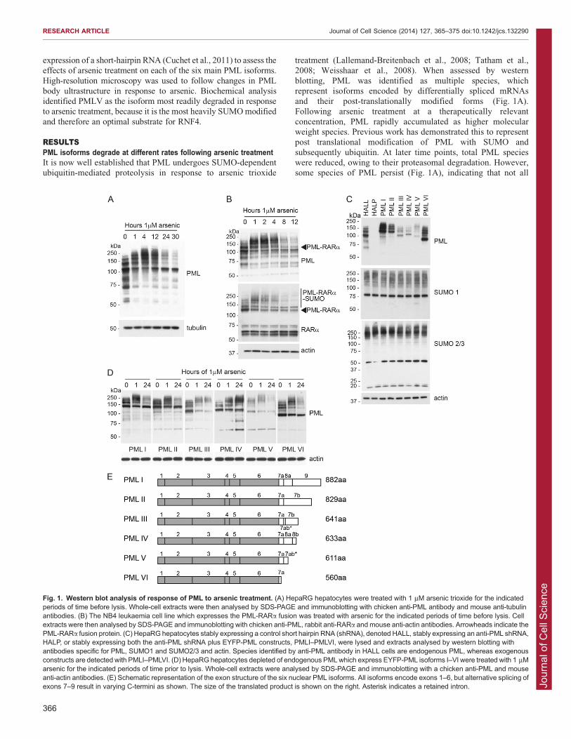
RESULTSPML isoforms degrade at different rates following arsenic treatmentIt is now well established that PML undergoes SUMO-dependentubiquitin-mediated proteolysis in response to arsenic trioxide
treatment (Lallemand-Breitenbach et al., 2008; Tatham et al.,2008; Weisshaar et al., 2008). When assessed by western
blotting, PML was identified as multiple species, whichrepresent isoforms encoded by differentially spliced mRNAsand their post-translationally modified forms (Fig. 1A).Following arsenic treatment at a therapeutically relevant
concentration, PML rapidly accumulated as higher molecularweight species. Previous work has demonstrated this to representpost translational modification of PML with SUMO and
subsequently ubiquitin. At later time points, total PML specieswere reduced, owing to their proteasomal degradation. However,some species of PML persist (Fig. 1A), indicating that not all
Fig. 1. Western blot analysis of response of PML to arsenic treatment. (A) HepaRG hepatocytes were treated with 1 mM arsenic trioxide for the indicatedperiods of time before lysis. Whole-cell extracts were then analysed by SDS-PAGE and immunoblotting with chicken anti-PML antibody and mouse anti-tubulinantibodies. (B) The NB4 leukaemia cell line which expresses the PML-RARa fusion was treated with arsenic for the indicated periods of time before lysis. Cellextracts were then analysed by SDS-PAGE and immunoblotting with chicken anti-PML, rabbit anti-RARa and mouse anti-actin antibodies. Arrowheads indicate thePML-RARa fusion protein. (C) HepaRG hepatocytes stably expressing a control short hairpin RNA (shRNA), denoted HALL, stably expressing an anti-PML shRNA,HALP, or stably expressing both the anti-PML shRNA plus EYFP-PML constructs, PMLI–PMLVI, were lysed and extracts analysed by western blotting withantibodies specific for PML, SUMO1 and SUMO2/3 and actin. Species identified by anti-PML antibody in HALL cells are endogenous PML, whereas exogenousconstructs are detected with PMLI–PMLVI. (D) HepaRG hepatocytes depleted of endogenous PMLwhich express EYFP-PML isoforms I–VI were treated with 1 mMarsenic for the indicated periods of time prior to lysis. Whole-cell extracts were analysed by SDS-PAGE and immunoblotting with a chicken anti-PML and mouseanti-actin antibodies. (E) Schematic representation of the exon structure of the six nuclear PML isoforms. All isoforms encode exons 1–6, but alternative splicing ofexons 7–9 result in varying C-termini as shown. The size of the translated product is shown on the right. Asterisk indicates a retained intron.
RESEARCH ARTICLE Journal of Cell Science (2014) 127, 365–375 doi:10.1242/jcs.132290
366
Jour
nal o
f Cel
l Sci
ence
forms of PML are degraded at the same rate in response to arsenictreatment.
The PML-RARa fusion product in acute promyelocyticleukaemia represents a unique PML isoform in which PML ispresent at the N-terminus, with RARa forming the C-terminusof the oncoprotein. We treated NB4 leukaemia cells that contain
the PML-RARAa fusion with arsenic trioxide and comparedthe degradation of PML-RARa with that of endogenous PML(Fig. 1B). Western blotting of whole-cell extracts with antibodies
specific for PML and RARa demonstrate that the PML-RARafusion is post-translationally modified and degraded at a similarrate to endogenous PML, suggesting the introduction of the
RARa at the C-terminus does not impair the ability of PML tobe SUMO modified or inhibit the action of the ubiquitin E3ligase RNF4.
Cells expressing a single PML isoformTo assess the role of the variable C-terminal regions of PMLisoforms in determining response to arsenic treatment, we used a
system in which a single EYFP-linked PML isoform is stablyexpressed in HepaRG hepatocytes in which endogenous PML hasbeen stably depleted by expression of an anti-PML short-hairpin
RNA (Cuchet et al., 2011). This system allows the true responseof a single PML isoform to be assessed in isolation by removingpossible interactions between isoforms that are demonstrated to
take place in systems where a single isoform is overexpressed ona background of endogenous PML. We can thus assess theresponse to arsenic of the six major PML isoforms, PMLI–
PMLVI, using these cell lines. Western blotting of cell extractswith an anti-PML antibody revealed a different profile of bandsfor each isoform, representing the PML isoform and its variouspost-translationally modified forms (Fig. 1C, top panel). The
expression levels of the different PML isoforms varied in thesecells (supplementary material Fig. S1A), as did the levels of theendogenous PML isoforms previously examined in various cell
lines (Condemine et al., 2006). These apparent differences inexpression reflect the varying degrees of post-translationalmodification of each isoform, as well as the relative stability of
the isoforms under basal conditions. The profile of totalSUMO1 conjugates does not differ significantly between celllines, although there might be slight variations in the patterns ofoverall SUMO2/3 conjugates in the different cell lines
(Fig. 1C).To assess the response of the different PML isoforms to arsenic
treatment, each cell line was exposed to arsenic for various
periods of time, and the resulting whole-cell extracts analysed bywestern blotting with an anti-PML antibody (Fig. 1D). Asanticipated, differences in the response to arsenic were
identified. All isoforms appeared to undergo additional post-translational modification in response to arsenic, as demonstratedby a change in electrophoretic mobility of PML observed after
1 hour of treatment, with the appearance of higher molecularweight PML species. After 24 hours of treatment, there wasextensive loss of these modified forms in most cases, but somePML isoforms had been degraded more than others
(supplementary material Fig. S1B). Treatment of PMLIV witharsenic trioxide resulted in marked accumulation of highmolecular weight PML species, but little or no apparent
degradation, whereas PMLIII and PMLV were initially SUMOmodified before being extensively degraded. PMLI, PMLII andPMLVI showed similar patterns of response: they were initially
modified, then these modified species were degraded, but there
was a reappearance of a major species that is likely to representnewly synthesised unmodified material that accumulates after
24 hours of treatment. Extending the length of arsenic treatmentdid not affect the patterns of degradation seen (data not shown).Although the PML isoforms (Fig. 1E) were expressed at differentlevels (Fig. 1C), the ability to be degraded did not correlate with
the levels of the expressed protein, because PML isoform IV wasexpressed at low levels (Fig. 1C) and yet was largely resistant toarsenic-induced degradation (Fig. 1D).
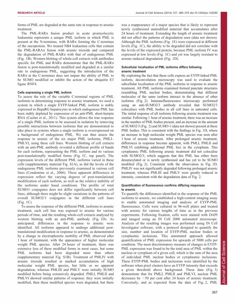
Subcellular localisation of PML isoforms differs followingarsenic treatmentBy exploiting the fact that these cells express an EYFP-linked PMLisoform, deconvolution microscopy was used to evaluate thesubcellular localisation of the PML isoforms in response to arsenic
treatment. All PML isoforms examined formed punctate structuresresembling PML nuclear bodies, demonstrating that differentmolecules of the same isoform interact in the absence of otherisoforms (Fig. 2). Immunofluorescence microscopy performed
using an anti-SUMO2/3 antibody revealed that SUMO2/3colocalises with PML bodies in all cell lines in untreated cells.The initial response of all isoforms following arsenic treatment was
similar. Following 1 hour of arsenic treatment, there was an increasein the number of PML bodies present, and an increase in the amountof SUMO2/3 (Fig. 2) and SUMO-1 (data not shown) present at these
PML bodies. This is consistent with the findings in Fig. 1D, wherean increase in high molecular weight PML species was seen after1 hour of arsenic treatment. After prolonged arsenic treatment,
differences in response become apparent, with PMLI, PMLII andPMLVI exhibiting additional PML foci in the cytoplasm. Thiscytoplasmic PML following arsenic treatment did not colocalisewith SUMO2/3, which suggests that this PML has either been
desumoylated or is newly synthesised and has yet to be SUMOmodified (Fig. 2). Consistent with the observation in Fig. 1D,PMLIV was not significantly degraded following prolonged arsenic
treatment, whereas PMLIII and PMLV were greatly reduced inintensity, consistent with the degradation data of Fig. 2.
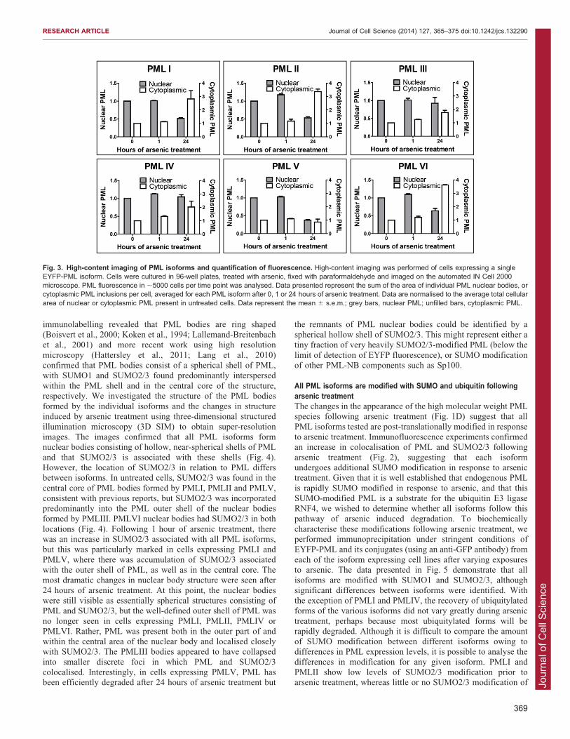
Quantification of fluorescence confirms differing responsesto arsenicTo quantify the differences identified in the response of the PMLisoforms to arsenic, we established a high-content imaging assay
to enable automated imaging and analysis of EYFP-PMLfluorescence. Cells were cultured in 96-well plates and treatedwith arsenic for various lengths of time as in the previous
experiments. Following fixation, cells were stained with DAPIand imaged using an IN Cell 2000 automated microscope.Analysis of the resulting images was performed using IN Cell
Investigator software, with a protocol designed to quantify thesize, number and location of EYFP-PML nuclear bodies orcytoplasmic inclusions. This automated approach allowed
quantification of PML expression for upwards of 5000 cells percondition. The most discriminatory measure of changes in EYFP-PML expression was found to be the total area of PML within thenucleus or cytoplasm of a given cell, which is the sum of the area
of individual PML nuclear bodies or cytoplasmic inclusions.These EYFP-PML bodies and inclusions were identified by thesoftware when pixel clusters have an EYFP intensity that exceeds
a given threshold above background. These data (Fig. 3)demonstrate that for PMLI, PMLII and PMLVI, nuclear PMLdecreases by ,50 per cent after 24 hours of arsenic treatment.
Conversely, and as expected from the data of Fig. 2, PML
RESEARCH ARTICLE Journal of Cell Science (2014) 127, 365–375 doi:10.1242/jcs.132290
367
Jour
nal o
f Cel
l Sci
enceaccumulated in the cytoplasm of these cells, increasing by up to
3.5-fold. PMLV was confirmed to be readily degraded by arsenic
treatment, with the amount of nuclear PML decreasing bytwo-thirds, with no increase in cytoplasmic PML. Considering thedata of Fig. 1D, Figs 2 and 3 together, it is possible that the
unmodified PMLI, PMLII and PMLVI detected by westernblotting of whole-cell extracts after 24 hours of arsenic treatmentrepresents the cytoplasmic fraction of PML, given that
cytoplasmic PML did not colocalise with SUMO2/3 byimmunofluorescence (Fig. 2). Of note, the PML isoforms did
not accumulate in the cytoplasm of these cells treated with botharsenic and the proteasome inhibitor MG132, indicating that
proteasomal function is required for this relocalisation to takeplace (data not shown).
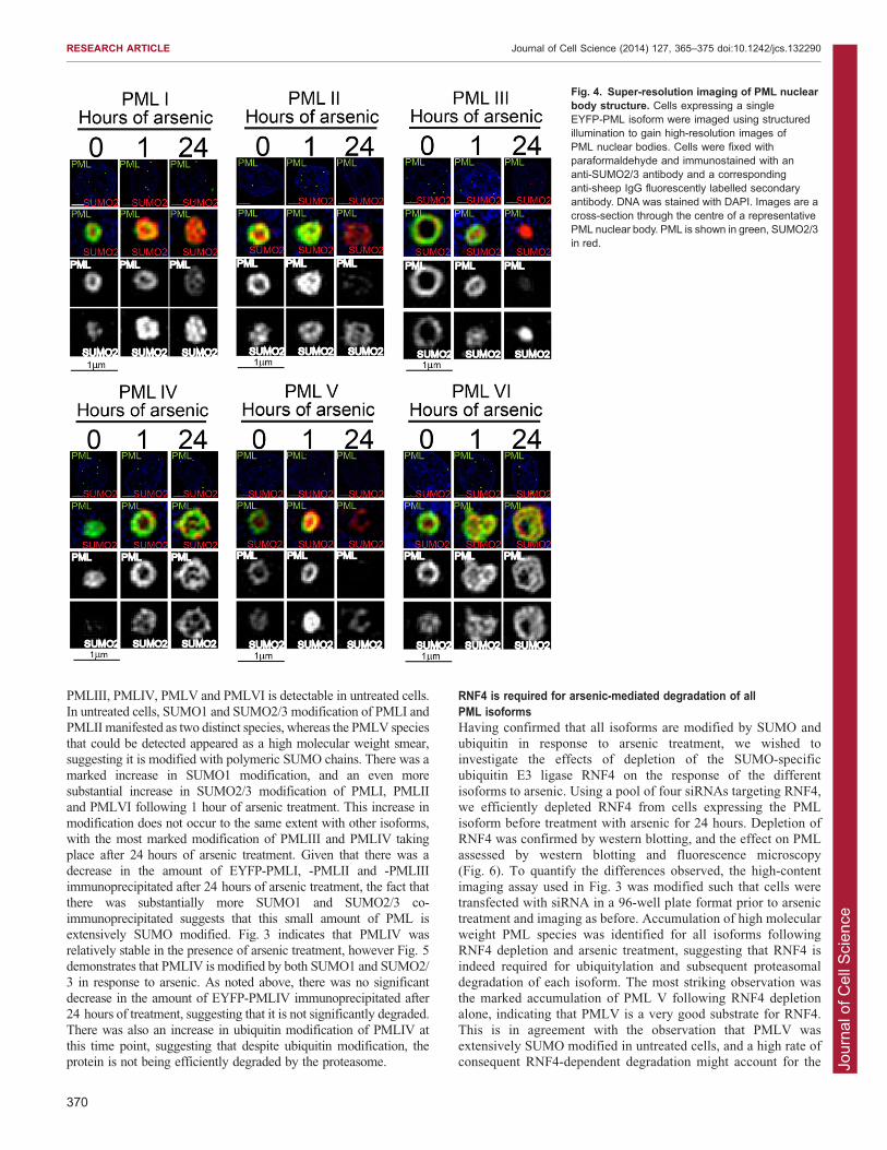
Super-resolution imaging reveals differences in PML body structureAs demonstrated in Fig. 2 and reported previously (Cuchetet al., 2011), the individually expressed PML isoforms formed
nucleate punctate structures that were associated with SUMOmodification. Previous electron microscopy studies with
Fig. 2. Immunofluorescence analysis of PML isoforms following arsenic treatment. Cells expressing a single EYFP-PML isoform were cultured oncoverslips and treated with 1 mM arsenic. Cells were fixed at the time points described and immunostained with a sheep anti-SUMO2/3 antibody, fluorescentlylabelled anti-sheep IgG secondary antibody and DAPI to stain DNA. EYFP-PML fluorescence is shown in green, SUMO2/3 in red. Images are presented asmaximal intensity projections of multiple z-slices.
RESEARCH ARTICLE Journal of Cell Science (2014) 127, 365–375 doi:10.1242/jcs.132290
368
Jour
nal o
f Cel
l Sci
ence
immunolabelling revealed that PML bodies are ring shaped(Boisvert et al., 2000; Koken et al., 1994; Lallemand-Breitenbachet al., 2001) and more recent work using high resolution
microscopy (Hattersley et al., 2011; Lang et al., 2010)confirmed that PML bodies consist of a spherical shell of PML,with SUMO1 and SUMO2/3 found predominantly interspersed
within the PML shell and in the central core of the structure,respectively. We investigated the structure of the PML bodiesformed by the individual isoforms and the changes in structureinduced by arsenic treatment using three-dimensional structured
illumination microscopy (3D SIM) to obtain super-resolutionimages. The images confirmed that all PML isoforms formnuclear bodies consisting of hollow, near-spherical shells of PML
and that SUMO2/3 is associated with these shells (Fig. 4).However, the location of SUMO2/3 in relation to PML differsbetween isoforms. In untreated cells, SUMO2/3 was found in the
central core of PML bodies formed by PMLI, PMLII and PMLV,consistent with previous reports, but SUMO2/3 was incorporatedpredominantly into the PML outer shell of the nuclear bodiesformed by PMLIII. PMLVI nuclear bodies had SUMO2/3 in both
locations (Fig. 4). Following 1 hour of arsenic treatment, therewas an increase in SUMO2/3 associated with all PML isoforms,but this was particularly marked in cells expressing PMLI and
PMLV, where there was accumulation of SUMO2/3 associatedwith the outer shell of PML, as well as in the central core. Themost dramatic changes in nuclear body structure were seen after
24 hours of arsenic treatment. At this point, the nuclear bodieswere still visible as essentially spherical structures consisting ofPML and SUMO2/3, but the well-defined outer shell of PML was
no longer seen in cells expressing PMLI, PMLII, PMLIV orPMLVI. Rather, PML was present both in the outer part of andwithin the central area of the nuclear body and localised closelywith SUMO2/3. The PMLIII bodies appeared to have collapsed
into smaller discrete foci in which PML and SUMO2/3colocalised. Interestingly, in cells expressing PMLV, PML hasbeen efficiently degraded after 24 hours of arsenic treatment but
the remnants of PML nuclear bodies could be identified by aspherical hollow shell of SUMO2/3. This might represent either atiny fraction of very heavily SUMO2/3-modified PML (below the
limit of detection of EYFP fluorescence), or SUMO modificationof other PML-NB components such as Sp100.
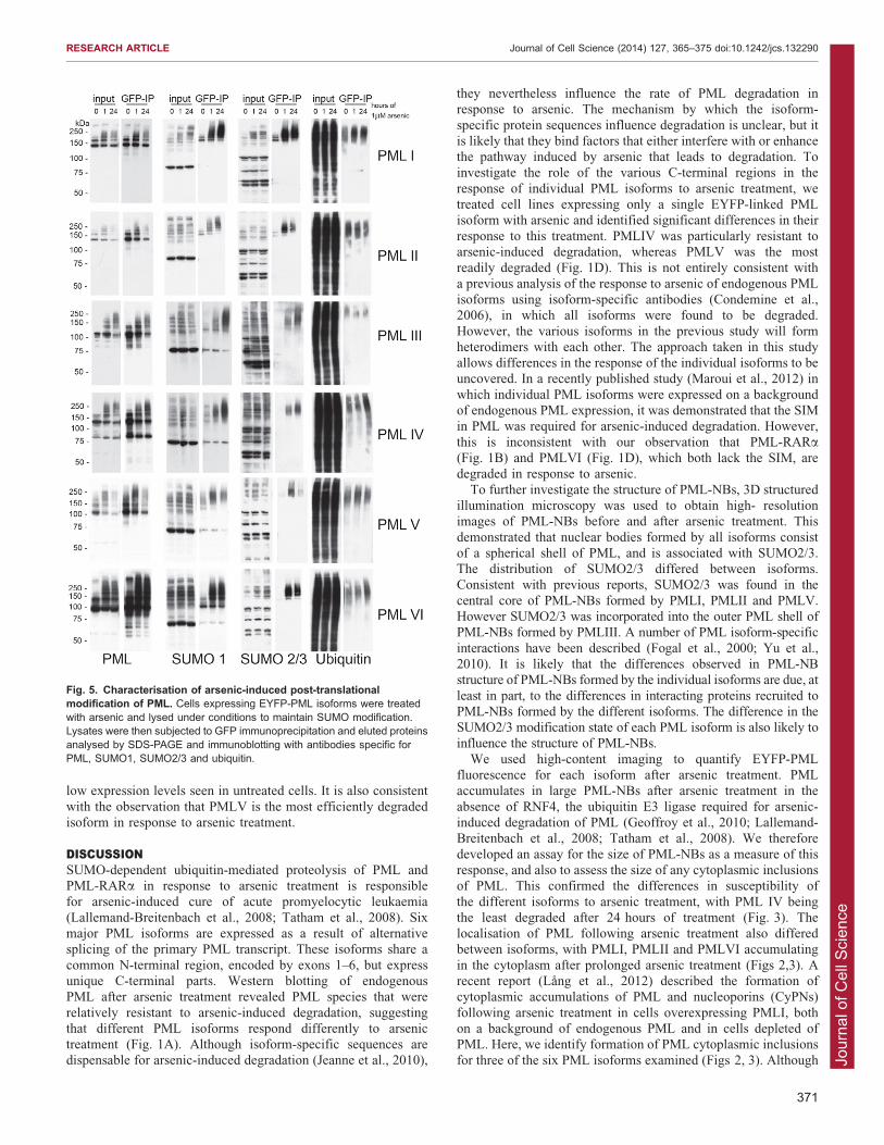
All PML isoforms are modified with SUMO and ubiquitin followingarsenic treatmentThe changes in the appearance of the high molecular weight PMLspecies following arsenic treatment (Fig. 1D) suggest that all
PML isoforms tested are post-translationally modified in responseto arsenic treatment. Immunofluorescence experiments confirmedan increase in colocalisation of PML and SUMO2/3 following
arsenic treatment (Fig. 2), suggesting that each isoformundergoes additional SUMO modification in response to arsenictreatment. Given that it is well established that endogenous PML
is rapidly SUMO modified in response to arsenic, and that thisSUMO-modified PML is a substrate for the ubiquitin E3 ligaseRNF4, we wished to determine whether all isoforms follow thispathway of arsenic induced degradation. To biochemically
characterise these modifications following arsenic treatment, weperformed immunoprecipitation under stringent conditions ofEYFP-PML and its conjugates (using an anti-GFP antibody) from
each of the isoform expressing cell lines after varying exposuresto arsenic. The data presented in Fig. 5 demonstrate that allisoforms are modified with SUMO1 and SUMO2/3, although
significant differences between isoforms were identified. Withthe exception of PMLI and PMLIV, the recovery of ubiquitylatedforms of the various isoforms did not vary greatly during arsenic
treatment, perhaps because most ubiquitylated forms will berapidly degraded. Although it is difficult to compare the amountof SUMO modification between different isoforms owing todifferences in PML expression levels, it is possible to analyse the
differences in modification for any given isoform. PMLI andPMLII show low levels of SUMO2/3 modification prior toarsenic treatment, whereas little or no SUMO2/3 modification of
Fig. 3. High-content imaging of PML isoforms and quantification of fluorescence. High-content imaging was performed of cells expressing a singleEYFP-PML isoform. Cells were cultured in 96-well plates, treated with arsenic, fixed with paraformaldehyde and imaged on the automated IN Cell 2000microscope. PML fluorescence in ,5000 cells per time point was analysed. Data presented represent the sum of the area of individual PML nuclear bodies, orcytoplasmic PML inclusions per cell, averaged for each PML isoform after 0, 1 or 24 hours of arsenic treatment. Data are normalised to the average total cellulararea of nuclear or cytoplasmic PML present in untreated cells. Data represent the mean 6 s.e.m.; grey bars, nuclear PML; unfilled bars, cytoplasmic PML.
RESEARCH ARTICLE Journal of Cell Science (2014) 127, 365–375 doi:10.1242/jcs.132290
369
Jour
nal o
f Cel
l Sci
ence
PMLIII, PMLIV, PMLV and PMLVI is detectable in untreated cells.
In untreated cells, SUMO1 and SUMO2/3 modification of PMLI andPMLII manifested as two distinct species, whereas the PMLV speciesthat could be detected appeared as a high molecular weight smear,suggesting it is modified with polymeric SUMO chains. There was a
marked increase in SUMO1 modification, and an even moresubstantial increase in SUMO2/3 modification of PMLI, PMLIIand PMLVI following 1 hour of arsenic treatment. This increase in
modification does not occur to the same extent with other isoforms,with the most marked modification of PMLIII and PMLIV takingplace after 24 hours of arsenic treatment. Given that there was a
decrease in the amount of EYFP-PMLI, -PMLII and -PMLIIIimmunoprecipitated after 24 hours of arsenic treatment, the fact thatthere was substantially more SUMO1 and SUMO2/3 co-
immunoprecipitated suggests that this small amount of PML isextensively SUMO modified. Fig. 3 indicates that PMLIV wasrelatively stable in the presence of arsenic treatment, however Fig. 5demonstrates that PMLIV is modified by both SUMO1 and SUMO2/
3 in response to arsenic. As noted above, there was no significantdecrease in the amount of EYFP-PMLIV immunoprecipitated after24 hours of treatment, suggesting that it is not significantly degraded.
There was also an increase in ubiquitin modification of PMLIV atthis time point, suggesting that despite ubiquitin modification, theprotein is not being efficiently degraded by the proteasome.
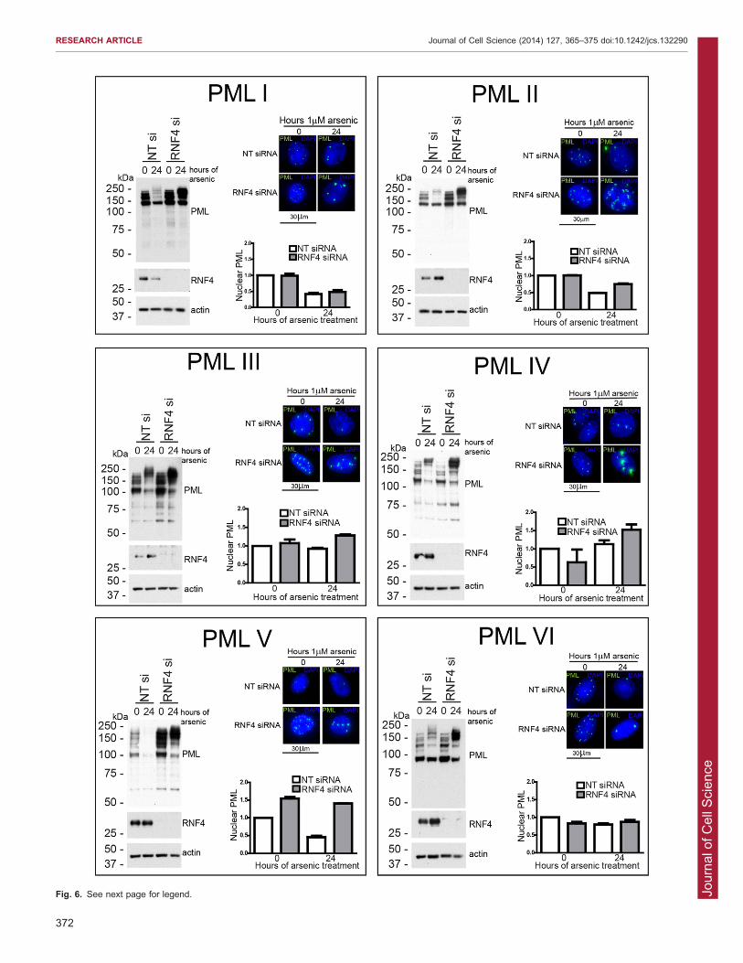
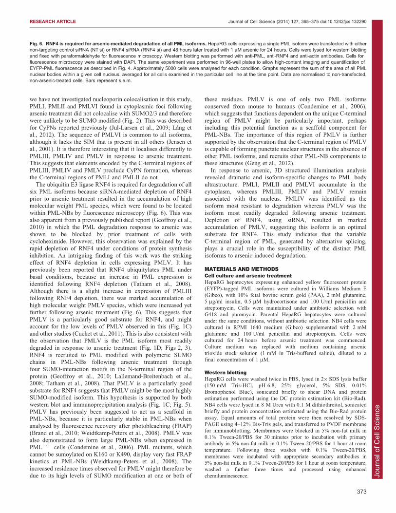
RNF4 is required for arsenic-mediated degradation of allPML isoformsHaving confirmed that all isoforms are modified by SUMO andubiquitin in response to arsenic treatment, we wished toinvestigate the effects of depletion of the SUMO-specific
ubiquitin E3 ligase RNF4 on the response of the differentisoforms to arsenic. Using a pool of four siRNAs targeting RNF4,we efficiently depleted RNF4 from cells expressing the PML
isoform before treatment with arsenic for 24 hours. Depletion ofRNF4 was confirmed by western blotting, and the effect on PMLassessed by western blotting and fluorescence microscopy
(Fig. 6). To quantify the differences observed, the high-contentimaging assay used in Fig. 3 was modified such that cells weretransfected with siRNA in a 96-well plate format prior to arsenic
treatment and imaging as before. Accumulation of high molecularweight PML species was identified for all isoforms followingRNF4 depletion and arsenic treatment, suggesting that RNF4 isindeed required for ubiquitylation and subsequent proteasomal
degradation of each isoform. The most striking observation wasthe marked accumulation of PML V following RNF4 depletionalone, indicating that PMLV is a very good substrate for RNF4.
This is in agreement with the observation that PMLV wasextensively SUMO modified in untreated cells, and a high rate ofconsequent RNF4-dependent degradation might account for the
Fig. 4. Super-resolution imaging of PML nuclearbody structure. Cells expressing a singleEYFP-PML isoform were imaged using structuredillumination to gain high-resolution images ofPML nuclear bodies. Cells were fixed withparaformaldehyde and immunostained with ananti-SUMO2/3 antibody and a correspondinganti-sheep IgG fluorescently labelled secondaryantibody. DNA was stained with DAPI. Images are across-section through the centre of a representativePML nuclear body. PML is shown in green, SUMO2/3in red.
RESEARCH ARTICLE Journal of Cell Science (2014) 127, 365–375 doi:10.1242/jcs.132290
370
Jour
nal o
f Cel
l Sci
ence
low expression levels seen in untreated cells. It is also consistent
with the observation that PMLV is the most efficiently degradedisoform in response to arsenic treatment.
DISCUSSIONSUMO-dependent ubiquitin-mediated proteolysis of PML andPML-RARa in response to arsenic treatment is responsiblefor arsenic-induced cure of acute promyelocytic leukaemia
(Lallemand-Breitenbach et al., 2008; Tatham et al., 2008). Sixmajor PML isoforms are expressed as a result of alternativesplicing of the primary PML transcript. These isoforms share a
common N-terminal region, encoded by exons 1–6, but expressunique C-terminal parts. Western blotting of endogenousPML after arsenic treatment revealed PML species that were
relatively resistant to arsenic-induced degradation, suggestingthat different PML isoforms respond differently to arsenictreatment (Fig. 1A). Although isoform-specific sequences aredispensable for arsenic-induced degradation (Jeanne et al., 2010),
they nevertheless influence the rate of PML degradation inresponse to arsenic. The mechanism by which the isoform-
specific protein sequences influence degradation is unclear, but itis likely that they bind factors that either interfere with or enhancethe pathway induced by arsenic that leads to degradation. Toinvestigate the role of the various C-terminal regions in the
response of individual PML isoforms to arsenic treatment, wetreated cell lines expressing only a single EYFP-linked PMLisoform with arsenic and identified significant differences in their
response to this treatment. PMLIV was particularly resistant toarsenic-induced degradation, whereas PMLV was the mostreadily degraded (Fig. 1D). This is not entirely consistent with
a previous analysis of the response to arsenic of endogenous PMLisoforms using isoform-specific antibodies (Condemine et al.,2006), in which all isoforms were found to be degraded.
However, the various isoforms in the previous study will formheterodimers with each other. The approach taken in this studyallows differences in the response of the individual isoforms to beuncovered. In a recently published study (Maroui et al., 2012) in
which individual PML isoforms were expressed on a backgroundof endogenous PML expression, it was demonstrated that the SIMin PML was required for arsenic-induced degradation. However,
this is inconsistent with our observation that PML-RARa(Fig. 1B) and PMLVI (Fig. 1D), which both lack the SIM, aredegraded in response to arsenic.
To further investigate the structure of PML-NBs, 3D structuredillumination microscopy was used to obtain high- resolutionimages of PML-NBs before and after arsenic treatment. This
demonstrated that nuclear bodies formed by all isoforms consistof a spherical shell of PML, and is associated with SUMO2/3.The distribution of SUMO2/3 differed between isoforms.Consistent with previous reports, SUMO2/3 was found in the
central core of PML-NBs formed by PMLI, PMLII and PMLV.However SUMO2/3 was incorporated into the outer PML shell ofPML-NBs formed by PMLIII. A number of PML isoform-specific
interactions have been described (Fogal et al., 2000; Yu et al.,2010). It is likely that the differences observed in PML-NBstructure of PML-NBs formed by the individual isoforms are due, at
least in part, to the differences in interacting proteins recruited toPML-NBs formed by the different isoforms. The difference in theSUMO2/3 modification state of each PML isoform is also likely toinfluence the structure of PML-NBs.
We used high-content imaging to quantify EYFP-PMLfluorescence for each isoform after arsenic treatment. PMLaccumulates in large PML-NBs after arsenic treatment in the
absence of RNF4, the ubiquitin E3 ligase required for arsenic-induced degradation of PML (Geoffroy et al., 2010; Lallemand-Breitenbach et al., 2008; Tatham et al., 2008). We therefore
developed an assay for the size of PML-NBs as a measure of thisresponse, and also to assess the size of any cytoplasmic inclusionsof PML. This confirmed the differences in susceptibility of
the different isoforms to arsenic treatment, with PML IV beingthe least degraded after 24 hours of treatment (Fig. 3). Thelocalisation of PML following arsenic treatment also differedbetween isoforms, with PMLI, PMLII and PMLVI accumulating
in the cytoplasm after prolonged arsenic treatment (Figs 2,3). Arecent report (Lang et al., 2012) described the formation ofcytoplasmic accumulations of PML and nucleoporins (CyPNs)
following arsenic treatment in cells overexpressing PMLI, bothon a background of endogenous PML and in cells depleted ofPML. Here, we identify formation of PML cytoplasmic inclusions
for three of the six PML isoforms examined (Figs 2, 3). Although
Fig. 5. Characterisation of arsenic-induced post-translationalmodification of PML. Cells expressing EYFP-PML isoforms were treatedwith arsenic and lysed under conditions to maintain SUMO modification.Lysates were then subjected to GFP immunoprecipitation and eluted proteinsanalysed by SDS-PAGE and immunoblotting with antibodies specific forPML, SUMO1, SUMO2/3 and ubiquitin.
RESEARCH ARTICLE Journal of Cell Science (2014) 127, 365–375 doi:10.1242/jcs.132290
371
Jour
nal o
f Cel
l Sci
ence
Fig. 6. See next page for legend.
RESEARCH ARTICLE Journal of Cell Science (2014) 127, 365–375 doi:10.1242/jcs.132290
372
Jour
nal o
f Cel
l Sci
ence
we have not investigated nucleoporin colocalisation in this study,PMLI, PMLII and PMLVI found in cytoplasmic foci followingarsenic treatment did not colocalise with SUMO2/3 and therefore
were unlikely to be SUMO modified (Fig. 2). This was describedfor CyPNs reported previously (Jul-Larsen et al., 2009; Lang etal., 2012). The sequence of PMLVI is common to all isoforms,although it lacks the SIM that is present in all others (Jensen et
al., 2001). It is therefore interesting that it localises differently toPMLIII, PMLIV and PMLV in response to arsenic treatment.This suggests that elements encoded by the C-terminal regions of
PMLIII, PMLIV and PMLV preclude CyPN formation, whereasthe C-terminal regions of PMLI and PMLII do not.
The ubiquitin E3 ligase RNF4 is required for degradation of all
six PML isoforms because siRNA-mediated depletion of RNF4prior to arsenic treatment resulted in the accumulation of highmolecular weight PML species, which were found to be located
within PML-NBs by fluorescence microscopy (Fig. 6). This wasalso apparent from a previously published report (Geoffroy et al.,2010) in which the PML degradation response to arsenic wasshown to be blocked by prior treatment of cells with
cycloheximide. However, this observation was explained by therapid depletion of RNF4 under conditions of protein synthesisinhibition. An intriguing finding of this work was the striking
effect of RNF4 depletion in cells expressing PMLV. It haspreviously been reported that RNF4 ubiquitylates PML underbasal conditions, because an increase in PML expression is
identified following RNF4 depletion (Tatham et al., 2008).Although there is a slight increase in expression of PMLIIIfollowing RNF4 depletion, there was marked accumulation ofhigh molecular weight PMLV species, which were increased yet
further following arsenic treatment (Fig. 6). This suggests thatPMLV is a particularly good substrate for RNF4, and mightaccount for the low levels of PMLV observed in this (Fig. 1C)
and other studies (Cuchet et al., 2011). This is also consistent withthe observation that PMLV is the PML isoform most readilydegraded in response to arsenic treatment (Fig. 1D; Figs 2, 3).
RNF4 is recruited to PML modified with polymeric SUMOchains in PML-NBs following arsenic treatment throughfour SUMO-interaction motifs in the N-terminal region of the
protein (Geoffroy et al., 2010; Lallemand-Breitenbach et al.,2008; Tatham et al., 2008). That PMLV is a particularly goodsubstrate for RNF4 suggests that PMLV might be the most highlySUMO-modified isoform. This hypothesis is supported by both
western blot and immunoprecipitation analysis (Fig. 1C; Fig. 5).PMLV has previously been suggested to act as a scaffold inPML-NBs, because it is particularly stable in PML-NBs when
analysed by fluorescence recovery after photobleaching (FRAP)(Brand et al., 2010; Weidtkamp-Peters et al., 2008). PMLV wasalso demonstrated to form large PML-NBs when expressed in
PML2/2 cells (Condemine et al., 2006). PML mutants, whichcannot be sumoylated on K160 or K490, display very fast FRAPkinetics at PML-NBs (Weidtkamp-Peters et al., 2008). Theincreased residence times observed for PMLV might therefore be
due to its high levels of SUMO modification at one or both of
these residues. PMLV is one of only two PML isoformsconserved from mouse to humans (Condemine et al., 2006),which suggests that functions dependent on the unique C-terminal
region of PMLV might be particularly important, perhapsincluding this potential function as a scaffold component forPML-NBs. The importance of this region of PMLV is furthersupported by the observation that the C-terminal region of PMLV
is capable of forming punctate nuclear structures in the absence ofother PML isoforms, and recruits other PML-NB components tothese structures (Geng et al., 2012).
In response to arsenic, 3D structured illumination analysisrevealed dramatic and isoform-specific changes to PML bodyultrastructure. PMLI, PMLII and PMLVI accumulate in the
cytoplasm, whereas PMLIII, PMLIV and PMLV remainassociated with the nucleus. PMLIV was identified as theisoform most resistant to degradation whereas PMLV was the
isoform most readily degraded following arsenic treatment.Depletion of RNF4, using siRNA, resulted in markedaccumulation of PMLV, suggesting this isoform is an optimalsubstrate for RNF4. This study indicates that the variable
C-terminal region of PML, generated by alternative splicing,plays a crucial role in the susceptibility of the distinct PMLisoforms to arsenic-induced degradation.
MATERIALS AND METHODSCell culture and arsenic treatmentHepaRG hepatocytes expressing enhanced yellow fluorescent protein
(EYFP)-tagged PML isoforms were cultured in Williams Medium E
(Gibco), with 10% fetal bovine serum gold (PAA), 2 mM glutamine,
5 mg/ml insulin, 0.5 mM hydrocortisone and 100 U/ml penicillin and
streptomycin. Cells were maintained under antibiotic selection with
G418 and puromycin. Parental HepaRG hepatocytes were cultured
under the same conditions, without antibiotic selection. NB4 cells were
cultured in RPMI 1640 medium (Gibco) supplemented with 2 mM
glutamine and 100 U/ml penicillin and streptomycin. Cells were
cultured for 24 hours before arsenic treatment was commenced.
Culture medium was replaced with medium containing arsenic
trioxide stock solution (1 mM in Tris-buffered saline), diluted to a
final concentration of 1 mM.
Western blottingHepaRG cells were washed twice in PBS, lysed in 26SDS lysis buffer
(150 mM Tris-HCl, pH 6.8, 25% glycerol, 5% SDS, 0.01%
Bromophenol Blue), sonicated briefly to shear DNA and protein
estimation performed using the DC protein estimation kit (Bio-Rad).
NB4 cells were lysed in 8 M Urea with 0.1 M dithiothreitol, sonicated
briefly and protein concentration estimated using the Bio-Rad protein
assay. Equal amounts of total protein were then resolved by SDS-
PAGE using 4–12% Bis-Tris gels, and transferred to PVDF membrane
for immunoblotting. Membranes were blocked in 5% non-fat milk in
0.1% Tween-20/PBS for 30 minutes prior to incubation with primary
antibody in 5% non-fat milk in 0.1% Tween-20/PBS for 1 hour at room
temperature. Following three washes with 0.1% Tween-20/PBS,
membranes were incubated with appropriate secondary antibodies in
5% non-fat milk in 0.1% Tween-20/PBS for 1 hour at room temperature,
washed a further three times and processed using enhanced
chemiluminescence.
Fig. 6. RNF4 is required for arsenic-mediated degradation of all PML isoforms. HepaRG cells expressing a single PML isoform were transfected with eithernon-targeting control siRNA (NT si) or RNF4 siRNA (RNF4 si) and 48 hours later treated with 1 mM arsenic for 24 hours. Cells were lysed for western blottingand fixed with paraformaldehyde for fluorescence microscopy. Western blotting was performed with anti-PML, anti-RNF4 and anti-actin antibodies. Cells forfluorescence microscopy were stained with DAPI. The same experiment was performed in 96-well plates to allow high-content imaging and quantification ofEYFP-PML fluorescence as described in Fig. 4. Approximately 5000 cells were analysed for each condition. Graphs represent the sum of the area of all PMLnuclear bodies within a given cell nucleus, averaged for all cells examined in the particular cell line at the time point. Data are normalised to non-transfected,non-arsenic-treated cells. Bars represent s.e.m.
RESEARCH ARTICLE Journal of Cell Science (2014) 127, 365–375 doi:10.1242/jcs.132290
373
Jour
nal o
f Cel
l Sci
ence
AntibodiesAffinity-purified chicken anti-PML, chicken anti-RNF4, sheep anti-
SUMO1 and sheep anti-SUMO2/3 antibodies were generated in house.
Anti-ubiquitin (DAKO), anti-b-actin (Sigma), and anti-tubulin (Amersham)
antibodies were purchased from commercial sources. Rabbit anti-RARA
115 was a kind gift from Cecile Egly (IGBMC, Strasbourg, France).
Horseradish-peroxidase-conjugated secondary antibodies against chicken,
sheep, rabbit and mouse IgG were purchased from Sigma. Secondary
antibodies used for immunofluorescence, Cy-5-conjugated and Dylight-
594-conjugated anti-sheep IgG were purchased from Jackson
Immunochemicals.
ImmunofluorescenceCells were cultured on coverslips, washed twice in PBS and then fixed
with 4% paraformaldehyde in PBS for 10 minutes at 37 C degrees. Cells
were permeabilised with 0.2% Triton X-100 in PBS, blocked in 5% BSA,
0.1% Tween-20 in PBS for 30 minutes at room temperature and
incubated in primary then secondary antibodies diluted in 1% BSA,
0.1% Tween-20 in PBS for 1 hour each, also at room temperature. Cells
were then stained with 0.1 mg/ml DAPI and mounted using Vectashield
mounting medium. Images were collected using a Deltavision DV3
microscope and processed using Softworx software (Applied precision).
Images are presented as maximal intensity projections of multiple
z-sections.
High-content imagingCells were cultured in black, clear-bottomed 96-well plates (Corning
CellBIND) in 100 ml culture medium for 24 hours. For arsenic treatment,
10 ml of 11 mM arsenic trioxide diluted in culture medium was added to
wells using a multichannel pipette, to give a final concentration of 1 mM.
At the desired time points, cells were washed twice with PBS, fixed with
4% paraformaldehyde, permeabilised and stained with DAPI as described
above. 100 ml of PBS was dispensed into wells and plates were thermally
sealed using an X-Seal Manual Thermal Sealer (Fluid X) ready for imaging.
Imaging was performed using an IN Cell 2000 microscope (GE
Healthcare) to acquire two fields of view per well with a 206 lens
(Nikon), capturing DAPI and EYFP fluorescence using 350 nm and
500 nm light sources respectively. Images were analysed using IN Cell
Investigator software (GE Healthcare), using a protocol designed to
identify PML inclusions by multi-scale top hat transformation. The
measure of total area of PML per cell nucleus or cytoplasm was selected
as the most discriminatory for changes in PML following arsenic
treatment. Data were obtained for .5000 cells per condition and
averaged to give a value for each isoform under three conditions, 0, 1 and
24 hours of arsenic treatment. Data were then normalised to the value for
0 hours of arsenic treatment for each isoform, for ease of comparison
across isoforms. Data presented represent the mean 6 s.e.m.
Structured illuminationSamples were prepared as for immunofluorescence prior to imaging
using the OMX version 2 system (Applied Precision) as previously
described (Hattersley et al., 2011). Images were acquired using a 1006,
1.4 NA, oil-immersion objective lens (Olympus, Center Valley, PA) and
back-illuminated Cascade II 5126512 electron-multiplying charge-
coupled device (EMCCD) camera (Photometrics, Tucson, AZ) on the
OMX version 2 system (Applied Precision) equipped with 405, 488 and
593 nm solid-state lasers. Samples were illuminated by a coherent
scrambled laser light source that had passed through a diffraction grating
to generate the structured illumination by interference of light orders in
the image plane to create a 3D sinusoidal pattern, with lateral stripes
,0.2 mm apart. The pattern was shifted laterally through five phases and
through three angular rotations of 609 for each Z-section, separated by
0.125 mm. Exposure times were typically between 200 and 500 ms, and
the power of each laser was adjusted to achieve optimal intensities of
between 2000 and 4000 counts in a raw image of 16-bit dynamic range, at
the lowest possible laser power to minimize photo bleaching. Raw
images were processed and reconstructed to reveal structures with greater
resolution. The channels were then aligned in x, y, and rotationally using
predetermined shifts as measured using a target lens and the Softworx
alignment tool (Applied Precision).
ImmunoprecipitationHepaRG cells expressing PML isoforms were cultured in 10 cm plates
and treated with arsenic as described above prior to harvesting by
scraping after two washes with PBS containing 100 mM iodoacetamide
on ice. Cells were pelleted by centrifugation at 400 g and lysed in ice-
cold RIPA buffer (50 mM Tris-HCl, pH 7.5, 150 mM NaCl, 1% NP-40,
0.5% deoxycholate) and 100 mM iodoacetamide with end-over-end
rotation for 20 minutes at 4 C. Lysates were clarified by centrifugation at
17 000 g for 10 minutes and precleared by incubation with Sepharose
beads for 1 hour, followed by overnight incubation with agarose beads
coupled to a single-chain, recombinant GFP antibody (a gift from the
Division of Signal Transduction Therapy, University of Dundee) with
constant end-over-end mixing at 4 C. Beads were then washed three
times with RIPA buffer and bound proteins eluted in 26SDS lysis buffer,
and analysed by SDS-PAGE and immunoblotting.
siRNA transfectionsCells were transfected with a pool containing an equal amount of four
siRNA duplexes targeting RNF4 (Dharmacon ON-TARGET plus; RNF4,
1-GCUAAUACUUGCCCAACUUUU; RNF4, 2-GAAUGGACGUCU
CAUCGUUUU; RNF4, 3-GACAGAGACGUAUAUGUGAUU; RNF4,
4-GCAAUAAAUUCUAGACAAGUU) to a final concentration of 10 nM,
or a non-targeting control duplex at the same concentration using
Lipofectamine RNAiMAX (Invitrogen) according to the manufacturer’s
instructions. Arsenic treatment was commenced 48 hours after transfection.
For high-content imaging of RNF4-depleted cells, cells were reverse
transfected with the RNF4 siRNA pool described above or a non-
targeting control duplex in 96-well plates, with a final siRNA
concentration of 10 nM. 10 ml of 100 nM siRNA was dispensed into
wells, followed by 10 ml of a 1:50 dilution of RNAiMAX/opti-MEM
(Invitrogen) serum-free medium mix. This was incubated for 15 minutes
at room temperature prior to the addition of 5000 cells in 80 ml of
antibiotic-free culture medium per well. Arsenic treatment was
commenced at 48 hours after transfection, and cells were fixed,
stained, imaged and analysed as described above.
AcknowledgementsUse of the OMX microscope was supported by the Scottish University LifeSciences Alliance (SULSA).
Competing interestsThe authors declare no competing interests.
Author contributionsD.C.L. and R.D.E. generated the EYFP-PML isoform cell lines. K.J.H. and R.T.H.discussed experimental design and results and wrote the manuscript. K.J.H.performed the experiments.
FundingK.J.H. was supported by a postgraduate fellowship for clinicians from theWellcome Trust. Work in the R.T.H. laboratory is supported by Cancer ResearchUK programme grant [number C434/A13067] and by Wellcome Trust SeniorInvestigator Award [number 098391/Z/12/Z]. Deposited in PMC for immediaterelease.
Supplementary materialSupplementary material available online athttp://jcs.biologists.org/lookup/suppl/doi:10.1242/jcs.132290/-/DC1
ReferencesBeech, S. J., Lethbridge, K. J., Killick, N., McGlincy, N. and Leppard, K. N.(2005). Isoforms of the promyelocytic leukemia protein differ in their effects onND10 organization. Exp. Cell Res. 307, 109-117.
Bernardi, R. and Pandolfi, P. P. (2007). Structure, dynamics and functions ofpromyelocytic leukaemia nuclear bodies. Nat. Rev. Mol. Cell Biol. 8, 1006-1016.
Boisvert, F.-M., Hendzel, M. J. and Bazett-Jones, D. P. (2000). Promyelocyticleukemia (PML) nuclear bodies are protein structures that do not accumulateRNA. J. Cell Biol. 148, 283-292.
RESEARCH ARTICLE Journal of Cell Science (2014) 127, 365–375 doi:10.1242/jcs.132290
374
Jour
nal o
f Cel
l Sci
ence
Brand, P., Lenser, T. and Hemmerich, P. (2010). Assembly dynamics of PMLnuclear bodies in living cells. PMC Biophys 3, 3.
Condemine, W., Takahashi, Y., Zhu, J., Puvion-Dutilleul, F., Guegan, S., Janin,A. and de The, H. (2006). Characterization of endogenous human promyelocyticleukemia isoforms. Cancer Res. 66, 6192-6198.
Condemine, W., Takahashi, Y., Le Bras, M. and de The, H. (2007). A nucleolartargeting signal in PML-I addresses PML to nucleolar caps in stressed orsenescent cells. J. Cell Sci. 120, 3219-3227.
Cuchet, D., Sykes, A., Nicolas, A., Orr, A., Murray, J., Sirma, H., Heeren, J.,Bartelt, A. and Everett, R. D. (2011). PML isoforms I and II participate in PML-dependent restriction of HSV-1 replication. J. Cell Sci. 124, 280-291.
Cuchet-Lourenco, D., Boutell, C., Lukashchuk, V., Grant, K., Sykes, A.,Murray, J., Orr, A. and Everett, R. D. (2011). SUMO pathway dependentrecruitment of cellular repressors to herpes simplex virus type 1 genomes. PLoSPathog. 7, e1002123.
de The, H., Chomienne, C., Lanotte, M., Degos, L. and Dejean, A. (1990). Thet(15;17) translocation of acute promyelocytic leukaemia fuses the retinoic acidreceptor alpha gene to a novel transcribed locus. Nature 347, 558-561.
Fogal, V., Gostissa, M., Sandy, P., Zacchi, P., Sternsdorf, T., Jensen, K., Pandolfi,P. P., Will, H., Schneider, C. and Del Sal, G. (2000). Regulation of p53 activity innuclear bodies by a specific PML isoform. EMBO J. 19, 6185-6195.
Geng, Y., Monajembashi, S., Shao, A., Cui, D., He, W., Chen, Z., Hemmerich, P.and Tang, J. (2012). Contribution of the C-terminal regions of promyelocyticleukemia protein (PML) isoform II and V to PML nuclear body formation. J. Biol.Chem. 287, 30729-30742.
Geoffroy, M.-C., Jaffray, E. G., Walker, K. J. and Hay, R. T. (2010). Arsenic-induced SUMO-dependent recruitment of RNF4 into PML nuclear bodies. Mol.Biol. Cell 21, 4227-4239.
Hattersley, N., Shen, L., Jaffray, E. G. and Hay, R. T. (2011). The SUMOprotease SENP6 is a direct regulator of PML nuclear bodies. Mol. Biol. Cell 22,78-90.
Hu, J., Liu, Y.-F.,Wu, C.-F., Xu, F., Shen, Z.-X., Zhu, Y.-M., Li, J.-M., Tang,W., Zhao,W.-L., Wu, W. et al. (2009). Long-term efficacy and safety of all-trans retinoic acid/arsenic trioxide-based therapy in newly diagnosed acute promyelocytic leukemia.Proc. Natl. Acad. Sci. USA 106, 3342-3347.
Ishov, A. M., Sotnikov, A. G., Negorev, D., Vladimirova, O. V., Neff, N.,Kamitani, T., Yeh, E. T., Strauss, J. F., III and Maul, G. G. (1999). PML iscritical for ND10 formation and recruits the PML-interacting protein daxx to thisnuclear structure when modified by SUMO-1. J. Cell Biol. 147, 221-234.
Jeanne, M., Lallemand-Breitenbach, V., Ferhi, O., Koken, M., Le Bras, M.,Duffort, S., Peres, L., Berthier, C., Soilihi, H., Raught, B. et al. (2010). PML/RARA oxidation and arsenic binding initiate the antileukemia response ofAs2O3. Cancer Cell 18, 88-98.
Jensen, K., Shiels, C. and Freemont, P. S. (2001). PML protein isoforms and theRBCC/TRIM motif. Oncogene 20, 7223-7233.
Jul-Larsen, A., Grudic, A., Bjerkvig, R. and Bøe, S. O. (2009). Cell-cycleregulation and dynamics of cytoplasmic compartments containing the promyelocyticleukemia protein and nucleoporins. J. Cell Sci. 122, 1201-1210.
Kamitani, T., Kito, K., Nguyen, H. P., Wada, H., Fukuda-Kamitani, T. and Yeh,E. T. (1998). Identification of three major sentrinization sites in PML. J. Biol.Chem. 273, 26675-26682.
Koken, M. H. M., Puvion-Dutilleul, F., Guillemin, M. C., Viron, A., Linares-Cruz,G., Stuurman, N., de Jong, L., Szostecki, C., Calvo, F., Chomienne, C. et al.(1994). The t(15;17) translocation alters a nuclear body in a retinoic acid-reversible fashion. EMBO J. 13, 1073-1083.
Lallemand-Breitenbach, V. and de The, H. (2010). PML nuclear bodies. ColdSpring Harb. Perspect. Biol. 2, a000661.
Lallemand-Breitenbach, V., Zhu, J., Puvion, F., Koken, M., Honore, N.,Doubeikovsky, A., Duprez, E., Pandolfi, P. P., Puvion, E., Freemont, P.et al. (2001). Role of promyelocytic leukemia (PML) sumolation in nuclear bodyformation, 11S proteasome recruitment, and As2O3-induced PML or PML/retinoic acid receptor a degradation. J. Exp. Med. 193, 1361-1372.
Lallemand-Breitenbach, V., Jeanne, M., Benhenda, S., Nasr, R., Lei, M., Peres,L., Zhou, J., Zhu, J., Raught, B. and de The, H. (2008). Arsenic degrades PMLor PML-RARalpha through a SUMO-triggered RNF4/ubiquitin-mediated path-way. Nat. Cell Biol. 10, 547-555.
Lang, M., Jegou, T., Chung, I., Richter, K., Munch, S., Udvarhelyi, A., Cremer, C.,Hemmerich, P., Engelhardt, J., Hell, S. W. et al. (2010). Three-dimensionalorganization of promyelocytic leukemia nuclear bodies. J. Cell Sci. 123, 392-400.
Lang, E., Grudic, A., Pankiv, S., Bruserud, Ø., Simonsen, A., Bjerkvig, R.,Bjøras, M. and Bøe, S. O. (2012). The arsenic-based cure of acutepromyelocytic leukemia promotes cytoplasmic sequestration of PML and PML/RARA through inhibition of PML body recycling. Blood 120, 847-857.
Maroui, M. A., Kheddache-Atmane, S., El Asmi, F., Dianoux, L., Aubry, M. andChelbi-Alix, M. K. (2012). Requirement of PML SUMO interacting motif forRNF4- or arsenic trioxide-induced degradation of nuclear PML isoforms. PLoSONE 7, e44949.
Mathews, V., George, B., Chendamarai, E., Lakshmi, K. M., Desire, S.,Balasubramanian, P., Viswabandya, A., Thirugnanam, R., Abraham, A.,Shaji, R. V. et al. (2010). Single-agent arsenic trioxide in the treatment of newlydiagnosed acute promyelocytic leukemia: long-term follow-up data. J. Clin.Oncol. 28, 3866-3871.
Negorev, D. and Maul, G. G. (2001). Cellular proteins localized at and interactingwithin ND10/PML nuclear bodies/PODs suggest functions of a nuclear depot.Oncogene 20, 7234-7242.
Powell, B. L., Moser, B., Stock, W., Gallagher, R. E., Willman, C. L., Stone, R.M., Rowe, J. M., Coutre, S., Feusner, J. H., Gregory, J. et al. (2010). Arsenictrioxide improves event-free and overall survival for adults with acutepromyelocytic leukemia: North American Leukemia Intergroup Study C9710.Blood 116, 3751-3757.
Tatham, M. H., Geoffroy, M. C., Shen, L., Plechanovova, A., Hattersley, N.,Jaffray, E. G., Palvimo, J. J. and Hay, R. T. (2008). RNF4 is a poly-SUMO-specific E3 ubiquitin ligase required for arsenic-induced PML degradation. Nat.Cell Biol. 10, 538-546.
Weidtkamp-Peters, S., Lenser, T., Negorev, D., Gerstner, N., Hofmann, T. G.,Schwanitz, G., Hoischen, C., Maul, G., Dittrich, P. and Hemmerich, P. (2008).Dynamics of component exchangeatPMLnuclear bodies. J.Cell Sci. 121, 2731-2743.
Weisshaar, S. R., Keusekotten, K., Krause, A., Horst, C., Springer, H. M.,Gottsche, K., Dohmen, R. J. and Praefcke, G. J. K. (2008). Arsenic trioxidestimulates SUMO-2/3 modification leading to RNF4-dependent proteolytictargeting of PML. FEBS Lett. 582, 3174-3178.
Xu, Z.-X., Zou, W.-X., Lin, P. and Chang, K.-S. (2005). A role for PML3 incentrosome duplication and genome stability. Mol. Cell 17, 721-732.
Yu, J., Lan, J., Wang, C., Wu, Q., Zhu, Y., Lai, X., Sun, J., Jin, C. and Huang, H.(2010). PML3 interacts with TRF1 and is essential for ALT-associated PMLbodies assembly in U2OS cells. Cancer Lett. 291, 177-186.
Zhu, J., Koken, M. H. M., Quignon, F., Chelbi-Alix, M. K., Degos, L., Wang, Z.Y., Chen, Z. and de The, H. (1997). Arsenic-induced PML targeting onto nuclearbodies: implications for the treatment of acute promyelocytic leukemia. Proc.Natl. Acad. Sci. USA 94, 3978-3983.
RESEARCH ARTICLE Journal of Cell Science (2014) 127, 365–375 doi:10.1242/jcs.132290
375