causes and differential diagnosis of parkinson’s disease
TRANSCRIPT

www.mghcme.org
Causes and Differential Diagnosis of Parkinson’s disease
Christopher D. Stephen, MB ChB, MRCP (UK), MSInstructor in Neurology, Harvard Medical School
Movement Disorders Unit, Dystonia Clinic, Ataxia CenterDepartment of Neurology, Massachusetts General Hospital
Performing Arts Clinic, Brigham and Women’s Hospital25 September, 2020

www.mghcme.org
Disclosures
Neither I nor my spouse/partner has a relevant financial relationship with a commercial interest to disclose.
www.mghcme.org
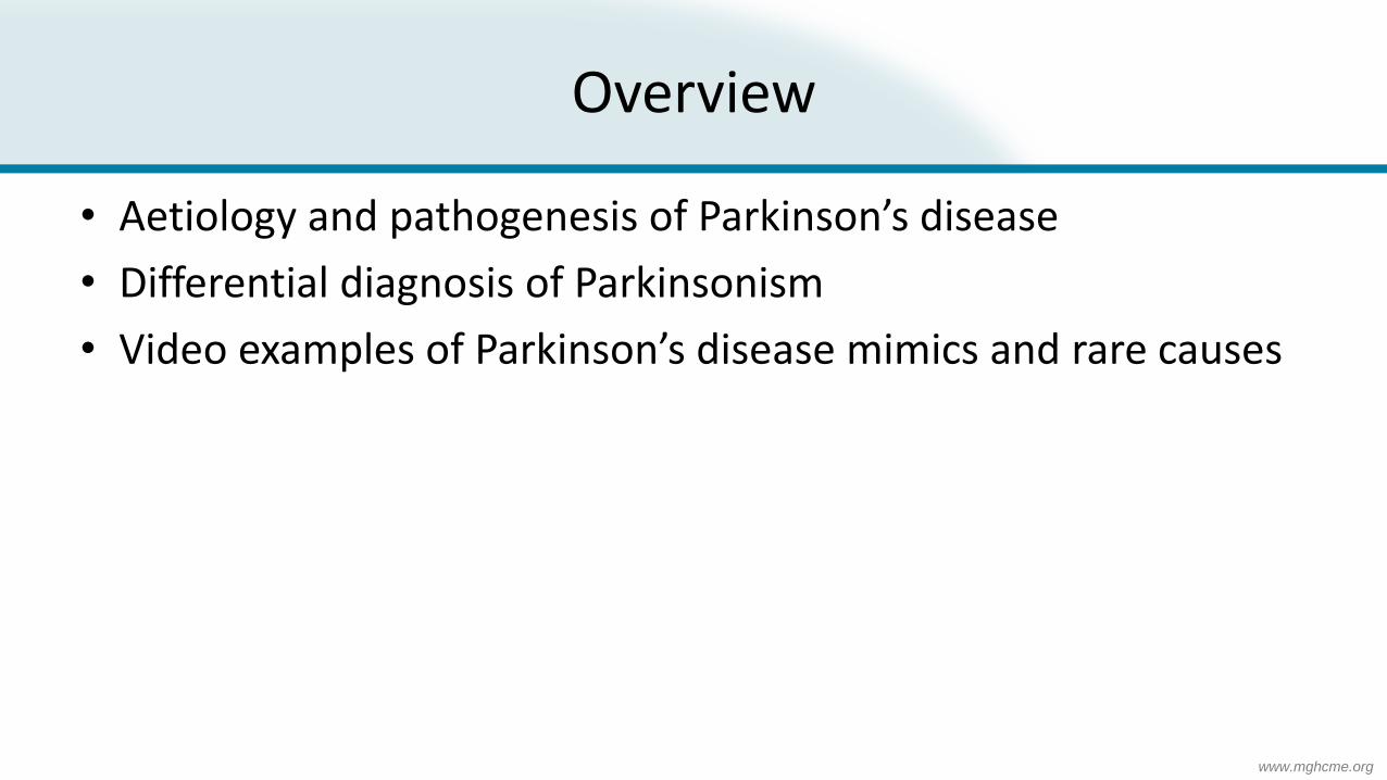
Overview
• Aetiology and pathogenesis of Parkinson’s disease
• Differential diagnosis of Parkinsonism
• Video examples of Parkinson’s disease mimics and rare causes

www.mghcme.org
Pathology
• Neuronal loss in substantia nigra and widespread α-synuclein deposition
• Results in striatal dopamine deficiency
• Dopamine plays an important role in the brain – Executive function, motivation, impulse
control (addiction behaviour), arousal, reinforcement and reward
– Motor control
– Lower-level functions including lactation, sexual gratification, nausea
• Pathological feature - Lewy bodies (aggregated α-synuclein)
• Spread of pathological α-synuclein protein (Braak hypothesis) Poewe, et al. Nat Rev Dis Primers 2017

www.mghcme.org
Pathophysiology
• Unclear cause
• Some have postulated a prion-like spread (Angot, Lancet Neurol 2010)– ? First sites gut enteric nerves and
olfactory bulb, resulting in prodromal hyposmia and constipation
• Complex pathophysiology
• Genetic risk genes being investigated include LRRK2 and GBA – potential additional therapeutic avenues
Poewe, et al. Nat Rev Dis Primers 2017

www.mghcme.org
Scope of the problem
• Incidence 5-35 per 100,000
• Incidence greatly increases after 6th decade
• A big problem in an ageing population
• Found in all regions
Poewe, et al. Nat Rev Dis Primers 2017
www.mghcme.org
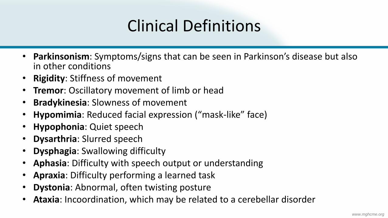
Clinical Definitions
• Parkinsonism: Symptoms/signs that can be seen in Parkinson’s disease but also in other conditions
• Rigidity: Stiffness of movement• Tremor: Oscillatory movement of limb or head• Bradykinesia: Slowness of movement• Hypomimia: Reduced facial expression (“mask-like” face)• Hypophonia: Quiet speech• Dysarthria: Slurred speech• Dysphagia: Swallowing difficulty• Aphasia: Difficulty with speech output or understanding• Apraxia: Difficulty performing a learned task• Dystonia: Abnormal, often twisting posture• Ataxia: Incoordination, which may be related to a cerebellar disorder

www.mghcme.org
Parkinsonism
• Triad of Tremor, Rigidity, Bradykinesia, (postural instability)
• Diagnosis is clinical
• Requires a complete medical history that includes timeline of symptoms, recognition of important clinical signs and consideration of differential diagnosis
• Clinical diagnosis can change over time due to emerging clinical signs

www.mghcme.org
Parkinson’s disease videos with a musical twist

www.mghcme.org
Differential diagnosis of Parkinsonism –Primary movement disorders
• Essential Tremor• Primary Parkinsonian disorders
– Idiopathic Parkinson’s disease (PD)– Atypical Parkinsonism (“Parkinson’s plus” syndrome)
• Progressive Supranuclear Palsy (PSP)• Dementia with Lewy Bodies (DLB)• Multiple System Atrophy (MSA)• Corticobasal degeneration/syndrome (CBD/CBS)
• Secondary Parkinsonism – Normal Pressure Hydrocephalus– Vascular parkinsonism– Other neurodegenerative diseases (Huntington’s disease, DRPLA, SCAs, Wilson's disease,
neurodegeneration with brain iron accumulation, neuroacanthocytosis etc) – Idiopathic basal ganglia calcification (Fahr’s disease)– Other secondary causes
• Functional (psychogenic) parkinsonism

www.mghcme.org
Secondary Parkinsonism
• Normal Pressure Hydrocephalus
• Vascular parkinsonism
• Other neurodegenerative diseases (Huntington’s disease, DRPLA, SCAs, Wilson's disease, neurodegeneration with brain iron accumulation, neuroacanthocytosis etc)
• Idiopathic basal ganglia calcification (Fahr’s disease)
• Other secondary causes
• (Functional/psychogenic parkinsonism)

www.mghcme.org
Other secondary Parkinsonism causes
• Drugs (classic and atypical antipsychotic agents, metoclopramide, prochlorperazine etc.)
• Toxins (carbon disulfide, carbon monoxide, cyanide, MPTP, manganese, organic solvents)
• Head trauma, isolated or repeated (dementia pugilistica)• Structural brain lesions that affect striatonigral circuits (hydrocephalus,
chronic subdural haematoma, tumour)• Metabolic and miscellaneous disorders (hypoparathyroidism,
pseudohypoparathyroidism, chronic liver failure, extrapontine myelinolysis)
• Infections (encephalitis lethargica or Economo's encephalitis, HIV/AIDS, neurosyphilis, prion disease, progressive multifocal leukoencephalopathy, toxoplasmosis)

www.mghcme.org
Essential tremor
• Most common cause of a pathological tremor• Estimated prevalence 5% of the population• Polygenic with family history common, so-called “familial tremor”• Onset usually 60s-70s (younger onset rarer)• Characteristics
– Bilateral, largely symmetrical postural and kinetic tremor involving the hands and forearms
– Worse on action– Can be associated with jaw (less common) and head tremor– Vocal tremor– Improves with alcohol

www.mghcme.org
Essential Tremor Video
Bhidayasiri and Tarsy, Movement Disorders: A Video Atlas, Springer 2012

www.mghcme.org
Progressive Supranuclear Palsy (PSP)
• Tauopathy (also found in AD, frontotemporal dementia, CBD)• Should be considered in all patients presenting with parkinsonism and:
– Poor response to levodopa therapy– Slowing of vertical saccades/ supranuclear vertical gaze palsy– Early postural instability with falls, executive dysfunction, dysarthria/dysphagia– (Eye-opening apraxia)
• Subtypes– Classic form –Steele-Richardson-Olszewski syndrome (PSP-RS)– PSP-parkinsonism (PSP-P)– PSP-pure akinesia with gait freezing (PSP-PAGF)– PSP-corticobasal syndrome (PSP-CBS)– PSP-frontal presentation (PSP-PNFA/PSP-bvFTD)– PSP-cerebellar features (PSP-C)

www.mghcme.org
Progressive Supranuclear Palsy (PSP)
• Mean age at diagnosis is 65 years
• Progression of disease and accumulation of disability in PSP is more rapid and severe than in PD
• Terminal stages of disease – Severe communication difficulties
– Immobility
– Severe axial rigidity
– Severe dysphagia
– Complete ophthalmoplegia
www.mghcme.org
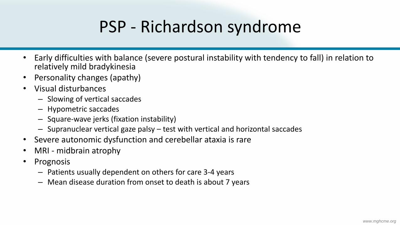
PSP - Richardson syndrome
• Early difficulties with balance (severe postural instability with tendency to fall) in relation to relatively mild bradykinesia
• Personality changes (apathy)• Visual disturbances
– Slowing of vertical saccades– Hypometric saccades– Square-wave jerks (fixation instability)– Supranuclear vertical gaze palsy – test with vertical and horizontal saccades
• Severe autonomic dysfunction and cerebellar ataxia is rare• MRI - midbrain atrophy• Prognosis
– Patients usually dependent on others for care 3-4 years– Mean disease duration from onset to death is about 7 years

www.mghcme.org
PSP – Parkinsonism
• Difficult to differentiate from PD in the earliest stages• Helpful pointers - rapid progression, prominent axial symptomatology, and
suboptimal response to levodopa despite typical clinical features of PD• Bradykinesia and limb rigidity at disease onset, which can be asymmetric and, in
some cases, associated with tremor• Axial rigidity is often a striking early feature, and limb rigidity is more common
and severe• Over time most will develop more typical PSP features - severe postural
instability, frontal cognitive decline, and vertical supranuclear gaze palsy• Disease duration to death is about 3 years longer in PSP-parkinsonism than
Richardson syndrome

www.mghcme.org
Other PSP variants
• Progressive supranuclear palsy– corticobasal syndrome
• Progressive supranuclear palsy– frontotemporal dementia
• PSP – pure akinesia with gait freezing
• PSP with predominant cerebellar features (PSP-C)
• Comprehensive diagnostic criteria for the various subtypes of PSP are found in the Movement Disorder Society Criteria (Höglinger, Mov Disord. 2017)

www.mghcme.org
PSP Video

www.mghcme.org
PSP Imaging
Midbrain atrophy and reduced midbrain:pons ratio

www.mghcme.org
“hummingbird sign” of PSP
Frederik Barkhof, et al., Neuroimaging in Dementia

www.mghcme.org
Multiple System Atrophy (MSA)
• Parkinsonism of MSA is usually symmetrical and classically responds poorly to dopaminergic therapies
• Alpha-synucleinopathy• Bradykinesia and rigidity progress somewhat faster than in PD, and as a
consequence, postural instability and falls usually emerge within the first 3 years of disease onset.
• Parkinsonism in the presence of increasing urinary urgency, constipation, postural hypotension, and erectile dysfunction in men
• Early progressive autonomic dysfunction precedes the evolution of motor symptoms by up to several years
• 2 subtypes– MSA-P – Predominant Parkinsonian symptoms (80%)– MSA-C – cerebellar syndrome with subtle Parkinsonism (20%)

www.mghcme.org
Revised MSA Diagnostic Criteria
Gilman S, et al. Neurology. 2008

www.mghcme.orgGilman S, et al. Neurology. 2008

www.mghcme.org
MSA-P Video

www.mghcme.org
MSA-P Imaging
T2 hyperintense putaminal rim

www.mghcme.org
MSA-P Imaging
Putaminal hypointensity on T2/T2*

www.mghcme.org
MSA-P Imaging
Cerebellar/MCP atrophy and linear hyperintensity in brainstem

www.mghcme.org
“Hot Cross Bun” Sign
Frederik Barkhof et al, Neuroimaging in Dementia

www.mghcme.org
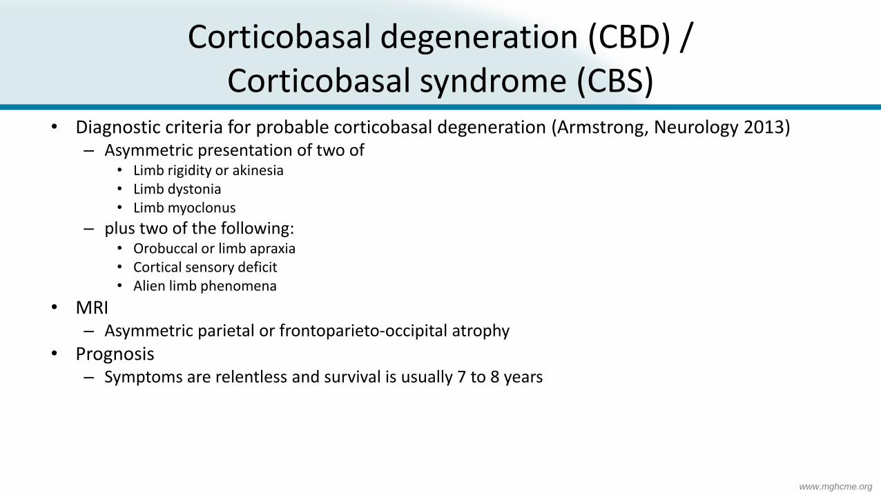
Corticobasal degeneration (CBD) / Corticobasal syndrome (CBS)
• Usually develops in the fifth to seventh decades of life• Presents with various phenotypes that include CBS, FTD, progressive nonfluent
aphasia• Definite diagnosis of CBD requires autopsy confirmation.• Symptoms
– Asymmetric ideomotor apraxia, associated with rigidity, myoclonus, dystonia– Alien-limb phenomenon - involuntary grasping, purposeless movements, or levitation in
an apraxic limb– When affecting the right extremities, often associated with a non-fluent aphasia– When affecting left extremities, often associated with visuospatial and visuoconstructive
deficits– Oculomotor disturbances
• Oculomotor apraxia (delayed latency of saccades with normal optokinetic nystagmus) -> supranuclear gaze palsy
www.mghcme.org
Corticobasal degeneration (CBD) / Corticobasal syndrome (CBS)
• Diagnostic criteria for probable corticobasal degeneration (Armstrong, Neurology 2013)– Asymmetric presentation of two of
• Limb rigidity or akinesia• Limb dystonia• Limb myoclonus
– plus two of the following:• Orobuccal or limb apraxia• Cortical sensory deficit• Alien limb phenomena
• MRI– Asymmetric parietal or frontoparieto-occipital atrophy
• Prognosis– Symptoms are relentless and survival is usually 7 to 8 years

www.mghcme.org
CBD Video

www.mghcme.org
CBD Imaging
Focal right posterior parietal atrophy

www.mghcme.org
Dementia with Lewy Bodies (DLB)
• Second most common type of degenerative dementia after Alzheimer disease• 10-22% of dementia cases• Pathology – alpha-synuclein, Lewy Bodies• Can co-exist with Alzheimer’s disease• Differential diagnosis – Parkinson’s disease dementia (dementia early in DLB)• Clinical features
– Dementia - early impairments in attention and executive and visuospatial function, memory later
– Fluctuations– Visual hallucinations in 2/3; well-formed images of people or animals; illusions– Parkinsonism – milder and more symmetric than PD– Neuroleptic sensitivity

www.mghcme.org

www.mghcme.org
Select secondary causes of parkinsonism

www.mghcme.org
Drug-induced Parkinsonism
• Features suggestive of DIP
– Subacute bilateral onset and progression of symptoms temporally associated with medication intake
– Early postural tremor
– Oro-buccal dyskinesia
• DIP usually resolves within weeks to months after stopping the offending drug; however, parkinsonism may persist or progress in 10%
• Diagnosis – clinical and DaTscan
• Treat by discontinuation of the offending drug
Alvarez et al, Neurology. 2008

www.mghcme.org
Drug-induced Parkinsonism
Shin and Chung, J Clin Neurol. 2012

www.mghcme.org
Normal pressure hydrocephalus (NPH)
• Form of communicating hydrocephalus with dilation of ventricles disproportional to cortical atrophy and without evidence of CSF obstruction
• Triad of dementia, gait disturbance (with parkinsonism) and urinary incontinence
• Diagnostic test with large volume lumbar puncture
• May be treated with CSF shunting

www.mghcme.org
NPH Video

www.mghcme.org
NPH Imaging
Ventriculomegaly and acute callosal angle

www.mghcme.org
Vascular parkinsonism
• 4.4-12% of all parkinsonism (Mehanna, Lancet Neurol 2013)• “lower body” parkinsonism, with a shuffling “magnetic” gait• Atypical features
– No resting tremor– Absent or poor response to dopamine replacement– Often in individuals with significant vascular disease
• Associated features (Zijlmans, Mov Disord 2004) – Diffuse vascular disease– Development of parkinsonism within one month of a stroke (rare)– Stroke in basal ganglia or diffuse subcortical whit mater disease– Vascular disease in 2 or more vascular territories– Vascular risk factors (hypertension, smoking, diabetes mellitus,
hyperlipidemia, presence of heart disease, family history of stroke, peripheral vascular disease)
– Stroke in two or more prior stroke or vascular risk factors
• Can co-exist with vascular dementia• Levodopa responsive in 30% (Miguel-Puga, Front Neurol
2017) Vizcarra, Mov Disord. 2015
www.mghcme.org
Select rare causes of parkinsonism

www.mghcme.org
Rare case 1

www.mghcme.org
Functional (psychogenic) parkinsonism
• 10% of Functional (Psychogenic) Movement Disorders (Hallett, J Neurol Sci 2011)
• Difficult to diagnose• Inconsistent with typical features of parkinsonism• May have sudden onset• Can have dramatic “on” or “off” periods inconsistent with those seen in PD• Functional tremor• “Rigidity” with gegenhalten and voluntary opposition• Bradykinesia characterised by ponderous, effortful slowness• Inconsistent postural instability with even slight perturbations in contrast to
better walking and balance than claimed• Normal DaT scan

www.mghcme.org
Rare case 2

www.mghcme.org
X-linked dystonia parkinsonism (XDP)
• Rare neurogenetic movement disorder, almost exclusively found in individuals with Filipino ancestry.
• Founder effect with origins in the Panay Islands
• Onset in the third to fifth decade (Lee, Medicine 1991)
• Significant phenotypic spectrum ranging from pure parkinsonism to varying combinations of parkinsonism and dystonia and rare development of chorea or myoclonus (Evidente 2018).
• Genetic cause is a hexameric repeat expansion within the SINE-VNTR-Alu (SVA) intronic region of the TAF-1 gene on the X-chromosome(Bragg, Proc Nat Acadm Sci 2017).
• Like other repeat expansion disorders, a larger repeat length correlated with an earlier age at onset

www.mghcme.org
Rare case 3
Gross calcifications in basal ganglia and cerebellum
Rohani, Parkinsonism Relat
Disord. 2017

www.mghcme.org
Idiopathic basal ganglia calcification
• Physiologic calcification in the basal ganglia are usually punctate and are located within the globus pallidus, the head of the caudate nucleus, and the putamen
• Especially if age <30., consider metabolic disorders, such as hyper/hypoparathyroidism, congenital disorders such as Fahr’s disease,andinfections
• Idiopathic basal ganglia calcification (Fahr’s disease)– Rare, autosomal dominant genetic disorder SCL20A2 most common, PDGFRB and PDGFB
(Batla, Parkinsonism Relat Disord. 2017)
– Characterised by abnormal deposits of calcium in basal ganglia, cerebellum and cortex
– Presents in third to fifth decade of life but may appear in childhood or later in life
– Neuropsychiatric disorder with mixed movement disorder (dystonia, chorea, parkinsonism in 20-30%)
– Can be associated with headache, seizures, dementia, mood disorders and psychosis

www.mghcme.org
Questions?