case study for ari - monochromatic x-rays
TRANSCRIPT
A Case Study for Accelerated Radical Innovation: Monochromatic X-rays
Aleksey Dubrovensky1, John A. Bers1, Frank Carroll2 1Vanderbilt University, Engineering Management Program, Nashville, TN 37235 USA
2Vanderbilt University, Radiology and Radiological Sciences, Nashville, TN 37235 USA Abstract--To illustrate the application of accelerated radi-
cal innovation (ARI), this paper examines a promising new can-cer therapy technique now at the juncture between Phase I (In-ception) and Phase (II Implementation) of the ARI industrial technology life cycle (ITLC). The new technology which uses tunable, monochromatic X-rays shows significant promise in the delivery of curative radiation therapy targeted specifically at the DNA of cancerous cells, sparing normal tissues. Factors that have inhibited its commercialization include the high capital cost of the equipment, the lack of an established supply chain for the key component of the system, and the reluctance of investors to gamble on what they perceive as a long-term high-risk in-vestment. The path of this innovation is studied in depth, both retrospectively and prospectively, through the perspective of ARI to determine how this promising radical innovation can be accelerated into widespread clinical use. The paper demon-strates that the ARI methodology offers a flexible set of tools and procedures that can be adapted to innovations at this criti-cal juncture of the ITLC, provides a structured approach to uncovering the challenges that all such risky endeavors face, and offers a template for dealing with them as they arise.
I. INTRODUCTION
The purpose of this paper is to apply the ARI methodol-
ogy framework described in a companion paper in this con-ference, PICMET ’07 paper: Principles and Practice of Ac-celerated Radical Innovation, to a specific radical innovation currently under way. From this case study we hope to accom-plish four objectives. First, how well does the prescribed methodology fair in analyzing a living example of a radical innovation? Second, does the ARI methodology aid in expos-ing and explaining the specific challenges the innovators have encountered as they drive toward commercialization? Third, does the model offer guidance in addressing these challenges and thereby accelerating the commercialization of the innova-tion? Finally, the case study is used to determine where and how the ARI methodology needs to be modified.
The innovation to be analyzed is being brought to the market by a high-tech start-up called MXISystems, Inc. This company is attempting to commercialize a novel approach to cancer diagnosis and therapy, with some potential for other applications, in the form of a pulsed tunable monochromatic X-ray beam device. The president of MXISystems and its technology champion, once discussed his innovation with an engineering management researcher, who observed that this device is an archetype of a disruptive innovation, since it em-bodies a completely different approach to radiotherapy from the standard approaches used today, and it also faces many of the same kinds of challenges.
The paper starts with a brief discussion of the field of ra-diotherapy in its current state, and then explains how MXI-Systems’ innovation works and how it could radically change current approaches. In the main body of the paper, the ARI methodology is applied to the innovation and is used to de-velop various problem-solving techniques that could be used to accelerate the MXISystems technology to commercial suc-cess. A. Background on radiotherapy and the monochromatic X-ray
The last quarter century has seen the growth and devel-opment of an astounding number of cancer treatment tech-niques. Once viewed as a nearly incurable scourge, cancer today is an often-treatable condition, with novel approaches and technologies constantly being brought to market to help combat this class of diseases. New surgical techniques have been developed to minimize the invasiveness of the proce-dure and maximize the ability of the affected tissues to heal after operation. Additionally, innovative new chemotherapy drugs have been developed that more effectively slow tumor growth and eventually kill off cancerous cells than have pre-vious [19].
However, these treatments are not without their limita-tions. The goal of oncology is the complete removal of the cancer without adverse effects on the remainder of the body, and adhering to this principle is where most of the conven-tional treatments find their shortcomings. There are three primary approaches to radiotherapy: surgery, which is the oldest form and this thus the best developed; chemotherapy, which has become very common in treating cancer, and brachytherapy, which is also well-developed. Surgery is in-herently limited by the fact that either primary or metasta-sized cancers can be virtually inoperable without irreparably damaging the surrounding tissues. Chemotherapy drugs lay waste to all fast-dividing cells in the body and have begun to reach efficacy limits in treating aggressive cancers [19].
Nonetheless, promising advances in oncology have been made in the field of radiation therapy. There are two forms currently in use: brachytherapy which is subdivided in to sealed source and unsealed source radiotherapy and external beam radiotherapy, which is also called teletherapy. Brachy-therapy involves the direct insertion via catheter, or injection, of a “hot” radioactive source, usually no larger than a rice grain, into a local tumor site. These materials may also be ingested and become distributed to tumors via the blood stream. After the cancerous tissue is allowed to absorb the
radioactivity for a predetermined length of time, the source is either removed from the patient or decays naturally [8].
This approach has a number of unique advantages over other treatment techniques. Brachytherapy is less invasive than most surgeries and runs a lower risk of creating any complications source. Additionally, it allows the oncologist to focus the treatment to the areas affected by the cancer rather than having to damage large amounts of normal, healthy tissue as chemotherapy does. However, brachyther-apy is not without its limitations as well. It can only be effec-tive in treating specific kinds of localized cancers and loses its effectiveness, while becoming increasingly difficult to administer, when treating cancers that are located deep within the body or within multiple organs. In addition, these radio-active sources must be remotely loaded into the patient via robot, and must be shielded from the technician administering the treatment, all of which is expensive and time consuming. It also adds to the possibility of additional radiation damage to the patient [7].
The search for more effective cancer treatments has thus led many researchers to the field of external beam radiother-apy (EBRT). Conventional EBRT uses megavoltage radia-tion, usually in the form of X-rays, to critically damage cells’ genetic material in the targeted tissue, making it impossible for these cells to continue to grow and divide. Because EBRT requires an immense dosage of radiation to achieve cancerous cell death, the doses are fractioned or spread out over time to allow the healthy surrounding tissue to heal [14]. Thus, to be effective, EBRT is generally applied to the patient five days a week for six to seven weeks. In addition, the side effects from even the most targeted applications of EBRT can significantly reduce the patient’s quality of life. These may include severe damage to the epithelial surfaces (skin, oral, pharyngeal and bowel mucosa, or to the urothelium), swell-ing, infertility and fatigue in the short term; as well as tissue fibrosis, permanent hair loss, chronic dryness in the salivary, tear and sweat glands, and the possible secondary malignan-cies stemming from radiation damage to surrounding tissue [1].
Given these circumstances, oncologists have had to bal-ance the effectiveness of various treatment options with their drawbacks and limitations, navigating between patient toler-ance and the need for effective dosing. This necessity has led to the emergence of multiple radical innovations within EBRT as a cancer treatment.
While a number of approaches have been attempted, among the most effective has been the use of proton beam therapy. This technique uses a specialized proton accelerator aimed at target tissue to ionize the cancerous DNA. Because protons are tuned to deliver their energy at more precise depths, the proton beam affects the tumor more than the sur-rounding healthy tissue. The primary advantage of proton therapy stems from the fact that a proton of a given energy level has a well-defined range and can not penetrate matter
beyond that distance. Essentially, the amount of energy ab-sorbed by the tissue depends only on the energy to which the particles were accelerated, allowing the beam to be distrib-uted in such a way as to cause cell damage predominately to the depth in the tissues where the tumor is situated, while mostly sparing the surrounding tissue from any lethal dam-age. The ability to accurately target and kill tumors is a sig-nificant advantage of this technology, which is why this tech-nique is one of the more promising forms of EBRT [13].
Yet like all other forms of cancer treatment, it is not without its limitations. In particular, proton therapy suffers from lacking the economics behind it to make it feasible for a wide range of applications. The equipment necessary to im-plement this form of treatment is both extremely large and heavy, and costs around $30 million. Thus far, it has only been developed for use in a very small number of experimen-tal applications, but its unique advantages have spurred on other researchers to find treatments capable of delivering similarly effective results at much lower cost and in a more feasible form. B. Tunable Monochromatic X-rays & MXISystems, Inc.
The technological and economic limits of preceding can-cer treatments have led researchers to a radically new type of EBRT called tunable monochromatic X-rays. Monochro-matic X-rays differ from their conventional electromagnetic counterparts in that they are emitted at a single wavelength, rather than as a broad range of wavelengths. Each individual wavelength of radiation is absorbed by different tissues or targets within the body. Softer X-rays that don’t penetrate the body are eliminated from the beam. If an X-ray-producing machine could be tuned to emit radiation at a single wave-length so as to interact in some way with the DNA of cancer-ous cells, it would then be able to induce cell death in these malignant cells while leaving the surrounding tissue virtually untouched. This can be done by labeling the cancer cells’ DNA with heavy metal-containing drugs and then tuning the X-rays to resonate with the metal in a unique way causing a cascade of radiation to occur right in the DNA, which is le-thal to the cell.
There exist numerous heavy metal-containing chemo-therapy drugs that are able to chemically bind to the dual strands of DNA on cancerous cells, without significantly bonding onto otherwise healthy, normal cells. While a num-ber of drugs have been approved by the US Food & Drug Administration (FDA), those showing greatest promise con-tain atoms of iodine, gadolinium, and platinum. Since these DNA-tagging drugs have already been approved for use as tools in therapeutic oncology, the focus of innovators has been to find a machine capable of producing the monochro-matic X-rays at an economically-viable cost.
MXISystems, Inc. arose from research being undertaken at the Vanderbilt University Medical Free Electron Laser program that was begun in 1991 [20]. This research proved
that pulsed, tunable monochromatic X-ray beams could be produced using high energy physics technology. In 1999, the research team detailed their methodology for creating sus-tainable, long-duration pulsed monochromatic X-ray beams. The application with the greatest potential for commercializa-tion has proven to be external beam radiation therapy (EBRT). The promise of the technology was to offer advan-tages similar to those of another recent innovation in radio-therapy, proton beam therapy, but at a much lower cost. 1) How Omni Works
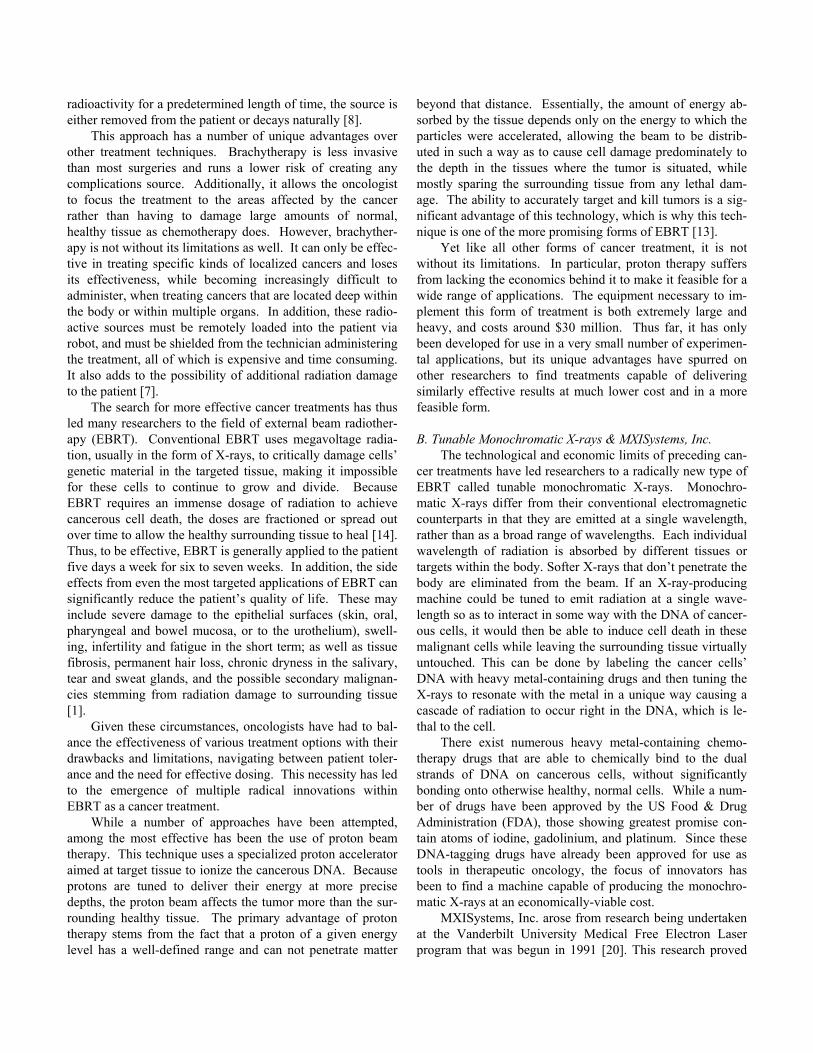
A portion of the light from the terawatt laser is used to generate electrons in the photocathode gun. These electrons
are accelerated in the linear accelerator (linac) and focused to a very small focal spot at the interaction zone (IZ). The re-mainder of the light from the laser is amplified and com-pressed in time and then sent back along the path of the elec-tron beam coming out of the linac. It too is focused to a very small spot at the IZ, where the electron beam and the laser beam collide head-on. X-rays are generated by this collision. They exit the machine in the direction that the electron beam was headed. Since the electron beam is tunable, the X-rays are tunable. Since the laser photons are all at the same fre-quency, the X-rays are all at the same wavelength (Figure 1) [6]
Figure 1. How Omni Works: Omni uses a new approach that collides a very energetic electron beam into a very intense laser beam. X-rays are produced through a mechanism called inverse Compton scattering. Laser photons go in to the device and monochromatic X-rays come out.
2) Radiotherapy as an Application for Monochromatic X-rays
The previously mentioned heavy metal containing DNA specific drugs are administered to a cancer patient, and they spread through the patient’s system within a matter of hours, selectively binding to the DNA in rapidly dividing cancerous cells. A pulsed monochromatic X-ray beam, tuned to the proper wavelength, is then focused on the metal atoms in the drugs in the patient’s tumor. The k-shell electrons on each the atoms of this chemical have a characteristic binding en-ergy associated with them, and when the monochromatic X-ray beam collides with them, they are ejected from their orbit.
This leads to a propagation of electrons in a process called Auger cascading, and the release of energy, along with an electron, from each metal atom causes irreparable damage to the immediately-surrounding molecules, in this case the double helix of the cancerous DNA to which it has bound.
This damage manifests itself in the form of double-strand breakage in the DNA, immediately leading to cell death. In brief, focusing a single-wavelength beam on the atoms has the effect of killing the cells with the tagged DNA while hav-ing very little effect on any healthy, normal surrounding cells. [3], [4], [5], [9], [10], [15]
II. APPLICATION OF THE ARI METHODOLOGY TO
MXISYSTEMS
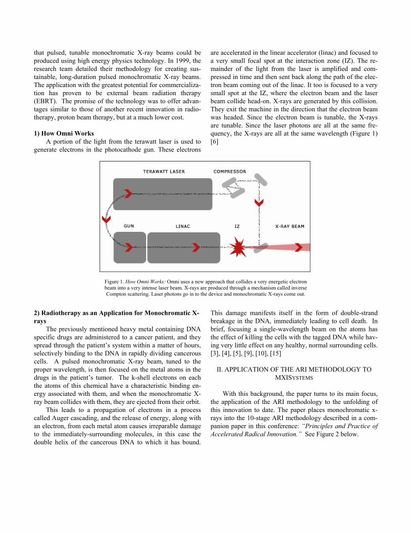
With this background, the paper turns to its main focus, the application of the ARI methodology to the unfolding of this innovation to date. The paper places monochromatic x-rays into the 10-stage ARI methodology described in a com-panion paper in this conference: “Principles and Practice of Accelerated Radical Innovation.” See Figure 2 below.
Figure 2: ARI Methodology Dynamics A. Step 1: 10X Crisis/Opportunity Definition: What is the crisis or opportunity that is driving this innovation?
The area that this technology would address is a portion of the $18 billion radiotherapy market consisting of patients with both primary and metastasized malignant tumors em-bedded deep in tissues that are not easily operable, particu-larly those based in the brain, liver, colorectal tract, mam-mary glands, prostate, and even the lungs. The ideal class of tumors that would best be treated with this technology are those that are deeply seated within tissues not easily amena-ble to surgical removal, or to those that have shown resis-tance to chemotherapy drugs, and are resistant to or impossi-ble to safely treat via conventional radiotherapy techniques. The numbers of patients that this encompasses varies by tu-mor type, but it affects millions per year. B. Step 2: Conceptual Breakthrough Innovation Area Definition: What is the innovation concept that addresses the 10x crisis/opportunity?
The promise of pulsed, tunable, monochromatic X-ray beams, described above, is that they can accurately deliver a lethal dose of radiation to cancerous cells while leaving the surrounding tissue relatively unharmed, and do so with a sig-nificantly lower total level of radiation. This innovation
would allow previously inoperable, life-threatening tumors to be successfully treated with a minimum of toxic radiation exposure to the patient.
One application for this innovation is the treatment of brain cancer in children. These kinds of tumors are inoper-able, and standard chemotherapy drugs have far too many side effects in children to be effective. EBRT must be used, but a conventional form has been shown to cause retardation in children and the development of other radiation induced tumors at a later age, so minimizing radiation exposure is critical in these kinds of applications [8]. One approach that has seen some success is the use of proton beam therapy, but as mentioned earlier, this is still in the experimental phase of development and would be inordinately expensive to apply. Using a pulsed, tunable, monochromatic X-ray beam in con-junction with drugs already utilized in treating these tumors would be ideal and appears to be economically viable to the parties involved in such treatment. C. Step 3: Grand Challenges Definition: What grand challenges that must be addressed to bring the radical innovation concept into being? The grand challenges fall into three categories: market/societal, scien-tific/technological, and business/organizational.
PPhhaassee 11 ((SStteeppss 11--66))
Phase 2 (Steps 7-10)
SStteepp 11
SStteepp 22
SStteepp 33
SStteepp 44
SStteepp 55
SStteepp 66
SStteepp 77
SStteepp 88 GGuuiiddeedd PPrroobbee &&
LLeeaarrnn
SStteepp 99
SStteepp 1100
ARI Methodology Dynamics©A Schematic Overview
Step 3a. Market/Societal Challenges The radiation oncology community has primarily been
focused on the incremental innovation of existing diagnostic and treatment technologies for at least the last fifty years, and they have had a great deal of success with that approach. Only a few relatively radical innovations have made some headway and had varying degrees of success in advancing the standard of practice for treatment.
Among the most notable success stories of this kind is that of computerized tomography (CT) scanning. Although on the surface it appeared to be an incremental innovation as it applied an emerging technology, computer modeling and integration of images, with an older technology, X-ray diag-nostic radiology tools, it actually served to usher in an en-tirely new method for medical diagnostic imaging and later greatly advanced the diagnostic process in radiation oncology [11].
CT scanning was radical in that it was able to produce surprisingly clear three-dimensional images for radiologists, a complete paradigm shift from the two-dimensional AP and lateral plain film images that were standard given existing technology when it was first brought to market in 1972. Even though this approach was met with a fair amount of doubt and resistance from the established radiological community, it was able to overcome these obstacles by proving to be an extremely useful tool with numerous immediate applications in medicine. These niches were instantly exploited by early adopters of this technology within the community, and soon gained wide acceptance on the way to becoming the gold standard for radiological imaging. Certainly, a number of other factors came into play in determining the eventual suc-cess of this radical innovation, particularly when faced with the prospect of dealing with the conservative radiological medicine community, but having a few immediately obvious early applications (brain scans for patients with concussions, neural hemorrhaging, brain tumors, and other neurological conditions for which existing radiation imaging techniques were woefully inadequate) gave it an edge in competing in this practice area.
Some of the lessons learned from that experience can be applied to the knowledge base that MXISystems is using. Specifically, it suggests an initial focus, once they gain FDA approval, on a specific niche in the radiation oncology indus-try that is underserved by incumbent approaches, places where this technology would have large technical and practi-cal advantages. Among the types of cancer, described above, best suited to this innovation, the field of pediatric brain can-cer treatment appears to be an excellent candidate for early adoption, as do colorectal tumors metastasized to the liver, inoperable malignant prostate cancers, and lung cancers (pri-marily because they are not only inoperable but they are also impossible to catheterize, ruling out brachytherapy). Step 3b. Scientific/Technological Challenges
The technical challenges that this innovation will have to overcome are plentiful and broad, and they are reflected in
the three kinds of thinking that need to be pursued to over-come them. Systems thinking involves looking outward, past the scope of simply what this innovation is trying to immedi-ately accomplish, to get at the problem at the root of the chal-lenge being addressed. Decomposition involves looking in-ward and breaking down the technology into its associated components, both in terms of the core innovation itself and the entire solution process that is going to be developed around it. Finally, coordination (“looking sideways”) in-volves seeking out, partnering with, and/or actively promot-ing complementary technologies and innovations that can contribute to the success of the innovation. Systems Thinking
The ultimate challenge at hand is to slow down the pro-gression of cancer within the patient. The immediate prob-lem at hand, then, is to develop a viable, compact, pulsed, tunable, monochromatic X-ray source that is able to induce Auger cascading in the targeted cancerous cells that have been molecularly tagged by previously administered drugs such as cis-platinum or oxaliplatin.
The development of a machine that can produce the de-sired X-rays has yet to reach an economically-viable state where it can be produced at low cost and provided to the nu-merous cancer treatment centers that would require it. Run-ning at a cost of $3.5 million each, such machines could ini-tially be afforded by only the largest and best-financed cancer research centers in the United States.
Seeking a way around this cost barrier leads to the search for another agent that is capable of inducing Auger cascades through a different process. One such approach is the use of a naturally-occurring or cyclotron-produced radionuclide that emits energy at a frequency capable of inducing the same Auger cascade. Conventional brachytherapy could be used to inject this source directly into the tumor to achieve a nearly equivalent result to a tunable, monochromatic X-ray beam, but without the expensive X-ray device. Such a radionuclide, Ytterbium has recently become available on the market, and this approach is undergoing a preliminary clinical trial [12]. Unfortunately, it is still not at an energy that best stimulates the cascade, and as with all radioactive materials, once the nucleus has decayed radioactively, it cannot do so again. Us-ing non-radioactive metals in drugs and causing them to cas-cade with a monochromatic beam can be repeated millions of times, since between cascades the atom returns to its normal stable state. So it is not a perfect substitute for monochro-matic X-rays; as with other uses of brachytherapy, this form of frequency-selective chemo-radiation would be best suited to localized cancers.
But following the logic systems thinking, if Auger cas-cading as a line of attack should fail, then perhaps there is another way to arrest the replication of DNA in cancerous tumors, such as finding a substance that selectively binds to or incapacitates its target.
Finally, if this too fails, then perhaps and even more outward-focused approach to halting the tumor’s cellular rep-
lication and growth mechanism can be affected by some other approaches that have yet to be developed. If nothing else, this kind of outward-focused thinking would allow MXISys-tems to view the rest of their competition in this space, so they would better be able to approach the market and the community of practice with their innovation. Decomposition
There appear to be four components to this innovation, when it is decomposed into its essential elements. The task for MXISystems is to find which elements in their value chain that they ought to control, which parts that they should participate in, and influence and which parts they should sim-ply outsource. This sort of analysis is important so that the company knows where they ought to deploy their scant re-sources to maximize their chances of market success.
The most important segment of their decomposed struc-ture is the special accelerator that produces the electron beam that is used in the process of producing pulsed, tunable monochromatic X-rays over long durations. All of the basic scientific research behind the principles that this technology operates on has been completed and validated. A second-generation version of the machine capable of producing the desired beams has also been developed. In no uncertain terms, the success of this innovation depends in some part on the successful development of a smaller, cheaper accelerator, more so than any other component in the decomposed struc-ture of the innovation [14].
However, the importance of a computer-terminal control unit for this machine should also not be understated. The primary advantage of this technology is its ability to be tuned to various individual wavelengths, so a reliable and simple method to control, measure, and capture data from this ma-chine must be created and engineered so that it can be easily manufactured separate from the accelerator unit. This would allow these two inter-connected components to become de-veloped separately rather than jointly. This way, neither component acts as a limiting technology to the successful development of the other, and would also spur on a greater number of innovators (both firms and entrepreneurs) to de-velop additional improvements onto either one of these plat-forms.
Another critical component of this system is the metal containing drugs that tag the cancerous DNA. This sub-industry within the field of pharmacology is already estab-lished and has won FDA approvals for clinical use in human patients. The continued growth and expansion of this indus-try is vital to the success and development of this innovation. It is encouraging that a large number of additional drugs are being researched and developed to serve as heavy metal con-taining DNA specific agents. There are already a number of promising chemicals that could potentially serve as such but with fewer patient side effects, greater efficacy rates, and at lower cost than current such substances are able to achieve.
One factor that could additionally hamper MXISystems path to FDA approval is any use of chemotherapy drugs not
already in use for cancer therapy, solely for the purposes of irradiating the tumors with monochromatic X-rays. Such uses draw the company down an ever tightening spiral of long-term approval processes as a combination device, rather than a simple radiation producing machine for which approval is now sought. In contrast to the software module described earlier, this part of the innovation runs on its own set of tracks and is pretty much beyond the control of the company.
The therapy planning software that determines how the machine will operate when a patient is brought into the ther-apy suite is another critical part of this system. The devel-opment of this kind of specialized software would help stan-dardize the treatment procedures that the radiological oncol-ogy team will need to implement.
Finally, perhaps the most often overlooked but critically important component that must be considered when the sys-tem is decomposed is the role of the radiation oncology team that treats the patient. This group of trained medical person-nel will be the end user of this innovation and so they must be included as a stakeholder. This team is composed of a radia-tion oncologist who serves as the team leader, a radiation physicist who works closely with the oncologist to develop a treatment plan for each patient, and a radiation technician who usually implements the prescribed therapy. This team approach to treatment delivery is rather unusual in medicine, and one of the well-engrained institutions within the radio-logical oncology community. Understanding how it works and how to gain influence with it is a critical way to affect change in this inherently conservative community of practice.
Having defined each component within the system that this innovation will work with, it is now possible to assess what kind of value a company such as MXISystems can add to this market. They must determine what position they will occupy for each point along their value chain, because even though they might prefer to maintain control over all compo-nents in this sequence, they have finite resources at their dis-posal and must use them wisely to succeed. That is where coordination across other companies’ and organizations’ value chains becomes key. Coordination
Since the success of this innovation depends mostly on the continued development of a commercially-viable machine to reliably produce the pulsed, tunable, monochromatic X-ray beams, it appears that finding other partners to aid in this part of the process could be very useful to the company. While MXISystems will still retain all of the intellectual property rights on this technology, they can share in the development and manufacture of these special accelerators with other companies that have experience in doing so for other com-plex medical equipment.
First of all, MXISystems does not need to devote a sub-stantial share of its resources to manufacturing these devices since assembling them would not add much value to their offering. Rather, they can partner with a knowledgeable firm, such as an overseas firm in Japan or Europe, to partly out-

source this function. In this way, they would gain another powerful stakeholder in this project that could also serve as a guide on ways to gain influence and acceptance within the radiological oncology community.
They don’t have to give up all their development func-tions, however, since the miniaturization and cost-reduction of this machine is paramount. The current version, albeit better than the first and second iterations, is still half the size of a tractor-trailer, can be difficult to maintain, and costs be-tween $3.2-3.5 million which makes purchase by private can-cer treatment centers less likely in the early years of devel-opment of this type of therapy. MXISystems would still hold all the patents on this innovation, but could utilize the deeper pool of financial and human resources of their technology partner.
It appears that MXISystems has a chicken-egg problem [14]. Manufacturers are reluctant to commit the resources to such a development until they have a strong business case for doing so – specifically, definitive clinical evidence of safety and efficacy. But of course it’s hard for MXISystems to get the clinical evidence without accelerators at its disposal. Ob-taining these results would require the use of even more costly facilities, so this presents a difficult obstacle that MXISystems must work through.
In addition to taking on a partner to develop and manu-facture the next generations of these accelerators, MXISys-tems could seek out partners to concurrently develop and manufacture the therapy planning and delivery software for these machines, with the two firms working to create a plat-form that easily hosts future innovations by outside technol-ogy firms. Moreover, since there is little value in producing this component themselves, MXISystems can outsource this function fully without losing any advantages to competing technologies, so long as they retain intellectual property rights to the technology.
As far as the distribution of metal-containing drugs goes, MXISystems would gain little if nothing if they were to enter this portion of their value chain, so the recommendation is to let other already established firms handle the development, production and distribution of these chemicals. MXISystems can serve to connect the users of their innovation to the sup-pliers of these drugs, but anything more than this kind of par-ticipation, however, would likely be a misuse of resources for the company.
The one component in the value chain that can be fully controlled by the company for maximum value addition to their offering is with respect to the oncology treatment team. Specifically, the practices and operating procedures of the radiology and radiation oncology communities have been developed and fine-tuned for many years, and this is well-engrained in their culture. That being said, the operating pro-tocols for delivering cancer treatment with this innovation must be developed and evaluated by experienced members of such treatment teams. This process can be developed during the phantom testing and animal testing phases of the devel-opment of this technology.
MXISystems would first need to assemble an ad hoc community of practice around them by pulling in the various oncology practitioners to co-develop and refine the exact procedures and techniques that oncologists, physicists, and technicians would follow to use this innovation in cancer treatment. This point should not be understated: bringing in potential end-users to scrutinize and validate a systematic protocol for radiotherapy treatment with this innovation would add incomparable value to this offering by making it readily suitable for use in the community.
The most important characteristic of such protocols would be that the teams in this community feel completely comfortable and confident using them. Additionally, oncolo-gists will need from MXISystems a set of indications to as-sure themselves that this radiotherapy treatment aligns with their patients’ needs, particularly given the narrow range of indications where this treatment option would apply (e.g., where the cancer is not easily treatable with surgery or other means).
MXISystems must create a knowledgeable base of users available. After having developed a treatment protocol, MXISystems would be in an ideal position to serve as the primary instructor for teaching the members of the oncology community how to use this system. Once this treatment en-ters the standard of care, MXISystems would be the natural leader for bringing it into the radiation oncology residencies. A good parallel is the development of the CT scanner by EMI Inc. The primary challenge to their product gaining further use was a dire lack of well-educated medical physicists quali-fied to oversee the physical and computing needs of the ma-chines. Once more medical physics graduate programs be-come established, CT scanning developed into an integral part of treatment for various diseases and conditions [17].
MXISystems also has an opportunity to develop the op-eration and maintenance protocols for the latest-generation of these devices, in collaboration with the outsourced equip-ment’s manufacturer, to ensure the reliability of these expen-sive capital-equipment purchases for their users.
By outsourcing or partnering on manufacturing while fo-cusing its own energies on these value-adding activities, MXISystems can shape their offering to the market. Instead of simply approaching potential adopters with an expensive piece of sophisticated capital equipment, MXISystems can offer a far more compelling value proposition: a complete radiotherapy solution consisting of a accelerator, a con-trol/software interface, connections to FDA approved metal-containing drug distributors, along with full operating proto-cols and the personnel to educate the entire radiology and oncology departments on how to properly use and maintain these machines. Step 3c. Organizational and Business Challenges Economics A MXISystems device would be a very capital-intensive in-vestment for any treatment center, and this limits the innova-

tion to large research hospitals, at least until the cost of the machine is reduced enough to become more affordable for private treatment centers [8]. Re-imbursement to the treat-ment clinics that use pulsed, tunable, monochromatic X-ray beam machines would have to come from Medicare/ Medi-caid or from private insurance companies. The decision to recognize this procedure as reimbursable would first fall to the Center for Medicare and Medicaid Services (CMS), a U.S. Federal agency, meaning MXISystems would have to enter the policy arena, either directly or through a proxy. CMS decisions regarding reimbursement are usually contin-gent on approval by the FDA. The lack of FDA approval on this technology has deterred many potential investors and partners in this technology. The extensive start-up capital required prevented this innovation from moving along faster in its development into clinical testing and actual use in ani-mals and later patients [2]. The experience of this technology stands in stark contrast to that of another medical innovation that has recently suc-ceeded in the market: drug-coated stents. Their potential was recognized by both Johnson & Johnson and Boston Scien-tific, while the required capital investment was minuscule by contrast, both companies aggressively jumped into the mar-ket. This, of course, is to be expected when investing in a business that makes a device whose manufacturing cost is roughly one percent of its selling price. Alignment
When an innovator lacks the resources or leverage to line up the necessary supporting industries behind it, it may have to resort to aligning its own objectives with the agendas of other stakeholders. When MXISystems approached estab-lished manufacturers of diagnostic and treatment equipment about a partnership, it ran into a buzz saw of opposition. The message delivered to the company was clear: with an imma-ture business organization, this technology was too early to show. [2]
The company also found itself out of phase with the ven-ture capital community at the time it approached them for seed-stage venture capital funding (2002-2003). This was a time when the venture capital community was pulling in its horns on early stage innovation, such as monochromatic X-rays, that is oriented toward manufacturing expensive capital equipment and faces a long period of technical uncertainty. Most potential investors and strategic partners were loathe to send $5-10 million into what they saw as a “black hole” of an investment when any number of other biomedical invest-ments, at far lower cost, were available to them, such as a prescription ordering systems or the aforementioned drug-coated stents, where exit strategies were measured in one or two years rather than five to eight years [2]. Amplification
One of the missing elements in the monochromatic X-ray system is an inexpensive, compact accelerator that can help to produce X-rays at required wavelengths, and a group of sup-
pliers of these kinds of machines. For this supply base to be economically viable, it would have required an established market of multiple industrial applications for such an accel-erator.
The experience for CT scanners was eerily similar at the start. When EMI first introduced a head-only CT scanner to the market in 1972, it was seen as a very risky venture of a relatively new technology. EMI proceeded to sell the units to physicians in large research institutions in the US at the as-tounding cost of $300,000 apiece. Because this innovation had a unique advantage over conventional radiological imag-ing techniques, there was an immediate application within the field of neurosurgery. Once physicians became accustomed to the unprecedented resolution and finely detailed imagery that CT scanners were able to provide, they quickly clamored for them.
In fact, CT scanners had found such a promising applica-tion in the field of medical imaging that soon every kind of physician demanded to have CT units in their hospital, and EMI was forced to produce them without an established manufacturing base. They made due, however, by setting up multiple production facilities in various locales around the world and later by licensing the manufacturing of these units to Toshiba, a strategic distribution partner in Japan. The most important decision that EMI made was to outsource the manufacturing of most of the various components of their machine to multiple outside firms, and decided to simply assemble and distribute the machines to their customers. This allowed them to fill a large portion of their demand within the first few years of introducing the scanners.
As demand for these machines grew, so did the profits for the company which invariably attracted numerous com-petitors. In spite of EMI’s intellectual property rights to this technology, manufacturers of medical equipment all over the world began to produce and distribute their own CT scanners. Before the end of the 1970’s, EMI found itself as one of sev-eral companies selling CT scanners and this technology was well on its way to becoming the gold standard for medical imaging around the world. EMI was able to achieve this ex-traordinary level of success by breaking down their machine into its various components and deciding that the most value-added to their offering was in assembling and distributing the machines, not in manufacturing every disparate part for themselves [11].
Focusing again on monochromatic X-ray machines, the foremost entities to utilize in bringing this innovation to mar-ket are the large research universities, primarily because they have the staff that private clinics lack to work through the development of a new technology. These facilities would be ideal to use as early adopters of this technology, similar to the way EMI was able to use them for early uses of its CT scan-ning devices. In light of this, there are a number of processes that MXI must still pass through to get clinical trials in uni-versities going. These include FDA advance clearance as an investigational device, institutional review board approval,
the development of legal consent forms, and finding willing physicians to participate [8].
MXISystems initially envisioned this technology as a “killer application” in the world of mammography imaging. The company saw that monochromatic X-rays could be used to painlessly image the mammary glands, and provide physi-cians with incredibly clear and detailed images. However, the requirements of the Mammogram Quality Standard Act (MQSA) made it virtually impossible for the company to get FDA fast-track approval for this device to be used in that sort of application. Additionally, they found it next to impossible to locate funding for continued development of these ma-chines if they were to be used for mammography imaging only, since the reimbursement rates for those kinds of proce-dures are so low as to make it entirely economically unviable. D. Step 4: Hurdles Definition: Having determined the grand challenges, the in-novator can identify the full range of hurdles to be addressed, establishing an agenda of organization and action for subse-quent stages, and helping to assure that there are no un-pleasant surprises going forward.
Although they are explained in the preceding sections of the paper, this serves to summarize the key challenges facing this innovation from each of the three key challenge areas.
Within the market and societal challenges, this innova-tion must find a viable niche market where it can become a standard of care for patients with specific types of cancer. Among the technical challenges they must prove that this is the best approach to solving the problem at hand, slowing down the progression of cancer; developing a compact and economically viable version of their machine to produce the beams; separately developing a reliable and intuitive control module that can tune and control the beams, produce neces-sary images of the cancerous tissue, and serve as a platform for other applications; develop a set of treatment plan-ning/operation and maintenance protocols for oncologists, medical physicists, and radiotherapy technicians to be able to use the machine effectively; and establish a simple and effec-tive process to train these treatment team members on using and maintaining these machines. The organizational and business challenges that this innovation must face are that it is a highly capital-intensive investment for any potential cus-tomers; as of yet, it lacks of FDA approval for use as a cancer treatment device on human patients; it does not align with the strategy of many potential manufacturing and distribution partners; it would require a large and risky capital investment to its FDA approval testing; and there is a dire lack of an es-tablished manufacturing base for accelerators able to produce the electron beams. E. Step 5: Information Retrieval, Pattern Recognition & Knowledge Management Definition: This involves the deep scanning of available knowledge using tools of information retrieval, pattern rec-ognition, and knowledge management.
Using the knowledge resources available to them, in the form of published literature on the subject and personal communications with clinicians and administrators, MXISys-tems came to the conclusion that their initial approach, in the form of diagnostic cancer imaging wouldn’t be economically viable, given the prevailing reimbursement rate of $100 per scan. This led them to redirect their efforts toward radiation therapy. F. Step 6: Envisioned Radical Innovation Definition: The envisioning stage reduces the grand chal-lenges to a specific working concept of the radical innova-tion.
MXISystems will bring to market an accelerator that is capable of producing and controlling pulsed, tunable, mono-chromatic X-ray beams. These beams are able to induce Au-ger cascades in the k-shell electrons of the cancerous DNA where heavy metals such as platinum or some other previ-ously administered metal-containing drug has bound, causing double-strand breakage and cell death to the cancerous cells. The relatively low dosage of radiation necessary for this to take place creates a distinct advantage over other radiother-apy treatments because there is greatly reduced risk of radia-tion illness to the patient; there is a reduced need for elabo-rate, expensive, and bulky radiation-shielding equipment; and there is a potentially higher precision of treatment for various primary and metastasized tumors. In addition, the surround-ing tissues absorb significantly less of the radiation that the machine applies to the tissue, so the dosage of radiation is intensely targeted. Framing
MXISystems found that when they tried to “diagnose and treat” various forms of cancer using their technology, they ran into innumerable road-blocks, some of which had to do with regulatory restrictions, and low reimbursement rates. However, when they redefined and narrowed the scope of what their technology was going to do, (i.e. - only treat vari-ous forms of cancer), they suddenly found new room to ma-neuver into the market. G. Steps 7-10: Implementation Phase Definition: The implementation phase encompasses all the steps required to move the envisioned radical innovation into realization. It encompasses a planned series of interdepend-ent activities, some jointly and others separately to advance the innovation through, testing, scale-up, and the organiza-tion of a commercial infrastructure.
One of the purposes of this paper was to determine whether the application of the ARI methodology might un-cover potential future paths that an unfolding radical innova-tion could take. MXISystems now stands at the crossroads between the Inception and Implementation phases. At this point the focus of the paper shifts from a retrospective analy-sis to an exploration of possible paths that the ARI methodol-

ogy would suggest for MXISystems as it moves into the Im-plementation phase. Joint Development Projects with a Partner
If an outside developer of this type of sophisticated equipment existed to be able to concurrently develop this technology, a massive burden would be lifted from MXISys-tems. The company could then focus on perfecting the proto-cols behind this innovation and then teaching them to the oncologists, medical physicists and technicians that will be using this technology.
Additionally, an outside developer could focus on im-proving the design of the accelerator to make it fit better with the needs of this innovation. Making it smaller, portable, easy to install, serviceable and easy to maintain, and much cheaper then the current version would be paramount con-cerns. This seems to be a competency that any number of electronics-based manufacturers already have and could pos-sibly supply to this innovation, but modifications to these accelerators has been problematic even for the most sophisti-cated national laboratories. Keeping current with the latest physics developments and partnering with national labs here would be a winning strategy.
Currently, however, there are not enough uses for accel-erators, especially the kind required for MXISystems to suc-ceed, for any outside developers to invest the necessary capi-tal to make this vision a reality. There is already an estab-lished industrial base for other radiotherapy treatment equip-ment, but these major players have already turned down the opportunity to work with, invest in, or use the MXISystems approach. One way to potentially entice new entrants into the sphere of accelerator manufacturing would be to obtain some highly promising clinical results to demonstrate the promise that this technology holds. An existing synchrotron facility, such as the kind at the Louisiana State University School of Veterinary Medicine, could be used to perform clinical test-ing on animals [14]. Once the commercial potential of this innovation can be proven to the market through hard lab data, any number of potential partners would then be clamoring to get on board. Testing & Simulation
The standard protocol for receiving FDA approval in-volves first using “phantom” radiation tests,1 followed by animal testing, and finally finding physicians who are willing to use the new innovation on any of their patients [16]. Most of the necessary testing behind k-shell induced Auger cascad-ing has already been completed, so no further research is needed to prove the basic science behind this innovation. Designing Early User Experiences
It would be difficult for MXISystems to develop a ver-sion of their early user experience without building a proto-type of their technology and finding some early adopters to refine the user experience. Unfortunately, this would require 1 Phantom testing involves conducting mock treatments on manikins instru-mented with radiation detectors to verify that the specific dosage is delivered to the target sites.
a large capital investment from MXISystems or one of its community partners.
Rather, MXISystems might refocus their early stage de-velopment on working out the bugs in their process during the various rounds of testing that they must complete before clinical approval from the FDA is awarded. These include “phantom” radiation tests, small mammal testing, and large mammal testing [16]. The primary goal behind this initial testing is to find the smallest amount of radiation necessary to achieve a critical level of cellular response in these mammals. This would create some well-defined tolerances with which to begin and then refine the early human testing. Standardization
The primary standardization that must occur for this is in the treatment protocol. The following processes must be-come standardized for this innovation to succeed in the medi-cal community: • Evaluation of potential patients by the oncologist • Determination of the treatment plan by the physicist and
the oncologist • Administration of the metal-containing drugs specific for
DNA • Delivery of the external beam radiotherapy to the patient
by the technician
Standardizing on a machine design would also be useful, but this can and should be achieved by another community member besides MXISystems. Specifically, the software modules ought to become a standard design that would also allow other companies to produce additional software appli-cations on top of this operating platform. Planned Scale-up
To reach the point of critical mass in the market, MXI-Systems needs to identify good candidates for adopting their innovation. The early adopters, besides those in the clinical trial phase, would likely be university research and treatment clinics that have both the financial resources and the treat-ment specialists capable of implementing this innovation [8].
The remaining candidates in this market should then be pursued for adoption, allowing this technology to “cross the chasm.” This is a relatively small group of users, comprised of oncologists and medical physicists working in research and training hospitals, and they communicate well and share in-formation across various fields. The advantage of this is that once a few respected research clinics have adopted this tech-nology and find early success with it, the rest of the commu-nity will be much more attuned to adopting it themselves. The key is just to find those early adopters that can make this happen and give them all the necessary tools and support to make this happen. They need to be provided with the plat-form, trained on its basic use, and then allowed the freedom to perfect its use.

Education of the Market / Users After establishing standardized protocols for using this
technology, the company should invest resources into educat-ing the users of their technology (oncologists, medical physi-cists, and radiotherapy technicians) on how to maximize the benefits of this innovation. A knowledgeable expert from MXISystems or a community partner should be present dur-ing the first few clinical applications so that any problems can be ferreted out quickly and the results of these treatments can be used in the development process. Once the advantages of the technology are established, the users will promote this innovation to the rest of the community of practice.
One other avenue that could be explored for additional support could be the enlistment of the cancer community in promoting this technology. The experience of the developers of positron emission tomography (PET) scanning technology offers a clear precedent. PET had been languishing for years due to cost constraints and difficulties persuading the FDA to approve the use of a required radioactive tracer drug. PET’s developers mobilized the cancer community to persuade key members of Congress to intervene with the Secretary of Health and Human Services, leading to fast-track approval, and a quick follow-on decision to make the procedure reim-bursable [18].
The highly segmented nature of these organizations does provide an additional challenge to gaining their support, but once a few key players become convinced of the importance of bringing this technology to market, many of the rest will likely follow suit and agree to support this initiative as well. Standard clinical trial testing for medical devices takes about three to five years and a few million dollars in order to gain FDA approval, but with some lobbying weight behind them, MXISystems could dramatically reduce the time and expense needed to gain this critical certification.
Their task would then be to prove the potential of this in-novation in treating various kinds of cancers, especially those that were previously inoperable and/or untreatable using con-ventional treatment methods. Promising initial clinical re-sults could be used to gain some support, and additional re-search on small mammals and larger mammals would add to this momentum.
III. GUIDED PROBE AND LEARN A. Assessment
There are numerous goals that MXISystems ought to achieve to be able to call this venture a success, at least ini-tially. They must organize their own community of practice including their supply chain partners, technology developers, university-level cancer research centers willing to act as early adopters, physicians that would use this technology on some of their patients, and regulatory experts that can help them navigate the FDA approval process. B. Planning
Within the broad goals that have been outlined above, a number of more specific steps should occur:
• An outside partner should undertake the responsibility of developing and manufacturing a smaller and cheaper ac-celerator than the kind available to the market today.
• Another partner, perhaps the same one as with the accel-erators, should develop the software modules and inter-face.
• A large, well-funded university-level cancer research center should act as a host for the initial pre-human test-ing for this innovation.
• Completion of phantom radiation testing • Completion of small mammal testing • Completion of larger mammal testing • Gaining FDA advance clearance as an investigational
device • Institutional review board approval • Development of legal consent forms • Finding willing physicians to participate in human trials • Initiating human treatment trials, a process that can usu-
ally last three to five years • Planned scale-up of manufacture of machines C. Organization
A new community of practice must be organized around MXISystems. They must find supply chain partners that can reliably produce the things they need for this innovation to be successful. Specifically, they must find: • A partner to co-develop the accelerator, as mentioned
above • A treatment software module developer, as elaborated
upon earlier • A regulatory expert to help them gain FDA advance
clearance for an investigational device and to draw up legal consent forms and other clinical trial paperwork
• A well-funded cancer clinic that can partner with them on the non-human testing and on the clinical trials
• Within this cancer center, they should find willing physi-cians for their clinical testing
D. Implementation/Validation
The development of the accelerator, its associated soft-ware modules and interface, and the pre-human testing should occur concurrently. Hopefully, these projects will find completion within a short time of each other, so that the planned expansion of the company can occur as immediately as possible following full FDA approval for radiotherapy applications. E. Learning
The initial results from the phantom testing, the small mammal experiments, and the large mammal experiments should be used to continually refine the technology offering to make it more reliable and something closer to what the market is demanding. F. Assessment
Once all these steps have taken place, the lessons learned can be applied to the continual development of the technol-ogy and the phased expansion of this innovation can begin.

IV. RADICAL INNOVATION PROTOTYPE DE-SIGN/PERFORMANCE DEFINITION/STANDARD DE-
SIGN
To proceed, the company must gain final regulatory ap-proval. The FDA will authorize the safety and efficacy of this machine for specified uses. This will require the submis-sion of all the human clinical trial data along with the engi-neering diagrams and schematics, whereupon the FDA will conduct a review and grant full approval for commercializa-tion.
Concurrently, the company must also gain approval from the United States Center for Medicare & Medicaid Services (CMS) for the reimbursement of the cost of the medical pro-cedures that their machines will perform. This is a critical task that will allow patients to have access to this break-through medical innovation. Once these two regulatory ap-proval needs are met, the company can move ahead with the full commercialization of their innovation.
V. CONCLUSIONS
The intent of this paper was to provide an analysis of a living example of a radical innovation in order to map it to a previously-defined methodology for accelerated radical inno-vation (ARI). This particular example appears to conform very closely to this established methodology, and provides tangible evidence of its validity.
Among the benefits gained from doing this is the classi-fication of all the challenges faced by this innovation, so that a particular class of problem-solving technique or project can be identified to help accelerate this innovation toward com-mercial success. Each of the obstacles that this innovation has dealt with or will have to confront in the future was iden-tified, classified, and characterized so that the technology champion, or another innovator, has a clear idea of what problems must be solved to bring this technology to market.
One of the most important insights that can be drawn from this analysis is that all the challenges faced by this in-novation can be classified into one of three categories: (1) dealing with the unfavorable economics of a risk-intensive investment for both financiers and customers; (2) trying to develop a novel technology without an established manufac-turing base for the machines used in this innovation; or (3) facing a conservative community of practice with well-established practices. Essentially, the innovators need to fo-cus on solving these problems foremost to have a chance to reach commercial success.
Also of particular significance was the systems thinking analysis in the market and societal challenges section. This was intended to be an analysis of other techniques for achiev-ing the same results as this innovation, and though it is out-wardly-focused, it allows an innovators to have a clear view of what problem they are trying to solve, specifically, which market niche and market opportunity they are approaching, so as to design the best offering for this market.
Another goal of this paper was to identify and describe various projects that can be undertaken by an innovator to accelerate this innovation, and numerous examples of such projects were provided here. It is encouraging, then, to know that many of these projects have already been attempted or are planned on being attempted by the company in the future.
Essentially, the purpose of the ARI methodology is to provide a well-defined set of tools and techniques that can potentially be used to accelerate technologies and paradigm-changing innovations to market. The basic premise behind this methodology is that an innovation requires a technology champion to take up the challenges faced by the innovation, an external crisis or opportunity to drive this innovation to market, a new value chain and the establishment of a new community of practice around this innovation, and multiple forays into solving the identified challenges through the proc-ess of “guided probe and learn”. This paper has demon-strated that the ARI methodology offers a flexible set of tools and procedures that can be adapted to innovations at the junc-ture between the inception phase and the implementation phase (a companion paper in this session, Paper 07R0397: Wind Energy Electrical Power Generation: Industrial Life Cycle of a Radical Innovation, demonstrates that the method-ology is also applicable to innovations at the later standard design stage). Going forward, the ARI methodology will be applied to other radical innovations, from different industry sectors and at different stages of the industrial technology life cycle, with the intent of demonstrating its general applicabil-ity to any kind of radical innovation. While applying the ARI methodology doesn’t guarantee the ultimate success of a radical innovation, it has been shown to provide a structured approach to uncovering the challenges that all such risky en-deavors face and a template for dealing with them as they arise. A. MXISystems Results & Continuing Developments
The team at MXISystems has been moving rapidly to in-tegrate many of the ideas that have been discussed hence, and many others that are of their own thinking, to bring their in-novation closer to commercial success. They are focusing on taking what they term an “enabling technology” approach to the market: inserting their innovation into the market without radically changing existing applications.
Currently, they are looking into skipping small mammal testing entirely and moving directly into large mammal test-ing with dogs, and potentially even primates. They hope to begin clinical testing as soon as they gain regulatory approval from the FDA. The company is also working with another corporation to create the therapy planning software, but in-tends to leave existing therapy procedures in place so as to minimize the disruption to the clinical processes that are al-ready in place and well established in the community.
They are also working diligently on the development of the accelerator. The third generation of the machine will likely utilize a revolutionary new type of accelerator called Laser-Wakefield type, and they are working in conjunction
with Lawrence Livermore National Laboratory along with another developer in Paris. This next version of the accelera-tor will be multitudes of times smaller and very portable, po-tentially being small enough to be moved around to patient target sites. However, this next generation of accelerator is at least five years away from commercial realization.
They are hesitant to pursue additional international de-velopers for several reasons. The US Department of Com-merce (DOC) has certain restrictions in place for certain “critical technologies”, and this machine could potentially be used to enrich uranium so it cannot be placed in areas of the world that are deemed high-risk for their geopolitical stability such as Israel and South Korea. They hope to have more success with some of their European partners.
They have secured a manufacturer in California for the current iteration of the accelerator, which is a key component to their innovation reaching commercial success. MXISys-tems has also gained a variety of financial supporters to help turn their dream into a reality. A wealthy philanthropist in Florida is working with them to help install one of their ac-celerators in a world-renowned cancer treatment center. They want to use their initial successes at a small number of initial sites to encourage the market to expand this innovation to other sites around the country. One practical reason for their use of an initial handful of “testing” sites is because of the need to train and support the teams that will use the ma-chines. They will educate their users through continuing medical education (CME) seminars, and graduate education courses that will be funded through medical societies and through the company.
ACKNOWLEDGMENTS
The authors gratefully acknowledge the many insights of the following individuals who have been involved in the launch of MXISystems and participated in the interviews regarding the scientific, clinical, and business aspects of MXISystems. • Mr. Christopher Calton, Vice President – Investment
Banking; and Mr. Philip Krebs, Senior Managing Part-ner, Avondale Partners (interviewed Jan. 2, 2007)
• Dr. Dennis M. Duggan, Associate Professor of Medical Physics, Vanderbilt University Medical Center (inter-viewed Dec. 22, 2006)
• Dr. Dennis E. Hallahan, Ingram Professor and Chairman, Vanderbilt Center for Radiation Oncology (interviewed Jan. 3, 2007)
• Dr. Kenneth Hogstrom, Professor of Physics and Direc-tor, Medical Physics and Health Physics Program, Lou-isiana State University (interviewed Feb. 14, 2007)
• Dr. Patrick Kupelian, Director of Clinical Research, De-partment of Radiation Oncology, M. D. Anderson Can-cer Center Orlando (interviewed Jan. 26, 2007)
REFERENCES
[1] Bucci M., A. Bevan, M. Roach (2005). "Advances in radiation therapy: conventional to 3D, to IMRT, to 4D, and beyond.". CA Cancer J Clin 55 (2): 117-34
[2] Carlton C., P. Krebs, Personal Communication, Avondale Partners, 1/02/07
[3] Carroll F.E., "Medical uses of monochromatic X-rays," in Proceedings of the 1995 Particle Accelerator Conference (Cat. No.95CH35843), Dallas, TX, USA, 1995, pp. 80-2.
[4] Carroll F.E., "Tunable Monochromatic X Rays: A New Paradigm in Medicine," in American Journal of Roentgenology. vol. 179, 2002, pp. 583-590.
[5] Carroll F.E., M. H. Mendenhall, R. H. Traeger, C. Brau, and J. W. Waters, "Pulsed Tunable Monochromatic X-Ray Beams from a Com-pact Source: New Opportunities," in American Journal of Roentgenol-ogy. vol. 181, 2003, pp. 1197-1202.
[6] Carroll F.E., S. Degenhardt, R. Traeger “MXISystems – Our Machines: Diagram”, Retrieved 2/20/07 World Wide Web, http://www.mxisystems.com/ourmachinesdiag.html
[7] Delfino M., and M.E. Day, Cancer: we live and die by radiation, Mo-Beta Publishers, 2006
[8] Duggan D., Personal Communication, 12/22/06 [9] Edwards G.S., R. H. Austin, F. E. Carroll, M. L. Copeland, M. E. Cou-
prie, W. E. Gabella, R. F. Haglund, B. A. Hooper, M. S. Hutson, E. D. Jansen, K. M. Joos, D. P. Kiehart, I. Lindau, J. Miao, H. S. Pratisto, J. H. Shen, Y. Tokutake, A. F. G. van der Meer, and A. Xie, "Free-electron-laser-based biophysical and biomedical instrumentation," Re-view of Scientific Instruments, vol. 74, pp. 3207-45, 2003.
[10] Frank C., "Tunable, monochromatic X-rays: An enabling technology for molecular/cellular imaging and therapy," in Journal of Cellular Biochemistry. vol. 90, 2003, pp. 502-508.
[11] Gray J.E., C.G. Orton " Medical Physics: Some Recollections in Diag-nostic X-ray Imaging and Therapeutic Radiology" in Radiology. 2000
[12] Hallahan D., Personal Communication, 1/03/07 [13] Hartford A.C., A. L. Zietman, et al. "Proton Radiotherapy," (1999), in
Radiothereutic Management of Carcinoma of the Prostate, London, UK, Arnold Publishers: 61-72.
[14] Hogstrom K., Personal Communication, 2/14/07 [15] Kuhar J., "Building a Better Beam," RT Image, vol. 15, pp. 16-21, Feb.
18, 2002 2002. [16] Kupelian P., Personal Communication, 1/26/07 [17] Mitchell W., J. Smith "Playing Leap-frog with Elephants: EMI Ltd. and
the CT Scanner Competition in the 1970's", University of Michigan School of Business, August 1989
[18] Rundle R.L., "Image Booster: How a Small Firm Pushed PET Scans Into Mainstream --- To Cutting-Edge Science, CTI Added Political In-fluence And Piggyback Marketing --- `Guy Thing' With Sen. Stevens," in Wall Street Journal, 2002, p. A.1.
[19] Tannock I.F., R.P. Hill et al The Basic Science of Oncology. (eds) 4th ed.2005 McGraw-Hill.
[20] Tompkins P.A., W. D. Andrews, C. A. Brau, J. W. Waters, F. E. Car-roll, D. R. Pickens, R. R. Price, and C. Roos F., "The Vanderbilt Uni-versity Free-Electron Laser X-ray facility," in Proc. SPIE - Int. Soc. Opt. Eng. (USA), San Diego, CA, USA, 1993, pp. 72-83.