1 generation of quasi-monochromatic soft x-rays using a table-top electron accelerator alexander...
TRANSCRIPT
1
Generationof quasi-monochromatic
soft x-rays usinga table-top electron accelerator
Alexander Lobko
Institute for Nuclear Problems Belarus State University
July 2009 X Intl Gomel HEP School

2
Light sources
http://www.lightsources.org/cms/?pid=1000166
NanoscienceLife sciences

3
Typical size of contemporary synchrotron

4
Budget of the SOLEIL synchrotron
Construction
• Investment…………….235 M€
• Operation………………..64 M€
• Salaries………………….150 M€
Total………………………….449 M€
Yearly………………………...53 M€
www.synchrotron-soleil.fr

5
Why do we need (quasi)-monochromatic
soft x-rays?

6
Soft X-Ray Spectroscopy Methods: Soft x-ray absorption spectroscopy (XAS), near-edge x-ray absorption fine structure (NEXAFS) spectroscopy, soft x-ray emission spectroscopy (SXES), resonant inelastic x-ray scattering (RIXS), x-ray magnetic circular dichroism (XMCD), x-ray photo-emission spectroscopy (XPS), Auger spectroscopy.
Problems:Complex materials Magnetic materials
Environmental scienceCatalysis
The photon energy tunability and its brilliance for some above listed applications are
essential.
7
Soft X-Ray Scattering
Methods: Soft x-ray emission spectroscopy (SXES), inelastic x-ray scattering (IXS), resonant x-ray inelastic scattering
(RIXS), speckle patterns, small-angle x-ray scattering (SAXS).
Problems:Strongly correlated materials
Magnetic materials Environmental science
Catalysis
The tunability of radiation and its brilliance for some above listed applications are essential.
8
Soft X-Ray Imaging
Methods: Soft x-ray imaging, photoelectron emission microscopy (PEEM), scanning transmission x-ray microscopy (STXM), full-field microscopy, x-ray diffraction imaging (XDI),
x-ray tomography, computer-aided tomography (CAT).
Problems:Cell biology
Nano-magnetism Environmental scienceSoft matter, polymers
The tunability of radiation is absolutely essential for the creation of contrast mechanisms.
9
Applications to the life sciences
• Potential to form high spatial resolution images in hydrated bio-material
• Ability to identify atomic elements by the coincidence between photon energy and atomic resonances of the constituents of organic materials
ConcernRadiation-induced damage: photon energy deposited per unit mass (dose) can cause
observable changes in structure
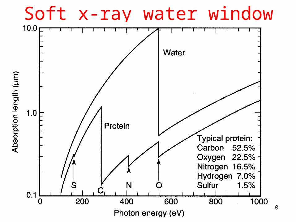
10
Soft x-ray water window
11
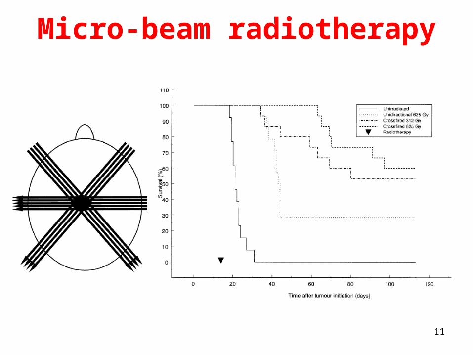
Micro-beam radiotherapy

12
Indirect radiation therapy
www.mpsd.de/irt
13
Monochromatic X ray medical imaging
By narrowing x-ray spectrum inside of the range required
for a specific medical imaging application, a patient’s
radiation-induced damage may be significantly reduced.
It has been evaluated that x-ray examinations performed
with quasi mono-energetic x-rays (even 15-20%) will deliver
a dose to the patient that will be up to 70% less than dose
deposited by a conventional x-ray system [P. Baldelli [et
al] // Phys. Med. Biol. 49 (2004) 4135].

14
Optimal X-Ray Energies forMedical Imaging
• mammography - 17-21 keV;
• radiography of chest,
extremities and head - 40-50 keV;
• abdomen and pelvis
radiography - 50-70 keV;
• digital angiography - ~33 keV.

15
How much monochromatic soft x-ray photons we need?

16
Evaluation of X-Ray Flow
for Medical Imaging
The Physics of Medical Imaging / S. Webb (Ed.), Bristol: Hilger, 1978.
2 2 21(1 )exp( ) /( ( ) )N k R t x x
xt
1
2
17
What do we need for high-quality in vivo imaging?Number of x-ray quanta needed to visualize
1.0 mm3 of biological tissue at 1% contrast is~3x10⁷ photons/mm2.
This evaluation made for film registration.
In case of digital detection 4×10⁴ photons per ~0.4 mm2 detector pixel are required. It leads to the
flux of~106 photons/mm2.
Due to heart beat and breathing, above photon flux must be provided within ~1/100 s.
Photons must penetrate considerable field of vision.

18
What do we exactly need for high-quality in vivo imaging?
We need, for example,
3x107 mm-2 * 100x100 mm2 / 10-2 s =
~3x10¹³ (1012) photons/s
with tunable x-ray energy in 10-70 keV range
Mono-chromaticity could be of ~10-2 for a patient’s dose reduction
Radiation background should be low

19
Spectral brilliance of x-ray sources
There is large gap between properties of common and high energy
accelerator-based x-ray sources
20
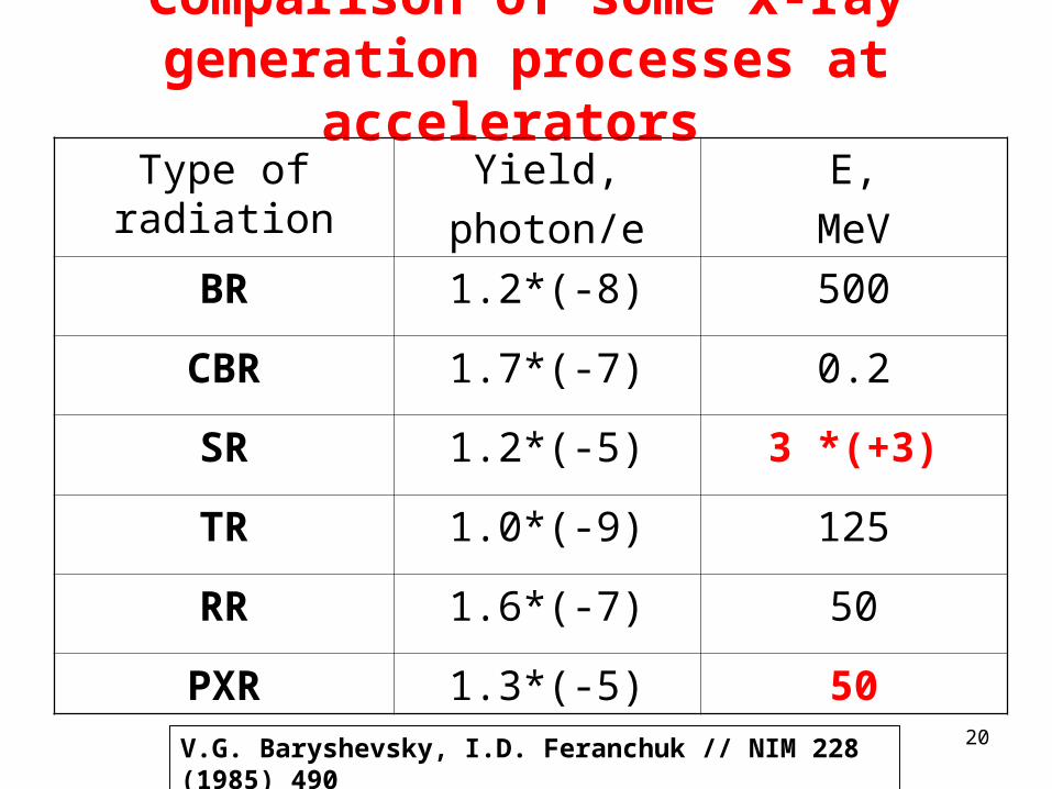
Comparison of some x-ray generation processes at accelerators
Type of radiation Yield,
photon/e
Е,
MeV
BR 1.2*(-8) 500
CBR 1.7*(-7) 0.2
SR 1.2*(-5) 3 *(+3)
TR 1.0*(-9) 125
RR 1.6*(-7) 50
PXR 1.3*(-5) 50
V.G. Baryshevsky, I.D. Feranchuk // NIM 228 (1985) 490

21
Compact x-ray source based on Compton back scattering
http://www.lynceantech.com/sci_tech_cls.html

22
Parametric x-rays
V. Baryshevsky, I. Feranchuk, A. Ulyanenkov Parametric X–ray Radiation in Crystals: Theory, Experiment and Applications // Springer, 2006, 176 p.
22
0, 0, , , , , , , , , , ,...nPXRB s
NNF eQ g L T X
k k
2
2( ) 1 1
2pn
1 cos 0vn k
sinnB
B
cn
d
Condition for the Cherenkov radiation emission

23
Motivation to use PXR• it is quasi-monochromatic x-rays• x-rays energy can be tuned smoothly by
single crystal target rotation• it is well directed and polarized x-rays• x-rays energy does not depend on energy of
incident electrons• radiation angle can be as large as
180 arc degrees - it means, one may work at virtually low background
• Optimal target thickness – 10-50 µm of light crystal material (diamond, silicone, graphite, LiF, quartz, etc) – weak multiple scattering
24
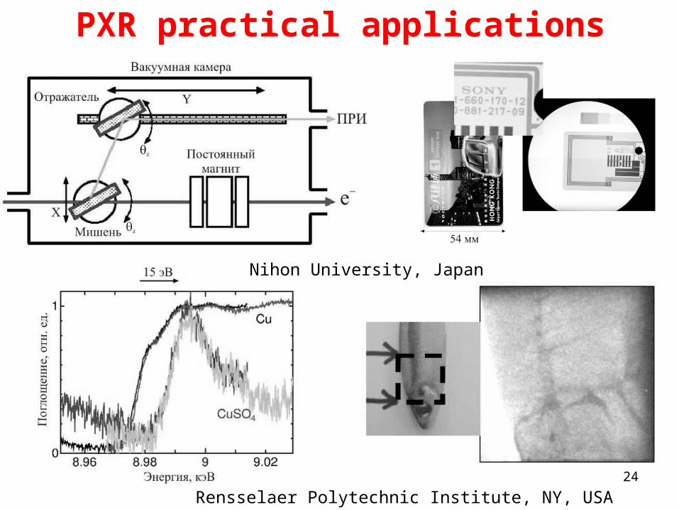
PXR practical applications
Rensselaer Polytechnic Institute, NY, USA
Nihon University, Japan

25
Racetrack microtron 70 MeV
http://nuclphys.sinp.msu.ru/nuc_techn/el_ac/index.html
2.2*1.8*0.9 м3
26
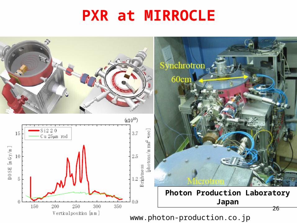
PXR at MIRROCLE
Photon Production Laboratory Japan
www.photon-production.co.jp
27
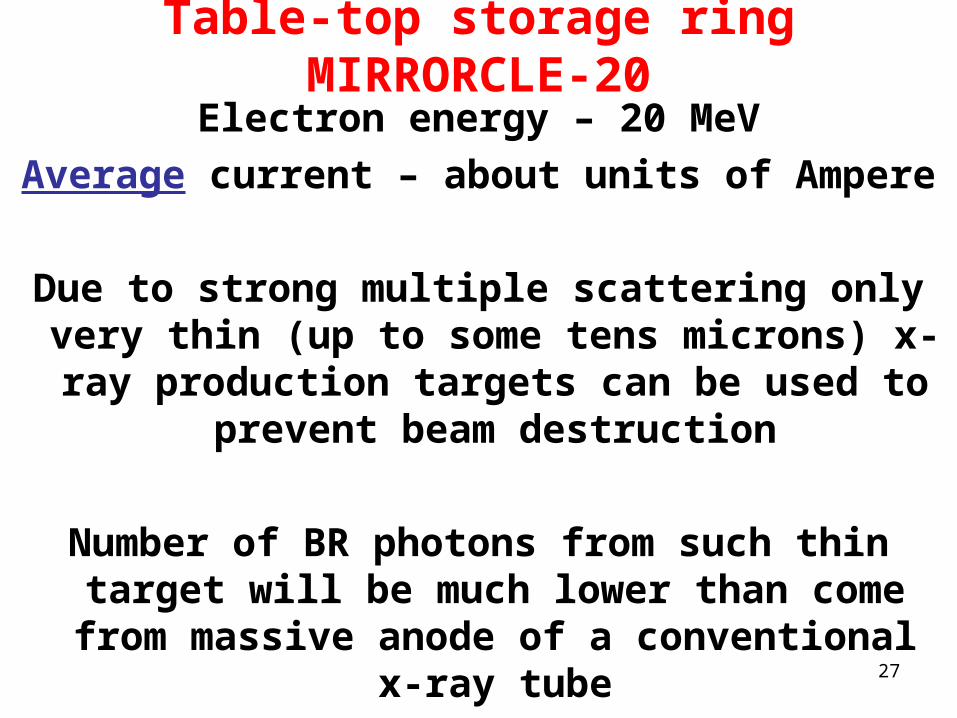
Table-top storage ring MIRRORCLE-20
Electron energy – 20 MeV
Average current – about units of Ampere
Due to strong multiple scattering only very thin (up to some tens microns) x-ray
production targets can be used to prevent beam destruction
Number of BR photons from such thin target will be much lower than come from massive
anode of a conventional x-ray tube

28
Evaluations of 33 keV PXR emissionfrom 20 MeV electrons
Ee = 20 MeV, Si target of L=0.01 cm thickness, 33 KeV x-rays, symmetrical Laue case for (111), (220), and (400). Angles between electron velocity direction and direction
to diffraction reflex are ~6.9, 11.2, and 15.9 degrees, respectively.
Si, L=0,01 cm
0,0E+00
5,0E-05
1,0E-04
1,5E-04
2,0E-04
2,5E-04
3,0E-04
3,5E-04
4,0E-04
0,0E+00 1,0E-02 2,0E-02 3,0E-02 4,0E-02 5,0E-02 6,0E-02 7,0E-02 8,0E-02 9,0E-02 1,0E-01
Polar angle, rad
Ang
ular
den
sity
, pho
tons
/(e- s
rad)
111
220
400
Dia 20 cm at 1.5 m~7·10-2 rad
Quantum Yield:(111) - 3·10-6 /e-
(220) – 4.5·10-7 /e-
(400) – 1.4·10-7 /e-
In some cases account of CB interference is needed

29
Asymmetric case
Angle between electron velocity and input plane normal is equal 55 arc degrees, angle between output plane normal and outgoing radiation is equal 35 arc degrees. Plane thickness was chosen equal to 0,00811 сm to provide electron path in crystal equal toL0=L/cos(55 arc degrees)=0,0141 cm

30
Optimal PXR crystal target - wedgeTo calculate optimal asymmetric geometries and wedge configurations – dynamical theory required
31
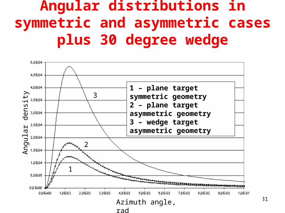
Angular distributions in symmetric and asymmetric cases plus 30 degree wedge
Azimuth angle, rad
1
2
31 – plane target symmetric geometry2 – plane target asymmetric geometry3 – wedge target asymmetric geometry
Ang
ular
den
sity

32
Soft PXR intensity at M-20
• Target Si (111) wedge shaped;
• Bragg angle = 45 arc degrees; EPXR = 2.8 keV;
• Absorption length 3.57 µm;
• Geometry – Symmetric Laue;
• Wedge thickness 0.01 cm;
• Wedge angle - 30 degrees;
• Energy resolution (integration) /= 10-3;
• Intensity of PXR+diffracted TR = ~210-6 ph/e-;
• Intensity of diffracted BR = ~510-6 ph/e-.

33
Wedge targets prototypes
At a moment wedge-shaped targets available of Si (111) and (100) base planes with length 3 through 24 mm (step 3 mm) and maximal thickness 450 or 350 mkm.Angle of the wedge and its material can be customized.

34
PXR reflex integral intensity at M-20
Depending on the beam fraction we can apply for PXR generation, in the ideal
integral flux may be as high as
10-5 ph/e * 1019 e/s.
It means 1014 s-1 X ray photons
of 10-3 monocromaticity
with tunable energy

35
M-20 beam shape

36
ConclusionsPXR radiation mechanism and table-top
accelerator can provide flux needed for contemporary soft x-ray applications in high-quality medical imaging and lowered dose radiation therapy.
Problems to be considered:Commissioning of the real beam shape as income for more exact evaluations and production of specific targetsTarget heatingPXR angular distributionX-ray harmonics filteringApplication of x-ray opticsTargets made of photonic crystals – way to T-rays
37
MinskYa. Kolas Sq.
1967

38
Many thanks for your attention