case report two-year follow-up results of c2/3 prestige-lp ...ijcem.com/files/ijcem0018962.pdf ·...
TRANSCRIPT

Int J Clin Exp Med 2016;9(4):7349-7353www.ijcem.com /ISSN:1940-5901/IJCEM0018962
Case Report Two-year follow-up results of C2/3 Prestige-LP cervical disc replacement: first report
Yi Yang1, Mengying Yang1, Shan Wu1, Ying Hong2, Litai Ma1, Beiyu Wang1, Chen Ding1, Yuxiao Deng1, Yueming Song1, Hao Liu1
1Department of Orthopaedics, West China Hospital, Sichuan University, Chengdu, Sichuan Province, P. R. China; 2Operation Room, West China Hospital, Sichuan University, Chengdu, Sichuan Province, P. R. China
Received November 1, 2015; Accepted March 18, 2016; Epub April 15, 2016; Published April 30, 2016
Abstract: A 49-year-old female patient presented multilevel cervical disc herniation with persisting neurological signs since 16 months ago. Mild spondylotic changes in segments C2/3 and C5/6 were detected on plain films. Magnetic resonance imaging (MRI) confirmed multilevel cervical disc herniation (C2/3, C3/4, C4/5, C5/6), compro-mising the neural foramen at the left C3 and C6 nerve roots seriously. A double-level cervical disc replacement C2/3 and C5/6 was performed to relief her pain. The surgery was carried out on a classic right approach after induction of general anesthesia. In this case, a 5*14 mm Prestige-LP was implanted in C2-3 and a 5*16 mm Prestige-LP was implanted in C5-6. Postoperative complications such as hoarseness, dysphagia, cerebrospinal fluid leakage and malposition of the prosthesis were not found. The patient’ clinical symptoms were totally relieved 6 months after surgery. The 24 months postoperative X-ray showed the good position of the implant, a satisfying cervical range of motion and cervical lordosis. The preliminary clinical and radiographic results of C2/3 Prestige-LP cervical disc replacement are good. In our opinion a good exposure is half of the success of CDR C2/3 and transnasal intubation anesthesia is recommended to achieve a larger exposed space. In most cases mandibulectomy is not needed. With a minor follow-up of 24 months, the clinical and radiographic results of this case are good and larger studies with longer follow-up duration are warranted to explore the safety and effectiveness of CDR C2/3.
Keywords: ACDF, cervical disc replacement, cervical spine, CDR, C2/3
Introduction
Anterior cervical discectomy and fusion (ACDF) is well regarded as the surgical gold standard for the treatment of cervical disc degenerative disease for several decades [1, 2]. ACDF offers the possibility to maintain segmental lordosis and preserve anatomical disc space height. However, previous biomechanical studies have reported higher intradiscal pressure, as well as increased segmental motion, in levels adjacent to a cervical fusion [3]. Cervical disc replace-ment (CDR) aims not only to allow the same neural decompression as traditional anterior surgery, but also to preserve motion at the treated level and decrease the incidence of adjacent segment disease. Many spinal sur-geons are familiar with the anterior approach to the upper cervical spine (occiput to C3) and experienced at performing discectomy and fusion with or without instrument in this region; however, how to perform cervical disc replace-
ment C2/3 has been little reported. Considering the little knowledge of CDR C2/3, we present this special technique case report to share our experience and to explore the safety and effec-tiveness of CDR C2/3 with a minor follow-up of 24 months.
Case description
This case describes a 49-year-old female pa- tient presenting multilevel cervical disc hernia-tion with persisting neurological signs since 16 months ago. The neck, shoulders and left arm pain has worsened in the last month despite a 6-month intensive conservative treatment. Currently she has some neck pain, shoulders pain and left side arm pain in the C3 and C6 roots distribution area. She does not have a neurologic deficit or walking disturbances. Mild spondylotic changes in segments C2/3 and C5/6 were detected on plain films. Dynamic flexion and extension X-rays showed the seg-

C2/3 Prestige-LP cervical disc replacement
7350 Int J Clin Exp Med 2016;9(4):7349-7353
mental movement well preserved. Magnetic resonance imaging (MRI) confirmed multilevel cervical disc herniation (C2/3, C3/4, C4/5, C5/6), compromising the neural foramen at the left C3 and C6 seriously.
The surgery was carried out on a classic right approach by a very experienced surgeon after induction of general anesthesia. The patient was carefully positioned supine on a radiolu-cent operating table with her head and neck in slightly lordotic cervical spine position (no rota-tion). A small towel roll was placed under the neck to assist with appropriate positioning of the neck and shoulders and to keep a physio-logic lordosis without creating a hyperlordosis. The head is placed on a folded towel to keep it from rolling during the procedure. Gentle trac-tion was given to the upper limbs and strapped by the side of body. Somatosensory evoked potential monitoring of cord function is sug-gested during the procedure. Fluoroscopy and metal markers were used to locate the correct incision point. A right-sided transverse skin inci-sion in the submandibular region with a vertical extension as long as required providing ade-quate exposure is made. Carry the dissection through the platysma muscle with the envelop-ing superficial fascia of the neck and mobilize flaps from this area. Mobilize the anterior bor-der of the sternocleidomastoid muscle by longi-tudinally dividing the superficial layer of the deep cervical fascia. Identify the digastric and stylohyoid muscles, and tag and divide the ten-don of the former. Retracting the trachea and esophagus to the left side opens the approach to the prevertebral fascia and the spine. Carotid
artery is covered by soft tissue. The disc level is confirmed with the help of fluoroscopy (Figure 1). The insertions of the longus colli muscle are divided from the cervical bodies with bipolar and scissors. C2-3 discectomy was done and long shaft Caspar screws for interbody retrac-tion were inserted into the middle of the adja-cent vertebral bodies. Posterior longitudinal ligament along with anterior, posterior and lat-eral ostophytes were resected with rongeurs. Meticulous hemostasis was used throughout this procedure to diminish the blood loss and minimize the risk of heterotopic ossification. After adequate decompression of C2-3, the affected level of C5-6 was also accomplished neurologic decompression using the same method listed above. The subchondral end-plates are preserved for the prevention of implant subsidence. After the endplate prepa-ration completed, the disc space was distract-ed and a trial implant of appropriate size was inserted under image control. The distance between the implant and the anterior and pos-terior rim of vertebral bodies should be at least 1-2 mm. The prosthesis was then mounted over the application instrument and inserted into intervertebral space with the help of fluorosco-py. Oversizing of the implant can be identified by the presence of distracted facet joints pos-teriorly. In this case, a 5*14 mm Prestige-LP was chosen for C2-3 and a 5*16 mm Prestige-LP was chosen for C5-6. Final imaging of the device implantation is performed before wound closure. Hemostasis is rechecked, and a drain-age was inserted. The skin was sutured sub- cutaneously.
Figure 1. Intraoperative fluoroscopy.

C2/3 Prestige-LP cervical disc replacement
7351 Int J Clin Exp Med 2016;9(4):7349-7353
The patient spent the first night in a recovery room, because of potential cervical wound bleeding. The patient was mobilised the next day and discharged home on the third day. She was obeyed to begin cervical function exercise 3 days after surgery and orthosis was not used.
One week after surgery she was obeyed to take anteroposterior, lateral and functional X-rays (Figure 2). Postoperative complications such as hoarseness, dysphagia, cerebrospinal fluid leakage and malposition of the prosthesis were not found. The patient’ clinical symptoms were
Figure 2. One week postoperative anteroposterior, lateral and functional X-rays.
C2/3 Prestige-LP cervical disc replacement
7352 Int J Clin Exp Med 2016;9(4):7349-7353
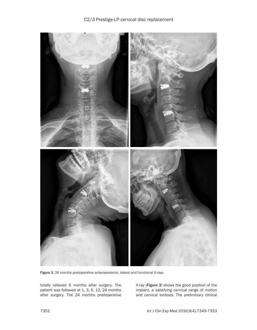
totally relieved 6 months after surgery. The patient was followed at 1, 3, 6, 12, 24 months after surgery. The 24 months postoperative
X-ray (Figure 3) shows the good position of the implant, a satisfying cervical range of motion and cervical lordosis. The preliminary clinical
Figure 3. 24 months postoperative anteroposterior, lateral and functional X-rays.
C2/3 Prestige-LP cervical disc replacement
7353 Int J Clin Exp Med 2016;9(4):7349-7353
and radiographic results of C2/3 Prestige-LP cervical disc replacement are good.
Discussion
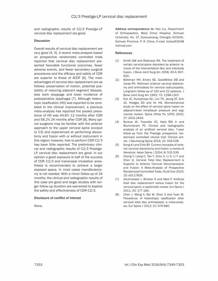
Overall results of cervical disc replacement are very good [4, 5]. A recent meta-analysis based on prospective randomized controlled trials reported that cervical disc replacement pre-sented favorable functional outcomes, fewer adverse events, and fewer secondary surgical procedures and the efficacy and safety of CDR are superior to those of ACDF [6]. The main advantages of cervical disc replacement are as follows: preservation of motion, potential pos-sibility of reducing adjacent segment disease, less work stoppage and lower incidence of postoperative dysphagia [7]. Although hetero-topic ossification (HO) was reported to be unre-lated to the clinical improvement, a previous meta-analysis has reported the pooled preva-lence of HO was 44.6% 12 months after CDR and 58.2% 24 months after CDR [8]. Many spi-nal surgeons may be familiar with the anterior approach to the upper cervical spine (occiput to C3) and experienced at performing discec-tomy and fusion with or without instrument in this region; however, how to perform CDR C2/3 has been little reported. The preliminary clini-cal and radiographic results of C2/3 Prestige-LP cervical disc replacement are good. In our opinion a good exposure is half of the success of CDR C2/3 and transnasal intubation anes-thesia is recommended to achieve a larger exposed space. In most cases mandibulecto-my is not needed. With a minor follow-up of 24 months, the clinical and radiographic results of this case are good and larger studies with lon-ger follow-up duration are warranted to explore the safety and effectiveness of CDR C2/3.
Disclosure of conflict of interest
None.
Address correspondence to: Hao Liu, Department of Orthopaedics, West China Hospital, Sichuan University, No. 37, Guoxuexiang, Chengdu 610041, Sichuan Province, P. R. China. E-mail: [email protected]
References
[1] Smith GW and Robinson RA. The treatment of certain cervical-spine disorders by anterior re-moval of the intervertebral disc and interbody fusion. J Bone Joint Surg Am 1958; 40-A: 607-624.
[2] Bohlman HH, Emery SE, Goodfellow DB and Jones PK. Robinson anterior cervical diskecto-my and arthrodesis for cervical radiculopathy. Long-term follow-up of 100 and 22 patients. J Bone Joint Surg Am 1993; 75: 1298-1307.
[3] Eck JC, Humphreys SC, Lim TH, Jeong ST, Kim JG, Hodges SD and An HS. Biomechanical study on the effect of cervical spine fusion on adjacent-level intradiscal pressure and seg-mental motion. Spine (Phila Pa 1976) 2002; 27: 2431-2434.
[4] Burkus JK, Traynelis VC, Haid RW Jr and Mummaneni PV. Clinical and radiographic analysis of an artificial cervical disc: 7-year follow-up from the Prestige prospective ran-domized controlled clinical trial: Clinical arti-cle. J Neurosurg Spine 2014; 21: 516-528.
[5] Song KJ and Choi BY. Current concepts of ante-rior cervical discectomy and fusion: a review of literature. Asian Spine J 2014; 8: 531-539.
[6] Zhang Y, Liang C, Tao Y, Zhou X, Li H, Li F and Chen Q. Cervical Total Disc Replacement is Superior to Anterior Cervical Decompression and Fusion: A Meta-Analysis of Prospective Randomized Controlled Trials. PLoS One 2015; 10: e0117826.
[7] Zechmeister I, Winkler R and Mad P. Artificial total disc replacement versus fusion for the cervical spine: a systematic review. Eur Spine J 2011; 20: 177-184.
[8] Chen J, Wang X, Bai W, Shen X and Yuan W. Prevalence of heterotopic ossification after cervical total disc arthroplasty: a meta-analy-sis. Eur Spine J 2012; 21: 674-680.