case report sclerokeratoplasty as the therapy for corneal...
TRANSCRIPT

Case ReportSclerokeratoplasty as the Therapy for Corneal Perforation due toExposure and Neurotrophic Keratopathy
Radoslaw Rozycki, Izabela Nowak-Gospodarowicz, Dominika Bialas,Rafal Pawlik, and Marek Rekas
Ophthalmology Department, Military Institute of Medicine, 128 Szaserow Street, 04-141 Warsaw, Poland
Correspondence should be addressed to Radoslaw Rozycki; [email protected]
Received 23 November 2013; Accepted 14 December 2013; Published 16 January 2014
Academic Editors: A. A. Bialasiewicz and V. Jhanji
Copyright © 2014 Radoslaw Rozycki et al. This is an open access article distributed under the Creative Commons AttributionLicense, which permits unrestricted use, distribution, and reproduction in any medium, provided the original work is properlycited.
A case report of exposure and neurotrophic keratopathy after acoustic neuroma surgery resulting in perforation if not managedappropriately and timely is presented. Sclerokeratoplasty on 360 degrees may be an effective treatment method of cornealperforation in complete anaesthetic cornea when the standard penetrating keratoplasty failed. At a 12-month follow-up, the patientis doing well. UCVA is 0.5, the IOP is normal, and the graft remains clear. Systemic immunosuppression is the main disadvantageof this method. Further investigation is needed to assess the effectiveness and safety of this method.
1. Introduction
Acoustic neuromas (Vestibular Schwannomas) (VS) areoncologically benign tumours which constitute more than90% of all cerebellopontine angle tumours and more than10% of all primary brain tumours. Surgical excision of thesetumours is one of the most challenging neurosurgical proce-dures because of their location close to vital structures suchas the anterior inferior cerebellar artery (AICA) or the 7thand 8th cranial nerves [1]. When the tumour exceeds 3 cm,it might involve the trigeminal nerve causing a depressedcorneal reflex, which is accompanied by peripheral facialnerve paresis leading to the development of exposure andneurotrophic keratopathy. This condition, especially withpoor Bell’s phenomenon, is usually resistant to conventionaltherapies and has a very unfavourable prognosis. Loss of thesensory innervation of the cornea decreased the number ofcorneal stem cells [2], decreased metabolic and mitotic ratesin the corneal epithelium, and reduced acetylcholine andcholine acetyltransferase concentrations [3, 4] resulting in thedevelopment of persistent epitheliopathy.
This chronic epithelial breakdown enables proteolyticenzymes to degrade the extracellular matrix componentsbecause they cannot protect corneal structural and signalingmatrix proteins anymore. This condition may progress tocorneal ulceration, perforation, and loss of the eye.
The ophthalmic goal of treatment is to protect the corneafrom external irritating factors, to stop its progressive degra-dation, and to support its healing.
2. Case Report
The patient was a 64-year-old female with a 4-year historyof exposure and neurotrophic keratopathy in the right eyedue to unresolved peripheral facial nerve and trigeminalnerve palsies after acoustic neuroma surgery. The patientunderwent bilateral cataract surgery at the age of 61 and,except for mild hypertension, remained healthy. After 2 yearsof satisfactory treatment of lagophthalmos with a gold eyelidweight, it was necessary to remove the weight from the rightupper eyelid in order to perform an MRI scan. Despite theuse ofmoisturizing drugs and eye taping, severe corneal ulcerdeveloped 6 months after the removal of the weight. After 2months of ineffective conservative treatment, the patient wasreferred to our clinic.
On admission, the corrected distance visual acuity(CDVA) was 0,01 (Snellen chart) and intraocular pressure(IOP) was 14mmHg in the right eye. CDVA was 1,0 in theleft eye.
Peripheral right facial nerve palsy, lagophthalmos of 5millimetres with paralytic ectropion, poor Bell’s phenom-enon, and complete corneal anaesthesia were noted in
Hindawi Publishing CorporationCase Reports in Ophthalmological MedicineVolume 2014, Article ID 467249, 4 pageshttp://dx.doi.org/10.1155/2014/467249
2 Case Reports in Ophthalmological Medicine
(a) (b) (c)
(d) (e) (f)
(g) (h) (i)
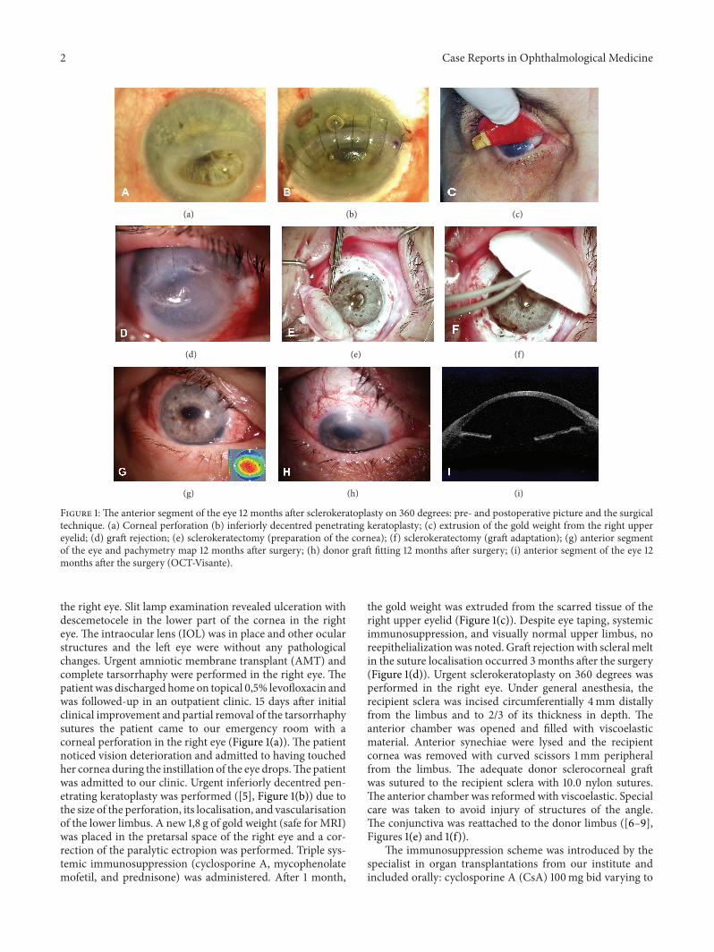
Figure 1: The anterior segment of the eye 12 months after sclerokeratoplasty on 360 degrees: pre- and postoperative picture and the surgicaltechnique. (a) Corneal perforation (b) inferiorly decentred penetrating keratoplasty; (c) extrusion of the gold weight from the right uppereyelid; (d) graft rejection; (e) sclerokeratectomy (preparation of the cornea); (f) sclerokeratectomy (graft adaptation); (g) anterior segmentof the eye and pachymetry map 12 months after surgery; (h) donor graft fitting 12 months after surgery; (i) anterior segment of the eye 12months after the surgery (OCT-Visante).
the right eye. Slit lamp examination revealed ulceration withdescemetocele in the lower part of the cornea in the righteye. The intraocular lens (IOL) was in place and other ocularstructures and the left eye were without any pathologicalchanges. Urgent amniotic membrane transplant (AMT) andcomplete tarsorrhaphy were performed in the right eye. Thepatientwas discharged homeon topical 0,5% levofloxacin andwas followed-up in an outpatient clinic. 15 days after initialclinical improvement and partial removal of the tarsorrhaphysutures the patient came to our emergency room with acorneal perforation in the right eye (Figure 1(a)). The patientnoticed vision deterioration and admitted to having touchedher cornea during the instillation of the eye drops.The patientwas admitted to our clinic. Urgent inferiorly decentred pen-etrating keratoplasty was performed ([5], Figure 1(b)) due tothe size of the perforation, its localisation, and vascularisationof the lower limbus. A new 1,8 g of gold weight (safe for MRI)was placed in the pretarsal space of the right eye and a cor-rection of the paralytic ectropion was performed. Triple sys-temic immunosuppression (cyclosporine A, mycophenolatemofetil, and prednisone) was administered. After 1 month,
the gold weight was extruded from the scarred tissue of theright upper eyelid (Figure 1(c)). Despite eye taping, systemicimmunosuppression, and visually normal upper limbus, noreepithelializationwas noted. Graft rejectionwith scleralmeltin the suture localisation occurred 3 months after the surgery(Figure 1(d)). Urgent sclerokeratoplasty on 360 degrees wasperformed in the right eye. Under general anesthesia, therecipient sclera was incised circumferentially 4mm distallyfrom the limbus and to 2/3 of its thickness in depth. Theanterior chamber was opened and filled with viscoelasticmaterial. Anterior synechiae were lysed and the recipientcornea was removed with curved scissors 1mm peripheralfrom the limbus. The adequate donor sclerocorneal graftwas sutured to the recipient sclera with 10.0 nylon sutures.The anterior chamber was reformed with viscoelastic. Specialcare was taken to avoid injury of structures of the angle.The conjunctiva was reattached to the donor limbus ([6–9],Figures 1(e) and 1(f)).
The immunosuppression scheme was introduced by thespecialist in organ transplantations from our institute andincluded orally: cyclosporine A (CsA) 100mg bid varying to

Case Reports in Ophthalmological Medicine 3
50mg bid depending on drug concentration level in blood,mycophenolate mofetil (MMF) 1000 bid, and prednisone40mg daily for 1 month and then tapered to 8mg per dayuntil completion in POM12; locally: prednisolone acetate 1%1 gtt q2h daily limited to 1 gtt qid from POM3.
At a 12-month follow-up, the patient is doingwell (Figures1(g), 1(h), and 1(i)). CDVA in the right eye is 0.5 (Snellenchart) and intraocular pressure (IOP) is 16mmHg. The graftremains clear. No complications of the systemic immunosup-pression have been noted.The patient receives regular follow-up in our clinic.
3. Discussion
This case shows how complicated the treatment of severeexposure and neurotrophic keratopathy after excision of thecerebellopontine angle tumour might be.
To our best knowledge, there are no standards for treat-ment of this condition.
Restoration of complete eyelid closure is crucial in orderto prevent the cornea from mechanical injuries and enableproper impact of therapeutic agents.
Depending on the severity of impairment of the ocularsurface, vitamins, collagenase inhibitors, anti-inflammatoriesand tear substitutes, cyclosporine, autologous serum, or graftsof the amniotic membrane may be used. Recent studies havealso shown promising results on the use of nerve growthfactor (NGF) [10–12], matrix proteins, and bioengineeredmatrix regenerating agent (RGTA) [13].
In this case, initial AMTwith tarsorrhaphy brought aboutclinical improvement and therefore corneal perforation cameas a surprise to us. The patient admitted that she might havetouched the cornea during instillation of the eye drops. Webelieve that this is the direct cause of the perforation.
Classic treatment of corneal perforation involves the useof glue, the smallest patch grafts, and tectonic grafts [14, 15].After the first urgent decentred penetrating keratoplasty, thevisual outcome was disappointing (the finger counting level).Despite systemic triple immunosuppression, there was noreepithelialization of the cornea and the graft was rejected.Moreover, apart from vascularization of the lower part of thecorneal graft and limbus, we observed scleralmelt on the graftrecipient border.These complications propelled us to look fora techniquewhichwould enable harvesting a graft big enoughto cover all the impairmentswhichwere to be removed,mightgive better visual outcomes due to positioning the suturesnot in the visual axis, and would graft limbal stem cells.That is why we decided to perform sclerokeratoplasty on 360degrees. We believe that the final effect of this procedure maycorroborate the rationale for our treatment paradigm.
Systemic immunosuppression is the main disadvantageof this method. There are no consistent details concerningimmunosuppressive treatment, dosage, and its duration.Frommany different schemes, we concluded that at least twosystemic immunosuppressive agents are necessary to preventsclerocorneal graft from rejection [16–21]. In our case, wefollowed the orders of the specialist in organ transplantationsfrom our institute based on recommendations of the Euro-pean Society of Organ Transplantation (ESOT) [22].
Although sclerokeratoplasty is not a standard procedurefor the treatment of corneal perforation, it may be effectivein some cases of the perforation of an anaesthetic cornea,especially when the standard penetrating keratoplasty is ofpoor prognosis or impossible to be performed due to theanatomy of the lesions. Further investigation is needed toconfirm the effectiveness and safety of this method.
This paperwas accepted as a scientific poster for the Euro-pean Society of Ophthalmology Congress (SOE), Copen-hagen, Denmark, 8–11 June 2013, and was presented at the5th International Symposium “Advances in diagnosis andtreatment of corneal diseases,”Wisla, Poland, 7–9March 2013.
Conflict of Interests
The authors declare that there is no conflict of interestsregarding the publication of this paper.
References
[1] V. Darrouzet, J. Martel, V. Enee, J. P. Bebear, and J. Guerin,“Vestibular Schwannoma surgery outcomes: our multidisci-plinary experience in 400 cases over 17 years,”Laryngoscope, vol.114, no. 4, pp. 681–688, 2004.
[2] V. Puangsricharern and S. C. G. Tseng, “Cytologic evidence ofcorneal diseases with limbal stem cell deficiency,” Ophthalmol-ogy, vol. 102, no. 10, pp. 1476–1485, 1995.
[3] S. Sigelman and J. S. Friedenwald, “Mitotic and wound-healingactivities of the corneal epithelium: effect of sensory denerva-tion,” AMA Archives of Ophthalmology, vol. 52, no. 1, pp. 46–57,1954.
[4] T. W. Mittag, J. S. Mindel, and J. P. Green, “Trophic functionsof the neuron. V. Familial dysautonomia. Choline acetyltrans-ferase in familial dysautonomia,” Annals of the New YorkAcademy of Sciences, vol. 228, pp. 301–306, 1974.
[5] J. B. Jonas, R. M. Rank, and W. M. Budde, “Tectonic scleroker-atoplasty and tectonic penetrating keratoplasty as treatment forperforated or predescemetal corneal ulcers,” American Journalof Ophthalmology, vol. 132, no. 1, pp. 14–18, 2001.
[6] A. A. Bialasiewicz and G. O. H. Naumann, “Tectonic kerato-plasty to treat perforated corneal ulcer in Sjogren’s syndrome,”Klinische Monatsblatter fur Augenheilkunde, vol. 193, no. 6, pp.554–564, 1988.
[7] A. Panda, “Lamellolamellar sclerokeratoplasty. Where do westand today?” Eye, vol. 13, part 2, pp. 221–225, 1999.
[8] M. Cobo, J. R. Ortiz, and S. G. Safran, “Sclerokeratoplasty withmaintenance of the angle,” American Journal of Ophthalmology,vol. 113, no. 5, pp. 533–537, 1992.
[9] D. J. Coster, “An alternative approach to corneoscleral repair,”British Journal of Ophthalmology, vol. 77, no. 6, p. 325, 1993.
[10] A. Lambiase, M. Sacchetti, and S. Bonini, “Nerve growth factortherapy for corneal disease,”Current Opinion inOphthalmology,vol. 23, no. 4, pp. 296–302, 2012.
[11] S. Bonini, A. Lambiase, P. Rama, G. Caprioglio, and L. Aloe,“Topical treatment with nerve growth factor for neurotrophickeratitis,” Ophthalmology, vol. 107, no. 7, pp. 1347–1351, 2000.
[12] A. Lambiase, P. Rama, S. Bonini, G. Caprioglio, and L. Aloe,“Topical treatment with nerve growth factor for corneal neu-rotrophic ulcers,”New England Journal of Medicine, vol. 338, no.17, pp. 1174–1180, 1998.

4 Case Reports in Ophthalmological Medicine
[13] C. Khammari Chebbi, K. Kichenin, N. Amar et al., “Pilot studyof a new matrix therapy agent (RGTA OTR4120) in treatment-resistant corneal ulcers and corneal dystrophy,” Journal Francaisd’Ophtalmologie, vol. 31, no. 5, pp. 465–471, 2008.
[14] V. Jhanji, A. L. Young, J. S. Mehta, N. Sharma, T. Agarwal, andR. B. Vajpayee, “Management of corneal perforation,” Survey ofOphthalmology, vol. 56, no. 6, pp. 522–538, 2011.
[15] N. Sharma, R. Sachdev, V. Jhanji, J. S. Titiyal, and R. B. Vaj-payee, “Therapeutic keratoplasty for microbial keratitis,” Cur-rent Opinion in Ophthalmology, vol. 21, no. 4, pp. 293–300, 2010.
[16] T. Reinhard, R. Sundmacher, and P. Heering, “Systemicciclosporin A in high-risk keratoplasties,” Graefe’s Archive forClinical and Experimental Ophthalmology, vol. 234, supplement1, pp. S115–S121, 1996.
[17] J. C. Hill, “Immunosuppression in corneal transplantation,” Eye,vol. 9, no. 2, pp. 247–253, 1995.
[18] J. C. Hill, “Systemic cyclosporine in high-risk keratoplasty:short-versus long-term therapy,” Ophthalmology, vol. 101, no. 1,pp. 128–133, 1994.
[19] J. Y. Niederkorn, “Corneal transplantation and immune priv-ilege,” International Reviews of Immunology, vol. 32, no. 1, pp.57–67, 2013.
[20] P. Nguyen, F. Barte, S. Shinada, and S. C. Yiu, “Management ofcorneal graft rejection—a case series report and review of theliterature,” Journal of Clinical & Experimental Ophthalmology,vol. 1, article 103, 2010.
[21] J. B. Randleman and R. D. Stulting, “Prevention and treatmentof corneal graft rejection: current practice patterns (2004),”Cornea, vol. 25, no. 3, pp. 286–290, 2006.
[22] L. Paczek, B. Foroncewicz, and K. Mucha, “Transplantologiapraktyczna,” in Postepy W Transplantologii, vol. 4, pp. 195–196,Wydawnictwo Naukowe PWN, 2013.

Submit your manuscripts athttp://www.hindawi.com
Stem CellsInternational
Hindawi Publishing Corporationhttp://www.hindawi.com Volume 2014
Hindawi Publishing Corporationhttp://www.hindawi.com Volume 2014
MEDIATORSINFLAMMATION
of
Hindawi Publishing Corporationhttp://www.hindawi.com Volume 2014
Behavioural Neurology
EndocrinologyInternational Journal of
Hindawi Publishing Corporationhttp://www.hindawi.com Volume 2014
Hindawi Publishing Corporationhttp://www.hindawi.com Volume 2014
Disease Markers
Hindawi Publishing Corporationhttp://www.hindawi.com Volume 2014
BioMed Research International
OncologyJournal of
Hindawi Publishing Corporationhttp://www.hindawi.com Volume 2014
Hindawi Publishing Corporationhttp://www.hindawi.com Volume 2014
Oxidative Medicine and Cellular Longevity
Hindawi Publishing Corporationhttp://www.hindawi.com Volume 2014
PPAR Research
The Scientific World JournalHindawi Publishing Corporation http://www.hindawi.com Volume 2014
Immunology ResearchHindawi Publishing Corporationhttp://www.hindawi.com Volume 2014
Journal of
ObesityJournal of
Hindawi Publishing Corporationhttp://www.hindawi.com Volume 2014
Hindawi Publishing Corporationhttp://www.hindawi.com Volume 2014
Computational and Mathematical Methods in Medicine
OphthalmologyJournal of
Hindawi Publishing Corporationhttp://www.hindawi.com Volume 2014
Diabetes ResearchJournal of
Hindawi Publishing Corporationhttp://www.hindawi.com Volume 2014
Hindawi Publishing Corporationhttp://www.hindawi.com Volume 2014
Research and TreatmentAIDS
Hindawi Publishing Corporationhttp://www.hindawi.com Volume 2014
Gastroenterology Research and Practice
Hindawi Publishing Corporationhttp://www.hindawi.com Volume 2014
Parkinson’s Disease
Evidence-Based Complementary and Alternative Medicine
Volume 2014Hindawi Publishing Corporationhttp://www.hindawi.com