cambios bioquÍmicos relacionados con la percepciÓn...
TRANSCRIPT

UNIVERSIDAD DE JAÉN FACULTAD DE CIENCIAS DE LA
SALUD DEPARTAMENTO DE CIENCIAS DE
LA SALUD
TESIS DOCTORAL
CAMBIOS BIOQUÍMICOS RELACIONADOS CON LA PERCEPCIÓN DEL DOLOR, LA
RESPUESTA AL ESTRÉS Y EL DAÑO TISULAR ASOCIADOS A UNA MANIPULACIÓN VERTEBRAL
PRESENTADA POR: ALEXANDER ACHALANDABASO OCHOA
DIRIGIDA POR: DR. D. ANTONIO MARTÍNEZ AMAT
DR. D. FRANCISCO J. MOLINA ORTEGA
JAÉN, 26 DE FEBRERO DE 2015
ISBN 978-84-8439-976-6


A mi familia de siempre, Aita, Mami y Naná;
A mi nueva familia Virginia y Lucia;
A mis amigos;
A vosotros en los que me apoyo
y os debo lo que soy.


DEPARTAMENTO DE CIENCIAS DE LA SALUD
FACULTAD DE CIENCIAS DE LA SALUD UNIVERSIDAD DE JAÉN
CAMBIOS BIOQUÍMICOS RELACIONADOS CON LA PERCEPCIÓN
DEL DOLOR, LA RESPUESTA AL ESTRÉS Y EL DAÑO TISULAR
ASOCIADOS A UNA MANIPULACIÓN VERTEBRAL
Alexander Achalandabaso Ochoa
Directores de Tesis
Dr. D. Antonio Martínez Amat Phd Profesor Contratado Doctor Universidad de Jaén
Dr. D. Francisco J. Molina Ortega Phd Profesor Contratado Doctor Universidad de Jaén
Jaén, 12 de Enero de 2015


Profesor Dr. Antonio Martínez Amat Profesor Contratado Doctor Profesor Dr. Francisco J. Molina Ortega Profesor Contratado Doctor ----- Departamento de Ciencias de la Salud Universidad de Jaén
AUTORIZACIÓN DE LOS DIRECTORES DE LA TESIS PARA SU PRESENTACIÓN
El Dr. Antonio Martínez Amat y el Dr. Francisco J. Molina Ortega como Directores de la
Tesis Doctoral titulada “Cambios bioquímicos relacionados con la percepción del dolor, la
respuesta al estrés y el daño tisular asociados a una Manipulación Vertebral“, realizada por
Don. ALEXANDER ACHALANDABASO OCHOA en el Departamento de Ciencias de la
Salud autorizan su presentación a trámite dado que reúne las condiciones necesarias para
su defensa. Lo firmo, para dar cumplimiento a los Reales Decretos 56/2005 y 778/98, en Jaén a 12
de Enero de 2015.
Dr. D. Antonio Martínez Amat Dr. D. Francisco J. Molina Ortega
Departamento de Ciencias de la Salud
Paraje Las Lagunillas, s/n – Edificio B3 - 23071 – Jaén
Tel. (+34) 953.21.18.51 - Fax (+34) 953 21 29 43

Achalandabaso A, 2015 Índice
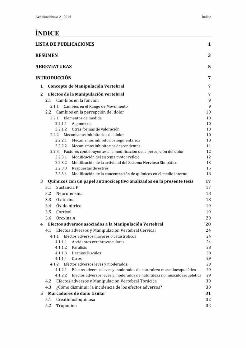
ÍNDICE . LISTA DE PUBLICACIONES . 1
RESUMEN . 3
ABREVIATURAS . 5
INTRODUCCIÓN . 7 1 Concepto de Manipulación Vertebral 7 2 Efectos de la Manipulación vertebral 7 2.1 Cambios en la función 9 2.1.1 Cambios en el Rango de Movimiento 9
2.2 Cambios en la percepción del dolor 10 2.2.1 Elementos de medida 10 2.2.1.1 Algometría 10 2.2.1.2 Otras formas de valoración 10
2.2.2 Mecanismos inhibitorios del dolor 10 2.2.2.1 Mecanismos inhibitorios segmentarios 10 2.2.2.2 Mecanismos inhibitorios descendentes 11
2.2.3 Factores contribuyentes a la modificación de la percepción del dolor 12 2.2.3.1 Modificación del sistema motor reflejo 12 2.2.3.2 Modificación de la actividad del Sistema Nervioso Simpático 13 2.2.3.3 Respuestas de estrés 15 2.2.3.4 Modificación de la concentración de químicos en el medio interno 16
3 Químicos con un papel antinociceptivo analizados en la presente tesis 17 3.1 Sustancia P 17 3.2 Neurotensina 18 3.3 Oxitocina 18 3.4 Óxido nítrico 19 3.5 Cortisol 19 3.6 Orexina A 20
4 Efectos adversos asociados a la Manipulación Vertebral 20 4.1 Efectos adversos y Manipulación Vertebral Cervical 24 4.1.1 Efectos adversos mayores o catastróficos 24 4.1.1.1 Accidentes cerebrovasculares 24 4.1.1.2 Parálisis 28 4.1.1.3 Hernias Discales 28 4.1.1.4 Otros 29
4.1.2 Efectos adversos leves y moderados: 29 4.1.2.1 Efectos adversos leves y moderados de naturaleza musculoesquelética 29 4.1.2.2 Efectos adversos leves y moderados de naturaleza no musculoesquelética 29
4.2 Efectos adversos y Manipulación Vertebral Torácica 30 4.3 ¿Cómo disminuir la incidencia de los efectos adversos? 30
5 Marcadores de daño tisular 31 5.1 Creatínfosfoquinasa 32 5.2 Troponina 32

Achalandabaso A, 2015 Índice
5.3 Mioglobina 32 5.4 Proteína C reactiva 32 5.5 Enolasa neuro-‐específica 33 5.6 Aldolasa 33 5.7 Lactato deshidrogenasa 33
BIBLIOGRAFÍA . 35
PROBLEMA DE INVESTIGACIÓN . 51
Planteamiento del problema 51 OBJETIVOS . 53
MATERIAL Y MÉTODOS . 55
RESULTADOS Y DISCUSIÓN . 61
Artículo I 63
Artículo II 75
Artículo III 85
CONCLUSIONES . 95
AGRADECIMIENTOS . 97

Achalandabaso A, 2015 Lista de publicaciones
1
LISTA DE PUBLICACIONES .
La siguiente memoria de tesis está compuesta por los siguientes artículos
científicos:
I. Plaza-Manzano G, Molina-Ortega F, Lomas-Vega R, Martínez-Amat A,
Achalandabaso A, Hita-Contreras F. Changes in Biochemical Markers of Pain
Perception and Stress Response After Spinal Manipulation. J Orthop Sports
Phys Ther. 2014 Apr;44(4):231–9.
II. Molina-Ortega F, Lomas-Vega R, Hita-Contreras F, Plaza-Manzano G,
Achalandabaso A, Ramos-Morcillo AJ, Martínez-Amat A. Immediate effects of
spinal manipulation on nitric oxide, substance P and pain perception. Manual
Therapy (2014), http://dx.doi.org/10.1016/j.math.2014.02.007
III. Achalandabaso A, Martínez-Amat A, Lomas-Vega R, Plaza-Manzano G,
Camacho MV, Gassó M, Hita-Contreras F, Molina-Ortega F. Tissue damage
markers after a spinal manipulation in healthy subjects: A preliminary report of
a randomized controlled trial. Disease Markers. Volume 2014, Article ID
815379, 7 pages, http://dx.doi.org/10.1155/2014/815379


Achalandabaso A, 2015 Resumen
3
RESUMEN .
La manipulación vertebral es una técnica ampliamente utilizada en el
tratamiento del dolor de origen musculoesquelético desde tiempos de Hipócrates (400
AC).
A pesar de su amplia utilización, aún no se conocen con certeza los mecanismos
por los cuales esta técnica es eficaz. Este hecho, ha dado pie a la especulación y
formulación de teorías que aun habiéndose descartado a través de estudios científicos,
siguen siendo de amplio uso dentro del ámbito clínico.
Cada vez son más las voces que se alzan advirtiendo de los posibles efectos
adversos del uso de estas técnicas. Algunos autores dudan de que sea una técnica inocua,
basándose para ello en los hallazgos de lesiones histológicas en los estudios en
cadáveres.
El objetivo general de esta tesis fue analizar y estudiar los posibles mecanismos
por los que la técnica es eficaz (artículos I y II) y si la técnica en si puede producir
daños en los tejidos (artículo III). Esto se realizará a través del análisis plasmático de
neuropéptidos relacionados con la analgesia y de marcadores de daño tisular.
Los principales resultados de la tesis sugieren que en sujetos sanos: a) La
aplicación de una manipulación vertebral cervical produce un incremento significativo
de la concentración plasmática de neurotensina, oxitocina, cortisol y sustancia P. b) La
aplicación de una manipulación vertebral torácica produce un incremento significativo
de la concentración plasmática de neurotensina y oxitocina. c) La aplicación de una
manipulación vertebral cervical o torácica, no produce cambios significativos en la
concentración plasmática de óxido nítrico. d) La aplicación de una manipulación
vertebral cervical produce un incremento significativo de los umbrales de dolor a la
presión locales (sobre la proyección de la articulación cigapofisaria C5-C6 y sobre el
epicóndilo lateral). d) La concentración de sustancia P previa a la manipulación cervical
es directamente dependiente de los cambios que se producen en los umbrales de dolor a
la presión tras la intervención de una manera positiva. e) La aplicación de una
manipulación vertebral cervical o torácica no produce cambios significativos en la
concentración plasmática de creatinfosfoquinasa, lactato deshidrogenasa, proteína C
reactiva, enolasa neuro-específica, aldolasa, troponina o mioglobina.

Achalandabaso A, 2015 Resumen
En síntesis, los resultados de la presente tesis Doctoral muestran que el empleo
de una manipulación vertebral tanto cervical, como torácica, sobre sujetos sanos
conlleva la liberación de sustancias químicas que pueden relacionarse con mecanismos
inhibitorios descendentes del dolor, pudiendo ser éste uno de los mecanismos que
explican los efectos analgésicos asociados a la manipulación vertebral. También se han
obtenido datos que indican que en sujetos sanos, el uso tanto de una manipulación
cervical como de una manipulación torácica, no parece generar un estrés mecánico los
suficientemente severo para suscitar. Estos hallazgos parecen indicar que los efectos
adversos asociados a la manipulación vertebral no tienen su base en lesiones
histológicas, si no en sistemas complejos del procesamiento de la información o en
procesos patológicos previos del paciente.
Terapeutas capacitados para el uso de las manipulaciones vertebrales deben de
sopesar los costes y los beneficios en cada paciente sobre el cual se vaya a aplicar la
técnica con el fin de minimizar los posibles efectos adversos asociados a la
manipulación vertebral y maximizar su papel antinociceptivo y analgésico.
Palabras clave: manipulación vertebral. Concentración plasmática de químicos.
Umbrales de dolor a la presión. Analgesia y antinocicepción. Efectos adversos.

Achalandabaso A, 2015 Abreviaturas
5
ABREVIATURAS .
ACI
ACV
AINES
ATP
AV
AVB
CPK
ECA
EMG
EMT
ENS
EVA
LDH
MV
MVC
MVL
MVT
NT
OMS
ON
OT
Arteria Carótida Interna
Accidente Cerebrovascular
Antiinflamatorios no Esteroideos
Adenosín Trifosfato
Arteria Vertebral
Arteria Vertebrobasilar
Creatinfosfoquinasa
Estudio Controlado Aleatorizado
Electromiografía
Estimulación Magnética Transcraneal
Enolasa Neuro-Específica
Escala Visual Analógica
Lactato Deshidrogenasa
Manipulación Vertebral
Manipulación Vertebral Cervical
Manipulación Vertebral Lumbar
Manipulación Vertebral Torácica
Neurotensina
Organización Mundial de la Salud
Oxido nítrico
Oxitocina

Achalandabaso A, 2015 Abreviaturas
PCR
PME
RM
SGPA
SNC
SNS
SP
UDP
Proteína C Reactiva
Potencial Motor Evocado
Rango de Movimiento
Sustancia Gris Periacueductal
Sistema Nervioso Central
Sistema Nervioso Simpático
Sustancia P
Umbral de Dolor a la Presión

Achalandabaso A, 2015 Introducción
7
INTRODUCCIÓN .
La fisioterapia es una disciplina cuya finalidad es la disminución de la
percepción de dolor por parte del paciente y la mejora o el mantenimiento de la función.
Para cumplir con estos objetivos la fisioterapia dispone de una gran variedad de técnicas,
entre las cuales se encuentran las técnicas manuales. Dentro de las técnicas manuales,
las técnicas articulares son de amplia utilización. Estas técnicas, pueden ser de baja
velocidad y gran amplitud de recorrido, o de alta velocidad y corta amplitud de
recorrido. Las técnicas de alta velocidad y corta amplitud, son comúnmente
denominadas; manipulación.
1 CONCEPTO DE MANIPULACIÓN VERTEBRAL
La manipulación vertebral (MV) es una técnica que ha sido utilizada desde
tiempos remotos por “recolocadores de huesos”, curanderos, chamanes, barberos y
practicantes. Hipócrates (400 AC) escribió sobre su valor en el tratamiento de las "mal
posiciones" vertebrales. Así mismo, existen datos de la utilización de la MV en China y
Grecia, ya en el periodo que comprende 2700-1500 AC. Hoy en día la MV es utilizada
por fisioterapeutas, quiropractores, osteópatas y médicos1 como parte importante del
tratamiento y manejo en pacientes con dolor de origen musculoesquelético, sobre todo
en las fases agudas y subagudas de dichos proceso2-5.
A pesar de ser una técnica ampliamente utilizada por diversos terapeutas, sigue
sin haber una definición consensuada entre los distintos profesionales, lo que hace que
el propio concepto de MV siga en revisión6. Gatterman en 1994 define la MV como un
procedimiento manual en el cual se realiza un movimiento de alta velocidad y corto
recorrido con la finalidad de mover a la articulación más allá de su rango de
movimiento (RM) fisiológico sin exceder los limites anatómicos de dicho RM7.
2 EFECTOS DE LA MANIPULACIÓN VERTEBRAL
La manipulación ha sido documentada como beneficiosa en el tratamiento del
cuello y de la espalda8. Existe numerosa evidencia de la eficacia de la utilización de la
manipulación en la región cervical para el tratamiento del dolor de origen
musculoesquelético en el cuello5,9,10. No obstante, numerosos clínicos expertos en el
tratamiento manual del dolor de cuello destacan la importancia del examen y del

Achalandabaso A, 2015 Introducción
8
tratamiento de la columna dorsal en pacientes con dolor de cuello, ya que en sí misma,
la relación biomecánica intrínseca existente entre la columna cervical y la columna
dorsal podría ser un factor contribuyente del dolor y de la disfunción de la región del
cuello. También habría que tener en cuenta que la literatura científica previene de las
intervenciones cervicales al final del RM así como, del uso de la manipulación vertebral
cervical (MVC) por el riesgo, aunque bajo, de efectos adversos11. Toda esta información
podría señalar a la manipulación vertebral torácica (MVT) como una alternativa eficaz a
la manipulación cervical en el tratamiento del dolor mecánico cervical.
En la actualidad, encontramos varios trabajos de relevancia que nos hablan de
los beneficios de añadir una MVT al tratamiento del dolor cervical. Por ejemplo,
Cleland y cols.12 en un ensayo clínico aleatorizado compararon la MVT con una MVT
fingida en pacientes con dolor de cuello. El grupo que recibió la MVT mostró mejoras
inmediatas claramente superiores en la intensidad del dolor. En la misma línea, en 2007
otro ensayo clínico aleatorizado, Cleland et al.13 mostraron como la MV aplicada a la
región dorsal alta y media, produjo mayor reducción de dolor y discapacidad que la
movilización pasiva de baja velocidad aplicada a la columna dorsal en pacientes con
dolor cervical subagudo. Igualmente, González-Iglesias et al.14,15 examinaron la
efectividad de añadir la MVT a un tratamiento de termoterapia superficial y
electroterapia analgésica en el manejo del dolor cervical agudo, encontrando en ambos
ensayos mejoras significativas en el grupo de la MVT en términos tanto de dolor como
de discapacidad. En una revisión sistemática reciente, Cross et al.16 concluyen que la
MVT es eficaz en el manejo del dolor cervical agudo y subagudo. En el año 2011 se ha
publicado el primer ensayo en una muestra de pacientes con dolor cervical crónico17
obteniéndose mejoras significativas tanto en la función, como en la percepción del dolor.
Así, el uso de la MV con la finalidad de disminuir el dolor y la discapacidad en los
pacientes está recogido en numerosas guías de práctica clínica18-21.
A pesar de que la MV es una técnica ampliamente utilizada, y que la literatura
consultada respalda el uso de la MV en el tratamiento del dolor y la disfunción de
origen musculoesquelético, aun no se conocen con exactitud los mecanismos
neurofisiológicos a través de los cuales se puede justificar la efectividad de dicha
técnica22. Numerosos investigadores han tratado de arrojar luz sobre esta cuestión,
pudiéndose observar dos líneas o corrientes de investigación. Investigadores que buscan
medir cambios en la función que se pueden justificar como cambios biomecánicos y en

Achalandabaso A, 2015 Introducción
9
el control motor, como es el aumento del RM; e investigadores que buscan determinar
el impacto que la MV tiene sobre la percepción del dolor.
2.1 Cambios en la función
En la actualidad, cada vez con más frecuencia los estudios que intentan arrojar
luz sobre aspectos relacionados con la patología de origen musculoesquelético se
centran en valorar el efecto que esta tiene sobre la función. Valorar cambios en la
función hace posible estimar el potencial efecto que la MV tiene en el tratamiento de
dicha patología. Con esta finalidad se utilizan diversos cuestionarios como el Neck
Disability Index, pruebas funcionales como el test de flexión craneocervical para la
musculatura flexora profunda cervical, o la valoración del movimiento fisiológico
uniplanar tanto pasivo como activo a través de un goniómetro.
2.1.1 Cambios en el Rango de Movimiento
El uso de la evaluación del movimiento fisiológico, tanto activo como pasivo de
la columna cervical, en los pacientes que presentan dolor en la región cervical y/o
cérvico-dorsal, forma parte integral de la exploración. Su registro permite tener un dato
objetivo a través del cual se puede valorar la magnitud de la disfunción del paciente y
del efecto del tratamiento. Tanto es así que la Guía de la Asociación de Médicos
Americana para la evaluación de la discapacidad permanente, cataloga esta medición
como un componente importante de la evaluación23. Dall’Alba et al.24 en un estudio
comparativo, fueron capaces de discriminar entre sujetos con latigazo cervical (dolor y
discapacidad moderados) y sujetos sanos utilizando como único dato la magnitud del
RM. Así mismo, Rudolfsson et al.25 determinaron a través de un estudio transversal, que
los sujetos con dolor cervical crónico tienen una limitación significativa del RM. Parece
ser que en los estudios sobre dolor cervical de origen musculoesquelético, el uso del
RM es la forma de medición objetiva más utilizada, afirmación avalada por varias
revisiones sistemáticas 26-28.
En una revisión sistemática reciente, Cross et al.16 concluyen que la MV
aumenta el RM tanto en pacientes con dolor cervical mecánico agudo, como subagudo.
Martínez-Segura et al.29 en un estudio controlado aleatorizado (ECA) obtuvieron un
aumento significativo del RM cervical tras una MVC. Dunning et al.30 en un estudio
clínico aleatorizado multicentro reciente también obtuvieron un aumento del RM
cervical, tanto al realizar una MVT, como una MVC.

Achalandabaso A, 2015 Introducción
10
2.2 Cambios en la percepción del dolor
2.2.1 Elementos de medida
En la historia clínica del paciente, uno de los datos más relevantes para el
fisioterapeuta es la percepción de dolor por parte del paciente, ya que es el motivo de
consulta más frecuente dentro del ámbito de la fisioterapia31. Para intentar objetivar una
experiencia tan subjetiva, los clínicos se ayudan de pruebas sensoriales cuantitativas
como la sensibilidad al dolor por presión mecánica medida con algometría o escalas
visuales analógicas (EVA), entre otras.
2.2.1.1 Algometría
La determinación de los umbrales de dolor a la presión (UDP) es una de las
modalidades más ampliamente utilizadas en la literatura científica para objetivar los
cambios en la percepción del dolor tras el uso de una intervención sobre un paciente. El
UDP es definido según la Asociación Internacional para el Estudio de Dolor (1986),
como la menor intensidad de estímulo a la presión en la cual un sujeto percibe el dolor32.
La determinación de los UDP a través de un algómetro de presión es una técnica de
medida de la sensibilidad mecánica de los tejidos periféricos que ha mostrado una
excelente reproducibilidad y validez en sujetos sanos33, además de una buena fiabilidad
intraobservador34. Al revisar la literatura científica, son numerosos los artículos
publicados que hacen referencia a cambios en los UDP como consecuencia de una MV
tanto en sujetos asintomáticos35-38, como en sujetos con dolor9,39-41.
2.2.1.2 Otras formas de valoración
La literatura científica recoge diversos métodos de medición del efecto de la MV
sobre la percepción del dolor como las EVA9,30,42 o la determinación del tamaño del
campo receptivo doloroso43.
2.2.2 Mecanismos inhibitorios del dolor
2.2.2.1 Mecanismos inhibitorios segmentarios
Históricamente se ha descrito que una parte de los mecanismos neurofisiológicos
que forman parte de las respuestas de analgésica asociadas a la manipulación parecen
estar justificados mediante la teoría del umbral de la compuerta descrita por Melzack22.
En esta teoría los autores postularon que los estímulos propioceptivos procedentes de

Achalandabaso A, 2015 Introducción
11
las fibras Aβ, de tacto discriminativo, tienden a inhibir la transmisión nociceptiva
(cierran la compuerta) en el asta dorsal de la médula espinal, mientras que la actividad
en las fibras nociceptivas Aδ y C tiende a facilitar dicha transmisión (abren la
compuerta)44. Ihdahl et al.45 en un modelo animal, observaron cómo la estimulación de
los propioceptores capsulares de una articulación cigapofisaria disminuye el reflejo
paraespinal asociado a la estimulación nociceptiva de la cápsula. Así mismo, Malisza et
al.46 en otro modelo animal, encontraron evidencia de mecanismos inhibitorios
segmentarios asociados al uso de terapia manual. Tanto el grupo de George47, como el
de Bialosky48 concluyeron que, en sujetos sanos, el efecto antinociceptivo observado
tras una MV es atribuible al asta dorsal de la médula.
2.2.2.2 Mecanismos inhibitorios descendentes
La literatura científica aporta datos sobre la influencia que tienen estructuras
supraespinales sobre el dolor (corteza anterior cingulada, amígdala, sustancia gris
periacueductal (SGPA) y la región rostral ventromedial del bulbo raquídeo)49. Las
investigaciones más recientes orientan a que los mecanismos centrales de modulación
descendente del dolor parecen ser los principales sistemas fisiológicos involucrados en
las respuestas de analgesia inmediata observadas tras la manipulación. Malisza et al.50
en un modelo realizado en ratas, determinaron a través de resonancia magnética
funcional las áreas cerebrales que se activaban como consecuencia del dolor producto
de una infiltración de capsaicina. Estos autores, observaron cómo, tras una movilización
articular de baja velocidad, disminuye la activación de las áreas cerebrales previamente
relacionadas con dolor. Estos resultados proporcionan confirmación directa de
mecanismos inhibitorios descendentes. Vicenzino et al.51 en sujetos con epicondilalgia
lateral sometidos a movilizaciones articulares de baja velocidad de la columna cervical
observaron cambios en la percepción del dolor y el sistema nervioso simpático (SNS) y
que estos cambios se correlacionaban, lo cual parece sugerir que parte de los efectos
atribuidos a la movilización articular de baja velocidad se deben a mecanismos
modulatorios centrales.
En la MV, estos mecanismos centrales difusos también se confirman al observar
cómo la manipulación dirigida a un segmento diagnosticado como hipomóvil no es más
beneficiosa que la manipulación realizada sobre un segmento elegido al azar, o que
diversos estudios recientes hayan mostrado cambios en la percepción de dolor en

Achalandabaso A, 2015 Introducción
12
regiones a distancia del área que se trató12. Numerosos autores han defendido esta
hipótesis de modulación descendente como el mecanismo responsable de la analgesia
inmediata producida tras la MV. Más concretamente, Skyba et al.52 sugirieron que el
efecto de la MV es una forma de analgesia no opioide dependiente, mediada por
receptores espinales serotonérgicos y noradrenérgicos que utilizan vías inhibitorias
descendentes, “neuronas off”, desde la SGPA, vía bulbo raquídeo rostral ventromedial y
tegmento pontino dorsolateral, hasta el asta dorsal de la medula.
2.2.3 Factores contribuyentes a la modificación de la percepción del dolor
2.2.3.1 Modificación del sistema motor reflejo
La perpetuación de la percepción del dolor se puede justificar por múltiples
mecanismos. Uno de ellos es el mantenimiento del ciclo dolor/espasmo/dolor. Diversos
autores defienden la hipótesis de que una de las claves del éxito de la MV, reside en una
inhibición motora refleja, la cual rompería este ciclo de dolor/espasmo/dolor51,53.
A pesar de la observación de cambios reflexógenos en el sistema motor
asociados a la MV, y de la cantidad de clínicos que se basan en esta premisa para
justificar parte de sus efectos objetivables, no existe consenso acerca de su potencial
efecto inhibitorio o excitatorio reflejo sobre el sistema motor. Para valorar el efecto
motor, se han utilizado diversos métodos:
− La electromiografía (EMG) se utiliza para evaluar y registrar la actividad
eléctrica muscular, de manera que detecta las diferencias de potencial eléctrico de las
fibras musculares al contraerse. Al revisar los cambios en la EMG que se producen tras
una MV, en la literatura científica observamos una tendencia clara a la modificación en
la señal electromiográfica. No obstante, parece que no existe consenso en si la
modificación comparta una disminución o un aumento de dicha señal. Numerosas
investigaciones avalan un aumento de la señal electromiográfica41,54-56, mientras otras
avalan una disminución de dicha señal38,57-60.
− El reflejo Hoffman tiene como finalidad determinar la competencia de la
médula espinal a través de la estimulación eléctrica del músculo, y ha sido ampliamente
utilizado para valorar de forma mínimamente invasiva la competencia del sistema
nervioso. De esta manera nos permite valorar cambios en la excitabilidad de las
neuronas espinales y corticoespinales. Fryer et al.61 en un estudio reciente con sujetos

Achalandabaso A, 2015 Introducción
13
asintomáticos, controlado y aleatorizado, de diseño cruzado, observaron una
disminución significativa del reflejo Hoffman tras una MV, situación que se
correlaciona con una disminución de la actividad a nivel espinal, produciéndose una
disminución significativa de la excitabilidad de la motoneurona alfa, lo que se traduce
un una disminución del tono. El grupo de Dishman et al.57,60,62,63 han obtenido
resultados similares.
− La estimulación magnética transcraneal (EMT) es un método no invasivo
para estimular eléctricamente el córtex cerebral y poder valorar la excitabilidad del
córtex motor y del tracto corticoespinal64, de manera que nos permite estudiar los
efectos de los inputs corticoespinales sobre la excitabilidad de la motoneurona alfa53.
Los potenciales que genera la EMT son referidos como potenciales motores evocados
(PME). La amplitud de estos PME en el músculo diana, refleja cambios en la
excitabilidad de células corticomotoneurales y cambios en la excitabilidad de la
motoneurona alfa. Dishman et al.65 valoraron cambios a nivel central de la excitabilidad
motora tras una MV a través de EMT, observándose un aumento significativo de la
excitabilidad de la motoneurona alfa desde mecanismos postsinápticos.
Una explicación de estas discrepancias de resultados en las mediciones de las
ondas H o de los PME, puede ser la diferencia metodológica de ambos procesos. No
obstante, algunos autores advierten que tras una facilitación central existe la posibilidad
de desencadenar una disminución de la actividad de las motoneuronas periféricas53.
2.2.3.2 Modificación de la actividad del Sistema Nervioso Simpático
Las respuestas que sugieren modificación en la actividad del SNS ante estímulos
aferentes no nocivos, han sido observadas tanto en investigaciones realizadas en
animales como en seres humanos. Numerosos estudios han evaluado los efectos que la
terapia manual tiene sobre el SNS con la finalidad de comprender sus posibles
mecanismos de acción. Modificaciones en la actividad del SNS podrían dar información
de la compleja respuesta que desde el sistema nervioso central (SNC) se le da a la MV.
La medición del ritmo cardiaco y la presión arterial son formas de sencillas medición y
ampliamente utilizadas para la valoración del impacto de una técnica sobre el SNS.
Otras formas utilizadas han sido la medición de cambios en la conductancia y de la
temperatura de la piel. En un ECA reciente, La Touche et al.66 demuestran que tras una
movilización de baja velocidad y gran amplitud de recorrido anteroposterior de la

Achalandabaso A, 2015 Introducción
14
región cervical superior, se produce un aumento significativo de la excitabilidad del
SNS. Para valorar la respuesta del SNS estos autores realizaron mediciones de la
conductancia y la temperatura de la piel, así como el ritmo respiratorio y cardiaco,
obteniendo un incremento significativo tanto de los ritmos respiratorio y cardiaco, como
de la conductancia dermal. Del mismo modo, Sterling et al.67 observaron un aumento de
la excitabilidad del SNS, determinado por un aumento de la conductancia dermal y un
descenso de la temperatura de la piel, al realizar una movilización de baja velocidad y
gran amplitud de recorrido posteroanterior de la región cervical baja. En otro ensayo
clínico, Vicenzino et al.51 observan resultados similares en sujetos con epicondilalgia
lateral crónica que fueron sometidos a movilizaciones rítmicas cervicales. Una revisión
sistemática reciente (2014)68 concluye que la movilización de baja velocidad y gran
amplitud de recorrido tiene como respuesta un incremento de la excitabilidad del SNS
independientemente del segmento movilizado.
Harris y Wagnon69 en su estudio con una muestra de 196 sujetos observaron un
aumento de la temperatura de la piel de la yema de los dedos tras una manipulación
vertebral lumbar (MVL), o una MVC. Así mismo, observaron un descenso de la
temperatura de la yema de los dedos tras una MVT. Debido a estos resultados, estos
autores concluyen que la MV tiene efectos sobre el SNS, y que estos efectos dependen
del segmento que es diana de la técnica.
A pesar de que la mayoría de las publicaciones sobre este tema avalan el
aumento de la excitabilidad del SNS como consecuencia de la terapia manual,
Knutson70 en un ECA, encontró un descenso significativo de la presión sistólica tras una
MVC. Otra muestra de divergencia en los resultados obtenidos tras una MVC la
tenemos en el estudio de Nansel et al.71 donde estos autores no encontraron ningún
efecto significativo sobre la presión arterial o el ritmo cardiaco.
Finalmente, Vicenzino et al. proponen que tras una MV el efecto excitatorio del
SNS y la analgesia inmediata observada se relacionaban de forma significativa72 lo que
sugiere un papel directo del SNS en la modulación del dolor.

Achalandabaso A, 2015 Introducción
15
2.2.3.3 Respuestas de estrés
La evidencia científica pone de manifiesto que el estrés puede producir efectos
analgésicos a través de la activación de mecanismos inhibitorios endógenos del dolor73.
La activación de estos mecanismos es dependiente de la magnitud y la duración del
estímulo estresor al cual es sometido el sujeto de tal manera que, los sujetos expuestos a
situaciones estresantes leves, activan sistemas modulatorios descendentes basados en la
activación del sistema opioide endógeno, mientras que los sujetos expuestos a
situaciones de gran estrés, activan sistemas modulatorios descendentes no opioides74.
También se ha observado que ante una situación de gran estrés la magnitud del tiempo a
la que el sujeto queda expuesto, parece también determinante a la hora de determinar si
la respuesta inhibitoria será opioide dependiente o no. Así, estímulos estresantes
intermitentes promueven la activación de los sistemas inhibitorios descendentes
opioides dependientes75,76, mientras que los estímulos estresantes que se mantienen en
el tiempo promueven los sistemas inhibitorios descendentes no opioides73,76. Esta
analgesia inducida por el estrés es de suma importancia para la supervivencia del
individuo, ya que es generada intrínsecamente en momentos críticos evitando que el
dolor pueda distraer al sujeto. Es decir, la atención se centra más en la amenaza que en
el dolor.
El cortisol es una hormona esteroidea que se ha correlacionado de forma positiva
con la magnitud de estrés. Diversos estudios han intentado relacionar la terapia manual
con cambios en la concentración de cortisol. En un estudio reciente en sujetos sanos
realizado por Lindgren et al. se observó un descenso significativo de los niveles de
cortisol en saliva tras cinco minutos de masaje77. Este dato está en concordancia con
varias revisiones78,79. Moraska et al. en su revisión concluyen que una sesión de masaje
produce una reducción significativa de los niveles de cortisol salivar, y que este efecto
se reproduce con cada sesión. No obstante, parece ser que el efecto no se mantiene en el
tiempo, ni es acumulable.
En un estudio en sujetos sanos realizado por Whelan et al.80 no se encontraron
cambios significativos en los niveles de cortisol en saliva tras una MV. Este resultado
está en concordancia con los resultados obtenidos por Tuchin PJ81
La literatura revisada parece indicar que la terapia manual no tiene un efecto
estresante sobre los sujetos y que el efecto analgésico asociado a la MV no parece ser

Achalandabaso A, 2015 Introducción
16
dependiente de mecanismos inhibitorios descendentes basados en una respuesta de
estrés.
2.2.3.4 Modificación de la concentración de químicos en el medio interno
Existen una gran variedad de mediadores químicos relacionados con el dolor.
Algunos de estos mediadores son secretados desde el SNC con una finalidad analgésica
o de control descendente del dolor, además de tener un papel de diversa importancia
sobre el SNS (noradrenalina y acetilcolina) o la respuesta de estrés (cortisol). Diversos
autores han estudiado con anterioridad el impacto que tiene la MV sobre la producción
de ciertos químicos. Vernon et al.82 observaron un aumento significativo de los niveles
sanguíneos de β-endorfinas a los 5 minutos tras una MVC. Sin embargo, Sanders et al.83
no encontraron cambios significativos en la concentración de β-endorfinas tras una
MVL. Así mismo, Christian et al.84 tampoco observaron diferencias significativas en la
concentración sanguínea de β-endorfinas tras una MVT o una MVC tanto en sujetos
asintomáticos, como sintomáticos.
Por otro lado, Nansel et al.71 estudiaron el efecto de una MVC sobre la
concentración de catecolaminas, no observando cambios significativos tras la
intervención.
Teodorczyk-Injeyan et al.85 estudiaron el efecto de una MVT sobre la
producción de citoquinas proinflamatorias, observando una disminución significativa de
la síntesis de las mismas tras la intervención. En una línea parecida, Roy et al.86
observaron en sujetos con dolor lumbar crónico una tendencia a la normalización de
interleukina 6 y proteína C reactiva al someterse a una MVL.

Achalandabaso A, 2015 Introducción
17
3 QUÍMICOS CON UN PAPEL ANTINOCICEPTIVO ANALIZADOS EN
LA PRESENTE TESIS
Diversos mediadores químicos secretados en sangre han sido relacionados tanto
con la nocicepción, como con la antinocicepción. De hecho, alguno de estos químicos
tienen un rol dual dependiente de la concentración y del contexto en el que se produce
su secreción.
3.1 Sustancia P
La sustancia P (SP) es un neuropéptido que se ha relacionado con la
proliferación celular, la producción de citoquinas, la vasodilatación y el aumento de la
permeabilidad vascular87. Estas respuestas proinflamatorias son causa de inflamación
neurogénica, la cual está asociada a la nocicepción y a la hiperalgesia88. Larson et al.89
en su estudio advierten del papel pro-nociceptivo de la SP en sujetos con síndromes de
dolor crónico como la fibromialgia, de manera que la SP contribuiría a la
sensibilización del asta dorsal, reduciendo los umbrales de dolor de estos sujetos.
También se ha observado que la SP tiene propiedades antinociceptivas, de hecho
se ha observado que tiene un potente efecto analgésico de larga duración al ser
administrada intraperitonealmente e intracranealmente en ratas90,91. Diversos autores,
sostienen que la SP puede actuar sobre la lamina V del asta dorsal produciendo un
efecto inhibitorio, de manera que se produzca un efecto analgésico sobre el
procesamiento de la señal nociceptiva92,93. Nakatsuka et al.92 interpretan este hecho
como un mecanismo de feed-forward inhibitorio de la actividad del asta dorsal,
concluyendo estos autores que la SP tiene un papel fundamental en el reclutamiento de
actividad inhibitoria para el procesamiento de la nocicepción. La relación entre la
concentración de SP y las terapias manuales sigue siendo controvertida, mientras al
masaje se le otorga un papel en la disminución de la concentración de SP en sujetos con
fibromialgia94, en la MV se observan incrementos de la concentración de SP95 o
ausencia de cambios en dicha concentración85. Estas discrepancias pueden ser fruto de
diferencias metodológicas.
Achalandabaso A, 2015 Introducción
18
3.2 Neurotensina
La neurotensina (NT), es un neuropéptido con un amplio rango de acciones entre
las cuales está la antinocicepción96. Esta afirmación se sustenta anatómicamente por la
alta densidad de receptores de NT localizados en la SGPA y la región rostral
ventromedial del bulbo raquídeo, ambas estructuras implicadas en mecanismos
descendentes. Además, un estudio realizado por Williams et al.97 realizado con
microdiálisis en ratas, mostró que la NT era liberada de forma endógena en la SGPA al
provocar una lesión inflamatoria. La NT, al igual que en la MV, tiene un efecto
analgésico independientes de la naloxona de manera que este efecto no es opiáceo
dependiente. Este efecto antinociceptivo se ha corroborado al inyectar el neuropéptido
en varias áreas cerebrales98. Así mismo la liberación de este neuropéptido parece jugar
un papel importante en la analgesia que se le atribuye a la electroacupuntura99.
3.3 Oxitocina
La oxitocina (OT), es un neuropéptido el cual desarrolla un potente control
antinociceptivo al regular de forma tónica a la SGPA, al núcleo central de la amígdala y
al núcleo del rafe magnus100. Ante estímulos nociceptivos, se ha observado su liberación
en la médula espinal desde proyecciones descendentes del eje hipotálamo-hipofisario.
Este efecto analgésico es ejercido a través de incrementar la liberación de péptidos
opioides como leucina-encefalinas, metionina-encefalinas o beta-endorfinas en la
SGPA101. Además de su efecto positivo sobre la tasa de secreción de péptidos opioides,
recientemente se ha demostrado que esta acción antinociceptiva está mediada en parte,
por un incremento de la inhibición sináptica de las láminas más superficiales de la
médula espinal102. Un estudio reciente realizado por Morhenn et al103 ha mostrado
incrementos significativos en la concentración sanguínea de OT tras 15 minutos de
masaje con una intensidad moderada.
Aparte de los efectos antinociceptivos, la OT puede producir efectos "antiestrés"
como la disminución de la presión arterial y de los niveles de cortisol, y promueve el
crecimiento y reparación de los tejidos104. También se ha observado que la
administración de OT en ratas induce un efecto antiinflamatorio105.

Achalandabaso A, 2015 Introducción
19
3.4 Óxido nítrico
El óxido nítrico (ON) es una pequeña molécula considerada como el mayor
vasodilatador local106. Se trata de un gas con la capacidad de difundir, y que reacciona
rápidamente con el oxígeno para formar derivados de sí mismo como son el nitrito y el
nitrato107. Esta pequeña molécula tiene dos papeles de gran importancia, por un lado
media en la supervivencia celular108, y por otro en la nocicepción109. Luo y Cizkova en
una revisión sistemática reciente advierten del efecto beneficioso de la liberación de
pequeñas cantidades de ON durante la inhibición de las vías nociceptivas110. No
obstante, Aley et al. en un modelo animal concluyen que la liberación de ON contribuye
a la hiperalgesia111. Estos resultados aparentemente contradictorios nos dan una idea del
complejo papel que juega el ON en la nocicepción. La liberación de ON por los tejidos
endoteliales y las células sanguíneas ha sido demostrada ante estímulos mecánicos
fisiológicos como es la puesta en tensión por estiramiento de los tejidos112,113. Su
liberación también ha sido demostrada tras la aplicación de diferentes formas de terapia
física como son el masaje103 o la acupuntura114, pudiendo ser este uno de los
mecanismos a través los cuales estas terapias producen efectos analgésicos.
3.5 Cortisol
El cortisol es una hormona esteroidea producida por la glándula suprarrenal en
situaciones de estrés115. Su liberación produce disminución de edemas y del dolor a
través de la inhibición de las primeras etapas del proceso inflamatorio. Altos niveles de
cortisol en sangre promueven la reparación tisular a través de una facilitación de la
gluconeogénesis80. Determinar los cambios en la concentración de cortisol parece ser un
buen indicador de estrés, ya que un estudio realizado por Basset et al.116 demostró una
correlación positiva entre los niveles de estrés y la cantidad de cortisol secretado. La
concentración de cortisol ha mostrado ser influida por la terapia manual, ya que
diferentes estudios han observado una disminución de su concentración tras una terapia
manual basada en masaje77-79. No obstante, diferentes estudios parecen indicar que la
concentración de cortisol no es influenciada por la MV80,81,84.

Achalandabaso A, 2015 Introducción
20
3.6 Orexina A
La orexina es un neuropéptido descubierto recientemente (1998) que secretado
por el hipotálamo participa de forma importante en la toma de alimentos, la homeostasis
energética, los procesos de adicción y búsqueda de recompensa, y la promoción del
estado de vigilia117. Se han identificado proyecciones orexinérgicas tanto en la SGPA,
como en la lámina I y X del asta dorsal de la médula o el ganglio de la raíz dorsal118-121.
Además, se ha localizado el receptor para la orexina A en las fibras C a su llegada a la
lámina I122.
Existe evidencia del papel de la orexina A en la regulación del procesamiento
nociceptivo a través de mecanismos tanto espinales como centrales, ya que la
administración intratecal o intraventricular de esta sustancia elimina tanto la alodinia
mecánica como la hipersensibilidad térmica en múltiples modelos de dolor123,124. En
modelos de dolor neuropático, Jeong and Holden125 han mostrado que la orexina ejerce
un papel antinociceptivo. Este papel antinociceptivo no parece estar mediado por el
sistema opioide, ya que la analgesia derivada de la administración de orexina A no
revierte con la naloxona123.En un estudio reciente usando ratones se ha mostrado un
incremento significativo de orexina A al aplicar electroacupuntura126.
4 EFECTOS ADVERSOS ASOCIADOS A LA MANIPULACIÓN
VERTEBRAL
El hecho de que exista evidencia de su eficacia en el manejo de ciertos dolores
de origen musculoesquelético, y que en una reciente revisión sistemática se haya
establecido una relación coste-beneficio positiva, tanto si se utiliza de forma aislada o
asociada a otros tipos de terapia como pueden ser otras formas de terapia manual o
ejercicio físico 127, hace tentadora la idea de que pueda ser el tratamiento de elección en
pacientes con dolor de origen musculoesquelético. No obstante, es importante destacar
que cada vez son más las voces que se alzan para advertir de que su utilización puede
estar asociada a una serie de efectos adversos para la salud del paciente.
La Organización Mundial de la Salud (OMS) define un efecto adverso como
“cualquier respuesta nociva, no intencionada, que se produce a dosis habituales para la
profilaxis, diagnóstico, o tratamiento128. Esta definición ha sido tomada prestada del
contexto farmacéutico. Nykoliation and Mierau129, definen efecto adverso como

Achalandabaso A, 2015 Introducción
21
“cualquier situación con relación causal probada de una acción o tratamiento que resulta
en detrimento del sujeto”.
En la literatura científica, el primer caso documentado de aparición de efectos
adversos asociados a la MV se publica en 1907, donde Roberts et al.130 describen una
fractura-luxación del atlas. Posterior a esta publicación varios estudios de casos y
algunos estudios retrospectivos han puesto en entredicho la seguridad de la MV,
obteniéndose pruebas de la posible de asociación de efectos adversos a la MV. No
obstante, estos efectos adversos rara vez llegan a ser mortales131-133.
Debido a la gran variedad de efectos adversos descritos en la literatura tras una
MV, se hace imprescindible una taxonomía que nos permita jerarquizar dichos efectos
adversos para poder realizar una valoración más fiable de la relación coste-beneficio
asociada al uso de la MV en el tratamiento del dolor y la disfunción de origen
musculoesquelético.
En un estudio reciente basado en una modificación del método Delphi, Carnes et
al.134 llegan a una taxonomía de dichos efectos adversos asociados a la terapia manual,
clasificándolos como:
− Efectos adversos mayores o catastróficos: se caracterizan por perpetuarse
en el tiempo, pudiéndose observar a medio y largo plazo. La sintomatología asociada
puede considerarse entre moderada y severa. Normalmente el propio efecto adverso
necesita de tratamiento para su mejora o resolución. Estos autores los consideran como
inaceptables y pueden ser causa de invalidez o defunción.
− Efectos adversos moderados: se caracterizan por perpetuarse en el tiempo,
pudiéndose observar a medio y largo plazo. La sintomatología asociada se identifica
como moderada. Normalmente el paciente requiere de tratamiento para la mejora o
resolución del efecto adverso, aunque las consecuencias de estos efectos tienen
características autolimitantes en el tiempo.
− Efectos adversos leves: se caracterizan por una sintomatología leve y de
corta duración, no considerándose de importancia. La función del paciente no se ve
comprometida. Sus efectos son transitorios, no necesitando de tratamiento para su
resolución ya que las consecuencias son autolimitantes.

Achalandabaso A, 2015 Introducción
22
El grueso de la literatura científica que relaciona efectos adversos a la terapia
manual hace referencia a efectos adversos producidos por técnicas articulares, más
concretamente a MV. En una revisión sistemática reciente, Carnes et al.135 estiman la
incidencia de efectos adversos leves o moderados tras un tratamiento manual en un 41%,
estableciéndose la mayor parte de ellos en las primeras 24 horas tras el tratamiento y
resolviéndose a las 48 horas tras el tratamiento. En la misma línea encontramos un
estudio prospectivo también reciente que establece que un 48% de los pacientes que
reciben MV describen un recrudecimiento de los síntomas o un síntoma nuevo tras una
primera intervención, y que hasta un 26% de dichos pacientes refieren algún efecto
adverso tras la segunda o tercera intervención. Estos efectos adversos se establecen en
las primeras 48 horas tras la intervención en el 90% de los pacientes y son leves a
moderados. Los efectos adversos más observados en este estudio son dolor de origen
musculoesquelético o efectos relacionados con causas musculoesqueléticas, 75% y 72%
respectivamente. También describen efectos adversos que no se relacionan con causas
musculoesqueléticas como cansancio, mareo, nauseas o acufenos en un 19% de los
sujetos136. Otro estudio prospectivo realizado en Noruega por Senstad et al.137 utilizando
como muestra 1.058 pacientes y 4.712 tratamientos, establece que el 55% de los sujetos
sometidos a MV referirán al menos un efectos adverso. Sin embargo, sólo el 25% de los
tratamientos tiene como resultado al menos un efecto adverso, y el 5% más de un efecto
adverso. De estos efectos, el 64% tienen comienzo a las 4 horas tras el tratamiento, y de
este 64%, el 74% desaparecen dentro de las primeras 24 horas. Tan solo el 20% de estos
efectos tienen comienzo a los 10 minutos del tratamiento. Parece ser que la aparición de
estos efectos adversos se relacionan con el género y con la duración de los síntomas, de
manera que, las mujeres tienen mayor probabilidad de sufrir un efecto adverso que los
hombres, así como pacientes con sintomatología de larga evolución138.
Del estudio de Senstad et al. también se obtiene que el 50% de los efectos
adversos asociados a la MV se relacionan con molestias locales, de manera que el 35%
de los pacientes que refieren efectos adversos serán considerados leves, el 50%
moderados, el 14% mayores y el 1% catastróficos. Otros efectos adversos asociados a la
MV que describen estos autores fueron cefalea (12%), sensación de cansancio (11%),
dolor en otra zona distinta (10%), mareos y nauseas (5%)137.
Walker et al.139 en un ECA reciente concluyen que el riesgo relativo de padecer
un efecto adverso tras una MV es similar al que se obtiene al utilizar un tratamiento

Achalandabaso A, 2015 Introducción
23
placebo, valorando que una amplia proporción de los efectos adversos experimentados
tras un MV puede ser debida al curso natural de la sintomatología del paciente.
Sin duda alguna los efectos adversos mayores y catastróficos descritos en la
literatura a pesar de su baja incidencia son una clara llamada de atención para realizar
una evaluación del ratio coste-beneficio de la MV haciendo especial hincapié en el
análisis de la aparición de efectos adversos mayores. En este sentido, Rubinstein et al.136
a pesar de tener una muestra considerable (n=529), no observaron ningún efecto
adverso mayor asociado a la MV. También Carnes et al.135 obtienen una estimación del
riesgo de padecer un efecto adverso mayor asociado a una MV muy bajo, estimando que
un 0,01% de los pacientes, o en un 0,007% de los tratamientos tendrá asociado un
efecto adverso mayor. Para entender la magnitud del riesgo de sufrir un efecto adverso
mayor tras una MV, se hace necesario realizar una comparación con otras formas de
terapia. Por ejemplo, se estima que la MVL es 37.000-148.000 veces más segura que el
consumo de antiinflamatorios no esteroideos (AINES) y 55.500-444.000 veces más
segura que la cirugía en el tratamiento de la hernia discal lumbar140. Así mismo, Fries en
1992, estimó que aproximadamente uno de cada 220 sujetos con dolor de origen
musculoesquelético medicados con AINES será hospitalizado por complicaciones
gastrointestinales y que uno de cada 2.200 fallecerá por una reacción adversa asociada
al consumo de estos141. Aunque la incidencia de aparición de efectos adversos leves
tras una MV es alta, realizar una valoración despreciativa del uso de la MV en base a
este dato puede ser engañosa, ya que estos investigadores también establecen que el
riesgo relativo de padecer un efecto leve a moderado debido a un tratamiento manual es
similar al del ejercicio físico, y menor que el de tomar AINES135.
A pesar de su amplia utilización a lo largo de la historia, de haber sido
documentada como beneficiosa en el tratamiento del dolor de cuello y espalda baja, y de
que en las últimas décadas se haya incrementado la frecuencia de su uso142, son cada
vez más los estudios que advierten de que la MV no es una técnica inocua para el
paciente y que puede estar asociada a efectos adversos de diversa índole.
Achalandabaso A, 2015 Introducción
24
4.1 Efectos adversos y Manipulación Vertebral Cervical
En el raquis, la columna cervical es la región que más incidencia tiene de efectos
adversos que puedan relacionarse con una MV. En una encuesta realizada en Sudáfrica
a fisioterapeutas se observó que el 92% de los efectos adversos que se producen tras una
manipulación se relacionaba con la MVC143. Eriksen et al.144 en un estudio multicentro
prospectivo estimaron que un 53% de los sujetos sometidos a una MVC alta referirá al
menos un efecto adverso.
Con el fin de poder valorar de forma más congruente el riesgo real de la
utilización de la MVC vamos a utilizar la taxonomía anteriormente descrita.
4.1.1 Efectos adversos mayores o catastróficos
Al realizar una revisión de la literatura científica, la columna cervical es la
región que más casos descritos de la asociación entre MV y efectos adversos severos o
catastróficos tiene. Thiel et al. estimaron en base a su estudio prospectivo que el riesgo
de padecer un efecto adverso mayor o catastrófico tras una MVC es de 6 cada 100,000
MVC 11. De estos efectos adversos, los catastróficos son los que más debate y
especulación han producido al poder ser causa de defunción. En la literatura
encontramos dos posturas muy enfrentadas. Por un lado, la que advierte del gran peligro
que conlleva el uso de esta técnica, y que por lo tanto, debería de evitarse o prohibirse
su utilización133,145,146. Por otro, autores que defienden la dificultad para poder
correlacionar directamente dichos eventos con la MV, o que defienden que la incidencia
de dichos eventos es menor que la incidencia obtenida al usar otros tipos de
intervenciones terapéuticas que son de aceptado uso por la comunidad
científica136,137,147-149. Entre los efectos adversos catastróficos, los accidentes
cerebrovasculares son sobre los que más atención y debate hay.
4.1.1.1 Accidentes cerebrovasculares
Numerosos son los procesos fisiopatológicos que pueden llevar a un accidente
cerebrovascular (ACV), entre ellos aterosclerosis, una hemorragia secundaria a un
aneurisma o a una malformación arteriovenosa, o disección arterial. En la columna
cervical, la disección arterial es un término que engloba tanto a la arteria carótida
interna (ACI) con a la arteria vertebral (AV)150. La disección arterial se produce por un
desgarro de la túnica íntima del endotelio del vaso, pudiendo afectarse o no la túnica
media, para que posteriormente el flujo sanguíneo acceda al espacio entre túnicas, lo

Achalandabaso A, 2015 Introducción
25
que provocará la disección y la producción de un seudoaneurisma. Posteriormente podrá
provocarse un trombo y la estenosis parcial o total del vaso. La MVC y los ACV por
disección de la AV o la ACI han sido relacionados desde el informe de Thornton en
1934. Desde entonces, varios estudios han revisado la relación existente entre la MVC y
los ACV, estableciéndose un relación de 1 entre 300,000 a 1 entre 5.85 millones de
manipulaciones cervicales151. Patijn en 1991 determinó que el 65,1% de todas los
efectos adversos catastróficos relacionados con la MVC eran debido a la lesión de la
AV152.
Existe evidencia de que los movimientos en rotación de la columna cervical,
sobre todo a finales del RM, estresan más a la AV que los movimiento en flexión lateral
de la columna cervical153. Numerosos autores, entre ellos Nicolai Bogduk, afirman que
las fuerzas de estrés por distensión de dichas arterias al someterse a estos
procedimientos pueden ser suficientes para producir la disección de la AV y/o la ACI.
No obstante, Symons et al. en un estudio anatómico en cadáveres demuestran que el
segmento V3 (tramo entre el foramen de C2 y el codo que realiza la arteria
vertebrobasilar (AVB) en C1 para llegar al foramen magno) durante una MVC
orientada a los segmentos C2-C4 se estira un 3-5% de su longitud. Este mismo tramo
llega a estirarse hasta un 13% en el movimiento cervical en rotación realizado de forma
pasiva. Además, estos autores establecen que el fracaso de la íntima de la AVB se
produce con fuerzas tensiles de un 58-62%154. Así mismo, Herzog et al. establecen un
estiramiento de un 2% en la ACI durante una MVC, un estiramiento de un 7% al
realizar una rotación contralateral, y que la ACI se diseca con un estiramiento de un
59% de su longitud de reposo155. Siguiendo estas premisas biomecánicas, Symons et al.
discuten la imposibilidad de que las fuerzas a las que se ven sometidas tanto la AVB y
la ACI durante una MVC sean suficientes para producir una ACV en un sujeto que no
tenga una lesión preexistente (disección prodrómica) o una condición preexistente
(enfermedad del colágeno o agenesia de la íntima), y que si la MVC puede
desencadenar un ACV, en ese mismo sujeto, los movimientos cervicales normales
pueden así mismo desencadenar dicho acontecimiento156.
Aproximadamente el 80% de los sujetos con disección de la AV y/o de la ACI
refieren cefalea occipital intensa y dolor cervical posterior ipsilateral como primeros
síntomas157-159, pudiendo ser estas confundidas con un cuadro de dolor de origen

Achalandabaso A, 2015 Introducción
26
musculoesquelético. De esta forma, al tratar la columna cervical se puede acelerar el
proceso, o incluso un proceso autolimitante transformarse en uno catastrófico.
Según Murphy et al. la evidencia actual parece sugerir que no existe una fuerte
fundamentación en una relación causal entre la MVC y los ACV, y que una explicación
a los casos de ACV tras MVC puede ser que sujetos que están padeciendo un episodio
de ACV acuden al fisioterapeuta para mejorar su cervicalgia o cefalea150. De manera
que tras la visita al fisioterapeuta el ACV sigue su curso natural.
Como consecuencia de la disección de la AV o de la ACI, se puede producir un
trombo. El mecanismo, sería un mecanismo fisiológico de reparación del endotelio,
donde la agregación plaquetaria daría lugar al trombo160. La aterosclerosis y las
enfermedades cardiovasculares relacionadas con la producción de placas de ateroma se
han relacionado con la producción de ACV. A pesar de que en teoría es posible que un
émbolo pueda liberarse de una placa arterioesclerótica tras una MVC, no existen
pruebas de que se haya dado ningún caso. También son numerosas las voces que
postulan cambios degenerativos en la columna cervical como factor precipitante del
ACV, al poderse lesionar la AV o la ACI por un osteofito. Sin embargo, Grant en
199472 describió que en 23 de los 26 casos de complicaciones vasculares donde se
realizaron radiografías sólo se observaron hallazgos normales o degenerativos leves. Así
mismo, Patijin encontró alteraciones radiográficas degenerativas sólo en 9 de los 59
casos de lesión de la AV tras una MVC, pero fueron sólo alteraciones leves y no se
observaron osteofitos152. Además, el hecho de que la edad media de los pacientes en
riesgo de ACV por MVC es relativamente baja (30 a 45 años), hace que numerosos
autores descarten estos mecanismos como fuentes de un ACV por MVC.
A pesar de las fatales consecuencias de dicho efecto adverso, todavía existe
controversia sobre la magnitud del riesgo de sufrir un ACV por causa directa de una
MVC alta. Mientras que algunos autores advierten de la fuerte asociación existente
entre el uso de estas técnicas y los ACV161, otros sugieren que no existe dicha magnitud
de asociación147.
Cassidy et al. en un estudio poblacional de casos-controles y casos-cruzados
(case-crossover) agrupados dentro de la misma investigación realizada en los hospitales
de Ontario desde 1993 hasta 2002 con una población de 109,020,875 personas a lo largo
de los 9 años del estudio, encuentra una asociación entre la MVC y los AVC. No

Achalandabaso A, 2015 Introducción
27
obstante, esta asociación no es distinta a la existente entre visitar un médico general y
padecer un ACV. Estos autores discuten como hipótesis la posibilidad de que al no ser
posible que los sujetos sufran de un ACV debido al tratamiento del médico general,
parece razonable pensar que los sujetos que van al médico general con dolor cervical
y/o cefalea y que desarrollan un ACV estaban en el inicio de un ACV y no que la propia
intervención produjese la ACV. Al ser las asociaciones similares, estos autores
concluyen que no existen un exceso de riesgo de padecer un ACV tras una MVC162.
En una revisión sistemática reciente llevada a cabo por Haynes et al. se concluye
que no existe evidencia de una fuerte asociación entre la MVC alta y el riesgo de sufrir
un ACV. No obstante, estos autores también concluyen que no existe evidencia de la
falta de asociación entre ambos acontecimientos148. Esta falta de consenso podría ser
explicada por una posible relación causal positiva en sujetos con predisposición a
padecer un ACV debido a agenesias de la íntima tanto en la AV como en la ACI, los
cuales pueden sufrir un ACV como consecuencia de la lesión de la íntima en
actividades de la vida diaria, como pueden ser el ejercicio físico aeróbico o las
relaciones sexuales163.
En una comunidad de Francia, la incidencia anual de disección espontánea de la
ACI se estima en tres por cada 100,000 sujetos164. Schievink (2001) establece la
incidencia anual de disección espontanea de la AV en uno por cada 100,000 a 1.5 por
cada 100,000 sujetos159. Aunque en la actualidad las causas no traumáticas son
desconocidas, se especula que puede haber un factor de predisposición genética,
asociándose la disección a agenesias del endotelio vascular, así como a enfermedades
como el síndrome de Ehlers-Danlos tipo IV o el síndrome de Marfan165,166. Un dato que
parece apoyar un factor genético es el hecho de que aproximadamente el 5% de los
sujetos con disección espontánea de la ACI o la AV tienen al menos un miembro en su
familia que ha sufrido de un episodio similar167. Entre los eventos no traumáticos
asociados a la disección de estas arterias encontramos eventos triviales como estornudar,
vomitar, toser, mantener relaciones sexuales, pintar techos o practicar yoga159,163.
En el 80% de los casos de disección de la AV o de la ACI en los periodos
iniciales sólo cursan con una importante cefalea y dolor cervical. Dado lo cual, parece
comprensible que esta sintomatología pueda confundirse con sintomatología de origen
musculoesquelético y que el uso de una MVC pueda iniciar un embolismo que resulte

Achalandabaso A, 2015 Introducción
28
fatal, o pueda perpetuar una situación que en principio pudiese ser autolimitante,
conduciendo a graves secuelas o muerte148.
A pesar del gran debate que suscita, y de que son numerosos los autores que
apoyan claramente ambas teorías, la mayoría de las revisiones sistemáticas realizadas en
la literatura muestran una falta de datos concluyentes con los que afirmar o negar la
existencia de una relación causa efecto entre la MV y los ACV148.
Por último, recordar que para establecer un ratio coste-beneficio más
realista, sería adecuado realizar una comparación de incidencias de este tipo de efectos
adversos asociados a la MVC, con efectos adversos de índole similar asociados a otros
tipos de medidas terapéuticas que se consideran seguras. En una revisión realizada por
Dabbs V y Lauretti WJ se concluye que la MVC es varios cientos de veces más seguro
en el tratamiento del dolor cervical que el uso de AINES, y que además no existe
evidencia que indique que el uso de AINES sea más efectivo que la MVC en el manejo
del dolor cervical168.
4.1.1.2 Parálisis
Otro efecto adverso severo asociado con la MVC seria la pérdida parcial o total
de la función motora. Recientemente han sido publicados en prensa y en publicaciones
científicas, casos de sujetos que tras someterse a una técnica de MVC han desarrollado
algún grado de parálisis en una o varias de sus extremidades169-171.
4.1.1.3 Hernias Discales
La MVC se ha asociado con el agravamiento o inicio de una hernia discal que
precipite el recrudecimiento o el inicio de una radiculopatía o mielopatía cervical172-175.
A pesar de que en la literatura se describen casos de agravamiento o inicio de
radiculopatías o mielopatías cervicales tras un MVC, estos datos están basados en
escasos estudios de casos lo que hace difícil establecer una relación causa-efecto y
generalizarla. Autores como Murphy y Beres, advierten que es complicada la relación
ya que el curso de la afectación del paciente por la que busca el tratamiento puede ser el
desencadenante de la radiculopatía o mielopatía176.

Achalandabaso A, 2015 Introducción
29
4.1.1.4 Otros
En la literatura científica se describen como efectos adversos mayores asociados
a una MVC; fractura cervical (proceso odontoideo)130,177, parálisis diafragmática178,179,
síndrome de hipotensión intracraneal180, hematoma epidural cervical181, pérdida de
líquido cefalorraquídeo182 y problemas oftalmológicos183.
4.1.2 Efectos adversos leves y moderados:
La incidencia de efectos adversos moderados se estima en uno de cada 40,000
MV184. Los efectos adversos no mayores o catastróficos que se correlacionan con la
MVC pueden ser de naturaleza musculoesquelética o de naturaleza no
musculoesquelética.
4.1.2.1 Efectos adversos leves y moderados de naturaleza musculoesquelética
La mayor parte de los efectos adversos leves y moderados que se correlacionan
con la MVC son de naturaleza musculoesquelética, siendo de más predominio el
recrudecimiento del dolor o la aparición de dolor musculoesquelético referido. Thiel et
al.11 en un estudio prospectivo basado en una encuesta nacional entre quiropractores del
Reino Unido concluyen que los efectos adversos que se producen pronto tras una MVC
son predominantemente de origen musculoesquelético. El efecto adverso más frecuente
es el recrudecimiento del dolor cervical (7.3% de los casos). Otros efectos adversos
observados por este grupo son: dolor en la extremidad superior (4.8%), disminución de
la movilidad del cuello y/o la extremidad superior (3.9%). En la misma línea, en un
estudio prospectivo realizado por Eriksen et al.144 se observa que el 38.1% de los
efectos adversos eran de naturaleza musculoesquelética con un valor 3.4 en la escala
numérica de dolor de 11 puntos (donde 0 corresponde a ausencia de dolor y 10 a dolor
máximo), y sólo el 5.1% de los pacientes refirieron un efecto adverso ≥8 en la escala
numérica de dolor. Así mismo, estos autores también observan que el 54% de los
efectos adversos tuvieron un comienzo dentro de las primeras 24 horas.
4.1.2.2 Efectos adversos leves y moderados de naturaleza no musculoesquelética
Numerosos efectos adversos de naturaleza no musculoesquelética se han
asociado al uso de la MVC. A pesar de que los efectos adversos de naturaleza simpático
refleja parecen predominar, ciertos efectos adversos parecen relacionarse con
mecanismos centrales. Thiel et al.11 observaron efectos adversos que pueden

Achalandabaso A, 2015 Introducción
30
relacionarse con sintomatología de origen neurológica como desvanecimiento (3.9%),
vértigos (1.3%) y desorientación (1.1%). Estos autores también encontraron tras 7 días
cefalea (4%), parestesias en la extremidad superior (1.5%) y
desvanecimiento/vértigo/desorientación (1.3%). Eriksen et al.144 también apreciaron
cansancio (10.4%), dolor irradiado (6.3%), inestabilidad (4.9%) y cefalea (4.2%).
Advirtiendo que síntomas de naturaleza no musculoesquelética como la nausea, el
vértigo o la inestabilidad son raros, constituyendo no más del 5% de todos los efectos
adversos descritos en la literatura.
4.2 Efectos adversos y Manipulación Vertebral Torácica
La información registrada en la literatura científica que relaciona la MVT con
efectos adversos es escasa. Esto puede deberse a una falta de registro de dichos eventos,
o a ser una región que debido a sus características anatómicas, la utilización de MV no
conlleva la aparición de efectos adversos.
Casi todos los efectos adversos registrados tras una MVT se basan en estudios
de casos. Un caso reciente relacionado con un efecto adverso severo es descrito por
Struewer et al.184 donde un varón de 17 años sufrió un hematotórax secundario a una
MVT sin la existencia de fracturas vertebrales asociadas. Otro caso de efecto adverso
severo tras una MVT es descrito por Masneri et al.185, y consiste en la aparición de un
neumotórax secundario a la utilización de una MVT en una mujer de 20 años.
Nykoliation y Mierau129 describen el caso de una mujer de 32 años con dolor
escapulotorácico crónico secundario a una MVT.
4.3 ¿Cómo disminuir la incidencia de los efectos adversos?
Con el fin de disminuir la frecuencia y magnitud de los efectos adversos
asociados a la MV, numerosos autores proponen unas pautas a la hora de realizar una
MV72:
- No realizar múltiples manipulaciones a lo largo de una sesión. Parece ser
que realizar varias intervenciones en una única sesión se asocia con un aumento de la
probabilidad de que se produzca un efecto adverso.
- No realizar una MV sin haber determinado previamente los efectos de la
movilización articular pasiva de baja velocidad sobre ese segmento. Numerosos autores
han puesto en evidencia que algunos sujetos son susceptibles de padecer efectos

Achalandabaso A, 2015 Introducción
31
adversos tras la utilización de maniobras articulares de baja velocidad. De manera que
parece lógico realizar estas maniobras previas a la utilización de una MV ya que con
estas maniobras se puede controlar la magnitud de la dosis.
- Realizar una reevaluación del paciente para determinar la eficacia de la
MV en la mejora de la sintomatología del paciente y si apareció algún efecto adverso
atribuible a ésta que contraindique una nueva exposición a la técnica, o la necesidad de
realizarla sobre otra región vertebral.
- Evitar utilizar la MV utilizando un amplio rango de movimiento
fisiológico. Para esto se propone utilizar al menos dos movimientos fisiológicos para la
puesta en tensión. En la columna cervical superior, evitar las técnicas en rotación pura o
rotación más extensión. Klougart et al186. concluyeron que en los procedimientos
basados en rotación de la columna cervical alta se tienen más probabilidad de padecer
un efecto adverso moderado a severo, que en los procedimientos sin rotación en esta
misma región.
- Evitar las técnicas con un componente de tracción (columna cervical). Ya
que este componente pone en mayor estrés el componente vascular.
- Aplicar la MV con la menor cantidad de fuerza necesaria para la
consecución del objetivo de la técnica. Tanto Grieve como Bogduk advierten de la
relación que existe en los casos publicados de complicaciones por manipulación entre la
utilización de fuerza excesiva empleada en la MV y las complicaciones secundarias
atribuibles a la MV72.
5 MARCADORES DE DAÑO TISULAR
La determinación de biomarcadores de daño tisular ha sido ampliamente
utilizada tanto en el ámbito biomédico, como en el ámbito deportivo para poner de
manifiesto lesiones tisulares de cualquier tipo. Estos biomarcadores son proteínas o
enzimas celulares que normalmente se localizan en el interior de determinadas células,
de manera que la detección de estos biomarcadores en plasma se considera clave para
poder determinar que dichas células se han disgregado y que por tanto existe lesión
histológica. Se conocen una gran variedad de biomarcadores en función del tejido y el
tipo de lesión que se quiera detectar. De todas ellas, algunas proteínas como la
creatinfosfoquinasa (CPK), la lactato deshidrogenasa (LDH), la aldolasa, la mioglobina

Achalandabaso A, 2015 Introducción
32
y la troponina han sido ampliamente utilizadas como marcadores de daño tisular al ser
detectados a niveles anormales en sangre187-191.
5.1 Creatínfosfoquinasa
La creatinfosfoquinasa (CPK) es uno de los biomarcadores más extensamente
utilizados para determinar si existe daño tisular en la musculatura estriada a través de
muestras sanguíneas, a pesar de que para la diferenciación entre lesiones histológicas de
músculo esquelético y de músculo cardiaco otros parámetros tienen mayor
sensibilidad192. La CPK es una enzima intracelular del metabolismo energético que
cataliza la reacción reversible de ceder un fosfato a la creatina para formar creatina
fosfato a partir de adenosín trifosfato (ATP).
5.2 Troponina
Se la considera uno de los biomarcadores de daño tisular más adecuado para la
diferenciación entre lesiones de músculo esquelético y cardiaco. La Troponina es un
complejo proteínico regulador de la función contráctil del músculo estriado que, medida
en sus subunidades, permite dicha distinción. Sorichter et al.193 concluyeron que la
troponina parece ser un biomarcador mejor que la CPK para la determinación de daño
tisular del músculo esquelético, siendo utilizada ampliamente en el ámbito deportivo,
más concretamente en ejercicios extenuantes194.
5.3 Mioglobina
La mioglobina es una hemo-proteína presente en el músculo estriado, cuya
función es almacenar oxígeno en la célula, a través de una unión reversible con el
oxígeno, y transportarlo hasta la mitocondria, jugando un papel importante en el
metabolismo aeróbico de la célula. Brancaccio et al.187 determinaron que la presencia de
mioglobina fuera de las células musculares estriadas es de gran valor para determinar la
presencia de daño histológico en dichas células.
5.4 Proteína C reactiva
La proteína C reactiva (PCR) es un biomarcador no específico de inflamación
sintetizado por los hepatocitos. Ante infección y/o inflamación, puede aumentarse de
forma significativa su producción. En la fase aguda inflamatoria este aumento puede ser
de entre 100 y 1000 veces su valor inicial195. De manera que la PCR es ampliamente

Achalandabaso A, 2015 Introducción
33
utilizada en la determinación de daño histológico al ser considerada un marcador
sensible de inflamación y daño tisular196.
5.5 Enolasa neuro-‐específica
La enolasa neuro-específica (ENS), es considerada como el marcador enzimático
más específico de daño neuronal197. La ENS es una isoenzima encontrada en las
neuronas tanto centrales como periféricas, observándose una elevación significativa
tanto en plasma como en líquido cefalorraquídeo tras una lesión de estas células198. Se
ha encontrado que la ENS tiene un gran valor predictivo de secuelas neurológicas tras
un accidente anóxico y que su liberación, tanto plasmática, como en líquido
cefalorraquídeo, se correlaciona con la extensión y duración de procesos isquémicos
cerebrales197,199.
5.6 Aldolasa
La aldolasa es una enzima glicolítica que desempeña un papel central en el
metabolismo energético200. Al ser una enzima citoplasmática, la determinación de los
niveles plasmáticos de dicha enzima se utiliza con frecuencia para la determinación de
lesiones histológicas.
5.7 Lactato deshidrogenasa
La lactato deshidrogenasa (LDH) es una enzima citoplasmática que interviene en
el metabolismo energético y se haya presente en la mayoría de los tejidos humanos. En
la glicolisis anaeróbica, actúa catalizando una reacción redox reversible a través de la
cual el piruvato es reducido a lactato, de manera que es esencial para la producción de
ATP. También participa en la glucogénesis en tejidos aeróbicos como el corazón, donde
el lactato es usado como fuente de energía201. Al ser una enzima citoplasmática, la
determinación de los niveles plasmáticos de dicha enzima se utiliza con frecuencia para
la determinación de lesiones histológicas.


Achalandabaso A, 2015 Bibliografía
35
BIBLIOGRAFÍA .
1. Potter L, McCarthy C, Oldham J. Physiological effects of spinal manipulation: a review of proposed theories. Phys Ther Rev. 2005 Sep 1;10(3):163–70.
2. van Tulder MWM, Tuut MM, Pennick VV, Bombardier CC, Assendelft WJJW. Quality of primary care guidelines for acute low back pain. Spine. 2004 Aug 31;29(17):E357–62.
3. Assendelft WJ, Morton SC, Emily IY, Suttorp MJ, Shekelle PG. Spinal manipulative therapy for low back pain. A meta-analysis of effectiveness relative to other therapies. Ann Intern Med. Am Coll Physicians; 2003;138(11):871–81.
4. Waddell GG. Chiropractic for low back pain. Evidence for manipulation is stronger than that for most orthodox medical treatments. BMJ. 1999 Jan 22;318(7178):262–2.
5. Gross A, Miller J, D’Sylva J, Burnie SJ, Goldsmith CH, Graham N, et al. Manipulation or mobilisation for neck pain: A Cochrane Review. Manual Therapy. Elsevier Ltd; 2010 Aug 1;15(4):315–33.
6. Evans DW, Lucas N. What is �manipulation�? A reappraisal. Manual Therapy. Elsevier Ltd; 2010 Jun 1;15(3):286–91.
7. Gatterman MI, Hansen DT. Development of chiropractic nomenclature through consensus. Journal of Manipulative and Physiological Therapeutics. 1994 Jun;17(5):302–9.
8. Bronfort GG. Spinal manipulation: current state of research and its indications. Neurol Clin. 1999 Jan 31;17(1):91–111.
9. Martínez-Segura R. Immediate Changes in Widespread Pressure Pain Sensitivity, Neck Pain, and Cervical Range of Motion After Cervical or Thoracic Thrust Manipulation in Patients With Bilateral Chronic Mechanical Neck Pain: A Randomized Clinical Trial. J Orthop Sports Phys Ther. 2012.
10. Posadzki PP, Ernst EE. Spinal manipulation: an update of a systematic review of systematic reviews. N Z Med J. 2011 Aug 11;124(1340):55–71.
11. Thiel HW, Bolton JE, Docherty S, Portlock JC. Safety of chiropractic manipulation of the cervical spine: a prospective national survey. Spine. 2007 Sep 30;32(21):2375–9.
12. Cleland JA, Childs MJD, McRae M, Palmer JA, Stowell T. Immediate effects of thoracic manipulation in patients with neck pain: a randomized clinical trial. Manual Therapy. 2005 May;10(2):127–35.
13. Cleland JAJ, Glynn PP, Whitman JMJ, Eberhart SLS, MacDonald CC, Childs JDJ. Short-term effects of thrust versus nonthrust mobilization/manipulation directed at the thoracic spine in patients with neck pain: a randomized clinical

Achalandabaso A, 2015 Bibliografía
36
trial. Physical Therapy [Internet]. 2007 Mar 31;87(4):431–40. Available from: http://ptjournal.apta.org/cgi/doi/10.2522/ptj.20060217
14. González-Iglesias J, Fernández-de-las-Peñas C, Cleland JA, Alburquerque-Sendín F, Palomeque-del-Cerro L, MEndez-SAnchez R. Inclusion of thoracic spine thrust manipulation into an electro-therapy/thermal program for the management of patients with acute mechanical neck pain: A randomized clinical trial. Manual Therapy. Elsevier Ltd; 2009 Jun 1;14(3):306–13.
15. González-Iglesias J. Thoracic Spine Manipulation for the Management of Patients With Neck Pain: A Randomized Clinical Trial. J Orthop Sports Phys Ther. 2008.
16. Cross KM. Thoracic Spine Thrust Manipulation Improves Pain, Range of Motion, and Self-Reported Function in Patients With Mechanical Neck Pain: A Systematic Review. J Orthop Sports Phys Ther. 2011 Sep.
17. Lau HMC, Chiu TTW, Lam T-H. The effectiveness of thoracic manipulation on patients with chronic mechanical neck pain - A randomized controlled trial. Manual Therapy [Internet]. Elsevier Ltd; 2011 Apr 1;16(2):141–7. Available from: http://pubget.com/site/paper/20813577?institution=
18. Childs JD. Neck Pain. J Orthop Sports Phys Ther. 2008 Sep.
19. McCulloch DK. Non-specific Back Pain Assessment, Management, and Follow-up Guideline. Group Health Cooperative. 2013 Dec 10;:1–14.
20. Balagué F, Mannion AF, Pellisé F, Cedraschi C. Non-specific low back pain. Lancet. 2012 Feb;379(9814):482–91.
21. Simon Dagenais DC P, PhD ACT, Scott Haldeman DC MP. Synthesis of recommendations for the assessment and management of low back pain from recent clinical practice guidelines. Spine J. Elsevier Inc; 2010 Jun 1;10(6):514–29.
22. Fernández-de-las-Peñas C, Alonso-Blanco C, Cleland JA, Rodríguez-Blanco C, Alburquerque-Sendín F. Changes in Pressure Pain Thresholds Over C5-C6 Zygapophyseal Joint After a Cervicothoracic Junction Manipulation in Healthy Subjects. Journal of Manipulative and Physiological Therapeutics. 2008 Jun;31(5):332–7.
23. Association AM. Guides to the Evaluation of Permanent Impairment. 1996.
24. Dall'Alba PTP, Sterling MMM, Treleaven JMJ, Edwards SLS, Jull GAG. Cervical range of motion discriminates between asymptomatic persons and those with whiplash. Spine. 2001 Sep 30;26(19):2090–4.
25. Rudolfsson T, Björklund M, Djupsjöbacka M. Range of motion in the upper and lower cervical spine in people with chronic neck pain. Manual Therapy. Elsevier Ltd; 2011 Sep 24;:1–7.
26. Nordin M, Carragee EJ, Hogg-Johnson S, Weiner SS, Hurwitz EL, Peloso PM,

Achalandabaso A, 2015 Bibliografía
37
et al. Assessment of Neck Pain and Its Associated Disorders. Spine. 2008 Feb;33(Neck Pain Suppl):S101–22.
27. Kjellman CV, Skargren E, Oberg B. A critical analysis of randomised clinical trials on neck pain and treatment efficacy. A review of the literature. Scandinavian journal of rehabilitation medicine. Stockholm, Sweden, Scandinavian University Press [etc.]; 1999;31(3):139–52.
28. Strimpakos N. The assessment of the cervical spine. Part 1: Range of motion and proprioception. Journal of Bodywork and Movement Therapies. Elsevier Ltd; 2011 Jan 1;15(1):114–24.
29. Martínez-Segura R, Fernández-de-las-Peñas C, Ruiz-Sáez M, López-Jiménez C, Rodríguez-Blanco C. Immediate Effects on Neck Pain and Active Range of Motion After a Single Cervical High-Velocity Low-Amplitude Manipulation in Subjects Presenting with Mechanical Neck Pain: A Randomized Controlled Trial. Journal of Manipulative and Physiological Therapeutics. 2006 Sep;29(7):511–7.
30. Dunning JR, Cleland JA, Waldrop MA, Arnot C, Young I, Turner M, et al. Upper Cervical and Upper Thoracic Thrust Manipulation Versus Nonthrust Mobilization in Patients With Mechanical Neck Pain: A Multicenter Randomized Clinical Trial. J Orthop Sports Phys Ther. 2012 Jan;42(1):5–18.
31. Guide to Physical Therapist Practice. Part 1: A description of patient/client management. Part 2: Preferred practice patterns. American Physical Therapy Association. Physical Therapy. 1997 Oct 31;77(11):1160–656.
32. Classification of chronic pain. Descriptions of chronic pain syndromes and definitions of pain terms. Prepared by the International Association for the Study of Pain, Subcommittee on Taxonomy. Pain Suppl. 1986;3:S1–226.
33. Fischer AA. Pressure algometry over normal muscles. Standard values, validity and reproducibility of pressure threshold. Pain. 1987 Jul;30(1):115–26.
34. Vanderweeen, Oostendorp, Vaes, Duquet. Pressure algometry in manual therapy. Manual Therapy. 1996 Nov 30;1(5):8–8.
35. Fryer, Carub, McIver. The effect of manipulation and mobilisation on pressure pain thresholds in the thoracic spine. Journal of Osteopathic Medicine. 2004 Mar 31;7(1):7–7.
36. Fernández-de-las-Peñas C. Immediate Effects on Pressure Pain Threshold Following a Single Cervical Spine Manipulation in Healthy Subjects. J Orthop Sports Phys Ther. 2007 Jun.
37. Ruiz-Sáez M, Fernández-de-las-Peñas C, Blanco CR, Martínez-Segura R, García-León R. Changes in Pressure Pain Sensitivity in Latent Myofascial Trigger Points in the Upper Trapezius Muscle After a Cervical Spine Manipulation in Pain-Free Subjects. Journal of Manipulative and Physiological Therapeutics. 2007 Oct;30(8):578–83.

Achalandabaso A, 2015 Bibliografía
38
38. Yu X, Wang X, Zhang J, Wang Y. Changes in Pressure Pain Thresholds and Basal Electromyographic Activity After Instrument-Assisted Spinal Manipulative Therapy in Asymptomatic Participants: A Randomized, Controlled Trial. Journal of Manipulative and Physiological Therapeutics. 2012 Jul;35(6):437–45.
39. Fernández-Carnero J, Fernández-de-las-Peñas C, Cleland JA. Immediate Hypoalgesic and Motor Effects After a Single Cervical Spine Manipulation in Subjects With Lateral Epicondylalgia. Journal of Manipulative and Physiological Therapeutics. 2008 Dec 31;31(9):675–81.
40. Fernández-Carnero J, Cleland JA, Arbizu RLT. Examination of Motor and Hypoalgesic Effects of Cervical vs Thoracic Spine Manipulation in Patients With Lateral Epicondylalgia: A Clinical Trial. Journal of Manipulative and Physiological Therapeutics. 2011 Sep;34(7):432–40.
41. Viviane Maduro de Camargo PT DO, Francisco Alburquerque-Sendín PT P, PhD FB, PT VCS, de Souza PT DPR, César Fernández-de-las-Peñas PT DP. Immediate Effects on Electromyographic Activity and Pressure Pain Thresholds After a Cervical Manipulation in Mechanical Neck Pain: A Randomized Controlled Trial. Journal of Manipulative and Physiological Therapeutics. National University of Health Sciences; 2011 May 1;34(4):211–20.
42. Suvarnnato T, Puntumetakul R, Kaber D, Boucaut R, Boonphakob Y, Arayawichanon P, et al. The effects of thoracic manipulation versus mobilization for chronic neck pain: a randomized controlled trial pilot study. J Phys Ther Sci. 2013 Jun 30;25(7):865–71.
43. Glover JR, Morris JG, Khosla T. Back pain: a randomized clinical trial of rotational manipulation of the trunk. Br J Ind Med. 1974 Jan;31(1):59–64.
44. McMahon SB, Martin Koltzenburg MD. Wall y Melzack Tratado Del Dolor. Harcourt Brace De Espana Sa; 2007. 1 p.
45. Indahl A, Kaigle AM, Reikerås O, Holm SH. Interaction between the porcine lumbar intervertebral disc, zygapophysial joints, and paraspinal muscles. Spine. LWW; 1997;22(24):2834–40.
46. Malisza KL, Stroman PW, Turner A, Gregorash L, Foniok T, Wright A. Functional MRI of the rat lumbar spinal cord involving painful stimulation and the effect of peripheral joint mobilization. J Magn Reson Imaging. 2003 Jul 21;18(2):152–9.
47. George SZS, Bishop MDM, Bialosky JEJ, Zeppieri GG, Robinson MEM. Immediate effects of spinal manipulation on thermal pain sensitivity: an experimental study. BMC Musculoskeletal Disorders. 2006;7:68–8.
48. Bialosky JE, Bishop MD, Robinson ME, Barabas JA, George SZ. The influence of expectation on spinal manipulation induced hypoalgesia: An experimental study in normal subjects. BMC Musculoskeletal Disorders. 2008;9(1):19.

Achalandabaso A, 2015 Bibliografía
39
49. Bialosky JE, Bishop MD, Price DD, Robinson ME, George SZ. The mechanisms of manual therapy in the treatment of musculoskeletal pain: a comprehensive model. Manual Therapy [Internet]. Elsevier Ltd; 2009 Oct 1;14(5):531–8. Available from: http://eutils.ncbi.nlm.nih.gov/entrez/eutils/elink.fcgi?dbfrom=pubmed&id=19027342&retmode=ref&cmd=prlinks
50. Malisza KL, Gregorash L, Turner A, Foniok T, Stroman PW, Allman A-A, et al. Functional MRI involving painful stimulation of the ankle and the effect of physiotherapy joint mobilization. Magnetic Resonance Imaging. 2003 Jun;21(5):489–96.
51. Vicenzino B, Collins D, Benson H, Wright A. An investigation of the interrelationship between manipulative therapy-induced hypoalgesia and sympathoexcitation. Journal of Manipulative and Physiological Therapeutics. 1998 Aug 31;21(7):448–53.
52. Skyba DA, Radhakrishnan R, Rohlwing JJ, Wright A, Sluka KA. Joint manipulation reduces hyperalgesia by activation of monoamine receptors but not opioid or GABA receptors in the spinal cord. Pain. 2003 Nov;106(1-2):159–68.
53. Dishman JD, Ball KA, Burke J. First prize central motor excitability changes after spinal manipulation: A transcranial magnetic stimulation study. Journal of Manipulative and Physiological Therapeutics. Elsevier; 2002;25(1):1–9.
54. Herzog W, Scheele D, Conway PJ. Electromyographic responses of back and limb muscles associated with spinal manipulative therapy. Spine. LWW; 1999;24(2):146–52.
55. Harvey M-P, Descarreaux M. Short term modulation of trunk neuromuscular responses following spinal manipulation: a control group study. BMC Musculoskeletal Disorders. BMC Musculoskeletal Disorders; 2013 Mar 13;14(1):1–1.
56. Dunning J, Rushton A. The effects of cervical high-velocity low-amplitude thrust manipulation on resting electromyographic activity of the biceps brachii muscle. Manual Therapy. Elsevier Ltd; 2009 Oct 1;14(5):508–13.
57. Dishman JD, Bulbulian R. Spinal reflex attenuation associated with spinal manipulation. Spine. 2000 Oct 1;25(19):2519–24–discussion2525.
58. Lehman GJG, McGill SMS. Spinal manipulation causes variable spine kinematic and trunk muscle electromyographic responses. JCLB. 2001 Apr 30;16(4):293–9.
59. DeVocht JW, Pickar JG, Wilder DG. Spinal manipulation alters electromyographic activity of paraspinal muscles: a descriptive study. Journal of Manipulative and Physiological Therapeutics. 2005 Sep;28(7):465–71.
60. Dishman JDJ, Bulbulian RR. Comparison of effects of spinal manipulation and massage on motoneuron excitability. Electromyogr Clin Neurophysiol. 2001

Achalandabaso A, 2015 Bibliografía
40
Feb 28;41(2):97–106.
61. Gary Fryer PhD BSc Osteo ND, Alan J Pearce PhD BSc Spt Sci Hons GD. The Effect of Lumbosacral Manipulation on Corticospinal and Spinal Reflex Excitability on Asymptomatic Participants. Journal of Manipulative and Physiological Therapeutics [Internet]. National University of Health Sciences; 2011 Oct 25;35(2):86–93. Available from: http://linkinghub.elsevier.com/retrieve/pii/S0161475411002284
62. Dishman JD, Burke J. Spinal reflex excitability changes after cervical and lumbar spinal manipulation: a comparative study. Spine J. 2003 Apr 30;3(3):204–12.
63. Dishman JD, Dougherty PE, Burke JR. Evaluation of the effect of postural perturbation on motoneuronal activity following various methods of lumbar spinal manipulation. The Spine Journal. 2005 Nov;5(6):650–9.
64. Ibiricu MA, Morales G. [Transcranial magnetic stimulation]. An Sist Sanit Navar. 2009;32 Suppl 3:105–13.
65. Dishman JD, Ball KA, Burke J. First prize central motor excitability changes after spinal manipulation: A transcranial magnetic stimulation study. Journal of Manipulative and Physiological Therapeutics. Elsevier; 2002;25(1):1–9.
66. La Touche R, Paris-Alemany A, Mannheimer JS, Angulo-Díaz-Parreño S, Bishop MD, Lopéz-Valverde-Centeno A, et al. Does mobilization of the upper cervical spine affect pain sensitivity and autonomic nervous system function in patients with cervico-craniofacial pain?: A randomized-controlled trial. Clin J Pain. LWW; 2013;29(3):205–15.
67. Sterling M, Jull G, Wright A. Cervical mobilisation: concurrent effects on pain, sympathetic nervous system activity and motor activity. Manual Therapy. 2001 May;6(2):72–81.
68. Kingston L, Claydon L, Tumilty S. The effects of spinal mobilizations on the sympathetic nervous system: A systematic review. Manual Therapy. Elsevier Ltd; 2014 Aug 1;19(4):281–7.
69. Harris W, Wagnon RJ. The effects of chiropractic adjustments on distal skin temperature. Journal of Manipulative and Physiological Therapeutics. 1987 Mar 31;10(2):57–60.
70. Knutson GA. Significant changes in systolic blood pressure post vectored upper cervical adjustment vs resting control groups: a possible effect of the cervicosympathetic and/or pressor reflex. Journal of Manipulative and Physiological Therapeutics. 2001 Jan 31;24(2):101–9.
71. Nansel D, Jansen R, Cremata E, Dhami MS, Holley D. Effects of cervical adjustments on lateral-flexion passive end-range asymmetry and on blood pressure, heart rate and plasma catecholamine levels. Journal of Manipulative and Physiological Therapeutics. 1991 Sep 30;14(8):450–6.

Achalandabaso A, 2015 Bibliografía
41
72. Boyling JD, Jull GA. Grieve's Modern Manual Therapy. 2004. 1 p.
73. Parikh D, Hamid A, Friedman TC, Nguyen K, Tseng A, Marquez P, et al. Stress-induced analgesia and endogenous opioid peptides: The importance of stress duration. European Journal of Pharmacology. 2011 Jan;650(2-3):563–7.
74. Butler RK, Finn DP. Stress-induced analgesia. Progress in Neurobiology. 2009 Jul;88(3):184–202.
75. Girardot MN, Holloway FA. Intermittent cold water stress-analgesia in rats: cross-tolerance to morphine. Pharmacol Biochem Behav. 1984 Apr;20(4):631–3.
76. Pavlovic Z, Bodnar RJ. Antinociceptive and hypothermic crosstolerance between continuous and intermittent cold-water swims in rats. Physiology & behavior. Elsevier; 1993;54(6):1081–4.
77. Lindgren L, Rundgren S, Winsö O, Lehtipalo S, Wiklund U, Karlsson M, et al. Autonomic Neuroscience: Basic and Clinical. Autonomic Neuroscience: Basic and Clinical. Elsevier B.V; 2010 Dec 8;158(1-2):105–10.
78. FIELD T, HERNANDEZ-REIF M, DIEGO M, SCHANBERG S, KUHN C. CORTISOL DECREASES AND SEROTONIN AND DOPAMINE INCREASE FOLLOWING MASSAGE THERAPY. Int J Neurosci. 2005 Jan;115(10):1397–413.
79. Moraska A, Pollini RA, Boulanger K, Brooks MZ, Teitlebaum L. Physiological Adjustments to Stress Measures Following Massage Therapy: A Review of the Literature. Evidence-Based Complementary and Alternative Medicine. 2010;7(4):409–18.
80. Whelan TL, Dishman JD, Burke J, Levine S, Sciotti V. The effect of chiropractic manipulation on salivary cortisol levels. Journal of Manipulative and Physiological Therapeutics. 2002 Mar;25(3):149–53.
81. Tuchin PJ. The effect of chiropractic spinal manipulative therapy on salivary cortisol levels. Australas Chiropr Osteopathy. 1998 Jun 30;7(2):86–92.
82. Vernon HT, Dhami MS, Howley TP, Annett R. Spinal manipulation and beta-endorphin: a controlled study of the effect of a spinal manipulation on plasma beta-endorphin levels in normal males. Journal of Manipulative and Physiological Therapeutics. 1986 May 31;9(2):115–23.
83. Sanders GE, Reinert O, Tepe R, Maloney P. Chiropractic adjustive manipulation on subjects with acute low back pain: visual analog pain scores and plasma beta-endorphin levels. Journal of Manipulative and Physiological Therapeutics. 1990 Aug 31;13(7):391–5.
84. Christian GFG, Stanton GJG, Sissons DD, How HYH, Jamison JJ, Alder BB, et al. Immunoreactive ACTH, beta-endorphin, and cortisol levels in plasma following spinal manipulative therapy. Spine. 1988 Nov 30;13(12):1411–7.

Achalandabaso A, 2015 Bibliografía
42
85. Teodorczyk-Injeyan JA, Injeyan HS, Ruegg R. Spinal Manipulative Therapy Reduces Inflammatory Cytokines but Not Substance P Production in Normal Subjects. Journal of Manipulative and Physiological Therapeutics. 2006 Jan;29(1):14–21.
86. Richard A Roy DC M, FACSM JPBP, PhD ASC. Inflammatory response following a short-term course of chiropractic treatment in subjects with and without chronic low back pain. Journal of Chiropractic Medicine. National University of Health Sciences; 2010 Sep 1;9(3):107–14.
87. Nicoletti M, Neri G, Maccauro G, Tripodi D, Varvara G, Saggini A, et al. Impact of neuropeptide substance P an inflammatory compound on arachidonic acid compound generation. Int J Immunopathol Pharmacol. 2012 Sep 30;25(4):849–57.
88. Chin MS, Lancerotto L, Helm DL, Dastouri P, Prsa MJ, Ottensmeyer M, et al. Analysis of Neuropeptides in Stretched Skin. Plastic and Reconstructive Surgery. 2009 Jul;124(1):102–13.
89. Larson AA, Giovengo SL, Russell IJ, Michalek JE. Changes in the concentrations of amino acids in the cerebrospinal fluid that correlate with pain in patients with fibromyalgia: implications for nitric oxide pathways. Pain. 2000 Aug;87(2):201–11.
90. Frederickson RCR, Burgis VV, Harrell CEC, Edwards JDJ. Dual actions of substance P on nociception: possible role of endogenous opioids. Science. 1978 Mar 23;199(4335):1359–62.
91. STEWART JM, GETTO CJ, NELDNER K, REEVE EB, KRIVOY WA, ZIMMERMANN E. Substance P and analgesia. Nature. 1976 Aug 26;262(5571):784–5.
92. Nakatsuka T, Chen M, Takeda D, King C, Ling J, Xing H, et al. Substance P-driven feed-forward inhibitory activity in the mammalian spinal cord. Mol Pain [Internet]. 2004 Dec 31;1:20–0. Available from: http://eutils.ncbi.nlm.nih.gov/entrez/eutils/elink.fcgi?dbfrom=pubmed&id=15987503&retmode=ref&cmd=prlinks
93. Wu L-JL, Xu HH, Ko SWS, Yoshimura MM, Zhuo MM. Feed-forward inhibition: a novel cellular mechanism for the analgesic effect of substance P. Mol Pain [Internet]. 2004 Dec 31;1:34–4. Available from: http://eutils.ncbi.nlm.nih.gov/entrez/eutils/elink.fcgi?dbfrom=pubmed&id=16297242&retmode=ref&cmd=prlinks
94. FIELD T, DIEGO M, Cullen C, HERNANDEZ-REIF M, Sunshine W, Douglas S. Fibromyalgia pain and substance P decrease and sleep improves after massage therapy. JCR: Journal of Clinical Rheumatology. 2002 Mar 31;8(2):72–6.
95. Brennan PC, Kokjohn K, Kaltinger CJ, Lohr GE, Glendening C, Hondras MA, et al. Enhanced phagocytic cell respiratory burst induced by spinal manipulation: potential role of substance P. Journal of Manipulative and

Achalandabaso A, 2015 Bibliografía
43
Physiological Therapeutics. 1991 Aug 31;14(7):399–408.
96. Rossi GC, Matulonis JE, Richelson E, Barbut D, Pasternak GW. Systemically and Topically Active Antinociceptive Neurotensin Compounds. Journal of Pharmacology and Experimental Therapeutics. 2010 Aug 19;334(3):1075–9.
97. Williams FG, Mullet MA, Beitz AJ. Basal release of Met-enkephalin and neurotensin in the ventrolateral periaqueductal gray matter of the rat: a microdialysis study of antinociceptive circuits. Brain Research. 1995 Sep 3;690(2):207–16.
98. St-Gelais FF, Jomphe CC, Trudeau L-EL. The role of neurotensin in central nervous system pathophysiology: what is the evidence? J Psychiatry Neurosci. 2006 Jun 30;31(4):229–45.
99. Liu W, Bai B, Wang H, Song C, Lin B. [The effect of intrathecal injection of neurotensin on acupuncture analgesia in rats]. Zhen Ci Yan Jiu. 1994 Dec 31;20(3):13–7.
100. Yang H-P, Wang L, Han L, Wang SC. Nonsocial Functions of Hypothalamic Oxytocin. ISRN Neuroscience. 2013;2013(4429):1–13.
101. Yang J, Liang J-Y, Zhang X-Y, Qiu P-Y, Pan Y-J, Li P, et al. Oxytocin, but not arginine vasopressin is involving in the antinociceptive role of hypothalamic supraoptic nucleus. Peptides. 2011 May;32(5):1042–6.
102. Breton J, Poisbeau P, Darbon P. Antinociceptive action of oxytocin involves inhibition of potassium channel currents in lamina II neurons of the rat spinal cord. Mol Pain. 2009;5(1):63.
103. Morhenn V, Beavin LE, Zak PJ. Massage increases oxytocin and reduces adrenocorticotropin hormone in humans. Alternative therapies in health and medicine. 2011;18(6):11–8.
104. Uvnäs-Moberg K, Bruzelius G, Alster P, Bileviciute I, Lundeberg T. Oxytocin increases and a specific oxytocin antagonist decreases pain threshold in male rats. Acta Physiol Scand. 1992 Mar 31;144(4):487–8.
105. Petersson MM, Wiberg UU, Lundeberg TT, Uvnäs-Moberg KK. Oxytocin decreases carrageenan induced inflammation in rats. Peptides. 2001 Aug 31;22(9):1479–84.
106. Takuwa H, Matsuura T, Bakalova R, Obata T, Kanno I. Contribution of nitric oxide to cerebral blood flow regulation under hypoxia in rats. J Physiol Sci. 2010 Oct 7;60(6):399–406.
107. Lundberg JO, Weitzberg E, Gladwin MT. The nitrate–nitrite–nitric oxide pathway in physiology and therapeutics. Nat Rev Drug Discov. 2008 Feb;7(2):156–67.
108. Cauwels A, Bultinck J, Brouckaert P. Dual role of endogenous nitric oxide in tumor necrosis factor shock: induced NO tempers oxidative stress. CMLS, Cell

Achalandabaso A, 2015 Bibliografía
44
Mol Life Sci. 2005 Jun 30;62(14):1632–40.
109. Millan MJ. Descending control of pain. Progress in Neurobiology. 2002 Apr;66(6):355–474.
110. Luo ZD, Cizkova D. The role of nitric oxide in nociception. Current Review of Pain. 2000 Dec;4(6):459–66.
111. Aley KO, McCarter G, Levine JD. Nitric oxide signaling in pain and nociceptor sensitization in the rat. Journal of Neuroscience. 1998 Aug 31;18(17):7008–14.
112. Zhang JS. Stretch-induced nitric oxide modulates mechanical properties of skeletal muscle cells. AJP: Cell Physiology. 2004 Mar 24;287(2):C292–9.
113. Baskurt OK, Ulker P, Meiselman HJ. Nitric oxide, erythrocytes and exercise. Clin Hemorheol Microcirc. 2010 Dec 31;49(1-4):175–81.
114. Tsuchiya M, Sato EF, Inoue M, Asada A. Acupuncture enhances generation of nitric oxide and increases local circulation. Anesthesia & Analgesia. 2007 Jan 31;104(2):301–7.
115. Hellhammer DH, Wüst S, Kudielka BM. Salivary cortisol as a biomarker in stress research. Psychoneuroendocrinology. 2009 Feb;34(2):163–71.
116. Bassett JRJ, Marshall PMP, Spillane RR. The physiological measurement of acute stress (public speaking) in bank employees. Int J Psychophysiol. 1987 Nov 30;5(4):265–73.
117. Kodadek T, Cai D. Chemistry and biology of orexin signaling. Mol BioSyst. 2010;6(8):1366.
118. Yan J-A, Ge L, Huang W, Song B, Chen X-W, Yu Z-P. Orexin affects dorsal root ganglion neurons: a mechanism for regulating the spinal nociceptive processing. Physiol Res. 2008;57(5):797–800.
119. van den Pol ANA. Hypothalamic hypocretin (orexin): robust innervation of the spinal cord. Journal of Neuroscience. 1999 Apr 14;19(8):3171–82.
120. Guan J-L, Wang Q-P, Shioda S. Immunoelectron microscopic examination of orexin-like immunoreactive fibers in the dorsal horn of the rat spinal cord. Brain Research. Elsevier; 2003;987(1):86–92.
121. Date Y, Mondal MS, Matsukura S, Nakazato M. Distribution of orexin-A and orexin-B (hypocretins) in the rat spinal cord. Neuroscience Letters. 1999 Dec 31;288(2):87–90.
122. Hervieu GJ, Cluderay JE, Harrison DC, Roberts JC, Leslie RA. Gene expression and protein distribution of the orexin-1 receptor in the rat brain and spinal cord. Neuroscience. 2001 Dec 31;103(3):777–97.
123. Bingham S, Davey PT, Babbs AJ, Irving EA, Sammons MJ, Wyles M, et al. Orexin-A, an hypothalamic peptide with analgesic properties. Pain. 2001 Apr
Achalandabaso A, 2015 Bibliografía
45
30;92(1-2):81–90.
124. Yamamoto T, Nozaki-Taguchi N, Chiba T. Analgesic effect of intrathecally administered orexin-A in the rat formalin test and in the rat hot plate test. British Journal of Pharmacology. 2002 Sep;137(2):170–6.
125. Jeong Y, Holden JE. The role of spinal orexin-1 receptors in posterior hypothalamic modulation of neuropathic pain. Neuroscience. 2009 Apr;159(4):1414–21.
126. Feng X-M, Mi W-L, Xia F, Mao-Ying Q-L, Jiang J-W, Xiao S, et al. Involvement of spinal orexin A in the electroacupuncture analgesia in a rat model of post-laparotomy pain. BMC Complement Altern Med. 2012;12(1):225.
127. Michaleff ZA, Lin CWC, Maher CG, van Tulder MW. Spinal manipulation epidemiology: Systematic review of cost effectiveness studies. Journal of Electromyography and Kinesiology. Elsevier Ltd; 2012 Oct 1;22(5):655–62.
128. Metha DK, Ryan RSM, Hogerzeil HV. Formulario modelo de la OMS 2004. Ars Medica; 2004. 1 p.
129. Nykoliation J, Mierau D. Adverse effects potentially associated with the use of mechanical adjusting devices: a report of three cases. The Journal of the Canadian Chiropractic Association. The Canadian Chiropractic Association; 1999;43(3):161.
130. Roberts JB. Fracture dislocation of the atlas without symptoms of spinal cord injury. Annals of Surgery. LWW; 1907;45(4):632–40.
131. Assendelft WJ, Bouter LM, Knipschild PG. Complications of spinal manipulation: a comprehensive review of the literature. J Fam Pract. 1996 Apr 30;42(5):475–80.
132. Sédat a J, Dib a M, Mahagne b MH, Lonjon b M, Paquis b P. Stroke after chiropractic manipulation as a result of extracranial postero-inferior cerebellar artery dissection. Journal of Manipulative and Physiological Therapeutics. 2002 Nov;25(9):588–90.
133. Ernst E. Deaths after chiropractic: a review of published cases. International Journal of Clinical Practice. 2010 Jun 16;64(8):1162–5.
134. Carnes D, Mullinger B, Underwood M. Defining adverse events in manual therapies: a modified Delphi consensus study. Manual Therapy. 2010 Jan 31;15(1):2–6.
135. Carnes D, Mars TS, Mullinger B, Froud R, Underwood M. Adverse events and manual therapy: A systematic review. Manual Therapy. Elsevier Ltd; 2010 Aug 1;15(4):355–63.
136. Rubinstein SM, Leboeuf-Yde C, Knol DL, de Koekkoek TE, Pfeifle CE, van Tulder MW. The Benefits Outweigh the Risks for Patients Undergoing
Achalandabaso A, 2015 Bibliografía
46
Chiropractic Care for Neck Pain: A Prospective, Multicenter, Cohort Study. Journal of Manipulative and Physiological Therapeutics. 2007 Jul;30(6):408–18.
137. Senstad O, Leboeuf-Yde C, Borchgrevink C. Frequency and characteristics of side effects of spinal manipulative therapy. Spine. 1997 Feb 14;22(4):435–431.
138. Senstad O, Leboeuf-Yde C, Borchgrevink C. Predictors of side effects to spinal manipulative therapy. Journal of Manipulative and Physiological Therapeutics. 1996 Aug 31;19(7):441–5.
139. Walker BF, Hebert JJ, Stomski NJ, Clarke BR, Bowden RS, Losco B, et al. Outcomes of Usual Chiropractic; Harm (OUCH). A randomised controlled trial. Spine. 2013 Jun;:1.
140. Oliphant D. Safety of Spinal Manipulation in the Treatment of Lumbar Disk Herniations: A Systematic Review and Risk Assessment. Journal of Manipulative and Physiological Therapeutics. 2004 Mar;27(3):197–210.
141. Fries JF. Assessing and understanding patient risk. Scand J Rheumatol Suppl. 1992;92:21–4.
142. Hurwitz EL. Epidemiology: Spinal manipulation utilization. Journal of Electromyography and Kinesiology. 2012 Oct;22(5):648–54.
143. Michaeli A. Reported occurrence and nature of complications following manipulative physiotherapy in South Africa. Australian Journal of Physiotherapy. 1993;39(4):309–15.
144. Eriksen K, Rochester RP, Hurwitz EL. Symptomatic reactions, clinical outcomes and patient satisfaction associated with upper cervical chiropractic care: A prospective, multicenter, cohort study. BMC Musculoskeletal Disorders. 2011;12(1):219.
145. Miley ML, Wellik KE, Wingerchuk DM, Demaerschalk BM. Does Cervical Manipulative Therapy Cause Vertebral Artery Dissection and Stroke? The Neurologist. 2008 Jan;14(1):66–73.
146. Albuquerque FC, Hu YC, Dashti SR, Abla AA, Clark JC, Alkire B, et al. Craniocervical arterial dissections as sequelae of chiropractic manipulation: patterns of injury and management. Journal of Neurosurgery. 2011 Dec;115(6):1197–205.
147. Reggars JW, French SD, Walker BF, Cameron M, Pollard H, Vitiello A, et al. RISK MANAGEMENT FOR CHIROPRACTORS AND OSTEOPATHS: Neck Manipulation & Vertebrobasilar Stroke. Australas Chiropr Osteopathy. 2003 Feb 28;11(1):9–15.
148. Haynes MJ, Vincent K, Fischhoff C, Bremner AP, Lanlo O, Hankey GJ. Assessing the risk of stroke from neck manipulation: a systematic review. International Journal of Clinical Practice. 2012 Sep 19;66(10):940–7.
Achalandabaso A, 2015 Bibliografía
47
149. Jeffrey J Hebert DC P, PhD NJS, Simon D French PhD MPH BC, Sidney M Rubinstein DC P. ANIPULATIVE. Journal of Manipulative and Physiological Therapeutics. National University of Health Sciences; 2013 Jun 14;:1–15.
150. Murphy DR. Current understanding of the relationship between cervical manipulation and stroke: what does it mean for the chiropractic profession? Chiropr Osteopat. 2010;18(1):22.
151. Haldeman S, Carey P, Townsend M, Papadopoulos C. Arterial dissections following cervical manipulation: the chiropractic experience. CMAJ. 2001 Oct 1;165(7):905–6.
152. Patijn J. Complications in manual medicine: a review of the literature. J Manual Med. 1991;6:89–92.
153. Haynes MJ. Doppler studies comparing the effects of cervical rotation and lateral flexion on vertebral artery blood flow. Journal of Manipulative and Physiological Therapeutics. 1996 Jun 30;19(6):378–84.
154. Symons BP, Leonard T, Herzog W. Internal forces sustained by the vertebral artery during spinal manipulative therapy. Journal of Manipulative and Physiological Therapeutics. 2002 Oct;25(8):504–10.
155. Herzog W, Tang C, Leonard T. Internal Carotid Artery Strains During High-Speed, Low-Amplitude Spinal Manipulations of the Neck. Journal of Manipulative and Physiological Therapeutics. 2012 Nov.
156. Symons B, Herzog W. Cervical artery dissection: a biomechanical perspective. J Can Chiropr Assoc. 2013 Nov 30;57(4):276–8.
157. Silbert PL, Mokri B, Schievink WI. Headache and neck pain in spontaneous internal carotid and vertebral artery dissections. Neurology. 1995 Jul 31;45(8):1517–22.
158. Sredni S, Sredni RAF. Headache caused by vertebral artery dissection: case report. Revista Dor. SciELO Brasil; 2012;13(4):382–4.
159. Schievink WI. Spontaneous dissection of the carotid and vertebral arteries. N Engl J Med. 2001 Mar 22;344(12):898–906.
160. Haynes MJ, Cala LA, Melsom A, Mastaglia FL, Milne N, McGeachie JK. Vertebral arteries and cervical rotation: modeling and magnetic resonance angiography studies. Journal of Manipulative and Physiological Therapeutics. 2002 Jun 30;25(6):370–83.
161. Ernst E. Vascular accidents after neck manipulation: cause or coincidence? International Journal of Clinical Practice. 2010 May;64(6):673–7.
162. Cassidy JD, Boyle E, Côté P, He Y, Hogg-Johnson S, Silver FL, et al. Risk of vertebrobasilar stroke and chiropractic care: results of a population-based case-control and case-crossover study. Spine. 2008 Feb 15;33(4 Suppl):S176–83.

Achalandabaso A, 2015 Bibliografía
48
163. Gozke E, Tastekin Toz H, Kahraman Koytak P, Alparslan F. Postcoital Internal Carotid Artery Dissection Presenting as Isolated Painful Horner Syndrome: A Case Report. Case Reports in Neurological Medicine. 2013;2013(7):1–3.
164. Giroud M, Fayolle H, André N, Dumas R, Becker F, Martin D, et al. Incidence of internal carotid artery dissection in the community of Dijon. J Neurol Neurosurg Psychiatry. 1994 Oct 31;57(11):1443–3.
165. Schievink WI, Bjornsson J, Piepgras DG. Coexistence of fibromuscular dysplasia and cystic medial necrosis in a patient with Marfan's syndrome and bilateral carotid artery dissections. Stroke. 1994 Dec 1;25(12):2492–6.
166. Schievink WI, Michels VV, Piepgras DG. Neurovascular manifestations of heritable connective tissue disorders. A review. Stroke. 1994 Apr 1;25(4):889–903.
167. Schievink WI, Mokri B, Piepgras DG, Kuiper JD. Recurrent spontaneous arterial dissections risk in familial versus nonfamilial disease. Stroke. Am Heart Assoc; 1996;27(4):622–4.
168. Dabbs VV, Lauretti WJW. A risk assessment of cervical manipulation vs. NSAIDs for the treatment of neck pain. Journal of Manipulative and Physiological Therapeutics. 1995 Sep 30;18(8):530–6.
169. Epstein N, Forte Esq C. Medicolegal corner: Quadriplegia following chiropractic manipulation. Surg Neurol Int. 2013;4(6):327.
170. Tabloie B. Quadraparesis following Chiropractic Manipulation: A Case Report. J Clin Case Rep. 2012;02(08).
171. Hartel MJ, Seidel U, Iselin L, Exadaktylos AK, Benneker LM. Partial tetraplegic syndrome as a complication of a mobilizing/manipulating procedure of the cervical spine in a man with Forestier’s disease: a case report. Journal of Medical Case Reports. BioMed Central Ltd; 2011 Oct 27;5(1):529.
172. Tseng S-H, Lin S-M, Chen Y, Wang C-H. Ruptured cervical disc after spinal manipulation therapy: report of two cases. Spine. LWW; 2002;27(3):E80–2.
173. Oppenheim JS, Spitzer DE, Segal DH. Nonvascular complications following spinal manipulation. The Spine Journal. 2005 Nov;5(6):660–6.
174. Lee KP, Carlini WG, McCormick GF, Albers GW. Neurologic complications following chiropractic manipulation: a survey of California neurologists. Neurology. 1995 May 31;45(6):1213–5.
175. Rivett DA, Milburn P. Complications Arising from Spinal Manipulative Therapy in New Zealand. Physiotherapy. 1997 Dec;83(12):626–32.
176. Murphy DR, Beres JL. Cervical Myelopathy: A Case Report of a “Near-Miss” Complication to Cervical Manipulation. Journal of Manipulative and Physiological Therapeutics. 2008 Sep;31(7):553–7.

Achalandabaso A, 2015 Bibliografía
49
177. Schmitz A, Lutterbey G, Engelhardt von L, Falkenhausen von M, Stoffel M. Pathological cervical fracture after spinal manipulation in a pregnant patient. Journal of Manipulative and Physiological Therapeutics. 2005 Oct;28(8):633–6.
178. Laniado-Laborín R, Cabrales-Vargas N. Parálisis unilateral del diafragma secundaria a manipulación quiropráctica cervical. Revista del Instituto Nacional de Enfermedades Respiratorias. 2003 Oct 10;16:1–4.
179. Schram DJ, Vosik W, Cantral D. Diaphragmatic paralysis following cervical chiropractic manipulation: case report and review. Chest. 2001 Feb;119(2):638–40.
180. Morelli N, Gallerini S, Gori S, Chiti A, Cosottini M, Orlandi G, et al. Intracranial hypotension syndrome following chiropractic manipulation of the cervical spine. J Headache Pain. 2006 Aug 11;7(4):211–3.
181. Segal DH, Lidov MW, Camins MB. Cervical epidural hematoma after chiropractic manipulation in a healthy young woman: case report. Neurosurgery. 1996 Oct 31;39(5):1043–5.
182. Lu D, Kusnezov N, Velani S. Cerebrospinal fluid leak secondary to chiropractic manipulation. Surg Neurol Int. 2013;4(3):118.
183. Ernst E. Ophthalmological adverse effects of (chiropractic) upper spinal manipulation: evidence from recent case reports. Acta Ophthalmologica Scandinavica. 2005 Jul 29;83(5):581–5.
184. Struewer J, Frangen TM, Ziring E, Hinterseher U, Kirazidis I. Massive hematothorax after thoracic spinal manipulation for acute thoracolumbar pain. Orthop Rev (Pavia). 2013 Jul 24;5(3).
185. Masneri DA, Helphenstine JS, Masneri SM, Miller MA, Levsky ME. Trauma-induced pneumothorax after “bear-hug back crack” home remedy: attempted spinal manipulation by a layperson. J Am Osteopath Assoc. 2007 Aug 31;107(9):411–3.
186. Klougart N, Leboeuf-Yde C, Rasmussen LR. Safety in chiropractic practice. Part II: Treatment to the upper neck and the rate of cerebrovascular incidents. Journal of Manipulative and Physiological Therapeutics. 1996 Oct 31;19(9):563–9.
187. Brancaccio P, Lippi G, Maffulli N. Biochemical markers of muscular damage. Clin Chem Lab Med. 2010 Jun;48(6):757–67.
188. Brancaccio P, Maffulli N, Limongelli FM. Creatine kinase monitoring in sport medicine. British Medical Bulletin. 2007 Feb 6;81-82(1):209–30.
189. Schneider CM, Dennehy CA, Rodearmel SJ, Hayward JR. Effects of physical activity on creatine phosphokinase and the isoenzyme creatine kinase-MB. Ann Emerg Med. 1995 Mar 31;25(4):520–4.

Achalandabaso A, 2015 Bibliografía
50
190. Van Nieuwenhoven FA, Kleine AH, Wodzig WH, Hermens WT, Kragten HA, Maessen JG, et al. Discrimination between myocardial and skeletal muscle injury by assessment of the plasma ratio of myoglobin over fatty acid-binding protein. Circulation. 1995 Nov 14;92(10):2848–54.
191. Kaundal M, Sharma S. Analysis of isoproterenol-induced changes in gastrocnemius muscle and serum lactate dehydrogenase expression in mice. Singapore medical journal. 2011;52(4):274–82.
192. Baird MF, Graham SM, Baker JS, Bickerstaff GF. Creatine-Kinase- and Exercise-Related Muscle Damage Implications for Muscle Performance and Recovery. Journal of Nutrition and Metabolism. 2012;2012(4):1–13.
193. Sorichter S, Mair J, Koller A, Gebert W, Rama D, Calzolari C, et al. Skeletal troponin I as a marker of exercise-induced muscle damage. Journal of Applied Physiology. American Physiological Society; 1997;83(4):1076–82.
194. Sorichter S, Mair J, Koller A, Calzolari C, Huonker M, Pau B, et al. Release of muscle proteins after downhill running in male and female subjects. Scandinavian Journal of Medicine & Science in Sports. Wiley Online Library; 2001;11(1):28–32.
195. Elkind. Levels of acute phase proteins remain stable after ischemic stroke. BMC Neurol. 2005 Dec 31;6:37–7.
196. Pepys MB, Hirschfield GM. C-reactive protein: a critical update. J Clin Invest. 2003 Jun 15;111(12):1805–12.
197. Persson L, Hardemark HG, Gustafsson J, Rundstrom G, Mendel-Hartvig I, Esscher T, et al. S-100 protein and neuron-specific enolase in cerebrospinal fluid and serum: markers of cell damage in human central nervous system. Stroke. 1987 Sep 1;18(5):911–8.
198. Papa L. Exploring the role of biomarkers for the diagnosis and management of traumatic brain injury patients. Proteomics–Human Diseases and Protein Functions. 2012;:89–106.
199. Royds JA, Timperley WR, Taylor CB. Levels of enolase and other enzymes in the cerebrospinal fluid as indices of pathological change. J Neurol Neurosurg Psychiatry. 1981 Nov 30;44(12):1129–35.
200. Sygusch J, Beaudry D, Allaire M. Molecular architecture of rabbit skeletal muscle aldolase at 2.7-A resolution. Proc Natl Acad Sci U S A. National Acad Sciences; 1987;84(22):7846–50.
201. Singh RP, Sastry KVH, Agarwal R, Pandey NK, Saxena VK, Mohan J. Molecular and kinetic characterization of lactate dehydrogenase enzyme in the heart and breast muscle of Japanese quail (Coturnix japonica). Turkish Journal of Veterinary and Animal Sciences. The Scientific and Technological Research Council of Turkey; 2012;36(6):628–34.

Achalandabaso A, 2015 Problema de Investigación
51
PROBLEMA DE INVESTIGACIÓN .
PLANTEAMIENTO DEL PROBLEMA
Se reconoce que los cuadros de dolor y disfunción de origen musculoesquelético
tanto lumbares, como dorsales o cervicales, generan un elevado coste social y sanitario.
Ésto es debido en una parte a la demora en la reincorporación a la vida laboral, y en otra
a una percepción empobrecida de su calidad de vida (problemas para conciliar el sueño
e irascibilidad entre otros aspectos).
El abordaje de esta patología es amplio, siendo competencia de varios
profesionales sanitarios como los médicos o los fisioterapeutas. Dentro de la fisioterapia,
una de las técnicas más utilizadas en el manejo de estas condiciones es la MV. A pesar
de ser una técnica utilizada desde antes de la era científica, de ser ampliamente utilizada
por diversos terapeutas, y de haber mostrado ser eficaz en el alivio del dolor y la mejora
la función, hoy en día los mecanismos por los cuales es eficaz aun no están claramente
descritos. Se valoran varias hipótesis de mecanismo de acción sobre el dolor como son;
los efectos inhibitorios segmentarios, o los efectos inhibitorios descendentes.
Numerosos autores a su vez advierten del peligro de usar la MV por los posibles
efectos adversos que puedan estar asociados a su utilización. Mientras que la MVC
sostiene prácticamente el grueso de la literatura sobre los efectos adversos asociados a
una MV, la MVT apenas a referido casos a pesar de ser la región torácica donde más
MV se realizan. Una de las hipótesis que explican la posible causa de estos efectos
adversos es la asociación entre lesiones de los tejidos y las MV. Parece necesario
corroborar dicha afirmación o bien refutarla para poder tener un conocimiento más
exhaustivo de las indicaciones de la MV.


Achalandabaso A, 2015 Objetivos
53
OBJETIVOS .
Objetivo General:
El objetivo general de esta tesis Doctoral es determinar si tras una MVT o una
MVC se producen cambios inmediatos en la concentración plasmática de químicos que
puedan justificar alguno de los mecanismos de la MV (Artículos I Y II), y si la
intervención produce lesiones histológicas (Artículo III).
Objetivos Específicos:
− Analizar la concentración plasmática de NT, OT, SP, ON, orexina A y
cortisol previa a la MVC y posterior a ella (Artículos I y II).
− Determinar si la MVC tiene como efecto, un cambio significativo en las
concentraciones plasmática de NT, OT, SP, ON, orexina A y cortisol (Artículos I y II).
− Analizar la concentración plasmática de NT, OT, SP, ON, orexina A y
cortisol previa a la MVT y posterior a ella (Artículos I y II).
− Determinar si la MVT tiene como efecto, un cambio significativo en las
concentraciones plasmática de NT, OT, SP, ON, orexina A y cortisol (Artículos I y II).
− Analizar la percepción del dolor en localizaciones proximales y distales,
valoradas a través de la obtención de los UDP en dichas localizaciones (Artículo II).
− Determinar si la MVC tiene como efecto un cambio significativo sobre
los UDP locales y a distancia(Artículo II).
− Determinar si la MVT tiene como efecto un cambio significativo sobre
los UDP locales y a distancia(Artículo II).
− Analizar las concentraciones plasmáticas de LDH, CPK, PCR, ENS,
troponina, mioglobina y aldolasa, previa a la MVC y posterior a ella (Artículo III).
− Determinar si la MVC tiene como efecto, un cambio significativo en las
concentraciones plasmática de LDH, CPK, PCR, ENS, troponina, mioglobina y aldolasa
(Artículo III).

Achalandabaso A, 2015 Objetivos
54
− Analizar las concentraciones plasmáticas de LDH, CPK, PCR, ENS,
troponina, mioglobina y aldolasa, previa a la MVT y posterior a ella (Artículo III).
− Determinar si la MVT tiene como efecto, un cambio significativo en las
concentraciones plasmática de LDH, CPK, PCR, ENS, troponina, mioglobina y aldolasa
(Artículo III).

Achalandabaso A, 2015 Material y Métodos
55
MATERIAL Y MÉTODOS .
Todos los estudios que componen la presente tesis fueron aprobados por el
Comité de Ética de la Universidad de Jaén.
El diseño de la presente tesis se describe a continuación. Los análisis
bioquímicos realizados en cada uno de los estudios, así como la metodología estadística
se encuentran descritos en los artículos tal cual han sido publicados.
Sujetos:
A todos los sujetos se les explicó por escrito y verbalmente en qué consistía el
experimento y firmaron un consentimiento informado, accediendo a participar en él de
manera libre y voluntaria.
Treinta sujetos cumplieron los criterios de inclusión y fueron aleatorizados en
tres grupos con un n=10.
Intervenciones:
Manipulación cervical: se realizó una movilización de alta velocidad y corta
amplitud de recorrido sobre la columna cervical segmentos (C4-C5). El paciente estaba
en decúbito supino y para llegar a final de rango fisiológico se usó la combinación de
una rotación izquierda de unos 30º e inclinación contralateral. El impulso consistió en
un movimiento de rotación puro de corto recorrido y gran velocidad.
Manipulación torácica: se realizó una movilización de alta velocidad y corta
amplitud de recorrido sobre la columna torácica segmentos (T3-T4). El paciente estaba
en decúbito supino con los brazos cruzados sobre el pecho. El impulso consistió en un
movimiento anteroposterior de corto recorrido y gran velocidad.
Extracción sanguínea: a todos los sujetos se les realizó una extracción de sangre
antes, después y a las dos horas de la intervención. Las muestras sanguíneas fueron
extraídas por venopuntura de la vena cefálica usando un sistema Vacutainer (Becton-
Dickinson, United Kindom).

Achalandabaso A, 2015 Material y Métodos
56
Estudio I: Changes in Biochemical Markers of Pain Perception and Stress
Response After Spinal Manipulation.
Determinación de la concentración plasmática de NT, OT, orexina a y Cortisol a
través del sistema Luminex®. La determinación de la concentración plasmática fue
llevada a cabo en la Universidad de Granada.
Estudio II: Immediate effects of spinal manipulation on nitric oxide, substance P
and pain perception.
Determinación de la concentración plasmática de SP a través del sistema
Luminex® y de ON por el procedimiento de Braman y Hendrix. La determinación de la
concentración plasmática de sustancia P fue llevada a cabo en la Universidad de
Granada. La determinación de ON se realizó en la Universidad de Jaén.
Determinación de los UDP a través de un algómetro de presión en tres
localizaciones: sobre la proyección de la articulación cigapofisaria (C5-C6), sobre el
epicóndilo lateral y sobre el músculo tibial anterior. En cada localización se realizaron
tres medidas y se estableció la media de las tres medidas como el valor real.
Estudio III: Tissue damage markers after a spinal manipulation in healthy subjects: A preliminary report of a randomized controlled trial.
Determinación de la concentración en suero de CPK, LDH, y aldolasa a través
de un procedimiento enzimático en el analizador OLYMPUS AU5400 (Beckman-
Coulter, USA). Determinación de la concentración en suero de Troponina-I y ENS a
través de quimioluminiscencia (Dimension EXL, Siemens, Germany). Determinación
de la concentración en suero de CRP a través de un procedimiento de turbidimetría
(OYMPUS AU5400 Analyzer, Beckman-Coulter, USA). Determinación de la
concentración en suero de mioglobina a través de un inmunoanálisis enzimático
(Dimension EXL, Siemens, Germany). Todos las determinaciones fueron realizadas en
el Hospital Médico-Quirúrgico del Complejo Hospitalario de Jaén.
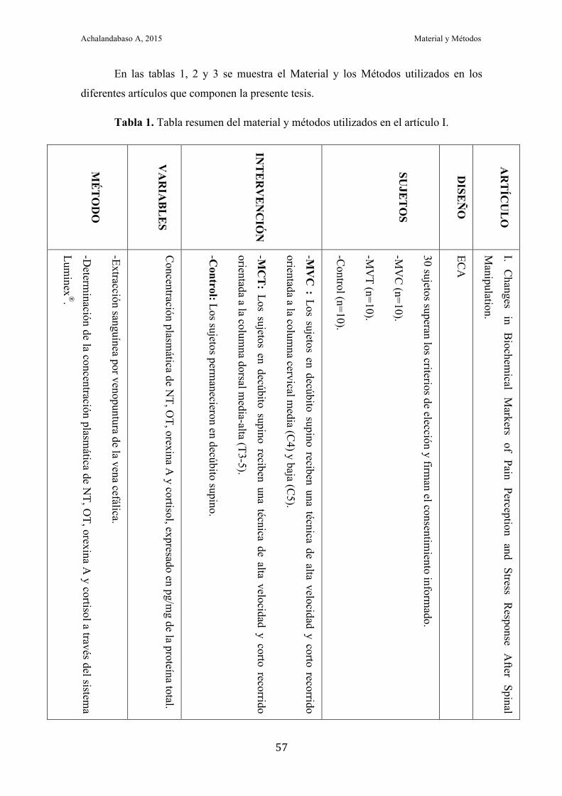
Achalandabaso A, 2015 Material y Métodos
57
En las tablas 1, 2 y 3 se muestra el Material y los Métodos utilizados en los
diferentes artículos que componen la presente tesis.
Tabla 1. Tabla resumen del material y métodos utilizados en el artículo I.
MÉ
TO
DO
VA
RIA
BL
ES
INT
ER
VE
NC
IÓN
SUJE
TO
S
DISE
ÑO
AR
TÍC
UL
O
-Extracción sanguínea por venopuntura de la vena cefálica.
-Determ
inación de la concentración plasmática de N
T, OT, orexina A
y cortisol a través del sistema
Luminex
®.
Concentración plasm
ática de NT, O
T, orexina A y cortisol, expresado en pg/m
g de la proteína total.
-MV
C : Los sujetos en decúbito supino reciben una técnica de alta velocidad y corto recorrido
orientada a la columna cervical m
edia (C4) y baja (C
5).
-MC
T: Los sujetos en decúbito supino reciben una técnica de alta velocidad y corto recorrido
orientada a la columna dorsal m
edia-alta (T3-5).
-Control: Los sujetos perm
anecieron en decúbito supino.
30 sujetos superan los criterios de elección y firman el consentim
iento informado.
-MV
C (n=10).
-MV
T (n=10).
-Control (n=10).
ECA
I. C
hanges in
Biochem
ical M
arkers of
Pain Perception
and Stress
Response
After
Spinal
Manipulation.

Achalandabaso A, 2015 Material y Métodos
58
Tabla 2. Tabla resumen del material y métodos utilizados en el artículo II.
MÉ
TO
DO
VA
RIA
BL
ES
INT
ER
VE
NC
IÓN
SUJE
TO
S
DISE
ÑO
AR
TÍC
UL
O
-Extracción sanguínea por venopuntura de la vena cefálica.
-Determ
inación de la concentración plasmática de SP a través del sistem
a Luminex
®, y de ON
por el procedim
iento de Bram
an y Hendrix.
-La determinación de los U
DP se realizó a través de un algóm
etro de presión en tres localizaciones: sobre la proyección de la articulación cigapofisaria (C
5-C6), sobre el epicóndilo lateral y sobre el m
úsculo tibial anterior. En cada localización se realizaron 3 m
edidas y se estableció la media de las tres m
edidas como el
valor real.
-Concentración plasm
ática de SP expresado en pg/mg de la proteína total.
-Concentración plasm
ática de ON
expresado en µmol/m
g de la proteína total.
-UD
P expresado en kg/cm2.
-MV
C : Los sujetos en decúbito supino reciben una técnica de alta velocidad y corto recorrido orientada a la
columna cervical m
edia (C4) y baja (C
5).
-MC
T: Los sujetos en decúbito supino reciben una técnica de alta velocidad y corto recorrido orientada a la
columna dorsal m
edia-alta (T3-5).
-Control: Los sujetos perm
anecieron en decúbito supino.
30 sujetos superan los criterios de elección y firman el consentim
iento informado.
-MV
C (n=10).
-MV
T (n=10).
-Control (n=10).
ECA
II. Imm
ediate effects of spinal manipulation on nitric oxide, substance P and pain perception.

Achalandabaso A, 2015 Material y Métodos
59
Tabla 3. Tabla resumen del material y métodos utilizados en el artículo III.
MÉ
TO
DO
VA
RIA
BL
ES
INT
ER
VE
NC
IÓN
SUJE
TO
S
DISE
ÑO
AR
TÍC
UL
O
-Extracción sanguínea por venopuntura de la vena cefálica.
-Determ
inación de la concentración en suero de CPK
, LDH
, PCR
y aldolasa a través del sistema O
LYM
PUS
AU
5400 Analyzer. La de troponina I y la EN
S a través de quimiolum
iniscencia. Y la de m
ioglobina a través de un inm
unoanálisis enzimático.
-Concentración plasm
ática de CPK
, LDH
, PCR
, aldolasa, troponina, ENS y m
ioglobina.
LDH
U/L; C
PK U
/L; PCR
mg/L; EN
S ng/mL; A
ldolasa U/L; Troponina I ng/m
L; Mioglobina ng/m
L.
-MV
C : Los sujetos en decúbito supino reciben una técnica de alta velocidad y corto recorrido orientada a la
columna cervical m
edia (C4) y baja (C
5).
-MC
T: Los sujetos en decúbito supino reciben una técnica de alta velocidad y corto recorrido orientada a la
columna dorsal m
edia-alta (T3-5).
-Control: Los sujetos perm
anecieron en decúbito supino.
30 sujetos superan los criterios de elección y firman el consentim
iento informado.
-MV
C (n=10).
-MV
T (n=10).
-Control (n=10).
ECA
III. Tissue damage m
arkers after a spinal manipulation in healthy subjects: A
preliminary report of a
randomized controlled trial.


Achalandabaso A, 2015 Resultados y Discusión
61
RESULTADOS Y DISCUSIÓN .
Los resultados y la discusión de la presente tesis se exponen en los artículos tal
cual han sido publicados.


Achalandabaso A, 2015 Artículos
63
ARTÍCULO I
CHANGES IN BIOCHEMICAL MARKERS OF PAIN
PERCEPTION AND STRESS RESPONSE AFTER SPINAL
MANIPULATION
.
Plaza-Manzano G, Molina-Ortega F, Lomas-Vega R, Martínez-Amat A, Achalandabaso
A, Hita-Contreras F
J Orthop Sports Phys Ther
2014 Apr;44(4):231–9


Achalandabaso A, 2015 Artículos
65
journal of orthopaedic & sports physical therapy | volume 44 | number 4 | april 2014 | 231
[ RESEARCH REPORT ]
Spinal manipulation (SM) is a common treatment approach for pain reduction in low back and neck disorders.37,38,41 The effectiveness of SM to treat musculoskeletal pain, such as spinal pain, has been summarized in recent Cochrane reviews.32,56
Overall, the evidence suggests that SM provides improvements in pain re-lief, though similar results have been de-scribed in other competing treatments, such as general practitioner manage-ment, medication, and exercise, in pa-tients with musculoskeletal pain.6,7 It has been shown that the presence of pain in-
duces changes in the anatomy and func-tion of the central and peripheral nervous systems.20,46,53 Therefore, research on an asymptomatic population may be impor-tant to accurately determine the antinoci-ceptive mechanism of SM. Several studies in asymptomatic subjects have shown that SM techniques induce changes in
physiological reflexes,28 increase neu-romuscular excitability,22 and modify sensitivity.30
The mechanisms through which SM alters musculoskeletal pain are still unknown. However, current evidence suggests an interaction between the mechanical stimulus and the associated neurophysiological responses,6,51 includ-ing rapid hypoalgesia with concurrent sympathetic nervous system and mo-tor system excitation, similar to those generated by direct stimulation of the periaqueductal gray matter.61,68 Recent animal studies show that the analgesia produced by joint mobilization involves serotonin and noradrenaline receptors in the spinal cord, thereby performing a supporting role for central mechanisms of pain modulation.60 Several neuropep-tides, such as neurotensin,23 oxytocin,29 or orexin A,3 have been associated with hypoalgesia and pain modulation, and it is well known that cortisol plays an anal-gesic role related to stress responses.4,44 Recent theories have also suggested that chronic pain could be partly maintained by maladaptive physiological responses of the organism facing a recurrent stressor, a situation related to high cortisol lev-els.45,66 To our knowledge, there is a lack of studies analyzing changes in these no-ciception-related biochemical markers in response to manual therapy.
! STUDY DESIGN: Controlled, repeated-mea-sures, single-blind randomized study.
! OBJECTIVES: To determine the effect of cervical or thoracic manipulation on neurotensin, oxytocin, orexin A, and cortisol levels.
! BACKGROUND: Previous studies have re-searched the effect of spinal manipulation on pain modulation and/or range of movement. However, there is little knowledge of the biochemical process that supports the antinociceptive effect of spinal manipulation.
! METHODS: Thirty asymptomatic subjects were randomly divided into 3 groups: cervical manipulation (n = 10), thoracic manipulation (n = 10), and nonmanipulation (control) (n = 10). Blood samples were extracted before, immediately after, and 2 hours after each intervention. Neurotensin, oxytocin, and orexin A were determined in plasma using enzyme-linked immuno assay. Cortisol was measured by microparticulate enzyme immuno assay in serum samples.
! RESULTS: Immediately after the intervention, significantly higher values of neurotensin (P<.05) and oxytocin (P<.001) levels were observed with both cervical and thoracic manipulation, whereas cortisol concentration was increased only in the cervical manipulation group (P<.05). No changes were detected for orexin A levels. Two hours after the intervention, no significant differences were observed in between-group analysis.
! CONCLUSION: The mechanical stimulus pro-vided by spinal manipulation triggers an increase in neurotensin, oxytocin, and cortisol blood levels. Data suggest that the initial capability of the tissues to tolerate mechanical deformation affects the capacity of these tissues to produce an induc-tion of neuropeptide expression. J Orthop Sports Phys Ther 2014;44(4):231-239. Epub 22 January 2014. doi:10.2519/jospt.2014.4996
! KEY WORDS: cortisol, neurotensin, orexin A, oxytocin, spinal manipulation
1Department of Physical Medicine and Rehabilitation, Faculty of Medicine, Universidad Complutense de Madrid, Madrid, Spain. 2Department of Health Sciences, Universidad de Jaén, Jaén, Spain. The protocol for this study was approved by the Ethical Committee in Clinical Research of the Universidad de Jaén, Jaén, Spain. The authors certify that they have no affiliations with or financial involvement in any organization or entity with a direct financial interest in the subject matter or materials discussed in the article. Address correspondence to Dr Fidel Hita-Contreras, Department of Health Sciences (B-3/272). Universidad de Jaén. Campus Las Lagunillas s/n, 23071 Jaén, Spain. E-mail: [email protected] ! Copyright ©2014 Journal of Orthopaedic & Sports Physical Therapy®
GUSTAVO PLAZA-MANZANO, PT1 • FRANCISCO MOLINA, PT, PhD2 • RAFAEL LOMAS-VEGA, PT, PhD2
ANTONIO MARTÍNEZ-AMAT, PhD2 • ALEXANDER ACHALANDABASO, PT1 • FIDEL HITA-CONTRERAS, MD, PhD2
Changes in Biochemical Markers of Pain Perception and Stress Response
After Spinal Manipulation
44-04 Plaza-Manzano.indd 231 3/19/2014 5:31:21 PM
Jour
nal o
f Orth
opae
dic
& S
ports
Phy
sica
l The
rapy
®
Dow
nloa
ded
from
ww
w.jo
spt.o
rg a
t on
Apr
il 4,
201
4. F
or p
erso
nal u
se o
nly.
No
othe
r use
s with
out p
erm
issi
on.
Cop
yrig
ht ©
201
4 Jo
urna
l of O
rthop
aedi
c &
Spo
rts P
hysi
cal T
hera
py®
. All
right
s res
erve
d.

Achalandabaso A, 2015 Artículos
66
232 | april 2014 | volume 44 | number 4 | journal of orthopaedic & sports physical therapy
[ RESEARCH REPORT ]There are controversial opinions re-
garding the antinociceptive effects of SM according to the site of application. Some authors have reported that cervical manipulation may produce better analge-sic effects than thoracic manipulation,52 and other authors have not detected differences in pain relief between the 2 techniques.43 To make better therapeu-tic decisions, professionals would profit from knowing whether one type of SM is better than others in terms of antino-ciceptive effects. Taking these data into account, our purpose was to determine whether cervical and thoracic manipu-lation would induce differences in neu-ropeptide production or have a similar biochemical response. The aim of this study was to evaluate the effects of cer-vical and thoracic SM on the plasmatic concentration of biochemical markers (neurotensin, orexin A, oxytocin, and cortisol). This study represents a prelimi-nary step in advancing the understand-ing of the underlying mechanisms of SM treatment and its effects.
METHODS
Subjects
The sample population consisted of graduate students who responded to advertisements placed in the Uni-
versity of Jaén (Spain). All subjects signed an informed consent form approved by the University of Jaén Institutional Review Board prior to participating in the study. Participants were verbally screened for their history of neck pain and for current use of any drug. Those who had 1 or more of the following con-ditions were excluded from the study: contraindication to manipulation, his-tory of whiplash or cervical surgery, pain related to cervical spine or arm in the previous month, headache in the previ-ous days, spinal manipulative therapy in the previous 2 months, or loss of standing balance. The study was approved by the Ethical Committee in Clinical Research of the University of Jaén, and the proto-col was performed following the Ethical
Principles for Medical Research in Hu-mans of the Declaration of Helsinki.
InterventionsSM procedures consisted of standard techniques performed as described by Gibbons and Tehan.31 The thoracic SM technique involved a high-velocity, end-range, anterior-posterior force through the elbows to the middle thoracic spine (T3-4) on the lower thoracic (T4-5) spine in a supine position, with the patient’s arms crossed (FIGURE 1). The cervical manipulation involved a high-velocity, midrange, left rotational force to the mid cervical spine (C4) on the lower cervical spine (C5) in supine, with left rotation and right sidebending (FIGURE 2).
Blood samples and active cervical ro-tation movement were obtained from all subjects before, immediately after, and 2 hours after the intervention. Confound-ing factors such as time of day (circadian rhythms), prior diet, and activity patterns were controlled in the 2-hour period prior to reassessment. All interventions were performed at the same time of day for each participant.
Outcome MeasuresExtracting Blood Samples and Obtain-ing Serum/Plasma Serum samples were extracted by venipuncture of the cephalic vein, according to a standardized proto-col36 that used a Vacutainer system (Bec-ton, Dickinson and Company, Franklin Lakes, NJ). Blood was collected in a tube for serum (Vacutainer SST II Advance,
model 367953) and a tube for plasma (Vacutainer PST II Advance, model 367374) separation. After blood extrac-tion, tubes stood at room temperature for 1 hour until the blood clotted. Afterward, the tubes were centrifuged for 10 min-utes at 2000g (Avanti J-30I; Beckman Coulter, Inc, Brea, CA). Supernatant was collected, aliquoted, and kept at –80°C until used.Neuropeptide Quantification It has been shown that neurotensin is implicated in analgesia via its actions within central and peripheral pain modulatory cir-cuits,23 oxytocin plays an antinociceptive role in the central nervous system,2 and orexin is involved in nociceptive sensory processes.3,24 Neuropeptides were de-termined by a Luminex (Luminex Cor-poration, Austin, TX) assay (Milliplex; EMD Millipore Corporation, Billerica, MA). This kit allows the simultaneous quantification of neurotensin, orexin A, and oxytocin (Milliplex HNP-35K; EMD Millipore Corporation). Plasma samples were thawed at room temperature and processed following recommendations from the manufacturer. Neuropeptide data were normalized with the total protein concentration of each sample, which was calculated using the Bradford assay.10 Cortisol has been found to corre-late inversely with pain intensity, and in this sense, a specific increase of cortisol has been proven to have an antinocicep-tive effect.1 Cortisol concentration was determined in serum samples using the microparticulate enzyme immuno assay in the AxSYM analyzer (Abbott Labo-
FIGURE 1. Thoracic spinal manipulation.FIGURE 2. Cervical spinal manipulation.
44-04 Plaza-Manzano.indd 232 3/19/2014 5:31:23 PM
Jour
nal o
f Orth
opae
dic
& S
ports
Phy
sica
l The
rapy
®
Dow
nloa
ded
from
ww
w.jo
spt.o
rg a
t on
Apr
il 4,
201
4. F
or p
erso
nal u
se o
nly.
No
othe
r use
s with
out p
erm
issi
on.
Cop
yrig
ht ©
201
4 Jo
urna
l of O
rthop
aedi
c &
Spo
rts P
hysi
cal T
hera
py®
. All
right
s res
erve
d.

Achalandabaso A, 2015 Artículos
67
journal of orthopaedic & sports physical therapy | volume 44 | number 4 | april 2014 | 233
ratories, Abbott Park, IL) following the manufacturer’s recommendations.
Statistical AnalysisData for continuous variables were ex-pressed as mean ! SD. Categorical data were expressed as frequencies and per-
centages. The Kolmogorov-Smirnov and Levene tests were performed to assess normality and homoscedasticity, respec-tively. A 3-by-3, mixed-model analysis of variance (ANOVA) was performed to test the effect of the factor (control, thoracic manipulation, and cervical manipula-
tion) on the dependent variables (range of motion and concentration of neuroten-sin, orexin A, oxytocin, and cortisol). The hypothesis of interest was the group-by-time interaction. Additionally, to find out if there was any significant interaction, a Bonferroni pairwise comparison was per-formed. Pearson correlation coefficients were used to analyze the relations be-tween continuous variables. Eta-square and adjusted R2 were used for measuring effect sizes. Management and data analy-sis were performed using the statistical package SPSS for Windows Version 17.0 (SPSS Inc, Chicago, IL) and MedCalc Version 12.5 (MedCalc Software bvba, Ostend, Belgium). The level of statistical significance was set at P<.05.
RESULTS
Out of the 35 participants screened, 30 subjects (46.7% women; mean ! SD age, 27.8 !
4.2 years) satisfied the eligibility criteria, agreed to participate, and were random-ized by sealed-envelope selection into the cervical SM group (n = 10), the thoracic SM group (n = 10), or a control group that did not receive any treatment (n = 10) (FIGURE 3). Baseline characteristics of participants are shown in TABLE 1.
Assessed for eligibility, n = 35Excluded, n = 5:• Previous spine manipulation, n = 4• Pharmacological treatment, n = 1
Randomization, n = 30
Follo
w-u
pAl
loca
tion
Enro
llmen
t
Control group, n = 10
Lost to follow-up, n = 0 Lost to follow-up, n = 0 Lost to follow-up, n = 0
Cervical manipulation group, n = 10 Thoracic manipulation group, n = 10
Analyzed, n = 10 Analyzed, n = 10 Analyzed, n = 10
Anal
ysis
FIGURE 3. Flow diagram of the study.
TABLE 1 Baseline Characteristics of Participants*
Abbreviation: BMI, body mass index.*Values are mean ! SD unless otherwise indicated.
All (n = 30) Control (n = 10)Thoracic Manipulation
(n = 10)Cervical Manipulation
(n = 10)
Age, y 27.80 ! 4.16 25.80 ! 3.22 29.80 ! 4.52 27.80 ! 3.99
Weight, kg 69.50 ! 12.94 70.80 ! 4.94 69.00 ! 14.95 68.70 ! 17.00
Height, m 1.73 ! 0.09 1.75 ! 0.07 1.73 ! 0.11 1.73 ! 0.08
BMI, kg/m2 22.98 ! 3.19 23.20 ! 1.41 22.93 ! 3.60 22.82 ! 4.23
Gender, n (%)
Male 16 (53.3) 6 (60.0) 5 (50.0) 5 (50.0)
Female 14 (46.7) 4 (40.0) 5 (50.0) 5 (50.0)
Student, n (%)
No 17 (56.7) 4 (40.0) 8 (80.0) 5 (50.0)
Yes 13 (43.3) 6 (60.0) 2 (20.0) 5 (50.0)
Married, n (%)
No 22 (73.3) 7 (70.0) 7 (70.0) 8 (80.0)
Yes 8 (26.7) 3 (30.0) 3 (30.0) 2 (20.0)
Smoker, n (%)
No 17 (56.7) 8 (80.0) 4 (40.0) 5 (50.0)
Yes 13 (43.3) 2 (20.0) 6 (60.0) 5 (50.0)
44-04 Plaza-Manzano.indd 233 3/19/2014 5:31:24 PM
Jour
nal o
f Orth
opae
dic
& S
ports
Phy
sica
l The
rapy
®
Dow
nloa
ded
from
ww
w.jo
spt.o
rg a
t on
Apr
il 4,
201
4. F
or p
erso
nal u
se o
nly.
No
othe
r use
s with
out p
erm
issi
on.
Cop
yrig
ht ©
201
4 Jo
urna
l of O
rthop
aedi
c &
Spo
rts P
hysi
cal T
hera
py®
. All
right
s res
erve
d.

Achalandabaso A, 2015 Artículos
68
234 | april 2014 | volume 44 | number 4 | journal of orthopaedic & sports physical therapy
[ RESEARCH REPORT ]Neurotensin Concentration in Blood SamplesThe 3-by-3, mixed-model ANOVA re-vealed a significant interaction of time by group for neurotensin concentration (P = .029), with an eta-square value of 18% (TABLE 2). On the other hand, with-in-group comparisons in cervical and thoracic manipulation groups showed a significant increase in neurotensin lev-els immediately postintervention com-pared with preintervention levels (P<.05) (TABLE 3). For the between-group analysis (FIGURE 4A), statistically significant differ-ences were found on posttreatment mea-surements between the control group and the thoracic manipulation group (mean difference, –3.87; 95% confidence interval [CI]: –6.00, –0.74; P = .012) and between the control and the cervical manipulation groups (mean difference, –4.86; 95% CI: –7.99, –1.74; P = .001).
Orexin A Concentration in Blood SamplesA 3-by-3, mixed-model ANOVA did not show a significant interaction of group by time for orexin A concentration in blood samples (P = .210) (TABLE 2). The effect size, measured by eta-square, was 10%. At the descriptive level, an important decrease in orexin A concentration was detected after the intervention in the tho-racic SM group in comparison with the control group (mean difference, 47.16; 95% CI: –4.78, 99.10; P = .085), although this decrease did not reach statistical sig-nificance (FIGURE 4B).
Oxytocin Concentration in Blood SamplesThe group-by-time interaction was sig-nificant for oxytocin plasma concentra-tion (P<.001). The effect size, measured by eta-square, was 62% (TABLE 2). An increase in oxytocin concentration was detected just after the intervention in both the cervical SM group (mean dif-ference, –200.58; 95% CI: –271.03, –130.12; P<.001) and the thoracic SM group (mean difference, –96.42; 95% CI: –166.87, –25.96; P = .005) when compared with the control group. In
TABLE 2Intergroup Comparison
of Neuropeptide Levels*
*Values are mean ! SD unless otherwise indicated.†Time by group.‡Concentration of neuropeptides in plasma samples (pg/mg of total protein).§Concentration of cortisol in serum samples (pg/mg of total protein).
Preintervention0 h
Postintervention2 h
Postintervention P Value† Effect Size, η2
Neurotensin‡ .029 0.179
Control 4.95 ! 1.24 5.47 ! 1.22 4.93 ! 1.30
Thoracic 5.15 ! 1.85 9.34 ! 2.33 6.16 ! 2.64
Cervical 6.58 ! 1.67 10.33 ! 3.95 8.01 ! 3.84
Orexin A‡ .210 0.101
Control 209.06 ! 23.85 205.13 ! 21.97 200.95 ! 26.76
Thoracic 207.51 ! 66.93 160.89 ! 46.19 153.78 ! 43.63
Cervical 211.72 ! 48.50 193.17 ! 66.96 167.03 ! 59.93
Oxytocin‡ <.001 0.622
Control 52.41 ! 15.73 50.77 ! 21.65 46.06 ! 13.64
Thoracic 53.12 ! 15.53 147.19 ! 51.11 58.39 ! 26.16
Cervical 58.45 ! 20.69 251.35 ! 91.36 85.68 ! 52.87
Cortisol§ <.001 0.326
Control 9.90 ! 3.48 9.60 ! 3.10 9.70 ! 3.13
Thoracic 9.80 ! 3.08 10.10 ! 3.67 6.70 ! 2.50
Cervical 10.51 ! 3.37 14.20 ! 3.58 8.50 ! 3.89
TABLE 3Within-Group Comparison
of Pretreatment Values and Values Immediately After Intervention
*Pretreatment minus immediately posttreatment values. Values in parentheses are 95% confidence interval.†Mean difference of neuropeptide concentration in plasma samples (pg/mg of total protein).‡Mean difference of cortisol in serum samples (pg/mg of total protein).
Mean Difference* P Value
Neurotensin†
Control –0.522 (–2.790, 1.745) 1.000
Thoracic –4.188 (–6.456, –1.921) <.001
Cervical –3.752 (–6.019, –1.485) .001
Orexin A†
Control 3.936 (–42.821, 50.692) 1.000
Thoracic 46.621 (–0.136, 93.377) .051
Cervical 18.552 (–28.205, 65.308) .961
Oxytocin†
Control 1.642 (–43.042, 46.326) 1.000
Thoracic –94.063 (–138.747, –49.379) <.001
Cervical –192.899 (–237.583, –148.215) <.001
Cortisol‡
Control 0.300 (–1.659, 2.259) 1.000
Thoracic –0.300 (–2.259, 1.659) 1.000
Cervical –3.690 (–5.649, –1.731) <.001
44-04 Plaza-Manzano.indd 234 3/19/2014 5:31:25 PM
Jour
nal o
f Orth
opae
dic
& S
ports
Phy
sica
l The
rapy
®
Dow
nloa
ded
from
ww
w.jo
spt.o
rg a
t on
Apr
il 4,
201
4. F
or p
erso
nal u
se o
nly.
No
othe
r use
s with
out p
erm
issi
on.
Cop
yrig
ht ©
201
4 Jo
urna
l of O
rthop
aedi
c &
Spo
rts P
hysi
cal T
hera
py®
. All
right
s res
erve
d.

Achalandabaso A, 2015 Artículos
69
journal of orthopaedic & sports physical therapy | volume 44 | number 4 | april 2014 | 235
the same way, the cervical SM group showed increased oxytocin values when compared with the thoracic SM group immediately postintervention (mean difference, –104.16; 95% CI: –174.62, –33.71; P<.002) (FIGURE 4C).
Likewise, in the within-group analy-sis, an increase in oxytocin plasma con-centration levels was detected in both the cervical manipulation and thoracic manipulation groups immediately post-intervention (P<.001) compared to pre-intervention levels (TABLE 3). At 2 hours after the intervention, an increase was found only in the cervical SM group (P<.05) when compared with preinter-vention levels (TABLE 4).
Cortisol Concentration in Blood SamplesUsing a mixed-model ANOVA, the group-by-time interaction for cortisol as a dependent variable was significant (P<.001). Eta-square analysis yielded a 32% effect size (TABLE 2).
Blood samples extracted from the cervical SM group showed a significant increase in cortisol plasma concentration immediately postintervention compared with baseline values (P<.001) (TABLE 3). On the other hand, a significant decrease was detected at 2 hours postintervention in the thoracic SM group when compared with the preintervention values (P<.05) (TABLE 4).
A significant increase in the between-group analysis was found immediately posttreatment in the cervical manipula-tion group compared with the control group (mean difference, 4.60; 95% CI: 0.65, 8.55; P = .018) and the thoracic ma-nipulation group (mean difference, 4.10; 95% CI: 0.15, 8.05; P<.040) (FIGURE 4D).
DISCUSSION
Several studies currently sup-port the idea that the analgesic effect of manual therapy is mediated by
central mechanisms of pain modulation through the modulation of neuropeptide production.5,27,60 To our knowledge, this is the first work to analyze neurotensin,
oxytocin, orexin A, and cortisol levels af-ter a cervical or a thoracic manipulation in asymptomatic subjects.
Neurotensin is a 13-amino acid pro-duced in several regions of the central nervous system, such as the substan-tia nigra, amygdala, hypothalamus, prefrontal cortex, periaqueductal gray matter, and the spinal cord,62 and it has several actions, including analgesia.14,23 Our data indicate an increase in neu-rotensin plasmatic concentration after an SM, suggesting that the mechanical stimulus provided by SM is enough to modulate the liberation of this neuro-peptide. In this sense, neurotensin has long been known to include analgesia among its actions.9,16,23 The analgesic ac-tions of neurotensin are readily distinct from those of the opioids, based on their insensitivity to the highly opioid-selective antagonist naloxone, thus ruling out an opioid mechanism.55 Neurotensin acts as
part of the peripheral and central mecha-nisms of pain modulation,23 because the antinociceptive effect of neurotensin has been reported after the injection of the peptide in many brain areas.62 There are anatomical data suggesting an in-teraction between neurotensin and se-rotonergic neurons. As a matter of fact, neurons of the rostral part of the raphe synthesize neurotensin, whereas neuro-tensin receptors are widely expressed in most of the raphe.18,40,57 The functional role of neurotensin in the raphe remains to be determined, but it may participate in the modulation of some of the known functions of the serotonergic system, in-cluding nociception13 and stress-related responses.19 It may also play a role in mediating stress-induced analgesia, as neurotensin knockout mice and rats pretreated with neurotensin antagonists show no increase in pain tolerance after stress.34 Recent studies with neurotensin
2
0Preintervention O h
postintervention2 h
postintervention
4
6
8
10
12
14
A
Neurotensin
0
100
50
150
200
250
300
Neur
opep
tide
Conc
entra
tion,
pg
/mg
B
Neur
opep
tide
Conc
entra
tion,
pg
/mg
Preintervention O h postintervention
2 h postintervention
Orexin A
*
*
50
0Preintervention O h
postintervention2 h
postintervention
100
150
200
250
300
350
C
Oxytocin
Control
0
42
68
1012141618
Neur
opep
tide
Conc
entra
tion,
pg
/mg
D
Neur
opep
tide
Conc
entra
tion,
pg
/mg
Preintervention O h postintervention
2 h postintervention
Cortisol
*
**
**
Thoracic Cervical
FIGURE 4. Mean and 95% confidence interval for neuropeptide concentration in blood samples. (A) neurotensin, (B) orexin A, (C) oxytocin, (D) cortisol. *P<.05.
44-04 Plaza-Manzano.indd 235 3/19/2014 5:31:26 PM
Jour
nal o
f Orth
opae
dic
& S
ports
Phy
sica
l The
rapy
®
Dow
nloa
ded
from
ww
w.jo
spt.o
rg a
t on
Apr
il 4,
201
4. F
or p
erso
nal u
se o
nly.
No
othe
r use
s with
out p
erm
issi
on.
Cop
yrig
ht ©
201
4 Jo
urna
l of O
rthop
aedi
c &
Spo
rts P
hysi
cal T
hera
py®
. All
right
s res
erve
d.

Achalandabaso A, 2015 Artículos
70
236 | april 2014 | volume 44 | number 4 | journal of orthopaedic & sports physical therapy
[ RESEARCH REPORT ]antagonists and knockout mice lacking neurotensin or neurotensin receptors have revealed that the neurotensiner-gic system plays a pivotal role in the nonopioid form of stress-induced anal-gesia.34,42,58 In summary, the antinocicep-tive effect of neurotensin after SM may increase the mechanical stress threshold that cervical tissues can tolerate.
It is well established that neurotensin affects the activity of oxytocin-positive cells in the supraoptic nucleus.39 Oxyto-cin is a nonapeptide that plays a major neuroendocrine role, modulating several physiological functions in mammals, like somatosensory transmission, nocicep-tion, and pain.2,64,65 Oxytocin is synthe-sized and secreted by a subpopulation of the paraventricular and supraoptic nuclei of the hypothalamus.64 In fact, several studies now support the idea that oxytocin exerts a potent antinocicep-tive control after its release in the spinal cord from hypothalamo-hypophysal de-scending projections.17,59,73 Breton et al12 have shown that this antinociceptive ac-tion is mediated, in part, by an increase in synaptic inhibition within the most superficial layers of the spinal cord. In addition, Robinson et al54 showed that oxytocin inhibits sensory glutamatergic transmission between afferent fibers and dorsal horn neurons. Along the same lines, Petersson et al50 hypothesized that an increase of oxytocin might possibly result in a greater synthesis of endog-enous opioids, because the antinocicep-tion observed after repeated injections of oxytocin was temporarily reversed by the opioid antagonist naloxone.
In studies involving human subjects, pain relief was reported in central neuro-genic pain and in low back pain72 after the intracerebroventricular and intrathecal administration of oxytocin. No previous study has evaluated whether SM has an ef-fect on oxytocin plasmatic concentration. Our results suggest that the increase of the plasmatic concentration of oxytocin fol-lowing an SM could be partly responsible for the analgesic effect linked to manual therapy techniques due to the activation
of descending pain-inhibitory pathways.Orexins are known to be a hypotha-
lamic peptide critical for feeding and normal wakefulness. Orexin A and B are distributed throughout the spinal cord, and orexin fibers are concentrated in lamina I of the dorsal horn and in lamina X surrounding the central canal.71 Orex-inergic projections were identified in periaqueductal gray matter, the rostral ventral medulla, the dorsal horn, and the dorsal root ganglion.21,33,67 Emerging evidence shows that the central nervous system administration (intracranial ven-tricle or intrathecal injection) of orexin A can suppress mechanical allodynia and thermal hypersensitivity in multiple pain models, suggesting the regulation of no-ciceptive processing via spinal and supra-spinal mechanisms.8,70
In addition, orexins showed antino-ciceptive effects on models of pain, such as neuropathic pain, carrageenan test, and postoperative pain.47,48 There is a lack of literature that analyzes the effect of physical therapy techniques on orexin
A expression. A recent study using a rat model reported a significant increase of orexin A following electroacupuncture therapy after a laparotomy.25 In contrast, our results did not show a statistically significant change in orexin A levels after a thoracic or cervical SM.
One of the actions of orexin A in stress situations is the activation of glu-cocorticoid production from adreno-cortical cells.11 Cortisol is therefore one of the biochemical factors delivered in stress situations35 that acts to decrease local edema and pain by blocking early stages of inflammation. In addition, it is also believed that high cortisol levels promote wound healing by stimulating gluconeogenesis.69 The response to stress is triggered by the stimulation of the hy-pothalamus-pituitary-adrenal axis. It has been proven that a subject’s level of stress can be correlated with secreted cortisol levels.4 Our results suggest that no plas-matic concentration changes of cortisol follow a thoracic SM, which agrees with the results of a recent review using mas-
TABLE 4Within-Group Comparison Between
Pretreatment Values and Values 2 Hours After Intervention
*Values in parentheses are 95% confidence interval.†Mean difference of neuropeptide concentration in plasma samples (pg/mg of total protein).‡Mean difference of cortisol in serum samples (pg/mg of total protein).
Mean Difference* P Value
Neurotensin†
Control 0.018 (–2.260, 2.296) 1.000
Thoracic –1.014 (–3.292, 1.264) .798
Cervical –1.431 (–3.709, 0.847) .362
Orexin A†
Control 8.117 (–38.213, 54.447) 1.000
Thoracic 53.729 (7.399, 100.060) .019
Cervical 44.683 (–1.647, 91.014) .062
Oxytocin†
Control 6.357 (–16.001, 28.714) 1.000
Thoracic –5.270 (–27.627, 17.087) 1.000
Cervical –27.236 (–49.594, –4.879) .013
Cortisol‡
Control 0.200 (–2.156, 2.556) 1.000
Thoracic 3.100 (0.744, 5.456) .007
Cervical 2.010 (–0.346, 4.366) .115
44-04 Plaza-Manzano.indd 236 3/19/2014 5:31:27 PM
Jour
nal o
f Orth
opae
dic
& S
ports
Phy
sica
l The
rapy
®
Dow
nloa
ded
from
ww
w.jo
spt.o
rg a
t on
Apr
il 4,
201
4. F
or p
erso
nal u
se o
nly.
No
othe
r use
s with
out p
erm
issi
on.
Cop
yrig
ht ©
201
4 Jo
urna
l of O
rthop
aedi
c &
Spo
rts P
hysi
cal T
hera
py®
. All
right
s res
erve
d.
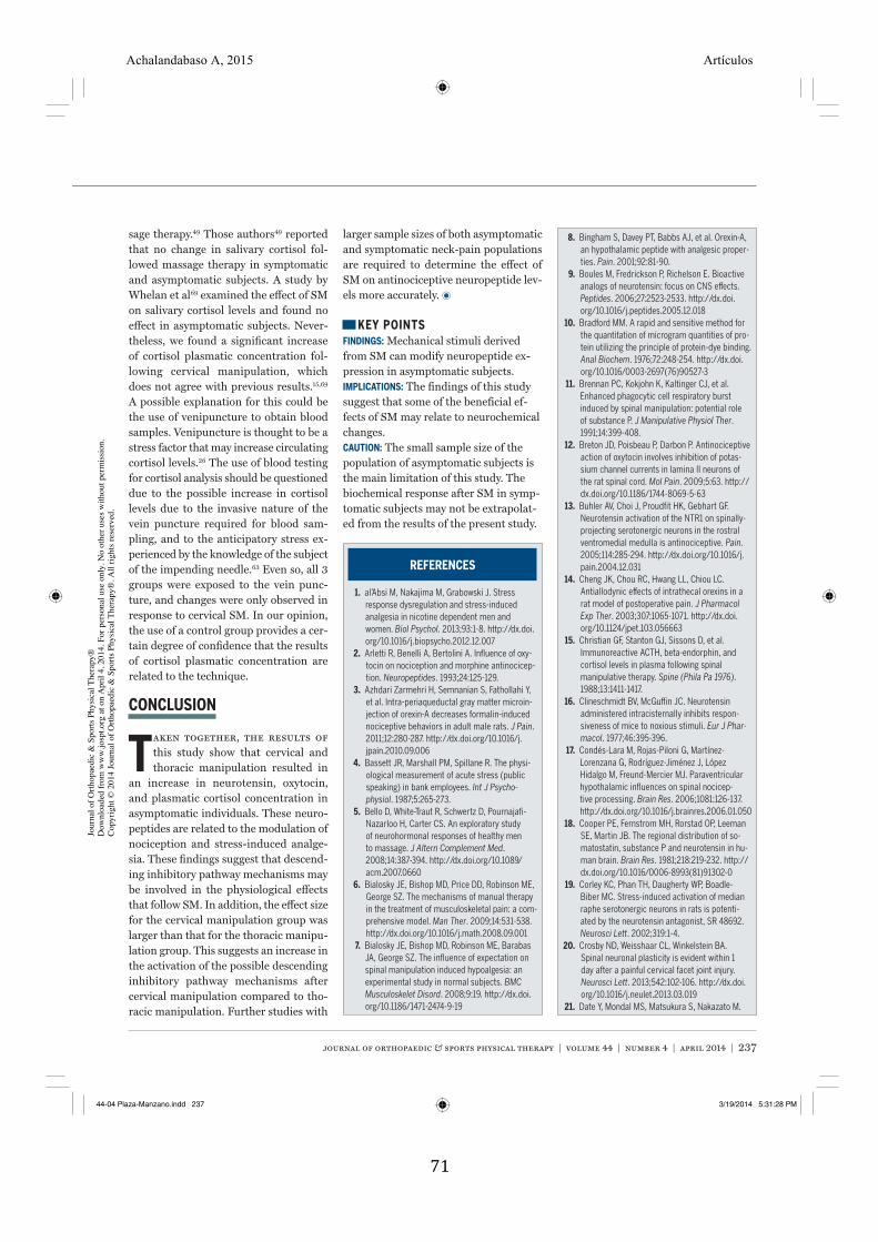
Achalandabaso A, 2015 Artículos
71
journal of orthopaedic & sports physical therapy | volume 44 | number 4 | april 2014 | 237
sage therapy.49 Those authors49 reported that no change in salivary cortisol fol-lowed massage therapy in symptomatic and asymptomatic subjects. A study by Whelan et al69 examined the effect of SM on salivary cortisol levels and found no effect in asymptomatic subjects. Never-theless, we found a significant increase of cortisol plasmatic concentration fol-lowing cervical manipulation, which does not agree with previous results.15,69 A possible explanation for this could be the use of venipuncture to obtain blood samples. Venipuncture is thought to be a stress factor that may increase circulating cortisol levels.26 The use of blood testing for cortisol analysis should be questioned due to the possible increase in cortisol levels due to the invasive nature of the vein puncture required for blood sam-pling, and to the anticipatory stress ex-perienced by the knowledge of the subject of the impending needle.63 Even so, all 3 groups were exposed to the vein punc-ture, and changes were only observed in response to cervical SM. In our opinion, the use of a control group provides a cer-tain degree of confidence that the results of cortisol plasmatic concentration are related to the technique.
CONCLUSION
Taken together, the results of this study show that cervical and thoracic manipulation resulted in
an increase in neurotensin, oxytocin, and plasmatic cortisol concentration in asymptomatic individuals. These neuro-peptides are related to the modulation of nociception and stress-induced analge-sia. These findings suggest that descend-ing inhibitory pathway mechanisms may be involved in the physiological effects that follow SM. In addition, the effect size for the cervical manipulation group was larger than that for the thoracic manipu-lation group. This suggests an increase in the activation of the possible descending inhibitory pathway mechanisms after cervical manipulation compared to tho-racic manipulation. Further studies with
larger sample sizes of both asymptomatic and symptomatic neck-pain populations are required to determine the effect of SM on antinociceptive neuropeptide lev-els more accurately. !
KEY POINTSFINDINGS: Mechanical stimuli derived from SM can modify neuropeptide ex-pression in asymptomatic subjects.IMPLICATIONS: The findings of this study suggest that some of the beneficial ef-fects of SM may relate to neurochemical changes.CAUTION: The small sample size of the population of asymptomatic subjects is the main limitation of this study. The biochemical response after SM in symp-tomatic subjects may not be extrapolat-ed from the results of the present study.
REFERENCES
1. al’Absi M, Nakajima M, Grabowski J. Stress response dysregulation and stress-induced analgesia in nicotine dependent men and women. Biol Psychol. 2013;93:1-8. http://dx.doi.org/10.1016/j.biopsycho.2012.12.007
2. Arletti R, Benelli A, Bertolini A. Influence of oxy-tocin on nociception and morphine antinocicep-tion. Neuropeptides. 1993;24:125-129.
3. Azhdari Zarmehri H, Semnanian S, Fathollahi Y, et al. Intra-periaqueductal gray matter microin-jection of orexin-A decreases formalin-induced nociceptive behaviors in adult male rats. J Pain. 2011;12:280-287. http://dx.doi.org/10.1016/j.jpain.2010.09.006
4. Bassett JR, Marshall PM, Spillane R. The physi-ological measurement of acute stress (public speaking) in bank employees. Int J Psycho-physiol. 1987;5:265-273.
5. Bello D, White-Traut R, Schwertz D, Pournajafi-Nazarloo H, Carter CS. An exploratory study of neurohormonal responses of healthy men to massage. J Altern Complement Med. 2008;14:387-394. http://dx.doi.org/10.1089/acm.2007.0660
6. Bialosky JE, Bishop MD, Price DD, Robinson ME, George SZ. The mechanisms of manual therapy in the treatment of musculoskeletal pain: a com-prehensive model. Man Ther. 2009;14:531-538. http://dx.doi.org/10.1016/j.math.2008.09.001
7. Bialosky JE, Bishop MD, Robinson ME, Barabas JA, George SZ. The influence of expectation on spinal manipulation induced hypoalgesia: an experimental study in normal subjects. BMC Musculoskelet Disord. 2008;9:19. http://dx.doi.org/10.1186/1471-2474-9-19
8. Bingham S, Davey PT, Babbs AJ, et al. Orexin-A, an hypothalamic peptide with analgesic proper-ties. Pain. 2001;92:81-90.
9. Boules M, Fredrickson P, Richelson E. Bioactive analogs of neurotensin: focus on CNS effects. Peptides. 2006;27:2523-2533. http://dx.doi.org/10.1016/j.peptides.2005.12.018
10. Bradford MM. A rapid and sensitive method for the quantitation of microgram quantities of pro-tein utilizing the principle of protein-dye binding. Anal Biochem. 1976;72:248-254. http://dx.doi.org/10.1016/0003-2697(76)90527-3
11. Brennan PC, Kokjohn K, Kaltinger CJ, et al. Enhanced phagocytic cell respiratory burst induced by spinal manipulation: potential role of substance P. J Manipulative Physiol Ther. 1991;14:399-408.
12. Breton JD, Poisbeau P, Darbon P. Antinociceptive action of oxytocin involves inhibition of potas-sium channel currents in lamina II neurons of the rat spinal cord. Mol Pain. 2009;5:63. http://dx.doi.org/10.1186/1744-8069-5-63
13. Buhler AV, Choi J, Proudfit HK, Gebhart GF. Neurotensin activation of the NTR1 on spinally-projecting serotonergic neurons in the rostral ventromedial medulla is antinociceptive. Pain. 2005;114:285-294. http://dx.doi.org/10.1016/j.pain.2004.12.031
14. Cheng JK, Chou RC, Hwang LL, Chiou LC. Antiallodynic effects of intrathecal orexins in a rat model of postoperative pain. J Pharmacol Exp Ther. 2003;307:1065-1071. http://dx.doi.org/10.1124/jpet.103.056663
15. Christian GF, Stanton GJ, Sissons D, et al. Immunoreactive ACTH, beta-endorphin, and cortisol levels in plasma following spinal manipulative therapy. Spine (Phila Pa 1976). 1988;13:1411-1417.
16. Clineschmidt BV, McGuffin JC. Neurotensin administered intracisternally inhibits respon-siveness of mice to noxious stimuli. Eur J Phar-macol. 1977;46:395-396.
17. Condés-Lara M, Rojas-Piloni G, Martínez-Lorenzana G, Rodríguez-Jiménez J, López Hidalgo M, Freund-Mercier MJ. Paraventricular hypothalamic influences on spinal nocicep-tive processing. Brain Res. 2006;1081:126-137. http://dx.doi.org/10.1016/j.brainres.2006.01.050
18. Cooper PE, Fernstrom MH, Rorstad OP, Leeman SE, Martin JB. The regional distribution of so-matostatin, substance P and neurotensin in hu-man brain. Brain Res. 1981;218:219-232. http://dx.doi.org/10.1016/0006-8993(81)91302-0
19. Corley KC, Phan TH, Daugherty WP, Boadle-Biber MC. Stress-induced activation of median raphe serotonergic neurons in rats is potenti-ated by the neurotensin antagonist, SR 48692. Neurosci Lett. 2002;319:1-4.
20. Crosby ND, Weisshaar CL, Winkelstein BA. Spinal neuronal plasticity is evident within 1 day after a painful cervical facet joint injury. Neurosci Lett. 2013;542:102-106. http://dx.doi.org/10.1016/j.neulet.2013.03.019
21. Date Y, Mondal MS, Matsukura S, Nakazato M.
44-04 Plaza-Manzano.indd 237 3/19/2014 5:31:28 PM
Jour
nal o
f Orth
opae
dic
& S
ports
Phy
sica
l The
rapy
®
Dow
nloa
ded
from
ww
w.jo
spt.o
rg a
t on
Apr
il 4,
201
4. F
or p
erso
nal u
se o
nly.
No
othe
r use
s with
out p
erm
issi
on.
Cop
yrig
ht ©
201
4 Jo
urna
l of O
rthop
aedi
c &
Spo
rts P
hysi
cal T
hera
py®
. All
right
s res
erve
d.

Achalandabaso A, 2015 Artículos
72
238 | april 2014 | volume 44 | number 4 | journal of orthopaedic & sports physical therapy
[ RESEARCH REPORT ]Distribution of orexin-A and orexin-B (hypo-cretins) in the rat spinal cord. Neurosci Lett. 2000;288:87-90.
22. Dishman JD, Ball KA, Burke J. First prize: central motor excitability changes after spinal ma-nipulation: a transcranial magnetic stimulation study. J Manipulative Physiol Ther. 2002;25:1-9.
23. Dobner PR. Neurotensin and pain modulation. Peptides. 2006;27:2405-2414. http://dx.doi.org/10.1016/j.peptides.2006.04.025
24. Erami E, Azhdari-Zarmehri H, Ghasemi-Dashkhasan E, Esmaeili MH, Semnanian S. Intra-paragigantocellularis lateralis injection of orexin-A has an antinociceptive effect on hot plate and formalin tests in rat. Brain Res. 2012;1478:16-23. http://dx.doi.org/10.1016/j.brainres.2012.08.013
25. Feng XM, Mi WL, Xia F, et al. Involvement of spi-nal orexin A in the electroacupuncture analgesia in a rat model of post-laparotomy pain. BMC Complement Altern Med. 2012;12:225. http://dx.doi.org/10.1186/1472-6882-12-225
26. Ferriani RA, Silva de Sá MF. Effect of venipunc-ture stress on plasma prolactin levels. Int J Gynaecol Obstet. 1985;23:459-462.
27. Field T, Hernandez-Reif M, Diego M, Schanberg S, Kuhn C. Cortisol decreases and serotonin and dopamine increase following massage therapy. Int J Neurosci. 2005;115:1397-1413. http://dx.doi.org/10.1080/00207450590956459
28. Fryer G, Pearce AJ. The effect of lumbosacral manipulation on corticospinal and spinal reflex excitability on asymptomatic participants. J Ma-nipulative Physiol Ther. 2012;35:86-93. http://dx.doi.org/10.1016/j.jmpt.2011.09.010
29. Gao L, Yu LC. Involvement of opioid receptors in the oxytocin-induced antinociception in the central nervous system of rats. Regul Pept. 2004;120:53-58. http://dx.doi.org/10.1016/j.regpep.2004.02.011
30. George SZ, Bishop MD, Bialosky JE, Zep-pieri G, Jr., Robinson ME. Immediate effects of spinal manipulation on thermal pain sensitivity: an experimental study. BMC Mus-culoskelet Disord. 2006;7:68. http://dx.doi.org/10.1186/1471-2474-7-68
31. Gibbons P, Tehan P. Manipulation of the Spine, Thorax and Pelvis: An Osteopathic Perspective. Edinburgh, UK: Churchill Livingstone; 2000.
32. Gross A, Miller J, D’Sylva J, et al. Manipulation or mobilisation for neck pain: a Cochrane Re-view. Man Ther. 2010;15:315-333. http://dx.doi.org/10.1016/j.math.2010.04.002
33. Guan JL, Wang QP, Shioda S. Immunoelectron microscopic examination of orexin-like immu-noreactive fibers in the dorsal horn of the rat spinal cord. Brain Res. 2003;987:86-92.
34. Gui X, Carraway RE, Dobner PR. Endogenous neurotensin facilitates visceral nociception and is required for stress-induced antinociception in mice and rats. Neuroscience. 2004;126:1023-1032. http://dx.doi.org/10.1016/j.neuroscience.2004.04.034
35. Hellhammer DH, Wüst S, Kudielka BM.
Salivary cortisol as a biomarker in stress research. Psychoneuroendocrinology. 2009;34:163-171. http://dx.doi.org/10.1016/j.psyneuen.2008.10.026
36. Himberger JR, Himberger LC. Accuracy of drawing blood through infusing intravenous lines. Heart Lung. 2001;30:66-73. http://dx.doi.org/10.1067/mhl.2001.110535
37. Huisman PA, Speksnijder CM, de Wijer A. The effect of thoracic spine manipulation on pain and disability in patients with non-specific neck pain: a systematic review. Disabil Rehabil. 2013;35:1677-1685. http://dx.doi.org/10.3109/09638288.2012.750689
38. Hurwitz EL. Epidemiology: spinal ma-nipulation utilization. J Electromyogr Kinesiol. 2012;22:648-654. http://dx.doi.org/10.1016/j.jelekin.2012.01.006
39. Johnstone LE, Leng G, Brown CH. Effects of neu-rotensin on the organization of activity in supra-optic nucleus cells in virgin and lactating rats. J Neuroendocrinol. 2004;16:605-611. http://dx.doi.org/10.1111/j.1365-2826.2004.01208.x
40. Kessler JP, Moyse E, Kitabgi P, Vincent JP, Beaudet A. Distribution of neurotensin bind-ing sites in the caudal brainstem of the rat: a light microscopic radioautographic study. Neuroscience. 1987;23:189-198. http://dx.doi.org/10.1016/0306-4522(87)90282-X
41. Kuczynski JJ, Schwieterman B, Columber K, Knupp D, Shaub L, Cook CE. Effectiveness of physical therapist administered spinal ma-nipulation for the treatment of low back pain: a systematic review of the literature. Int J Sports Phys Ther. 2012;7:647-662.
42. Lafrance M, Roussy G, Belleville K, et al. Involve-ment of NTS2 receptors in stress-induced anal-gesia. Neuroscience. 2010;166:639-652. http://dx.doi.org/10.1016/j.neuroscience.2009.12.042
43. Martínez-Segura R, de-la-Llave-Rincón AI, Ortega-Santiago R, Cleland JA, Fernández-de-las-Peñas C. Immediate changes in widespread pressure pain sensitivity, neck pain, and cervical range of motion after cervical or thoracic thrust manipulation in patients with bilateral chronic mechanical neck pain: a randomized clinical trial. J Orthop Sports Phys Ther. 2012;42:806-814. http://dx.doi.org/10.2519/jospt.2012.4151
44. Matousek RH, Dobkin PL, Pruessner J. Cortisol as a marker for improvement in mindfulness-based stress reduction. Complement Ther Clin Pract. 2010;16:13-19. http://dx.doi.org/10.1016/j.ctcp.2009.06.004
45. Melzack R. Pain and the neuromatrix in the brain. J Dent Educ. 2001;65:1378-1382.
46. Melzack R, Coderre TJ, Katz J, Vaccarino AL. Central neuroplasticity and pathological pain. Ann N Y Acad Sci. 2001;933:157-174.
47. Mobarakeh JI, Takahashi K, Sakurada S, et al. Enhanced antinociception by intracerebroven-tricularly administered orexin A in histamine H1 or H2 receptor gene knockout mice. Pain. 2005;118:254-262. http://dx.doi.org/10.1016/j.pain.2005.08.024
48. Mobarakeh JI, Takahashi K, Sakurada S, et al. Enhanced antinociception by intracerebroven-tricularly and intrathecally-administered orexin A and B (hypocretin-1 and -2) in mice. Peptides. 2005;26:767-777. http://dx.doi.org/10.1016/j.peptides.2005.01.001
49. Moyer CA, Seefeldt L, Mann ES, Jackley LM. Does massage therapy reduce cortisol? A com-prehensive quantitative review. J Bodyw Mov Ther. 2011;15:3-14. http://dx.doi.org/10.1016/j.jbmt.2010.06.001
50. Petersson M, Alster P, Lundeberg T, Uvnäs-Mo-berg K. Oxytocin increases nociceptive thresh-olds in a long-term perspective in female and male rats. Neurosci Lett. 1996;212:87-90. http://dx.doi.org/10.1016/0304-3940(96)12773-7
51. Pickar JG. Neurophysiological effects of spinal manipulation. Spine J. 2002;2:357-371.
52. Puentedura EJ, Landers MR, Cleland JA, Mint-ken PE, Huijbregts P, Fernández-de-las-Peñas C. Thoracic spine thrust manipulation versus cervical spine thrust manipulation in patients with acute neck pain: a randomized clinical trial. J Orthop Sports Phys Ther. 2011;41:208-220. http://dx.doi.org/10.2519/jospt.2011.3640
53. Quinn KP, Dong L, Golder FJ, Winkelstein BA. Neuronal hyperexcitability in the dorsal horn after painful facet joint injury. Pain. 2010;151:414-421. http://dx.doi.org/10.1016/j.pain.2010.07.034
54. Robinson DA, Wei F, Wang GD, et al. Oxytocin mediates stress-induced analgesia in adult mice. J Physiol. 2002;540:593-606.
55. Rossi GC, Matulonis JE, Richelson E, Barbut D, Pasternak GW. Systemically and topically active antinociceptive neurotensin compounds. J Phar-macol Exp Ther. 2010;334:1075-1079. http://dx.doi.org/10.1124/jpet.109.165282
56. Rubinstein SM, van Middelkoop M, Assendelft WJ, de Boer MR, van Tulder MW. Spinal ma-nipulative therapy for chronic low-back pain: an update of a Cochrane review. Spine (Phila Pa 1976). 2011;36:E825-E846. http://dx.doi.org/10.1097/BRS.0b013e3182197fe1
57. Sarret P, Perron A, Stroh T, Beaudet A. Immuno-histochemical distribution of NTS2 neurotensin receptors in the rat central nervous system. J Comp Neurol. 2003;461:520-538. http://dx.doi.org/10.1002/cne.10718
58. Seta KA, Jansen HT, Kreitel KD, Lehman M, Beh-behani MM. Cold water swim stress increases the expression of neurotensin mRNA in the lateral hypothalamus and medial preoptic re-gions of the rat brain. Brain Res Mol Brain Res. 2001;86:145-152.
59. Shiraishi T, Onoe M, Kojima T, Sameshima Y, Kageyama T. Effects of hypothalamic paraven-tricular nucleus: electrical stimulation produce marked analgesia in rats. Neurobiology (Bp). 1995;3:393-403.
60. Skyba DA, Radhakrishnan R, Rohlwing JJ, Wright A, Sluka KA. Joint manipulation reduces hyper-algesia by activation of monoamine receptors but not opioid or GABA receptors in the spinal
44-04 Plaza-Manzano.indd 238 3/19/2014 5:31:29 PM
Jour
nal o
f Orth
opae
dic
& S
ports
Phy
sica
l The
rapy
®
Dow
nloa
ded
from
ww
w.jo
spt.o
rg a
t on
Apr
il 4,
201
4. F
or p
erso
nal u
se o
nly.
No
othe
r use
s with
out p
erm
issi
on.
Cop
yrig
ht ©
201
4 Jo
urna
l of O
rthop
aedi
c &
Spo
rts P
hysi
cal T
hera
py®
. All
right
s res
erve
d.

Achalandabaso A, 2015 Artículos
73
journal of orthopaedic & sports physical therapy | volume 44 | number 4 | april 2014 | 239
MORE INFORMATIONWWW.JOSPT.ORG@
cord. Pain. 2003;106:159-168. 61. Sterling M, Jull G, Wright A. Cervical mobilisa-
tion: concurrent effects on pain, sympathetic nervous system activity and motor activity. Man Ther. 2001;6:72-81. http://dx.doi.org/10.1054/math.2000.0378
62. St-Gelais F, Jomphe C, Trudeau LE. The role of neurotensin in central nervous system patho-physiology: what is the evidence? J Psychiatry Neurosci. 2006;31:229-245.
63. Tuchin PJ. The effect of chiropractic spinal manipulative therapy on salivary cortisol levels. Australas Chiropr Osteopathy. 1998;7:86-92.
64. Uvnäs-Moberg K, Bruzelius G, Alster P, Bi-leviciute I, Lundeberg T. Oxytocin increases and a specific oxytocin antagonist decreases pain threshold in male rats. Acta Physiol Scand. 1992;144:487-488. http://dx.doi.org/10.1111/j.1748-1716.1992.tb09327.x
65. Uvnäs-Moberg K, Petersson M. [Oxytocin, a mediator of anti-stress, well-being, social inter-
action, growth and healing]. Z Psychosom Med Psychother. 2005;51:57-80.
66. Vachon-Presseau E, Roy M, Martel MO, et al. The stress model of chronic pain: evidence from basal cortisol and hippocampal structure and function in humans. Brain. 2013;136:815-827. http://dx.doi.org/10.1093/brain/aws371
67. van den Pol AN. Hypothalamic hypocretin (orexin): robust innervation of the spinal cord. J Neurosci. 1999;19:3171-3182.
68. Vicenzino B, Collins D, Benson H, Wright A. An investigation of the interrelationship between manipulative therapy-induced hypoalgesia and sympathoexcitation. J Manipulative Physiol Ther. 1998;21:448-453.
69. Whelan TL, Dishman JD, Burke J, Levine S, Sci-otti V. The effect of chiropractic manipulation on salivary cortisol levels. J Manipulative Physiol Ther. 2002;25:149-153.
70. Yamamoto T, Nozaki-Taguchi N, Chiba T. Analge-sic effect of intrathecally administered orexin-A
in the rat formalin test and in the rat hot plate test. Br J Pharmacol. 2002;137:170-176. http://dx.doi.org/10.1038/sj.bjp.0704851
71. Yan JA, Ge L, Huang W, Song B, Chen XW, Yu ZP. Orexin affects dorsal root ganglion neurons: a mechanism for regulating the spinal nociceptive processing. Physiol Res. 2008;57:797-800.
72. Yang J. Intrathecal administration of oxytocin induces analgesia in low back pain involving the endogenous opiate peptide system. Spine (Phila Pa 1976). 1994;19:867-871.
73. Yirmiya R, Ben-Eliyahu S, Shavit Y, Marek P, Liebeskind JC. Stimulation of the hypothalamic paraventricular nucleus produces analgesia not mediated by vasopressin or endogenous opi-oids. Brain Res. 1990;537:169-174.
PUBLISH Your Manuscript in a Journal With International Reach
JOSPT offers authors of accepted papers an international audience. The Journal is currently distributed to the members of APTA’s Orthopaedic and Sports Physical Therapy Sections and 31 orthopaedics, manual therapy, and sports groups in 25 countries who provide online access either as a member benefit or at a discount. As a result, the Journal is now distrib-uted monthly to more than 28 500 individuals around the world who special-ize in musculoskeletal and sports-related rehabilitation, health, and wellness. In addition, JOSPT reaches students and faculty, physical therapists and physicians at more than 1,250 institutions in 57 countries. Please review our Information for and Instructions to Authors at www.jospt.org in the Info Center for Authors and submit your manuscript for peer review at http://mc.manuscriptcentral.com/jospt.
44-04 Plaza-Manzano.indd 239 3/19/2014 5:31:30 PM
Jour
nal o
f Orth
opae
dic
& S
ports
Phy
sica
l The
rapy
®
Dow
nloa
ded
from
ww
w.jo
spt.o
rg a
t on
Apr
il 4,
201
4. F
or p
erso
nal u
se o
nly.
No
othe
r use
s with
out p
erm
issi
on.
Cop
yrig
ht ©
201
4 Jo
urna
l of O
rthop
aedi
c &
Spo
rts P
hysi
cal T
hera
py®
. All
right
s res
erve
d.


Achalandabaso A, 2015 Artículos
75
ARTÍCULO II
IMMEDIATE EFFECTS OF SPINAL MANIPULATION
ON NITRIC OXIDE, SUBSTANCE P AND PAIN
PERCEPTION
.
Molina-Ortega F, Lomas-Vega R, Hita-Contreras F, Plaza-Manzano G,
Achalandabaso A, Ramos-Morcillo AJ, Martínez-Amat A
Manual Therapy
2014 http://dx.doi.org/10.1016/j.math.2014.02.007


Achalandabaso A, 2015 Artículos
77
Original article
Immediate effects of spinal manipulation on nitric oxide, substance Pand pain perception
Francisco Molina-Ortega a, Rafael Lomas-Vega a, Fidel Hita-Contreras a,*,Gustavo Plaza Manzano b, Alexander Achalandabaso a, Antonio J. Ramos-Morcillo a,Antonio Martínez-Amat aaDepartment of Health Sciences, University of Jaén, Campus Las Lagunillas s/n, 23071 Jaén, SpainbDepartment of Physical Medicine and Rehabilitation, Complutense University School of Medicine, Avda. de Séneca, 2. Ciudad Universitaria, 28040 Madrid,Spain
a r t i c l e i n f o
Article history:Received 6 September 2013Received in revised form15 February 2014Accepted 23 February 2014
Keywords:Spinal manipulationSubstance PNitric oxidePressure pain threshold
a b s t r a c t
Previous studies have analyzed the effects of spinal manipulation on pain sensitivity by using severalsensory modalities, but to our knowledge, no studies have focused on serum biomarkers involved in thenociceptive pathway after spinal manipulation. Our objectives were to determine the immediate effect ofcervical and dorsal manipulation over the production of nitric oxide and substance P, and establishingtheir relationship with changes in pressure pain thresholds in asymptomatic subjects. In this single-blindrandomized controlled trial, 30 asymptomatic subjects (16 men) were randomly distributed into 3groups (n ¼ 10 per group): control, cervical and dorsal manipulation groups. Blood samples wereextracted to obtain serum. ELISA assay for substance P and chemiluminescence analysis for nitric oxidedetermination were performed. Pressure pain thresholds were measured with a pressure algometer atthe C5eC6 joint, the lateral epicondyle and the tibialis anterior muscle. Outcome measures were ob-tained before intervention, just after intervention and 2 h after intervention. Our results indicated anincrease in substance P plasma level in the cervical manipulation group (70.55%) when compared withother groups (p < 0.05). This group also showed an elevation in the pressure pain threshold at C5eC6(26.75%) and lateral epicondyle level (21.63%) immediately after the intervention (p < 0.05). No changesin nitric oxide production were observed. In conclusion, mechanical stimulus provided by cervicalmanipulation increases substance P levels and pressure pain threshold but does not change nitric oxideconcentrations. Part of the hypoalgesic effect of spinal manipulation may be due to the action of sub-stance P.
! 2014 Elsevier Ltd. All rights reserved.
1. Introduction
Manipulation of the spine is a manual therapy technique per-formed to increase range of motion in a joint with decreased jointplay, with the intention of relieving the pain of patients. Spinalmanipulation (SM) involves a high velocity “impulse” or “thrust” ofshort amplitude which is applied to interapophyseal joints. Theeffectiveness of SM to treat musculoskeletal pain has been sum-marized in recent systematic reviews. Overall, evidence suggeststhat SM provides greater relief for pain and function than a placebo
or no treatment (Gross et al., 2010; van Middelkoop et al., 2011).Although SM is widely used in the management of pain, thephysiological basis of its effectiveness remains unknown. It hasbeen proposed that the mechanical stimuli generated by SM couldactivate the liberation of many biochemical mediators from neuraltissue (Skyba et al., 2003).
The perception of pain is clearly a complex process due to thehigh number of biochemical mediators involved. Nitric oxide (NeO), considered as themajor local vasodilator (Takuwa et al., 2010), isa small molecule with a dual role in cell survival (Cauwels et al.,2005) and nociception (Millan, 2002). Nitric oxide is a diffusiblegas that rapidly reacts with oxygen to form nitric oxide derivatessuch as nitrite and nitrate (Lundberg et al., 2008). Although evi-dence exists regarding the beneficial effects of the release of smallamounts of NeO during the inhibition of nociceptive pathways
* Corresponding author. Department of Health Sciences (B-3/272), University ofJaén, Campus Las Lagunillas s/n, 23071 Jaén, Spain. Tel.: þ34 953 212921; fax: þ34953 211875.
E-mail address: [email protected] (F. Hita-Contreras).
Contents lists available at ScienceDirect
Manual Therapy
journal homepage: www.elsevier .com/math
http://dx.doi.org/10.1016/j.math.2014.02.0071356-689X/! 2014 Elsevier Ltd. All rights reserved.
Manual Therapy xxx (2014) 1e7
Please cite this article in press as: Molina-Ortega F, et al., Immediate effects of spinal manipulation on nitric oxide, substance P and painperception, Manual Therapy (2014), http://dx.doi.org/10.1016/j.math.2014.02.007

Achalandabaso A, 2015 Artículos
78
(Luo and Cizkova, 2000), other research suggests that NeO couldcontribute to hyperalgesia (Aley et al., 1998). Physiological me-chanical stimuli such as blood shear stress (Binti et al., 2011) ortissue stretch (Zhang et al., 2004) induce the liberation of NeO fromendothelial tissue and blood cells (Baskurt et al., 2011). It has beenproven that the mechanical stimulus provided by physical therapytechniques, such as massage (Morhenn et al., 2012) or acupuncture(Tsuchiya et al., 2007), modulates pain perception by modifying NeO production.
The vasodilation triggered by NeO is an intricate process inwhich several pro-algogenic peptides such as Substance P (SP)(Tagawa et al., 1997) and glutamate (Meng et al., 1995) are involved.Substance P is a neuropeptide which mediates multiple activities invarious cell types, including cell proliferation and inflammatoryprocesses Nicoletti et al., 2012). Although SP has been found to bewidespread in the central nervous system, it is most abundant inthe dorsal spinal roots as well as in unmyelinated sensory nervefibers (Salt et al., 1983). Substance P can modulate inhibitorytransmission in lamina V of the spinal cord thoracic horn, therebyexerting an analgesic effect on nociceptive sensory processing(Nakatsuka et al., 2005; Wu et al., 2005). Other studies have sug-gested that SP may cause generalized spinal cord hyperexcitability,reducing the patient’s pain threshold in several chronic pain syn-dromes such as fibromyalgia (Larson et al., 2000).
Pain sensitivity has been measured through the application ofdifferent sensory modalities (Staahl and Drewes, 2004; Arendt-Nielsen and Yarnitsky, 2009). One of these modalities is the pres-sure pain threshold (PPT), which is defined as the minimal amountof pressure where a sense of pressure first changes to pain (Fischer,1990). Pressure pain threshold is a validated measurement tech-nique with good intra-observer reliability (Vanderweeen et al.,1996), used in physical therapy to determine mechanical stresssensitivity of the peripheral tissues. Previous studies have used PPTto assess pain sensitivity after spinal manipulation in both symp-tomatic (Fernandez-Carnero et al., 2011; Maduro de Camargo et al.,2011) and asymptomatic (Fryer et al., 2004; Orakifar et al., 2012)individuals.
Studies dealing with the correlations between NeO, SP, and PPTare scarce, and their results are disparate. For some authors, neu-rons with higher mechanical activation thresholds were activatedby NeO donors (Levy and Strassman, 2004), while a decrease innociceptive threshold was observed in NeO donors (Thomsen et al.,1997). The analysis of the relation between SP and PPT is alsocontroversial, and as Basi et al. (2012) did not find any significantcorrelation, other authors have described negative (Appelgrenet al., 1998) and positive correlations (Mazzotta et al., 1997) be-tween them.
As mentioned above, previous research has analyzed the effectsof SM on pain sensitivity by using several sensory modalities, but toour knowledge, no studies have focused on serum biomarkersinvolved in the nociceptive pathway after SM. On these bases, themain goal of the present study was to examine the immediate ef-fects of a cervical or thoracic manipulation on pressure painthreshold (PPT) and serum NeO and SP levels in asymptomaticsubjects. The secondary objective of this study was to analyze thecorrelation between NeO, SP, and PPT after SM in asymptomaticsubjects.
2. Material and methods
2.1. Participants and experimental design
This was a single-blind randomized (sealed envelope system)controlled trial, performed in May 2012, in which 40 healthy sub-jects from the University of Jaén voluntarily took part in the
experiment and signed the agreement. A previous orthopedicscreening process was performed to verify the absence of muscu-loskeletal pain at the time of the study. On the other hand, it hasbeen shown that the presence of pain induces changes in theanatomy and function of the central and peripheral nervous system(Quinn et al., 2010; Crosby et al., 2013). Therefore, research on anasymptomatic population may be important to accurately deter-mine the anti-nociceptive mechanism of SM. Subjects with a his-tory of whiplash or cervical surgery, contraindication to SM, painrelated to cervical spine or arm in the previous month, headache inthe previous days, having undergone spinal manipulative therapyin the previous two months, regular use of analgesic or anti-inflammatory drugs, or loss of standing balance were excludedfrom the study. Thirty subjects (16 men, age: 27.8 ! 4.16) met theinclusion criteria and were randomized into three groups (controlgroup, cervical manipulation group, and thoracic manipulationgroup) with 10 subjects per group (Fig. 1) (Allison et al., 2002). Thestudy was approved by the Ethics Committee of the University ofJaén (Spain) and the protocol was performed following the EthicalPrinciples for Medical Research in Humans of the Declaration ofHelsinki (41st World Medical Assembly. Declaration of Helsinki,1990).
2.2. Interventions
In the intervention group, cervical manipulation was appliedunilaterally on the posterior joint at the C5 to C6 vertebral level,according to the protocol provided by Dunning and Rushton (2009).The subject was supine with the cervical spine in neutral position.The index finger of the therapist applied a contact over theposterolateral aspect of the zygapophyseal joint of C5. The therapistcradled the subject’s head with the opposite hand. Gentle ipsilat-eral side flexion and contralateral rotation to the targeted side wereintroduced until slight tension was perceived in the tissues at thecontact point. A high-velocity-low-amplitude thrust manipulationwas directed upward and medially in the direction of the subject’scontralateral eye. Control participants were treated following thecervical manipulation protocol with regard to hand contact, excepttherewas no application of tissue tension and thrust by the treatingclinician.
Thoracic manipulation was performed with the subject in su-pine position, arms crossed over the chest and hands wrappedaround the shoulders. The thoracic spine was in a neutral position.The clenched hand of the therapist made contact over the spinousprocess of T4. Gentle anterior to posterior pressure was appliedwith the chest of the therapist over the subject’s elbow. Finally, a
Fig. 1. Flow chart of participants.
F. Molina-Ortega et al. / Manual Therapy xxx (2014) 1e72
Please cite this article in press as: Molina-Ortega F, et al., Immediate effects of spinal manipulation on nitric oxide, substance P and painperception, Manual Therapy (2014), http://dx.doi.org/10.1016/j.math.2014.02.007

Achalandabaso A, 2015 Artículos
79
high -velocity-low -amplitude thrust was applied following thesame anterior to posterior direction.
All the evaluators were blinded to the intervention of thetherapist.
2.3. Outcome measures
Pressure pain threshold measures and blood samples were ob-tained before intervention (pre-intervention), immediately justafter intervention (0 h post-intervention) and 2 h after intervention(2 h post-intervention).
2.3.1. Blood sample preparationTotal blood samples were extracted by venipuncture of the ce-
phalic vein using a Vacutainer system (BectoneDickinson, UnitedKingdom). Blood was collected in tubes for serum separation (BDVacutainer SST II Advance, ref. 367953). After blood extraction,tubes were let stand at room temperature for 1 h until the bloodclotted. Afterward, tubes were centrifuged for 10 min at 2000 g(Avanti J-30I, Beckman Coulter, USA). Supernatant was collected,aliquoted and kept at !80 "C until used.
2.3.2. Nitric oxide determinationTo carry out the analysis, the thawed serum aliquots were mixed
in 1/2/2 (w/v/v) deproteinization buffer (0.5 N NaOH, 10% ZnSO4),briefly shaken and let stand at room temperature for 15 min. Afterthat, samples were centrifuged for 5 min at 13,500 rpm and su-pernatants were collected and maintained at 4 "C until analyzed.
The total amount of nitric oxide and NeO derivates was deter-mined by a modification of the procedure described by Braman andHendrix (1989) using NOA 280i the purge system of Sievers In-struments, model NOA 280i (GE Analytical Instruments, USA). Ni-tric oxide and NeO derivates concentrations were calculated bycomparison with standard solutions of sodium nitrate. Nitric oxideand NeO derivates data were normalized with total protein con-centration of each sample determined using the Bradford assay(Bradford, 1976).
2.3.3. Substance P determinationPlasma determination of SP was determined by using Luminex!
technology with a specific ELISA kit (Milliplex Ref: HNP-35K, Mil-lipore, USA). Substance P data were normalized with total protein
concentration of each sample calculated by the Bradford method(Bradford, 1976).
2.3.4. Pressure pain thresholdA pressure algometer (Pain Test" FPN 100,Wagner Instruments,
USA) was used to measure PPT. In this study several PPT pointswere used to determine the local or regional (C5eC6 zygapophysealjoint, lateral epicondyle) and global effects (tibialis anterior) of thespinal manipulation to verify the presence of segmental and/orcentral modulation of pain (Urban and Gebhart, 1999; Schaible,2007). All measurements were carried out by a well-trained expert.
2.4. Statistical analysis
Demographic and experimental data were treated with theSPSS! 19.0 (IBM, USA) and MedCalc 12.3 (MedCalc, Belgium)
Table 1Baseline characteristics of participants.
Characteristics Control (n ¼ 10) Thoracic (n ¼ 10) Cervical (n ¼ 10)
Mean $ SD Mean $ SD Mean $ SD p-value
Age 25.80 $ 3.22 29.80 $ 4.52 27.80 $ 3.99 0.095Weight 63.60 $ 8.47 73.70 $ 14.33 71.20 $ 14.19 0.196Height 1.71 $ 0.07 1.75 $ 0.06 1.75 $ 0.12 0.528Substance Pa 35.84 $ 9.36 36.23 $ 15.83 38.23 $ 18.34 0.930Nitric oxideb 0.19 $ 0.02 0.12 $ 0.03 0.25 $ 0.17 0.021*PPT C5eC6c 3.76 $ 0.51 3.44 $ 1.54 3.29 $ 0.98 0.622PPT Epic 5.60 $ 3.44 4.63 $ 1.61 4.30 $ 1.65 0.462PPT Tibc 8.54 $ 1.85 7.89 $ 2.23 7.85 $ 2.25 0.721Protein
contentd129.68 $ 35.84 128.79 $ 29.44 123.53 $ 27.07 0.892
*p < 0.05.Abbreviations: PPT C5-C6 (pressure pain threshold at C5eC6 zygapophyseal joint);PPT Epi (pressure pain threshold at right lateral epicondyle); PPT Tib (pressure painthreshold at tibialis anterior muscle).
a Substance P is expressed as pg/mg total protein.b Nitric oxide is expressed as mmol/mg total protein.c PPTs are expressed as kg/cm2.d Protein content is expressed as mg/ml.
Table 2Test-retest reliability for outcomes variables.
Variable CCI SEM MDC
Substance P 0.679 5.303 10.39Nitric oxide 0.620 0.012 0.02PPT C5eC6 0.781 0.239 0.47PPT Epi 0.736 1.768 3.46PPT Tib 0.913 0.546 1.07
Abbreviations: CCI (Intraclass Correlation Coefficient); SEM (Standard Error ofMeasurement); MDC (Minimal Detectable Change); PPT C5eC6 (pressure painthreshold at C5eC6 zygapophyseal joint); PPT Epi (pressure pain threshold at rightlateral epicondyle); PPT Tib (pressure pain threshold at tibialis anterior muscle).
Fig. 2. Mean plots for primary measures in each group and each time point.
F. Molina-Ortega et al. / Manual Therapy xxx (2014) 1e7 3
Please cite this article in press as: Molina-Ortega F, et al., Immediate effects of spinal manipulation on nitric oxide, substance P and painperception, Manual Therapy (2014), http://dx.doi.org/10.1016/j.math.2014.02.007

Achalandabaso A, 2015 Artículos
80
software packages. All analyses were performed with a 95% confi-dence interval (p < 0.05).
Test-retest reliability was assessed by taking as a reference thethree measurements taken on the control group. We calculated theintraclass correlation coefficient (ICC(3,1) by Shrout and Fleiss,1979), the standard error of measurement (SEM) and the mini-mal detectable change (MDC). Reliability was considered poor forICC <0.40, moderate between 0.40 and 0.75, substantial between0.75 and 0.90, and excellent when ICC >0.90 (Shrout and Fleiss,1979).
A repeated measures multivariate analysis of variance (RM-MANOVA) was used to analyze overall effects in the primarymeasures (NeO and SP plasma concentrations) and secondarymeasures (PPT in C5eC6, epicondyle, and tibialis anterior muscles)at follow-up periods. The model was comprised of three groups,three time points, and five outcome variables. Given the highnumber of parameters to be estimated in relation with the numberof subjects included, a multivariate mixed model (Park et al., 2001;Vossoughi et al., 2012) was also applied, correcting with the lowerbound of epsilon. The normality of the distribution dependentvariables at each time point was analyzed for all groups with theKolmogoroveSmirnov test. Linearity was examined using bivariatescatter plots of observed residual values against the expectedvalues. Box’s M test was used to test the equality of the covariancematrix, and the assumption of sphericity was tested with theMauchly test. Pairwise Bonferroni comparisons were performed toexplore between-group differences. The relations between vari-ables were analyzed through Pearson’s regression and correlation.Effect sizes were calculated with Eta2 for the interaction group-by-time and determination coefficient (adjusted R2) for Pearson’sregression analysis.
3. Results
Baseline characteristics of participants are presented in Table 1.There were no significant differences in variables between groupsat baseline level except for NeO.
In the test-retest reliability analysis, ICCs could be considered asmoderate for substance P, Nitric Oxide and PPT in epicondylemuscle, substantial for PPT C5eC6 and excellent for PPT in tibialismuscle (Table 2).
As for the overall effect, there no was a group-by-time interac-tion for the composite dependent variable with Pillai Trace ¼ 0.765(F ¼ 1.628, p ¼ 0.103, partial Eta2 ¼ 0.383). In the multivariatemixed model, a significant group-by-time interactionwas observedfor the composite variable (F ¼ 3,678, p ¼ 0.039, Eta2 ¼ 0.214).
Bonferroni’s pairwise comparison showed a significant differ-ence in SP plasma concentrations between the control and thecervical manipulation groups at zero hours post-treatment(p ¼ 0.032) and at 2 h post-treatment (p ¼ 0.002) (Fig. 2). No sig-nificant differences between groups were observed at post-treatment time points in any outcome measures.
In the regression and correlation analysis, a significant r coeffi-cient between SP at baseline with PPT at the lateral epicondyleappeared at 2 h in the cervical manipulation group (adjustedR2 ¼ 0.367) (Table 3). NeO plasma concentrations at zero hourspost-treatment showed a significant r coefficient with C5eC6zygapophyseal joint PPT (adjusted R2 ¼ 0.356) and lateral epi-condyle PPT at 2 h post-treatment (adjusted R2 ¼ 0.328) in thecontrol group (Table 4). No other significant correlations wereobserved. The bivariate dispersion charts show a linearity affectedby a slight elliptical or sigmoid trend, induced by some extremevalue. The R2 coefficients did not experience significant
Table 3Pearson Correlation coefficient between Substance P plasma concentrations and PPTs values at baseline, 0 h and 2 h after manipulation.
Control group Thoracic manipulation Cervical manipulation
Correlation p-value n Correlation p-value n Correlation p-value n
PPT C5eC6 Baseline "0.134 0.712 10 "0.013 0.971 10 0.391 0.264 10PPT C5eC6 Post 0 h "0.236 0.512 10 "0.194 0.592 10 0.584 0.076 10PPT C5eC6 Post 2 h "0.233 0.518 10 "0.283 0.428 10 0.451 0.191 10PPT Epi Baseline "0.138 0.703 10 "0.098 0.787 10 0.385 0.272 10PPT Epi Post 0 h "0.366 0.298 10 0.250 0.486 10 0.572 0.084 10PPT Epi Post 2 h "0.438 0.206 10 0.267 0.456 10 0.662* 0.037 10PPT Tib Baseline 0.319 0.369 10 0.076 0.834 10 "0.139 0.702 10PPT Tib Post 0 h 0.157 0.666 10 0.156 0.666 10 0.086 0.813 10PPT Tib Post 2 h 0.298 0.402 10 0.216 0.549 10 0.079 0.827 10
*p < 0.05.Abbreviations: PPT C5eC6 (pressure pain threshold at C5eC6 zygapophyseal joint); PPT Epi (pressure pain threshold at right lateral epicondyle); PPT Tib (pressure painthreshold at tibialis anterior muscle).
Table 4Pearson Correlation coefficient between nitric oxide plasma concentrations and PPTs values at baseline, 0 h and 2 h after manipulation.
Control group Thoracic manipulation Cervical manipulation
Correlation p-value n Correlation p-value n Correlation p-value n
PPT C5eC6 Baseline "0.231 0.522 10 0.016 0.964 10 "0.138 0.704 10PPT C5eC6 Post 0 h "0.490 0.150 10 0.013 0.972 10 "0.112 0.759 10PPT C5eC6 Post 2 h "0.654* 0.040 10 "0.143 0.693 10 "0.270 0.450 10PPT Epi Baseline "0.333 0.348 10 "0.152 0.676 10 "0.113 0.756 10PPT Epi Post 0 h "0.361 0.306 10 0.063 0.862 10 "0.188 0.603 10PPT Epi Post 2 h "0.634* 0.049 10 0.073 0.842 10 "0.220 0.541 10PPT Tib Baseline "0.002 0.996 10 0.055 0.880 10 0.543 0.105 10PPT Tib Post 0 h "0.248 0.490 10 0.050 0.891 10 0.418 0.229 10PPT Tib Post 2 h "0.030 0.935 10 "0.233 0.516 10 0.465 0.175 10
*p < 0.05.Abbreviations: PPT C5eC6 (pressure pain threshold at C5eC6 zygapophyseal joint); PPT Epi (pressure pain threshold at right lateral epicondyle); PPT Tib (pressure painthreshold at tibialis anterior muscle).
F. Molina-Ortega et al. / Manual Therapy xxx (2014) 1e74
Please cite this article in press as: Molina-Ortega F, et al., Immediate effects of spinal manipulation on nitric oxide, substance P and painperception, Manual Therapy (2014), http://dx.doi.org/10.1016/j.math.2014.02.007

Achalandabaso A, 2015 Artículos
81
improvement when curvilinear relations were explored. For thisreason, linear relations were selected in order to calculate theregression equations (Fig. 3).
4. Discussion
This study examined the effects of cervical and thoracicmanipulation on substance P and nitric oxide production, as well astheir anti-nociceptive role, through pressure pain threshold anal-ysis. The main outcome of the present research was an increase in
cervical and epicondyle PPT accompanied with a rise in SP levelsonly after cervical manipulation, without changes in the otherexperimental groups analyzed.
A number of works have used PPT to study the analgesic effect ofspinal manipulation but literature findings are contradictory (Fryeret al., 2004; Ruiz-Sáez et al., 2007; Thomson et al., 2009; Orakifaret al., 2012). Our results show that spinal manipulation producedan increase in PPT on C5eC6 and the lateral epicondyle in thecervical manipulation group but no changes were observed in thetibialis anterior PPT. These findings are in accordancewith previousstudies which analyzed the hypoalgesic effect of cervical manipu-lation in healthy subjects (Fernández-de-las-Peñas et al., 2007;Fernández-de-Las-Peñas et al., 2008). Plausible mechanisms forthe observed anti-nociceptive role of SM include both centralinhibitory and segmental responses in the pain modulatory system(Wall, 1980; Vicenzino et al., 2001; Skyba et al., 2003). Thus,segmental stimulation elicited by cervical manipulation may exertan influence on neural afferent activity, which could in turn in-crease the lateral epicondyle PPT.
One of the main goals of our research was the assessment of theimmediate effects of spinal manipulation on NeO, as they have notreceived sufficient attention in the scientific literature. Morhennet al. (2012) suggested that 15 min of massage reduce plasma NeO levels, and it has been reported that the use of acupuncture andelectro-acupuncture can modify NeO levels in several situations(Zhao et al., 2000; Kim et al., 2001; Ma, 2003; Anil et al., 2012). Tothe best of our knowledge, this is the first research that does notshow any change in serumNeO concentration for the control groupor for the cervical or thoracic manipulation groups. On the otherhand, it is important to take into account that several factors, suchas gender, age, or phase of estrus cycle, can influence basal levels ofplasma NeO in healthy people (Peinado et al., 2007; Ghasemi et al.,2010), and this could explain our differences observed in NeOplasma levels at baseline. Contrary to our results, Salamon et al.(2004) suggested that spinal manipulation augments blood flowand vasodilation by stimulating the release of NeO. Instead, wefound a correlation between pre-intervention NeO levels and PPTsin the control group in the same way that the study performed byHamza et al. (2010), who suggested that NeO concentration cor-relates inversely with pain. In this sense, the author states that atthis early stage of inflammatory pain, NeO is analgesic in the pe-riphery. Furthermore, low levels of NeO at the site of injury showanti-nociceptive effects (Aley et al., 1998), whereas higher con-centrations have nociceptive effects. Our results indicate that NeOmust be involved in the pain perception process but not in the anti-nociceptive effects of spinal manipulation.
This relationship of NeO with pain modulation might take placethrough the activation of cell glutamate receptors and the conse-quent modification of the SP release (Joad et al., 1997; Caruso et al.,2006). However, the role of SP in pain perception is still unclear.Although some researchers propose an analgesic function of SP (Wuet al., 2005), a pro-nociceptive role is suggested by others (Nakamuraet al., 2012). It has been demonstrated that manual therapymodifiesplasma SP concentrations through mechanical tissue stimulation(Backman et al., 2011) as a single thoracic manipulation resulted in ashort-term rise of plasma SP levels (Brennan et al., 1991), althoughpassive low velocity mobilization may reduce such levels (Backmanet al., 2011). In contrast with Brennan et al. (1991), our results did notreveal a statistically significant increase of SP in the thoracicmanipulation group (may be due to sample size) although anincreasing trend was observed. Nevertheless, we detect a rise in SPlevels just after intervention and in the 2 h follow-up for the cervicalmanipulation group. Furthermore, we found a positive correlationbetween SP concentration andPPTwhich suggests that high levels ofserum SP before cervical manipulation imply that the subject is
Fig. 3. Scatter plots for the relations with significant values. Linear regression equa-tions are also displayed.
F. Molina-Ortega et al. / Manual Therapy xxx (2014) 1e7 5
Please cite this article in press as: Molina-Ortega F, et al., Immediate effects of spinal manipulation on nitric oxide, substance P and painperception, Manual Therapy (2014), http://dx.doi.org/10.1016/j.math.2014.02.007

Achalandabaso A, 2015 Artículos
82
undergoing an increase in epicondyle PPT after the SM. Therefore,our data could be interpreted as a hypoalgesic action of SP after SM inasymptomatic subjects. Similar results were obtained using a modelof inflammatory pain in which supra-spinal treatment with a highdose of SP before an irritant injection reduced PPT in rats (Parentiet al., 2012). In addition, it has been hypothesized that the anal-gesic effect of thoracic column stimulation with electrical currentsmay be due to the increase of SP liberation in the dorsal horn(Linderoth et al., 1992).
4.1. Study limitations
The current work was conducted in a relatively small samplesize of young and healthy subjects, and thereby our results may notbe generalized and should be considered with caution. Furtherstudies with a larger sample size and symptomatic subjects from awide age range should be performed to further clarify the effectsand the clinical benefits of the mechanical-stress-induced NeO andSP production by SM, and their role as nociceptive/antinociceptivemediators.
5. Conclusions
In summary, mechanical stimuli through cervical manipulationincreased SP in asymptomatic subjects. This increase in SP wasaccompanied with an increase in upper-limb PPTs. Moreover, aprevious SP concentration predicted larger epicondyle PPT aftercervical manipulation. Thus, part of the anti-nociceptive effect ofspinal manipulationwas due to the action of SP. On the other hand,spinal manipulation did not modify the plasma NeO concentration.In addition, the existing correlation between NeO and upper-limbPPTs indicates that NeO plays a role in musculoskeletal painperception. Some of our findings should be interpreted withcaution, as the inherent differences in between-subject NeO levelscould alter SP responses to manual therapy techniques.
References
41st World Medical Assembly. Declaration of Helsinki: recommendations guidingphysicians in biomedical research involving human subjects. Bull Pan AmHealth Organ 1990;24:606e9.
Aley KO, McCarter G, Levine JD. Nitric oxide signaling in pain and nociceptorsensitization in the rat. J Neurosci 1998;18:7008e14.
Allison GT, Nagy BM, Hall T. A randomized clinical trial of manual therapy forcervico-brachial pain syndrome e a pilot study. Man Ther 2002;7:95e102.
Anil A, Peker T, Göktas T, Kilic S, Erbas D. Importance of acupuncture on premen-strual syndrome. Clin Exp Obstet Gynecol 2012;39:209e13.
Appelgren A, Appelgren B, Kopp S, Lundeberg T, Theodorsson E. Substance P-associated increase of intra-articular temperature and pain threshold in thearthritic TMJ. J Orofac Pain 1998;12(2):101e7.
Arendt-Nielsen L, Yarnitsky D. Experimental and clinical applications of quanti-tative sensory testing applied to skin, muscles and viscera. J Pain 2009;10:556e72.
Backman LJ, Fong G, Andersson G, Scott A, Danielson P. Substance P is a mecha-noresponsive, autocrine regulator of human tenocyte proliferation. PLoS One2011;6:e27209.
Basi DL, Velly AM, Schiffman EL, Lenton PA, Besspiata DA, Rankin AM, et al. Humantemporomandibular joint and myofascial pain biochemical profiles: a case-control study. J Oral Rehabil 2012;39(5):326e37.
Baskurt OK, Ulker P, Meiselman HJ. Nitric oxide, erythrocytes and exercise. ClinHemorheol Microcirc 2011;49:175e81.
Binti Md Isa K, Kawasaki N, Ueyama K, Sumii T, Kudo S. Effects of cold exposure andshear stress on endothelial nitric oxide synthase activation. Biochem BiophysRes Commun 2011;412:318e22.
Bradford MM. A rapid and sensitive method for the quantitation of microgramquantities of protein utilizing the principle of protein-dye binding. Anal Bio-chem 1976;72:248e54.
Braman RS, Hendrix SA. Nanogram nitrite and nitrate determination in environ-mental and biological materials by vanadium (III) reduction with chem-iluminescence detection. Anal Chem 1989;61:2715e8.
Brennan PC, Kokjohn K, Kaltinger CJ, Lohr GE, Glendening C, Hondras MA, et al.Enhanced phagocytic cell respiratory burst induced by spinal manipulation:potential role of substance P. J Manip Physiol Ther 1991;14:399e408.
Caruso C, Durand D, Watanobe H, Lasaga M. NMDA and group I metabotropicglutamate receptors activation modulates substance P release from the arcuatenucleus and median eminence. Neurosci Lett 2006;393(1):60e4.
Cauwels A, Bultinck J, Brouckaert P. Dual role of endogenous nitric oxide in tumornecrosis factor shock: induced NO tempers oxidative stress. Cell Mol Life Sci2005;62:1632e40.
Crosby ND, Weisshaar CL, Winkelstein BA. Spinal neuronal plasticity is evident within1 day after a painful cervical facet joint injury. Neurosci Lett 2013;542:102e6.
Dunning J, Rushton A. The effects of cervical high-velocity low-amplitude thrustmanipulation on resting electromyographic activity of the biceps brachii mus-cle. Man Ther 2009;14(5):508e13.
Fernández-Carnero J, Cleland JA, Arbizu RL. Examination of motor and hypoalgesiceffects of cervical vs thoracic spine manipulation in patients with lateral epi-condylalgia: a clinical trial. J Manip Physiol Ther 2011;34(7):432e40.
Fernández-de-las-Peñas C, Pérez-de-Heredia M, Brea-Rivero M, Miangolarra-Page JC. Immediate effects on pressure pain threshold following a single cer-vical spine manipulation in healthy subjects. J Orthop Sports Phys Ther2007;37:325e9.
Fernández-de-Las-Peñas C, Alonso-Blanco C, Cleland JA, Rodríguez-Blanco C,Alburquerque-Sendín F. Changes in pressure pain thresholds over C5-C6 zyg-apophyseal joint after a cervicothoracic junction manipulation in healthy sub-jects. J Manip Physiol Ther 2008;31(5):332e7.
Fischer AA. Application of pressure algometry in manual medicine. J Man Med1990;5:145e50.
Fryer G, Carub J, McIver S. The effect of manipulation and mobilisation on pressurepain thresholds in the thoracic spine. J Osteopath Med 2004;7(1):8e14.
Ghasemi A, Zahediasl S, Azizi F. Reference values for serum nitric oxide metabolitesin an adult population. Clin Biochem 2010;43(1e2):89e94.
Gross A, Miller J, D’Sylva J, Burnie SJ, Goldsmith CH, Graham N, et al. Manipulationor mobilisation for neck pain: a Cochrane Review. Man Ther 2010;15:315e33.
Hamza M, Wang XM, Wu T, Brahim JS, Rowan JS, Dionne RA. Nitric oxide is nega-tively correlated to pain during acute inflammation. Mol Pain 2010;6:55.
Joad JP, Kott KS, Bonham AC. Nitric oxide contributes to substance P-induced in-creases in lung rapidly adapting receptor activity in guinea-pigs. J Physiol1997;503(Pt 3):635e43.
Kim YS, Kim C, Kang M, Yoo J, Huh Y. Electroacupuncture-related changes ofNADPH-diaphorase and neuronal nitric oxide synthase in the brainstem ofspontaneously hypertensive rats. Neurosci Lett 2001;312:63e6.
Larson AA, Giovengo SL, Russell IJ, Michalek JE. Changes in the concentrations ofamino acids in the cerebrospinal fluid that correlate with pain in patients withfibromyalgia: implications for nitric oxide pathways. Pain 2000;87:201e11.
Levy D, Strassman AM. Modulation of dural nociceptor mechanosensitivity by thenitric oxide-cyclic GMP signaling cascade. J Neurophysiol 2004;92:766e72.
Linderoth B, Gazelius B, Franck J, Brodin E. Dorsal column stimulation inducesrelease of serotonin and substance P in the cat dorsal horn. Neurosurgery1992;31:89e96.
Lundberg JO, Weitzberg E, Gladwin MT. The nitrate-nitrite-nitric oxide pathway inphysiology and therapeutics. Nat Rev Drug Discov 2008;7:156e67.
Luo ZD, Cizkova D. The role of nitric oxide in nociception. Curr Rev Pain 2000;4:459e66.
Ma SX. Enhanced nitric oxide concentrations and expression of nitric oxide synthasein acupuncture points/meridians. J Altern Complement Med 2003;9:207e15.
Maduro de Camargo V, Alburquerque-Sendin F, Berzin F, Stefanelli VC, De Souza DP,Fernandez-de-Las Penas C. Immediate effects on electromyographic activity andpressure pain thresholds after a cervical manipulation in mechanical neck pain:a randomized controlled trial. J Manip Physiol Ther 2011;34(4):211e20.
Mazzotta G, Sarchielli P, Gaggioli A, Gallai V. Study of pressure pain and cellularconcentration of neurotransmitters related to nociception in episodic tension-type headache patients. Headache 1997;37(9):565e71.
MengW, Tobin JR, Busija DW. Glutamate-induced cerebral vasodilation ismediated bynitric oxide through N-methyl-D-aspartate receptors. Stroke 1995;26:857e62.
Millan MJ. Descending control of pain. Prog Neurobiol 2002;66:355e474.Morhenn V, Beavin LE, Zak PJ. Massage increases oxytocin and reduces adreno-
corticotropin hormone in humans. Altern Ther Health Med 2012;18:11e8.Nakamura Y, Une Y, Miyano K, Abe H, Hisaoka K, Morioka N, et al. Activation of
transient receptor potential ankyrin 1 evokes nociception through substance Prelease from primary sensory neurons. J Neurochem 2012;120:1036e47.
Nakatsuka T, Chen M, Takeda D, King C, Ling J, Xing H, et al. Substance P-drivenfeed-forward inhibitory activity in the mammalian spinal cord. Mol Pain2005;1:20.
Nicoletti M, Neri G, Maccauro G, Tripodi D, Varvara G, Saggini A, et al. Impact ofneuropeptide substance P an inflammatory compound on arachidonic acidcompound generation. Int J Immunopathol Pharmacol 2012;25:849e57.
Orakifar N, Kamali F, Pirouzi S, Jamshidi F. Sacroiliac joint manipulation attenuatesalpha-motoneuron activity in healthy women: a quasi-experimental study.Arch Phys Med Rehabil 2012;93(1):56e61.
Parenti C, Aricò G, Ronsisvalle G, Scoto GM. Supraspinal injection of substance Pattenuates allodynia and hyperalgesia in a rat model of inflammatory pain.Peptides 2012;34:412e8.
Park T, Park JK, Davis CS. Effects of covariance model assumptions on hypothesistests for repeated measurements: analysis of ovarian hormone data andpituitary-pteryomaxillary distance data. Stat Med 2001;20(16):2441e53.
Peinado MA, López-Ramos JC, Camacho MV, Molina FJ, Martínez-Romero R,Hernández R, et al. Age and sex-related serum changes in nitric oxide: corre-lations with serological markers. Int J Cardiol 2007;121(1):88e90.
F. Molina-Ortega et al. / Manual Therapy xxx (2014) 1e76
Please cite this article in press as: Molina-Ortega F, et al., Immediate effects of spinal manipulation on nitric oxide, substance P and painperception, Manual Therapy (2014), http://dx.doi.org/10.1016/j.math.2014.02.007

Achalandabaso A, 2015 Artículos
83
Quinn KP, Dong L, Golder FJ, Winkelstein BA. Neuronal hyperexcitability in thedorsal horn after painful facet joint injury. Pain 2010;151(2):414e21.
Ruiz-Sáez M, Fernández-de-las-Peñas C, Blanco CR, Martínez- Segura R, García-León R. Changes in pressure pain sensitivity in latent myofascial trigger pointsin the upper trapezius muscle after a cervical spine manipulation in pain-freesubjects. J Manip Physiol Ther 2007;30:578e83.
Salamon E, Zhu W, Stefano GB. Nitric oxide as a possible mechanism for under-standing the therapeutic effects of osteopathic manipulative medicine (Re-view). Int J Mol Med 2004;14:443e9.
Salt TE, Morris R, Hill RG. Distribution of substance P-responsive and nociceptiveneurones in relation to substance P-immunoreactivity within the caudal tri-geminal nucleus of the rat. Brain Res 1983;273:217e28.
Schaible HG. Peripheral and central mechanisms of pain generation. Handb ExpPharmacol 2007;177:3e28.
Shrout PE, Fleiss JL. Intraclass correlations: uses in assessing rater reliability. PsycholBull 1979;86(2):420e8.
Skyba DA, Radhakrishnan R, Rohlwing JJ, Wright A, Sluka KA. Joint manipulationreduces hyperalgesia by activation of monoamine receptors but not opioid orGABA receptors in the spinal cord. Pain 2003;106:159e68.
Staahl C, Drewes AM. Experimental human pain models: a review of standardisedmethods for preclinical testing of analgesics. Basic Clin Pharmacol Toxicol2004;95(3):97e111.
Tagawa T, Mohri M, Tagawa H, Egashira K, Shimokawa H, Kuga T, et al. Role of nitricoxide in substance P-induced vasodilation differs between the coronary andforearm circulation in humans. J Cardiovasc Pharmacol 1997;29:546e53.
Takuwa H, Matsuura T, Bakalova R, Obata T, Kanno I. Contribution of nitric oxide tocerebral blood flow regulation under hypoxia in rats. J Physiol Sci 2010;60:399e406.
Thomsen LL, Brennum J, Iversen HK, Olesen J. Effects of nitrogen oxide donorglyceryl trinitrate on pressure pain threshold in humans. Ugeskr Laeger1997;159(30):4645e8.
Thomson O, Haig L, Mansfield L. The effects of high-velocity low amplitude thrustmanipulation and mobilisation techniques on pressure pain threshold in thelumbar spine [abstract]. Int J Osteopath Med 2009;12:56e62.
Tsuchiya M, Sato EF, Inoue M, Asada A. Acupuncture enhances generation of nitricoxide and increases local circulation. Anesth Analg 2007;104:301e7.
Urban MO, Gebhart GF. Central mechanisms in pain. Med Clin North Am 1999;83:585e96.
van Middelkoop M, Rubinstein SM, Kuijpers T, Verhagen AP, Ostelo R, Koes BW,et al. A systematic review on the effectiveness of physical and rehabilitationinterventions for chronic non-specific low back pain. Eur Spine J 2011;20:19e39.
Vanderweeen L, Oostendorp RB, Vaes P, Duquet W. Pressure algometry in manualtherapy. Man Ther 1996;1:258e65.
Vicenzino B, Paungmali A, Buratowski S, Wright A. Specific manipulative therapytreatment for chronic lateral epicondylalgia produces uniquely characteristichypoalgesia. Man Ther 2001;6:205e12.
Vossoughi M, Ayatollahi SM, Towhidi M, Ketabchi F. On summary measure analysisof linear trend repeated measures data: performance comparison with twocompeting methods. BMC Med Res Methodol 2012;12:33.
Wall PD. The role of substantia gelatinosa as a gate control. Res Publ Assoc Res NervMent Dis 1980;58:205e31.
Wu LJ, Xu H, Ko SW, Yoshimura M, Zhuo M. Feed-forward inhibition: a novelcellular mechanism for the analgesic effect of substance P. Mol Pain 2005;1:34.
Zhang JS, Kraus WE, Truskey GA. Stretch-induced nitric oxide modulates mechan-ical properties of skeletal muscle cells. Am J Physiol Cell Physiol 2004;287:C292e9.
Zhao P, Huang ZN, Chen G, Cheng JS. Electro-acupuncture attenuates nitric oxiderelease from rat striatum after transient middle cerebral artery occlusion.Acupunct Electrother Res 2000;25:101e7.
F. Molina-Ortega et al. / Manual Therapy xxx (2014) 1e7 7
Please cite this article in press as: Molina-Ortega F, et al., Immediate effects of spinal manipulation on nitric oxide, substance P and painperception, Manual Therapy (2014), http://dx.doi.org/10.1016/j.math.2014.02.007


Achalandabaso A, 2015 Artículos
85
ARTÍCULO III
TISSUE DAMAGE MARKERS AFTER A SPINAL
MANIPULATION IN HEALTHY SUBJECTS: A
PRELIMINARY REPORT OF A RANDOMIZED
CONTROLLED TRIAL.
.
Achalandabaso A, Martínez-Amat A, Lomas-Vega R, Plaza-Manzano G,
Camacho MV, Gassó M, Hita-Contreras F, Molina-Ortega FDisease Markers
2014 http://dx.doi.org/10.1155/2014/815379


Achalandabaso A, 2015 Artículos
87
Research ArticleTissue Damage Markers after a Spinal Manipulation inHealthy Subjects: A Preliminary Report of a RandomizedControlled Trial
A. Achalandabaso,1 G. Plaza-Manzano,2 R. Lomas-Vega,3 A. Martínez-Amat,3
M. V. Camacho,4 M. Gassó,4 F. Hita-Contreras,3 and F. Molina3
1Centro de Fisioterapia y Psicologıa Soluciona, 18002 Granada, Spain2Department of Medicine, Universidad Complutense de Madrid, 28040Madrid, Spain3Department of Health Sciences, Universidad de Jaen, Paraje Las Lagunillas s/n, 23071 Jaen, Spain4Servicio de Analisis Clınicos, Hospital Medico-Quirurgico del Complejo Hospitalario de Jaen, Servicio Andaluz de Salud,23007 Jaen, Spain
Correspondence should be addressed to F. Molina; [email protected]
Received 25 September 2014; Accepted 10 December 2014; Published 25 December 2014
Academic Editor: Benoit Dugue
Copyright © 2014 A. Achalandabaso et al. This is an open access article distributed under the Creative Commons AttributionLicense, which permits unrestricted use, distribution, and reproduction in any medium, provided the original work is properlycited.
Spinal manipulation (SM) is a manual therapy technique frequently applied to treat musculoskeletal disorders because of itsanalgesic effects. It is defined by a manual procedure involving a directed impulse to move a joint past its physiologic range ofmovement (ROM). In this sense, to exceed the physiologic ROM of a joint could trigger tissue damage, which might representan adverse effect associated with spinal manipulation. The present work tries to explore the presence of tissue damage associatedwith SM through the damage markers analysis. Thirty healthy subjects recruited at the University of Jaen were submitted to aplacebo SM (control group; ! = 10), a single lower cervical manipulation (cervical group; ! = 10), and a thoracic manipulation(! = 10). Before the intervention, blood samples were extracted and centrifuged to obtain plasma and serum.The procedure wasrepeated right after the intervention and two hours after the intervention. Tissue damage markers creatine phosphokinase (CPK),lactate dehydrogenase (LDH), C-reactive protein (CRP), troponin-I, myoglobin, neuron-specific enolase (NSE), and aldolase weredetermined in samples. Statistical analysis was performed through a 3 × 3 mixed-model ANOVA. Neither cervical manipulationnor thoracic manipulation did produce significant changes in the CPK, LDH, CRP, troponin-I, myoglobin, NSE, or aldolase bloodlevels. Our data suggest that the mechanical strain produced by SM seems to be innocuous to the joints and surrounding tissues inhealthy subjects.
1. Introduction
Spinal manipulation (SM) is a common form of interventionused by a wide range of practitioners used to relieve pain anddisability of the musculoskeletal system. A precise definitionof SM is still under review.The SM is frequently defined as amanual procedure that involves a directed impulse to move ajoint past its physiologic ROMwithout exceeding its anatom-ical limit [1, 2]. Although its effectiveness has been demon-strated in some spinal syndromes such as [3, 4], severalstudies show concomitant organic complications with theapplication of cervical spinal manipulation (CSM) [2, 5, 6].
Mild to moderate adverse effects occur in a large proportionof patients receiving spinal manipulation [7]. Although themajority of the adverse effects are transient and nonserious[7], severe adverse events such as cerebrovascular accidentsand paraplegia have been associated with SM [8]. Theseinjuries are typically described following an upper-backCSM.Although some authors have proposed that, in the presenceof a marked degree of atherosclerosis, the mechanical stret-ching and compression effects of rotational manipulativetechniques [9] may impose a further risk factor not only forvertebrobasilar insufficiency but also for lesions of the endo-thelium [10], recent studies suggest that SM may induce less
Hindawi Publishing Corporation
Disease Markers
Volume 2014, Article ID 815379, 7 pages
http://dx.doi.org/10.1155/2014/815379

Achalandabaso A, 2015 Artículos
88
2 Disease Markers
arterial strain than the range of motion test when cervicalrotation is examined [11].
Therefore, there are controversial data about the possi-bility of CSM inducing pathological mechanical stress thatmight in turn provoke vascular and neurological accidents[11–14]. Some evidence supports thoracic spinalmanipulation(TSM) as an alternative to CSM to relieve pain and disabilityin the cervical spine [15].There is no evidence of serious adv-erse events related to TSM.Given this situation, it is necessaryto determine whether spinal manipulation is an innocuoustechnique.
Several proteins have been widely used in medicine asmarkers of tissue damage. These damage biomarkers are cellproteins or enzymes normally located inside specific cells.The detection of these proteins in serum and cerebrospinalfluid is a tell-tale of cell breakage produced by tissue damage.Proteins like creatine phosphokinase (CPK), lactate dehydro-genase (LDH), aldolase, myoglobin, and troponin-I [16] havebeen described as tissue damage markers in conditions suchas strenuous exercise, brain injury, and heart damage [16–21],when altered levels of theses markers were detected in bloodsamples. CPK [22] and myoglobin [16] are the most widelyused blood parameters for the detection of striated muscleinjury although other parameters seem to be more sensitiveto the difference between cardiac muscle and skeletal muscledamage. In this respect, the skeletal muscle troponin-I sub-unit may be an earlier and more suitable marker for skeletalmuscle damage than CPK [23] and it is widely used as a mus-cle damage maker after strenuous exercise [24].
On the other hand, neuron-specific enolase (NSE) is alsoa glycolytic enzyme, which occurs in neurons and axons andis an appropriate marker for neuronal damage [25].
Finally, C-reactive protein (CRP) is a widely employedsystemic marker of inflammation and tissue damage [26].
The aim of this preliminary study is to determine the pos-sible noxious effects of spinal manipulation. To the extent ofour knowledge, this is the first work that focused on the studyof SM and mechanically induced tissue damage through theanalysis of damage biomarkers in blood samples.
2. Methods
2.1. Design Overview. It is randomized repeated-measurescontrolled trial.
2.2. Setting and Participants. The study was approved by theEthics Committee of the University of Jaen (Spain), and allsubjects provided written informed consent. A total of 40healthy students from the University of Jaen were previouslyselected for this study. Subjects who had one or more of thefollowing conditions were excluded from the current study:contraindication to manipulation, history of whiplash orcervical surgery, pain related to cervical spine or arm in theprevious month, subject under pharmacological treatment,subject who has practice strenuous exercise 7 days prior tothe experiment, headache in the previous days, having under-gone spinal manipulative therapy in the previous 2 months,or loss of standing balance. Information about eating and
physical exercise habits was obtained from participants afterthe experiment was conducted.
2.3. Randomization and Intervention. A number was assign-ed to each participant by an external consultant, who did nothave any further involvement in the research. After that, alist of random numbers ranging from 0 to 30 was generatedby Microsoft Excel software. The first ten numbers of thislist were assigned to the control group, the next ten numbersto the cervical manipulation group, and the last ten to thethoracic manipulation group. Randomization was done byan external assessor who did not participate in the research.
Thoracic SM technique involved a high-velocity, end-ran-ge, anterior-posterior force through the elbows to the middlethoracic spine (T3-T4) on the lower thoracic (T4-T5) spine ina supine position with patient’s arms crossed. In the cervicalmanipulation, a high-velocity, midrange left rotational forceto themidcervical spine (C4) on the lower (C5) cervical spinein supine, with left rotation and right side bending.
Control participants were treated following the cervicalmanipulation protocol with regard to hand contact, but with-out intention of mobilization, nor application of tissue ten-sion by the treating clinician.
The time devoted to HVLA thrust manipulations in theintervention groups and that to the simulated procedures incontrol groupwere similar, in order tominimize the potentialfor an attention effect. All the researchers were blinded to thetherapist’s intervention.
2.4. Blood Extraction. Serum samples were extracted beforeintervention, right after intervention, and two hours aftermanipulation by venipuncture of the cephalic vein using aVacutainer system (Becton-Dickinson, United Kingdom).Blood was collected in two different tubes for both serum(BDVacutainer SST II Advance, ref. 367953) and plasma (BDVacutainer LH PST II Advance, ref. 367374) separation. Afterblood extraction, tubes were let stand at room temperaturefor one hour until the blood clotted. Afterwards, tubes werecentrifuged for ten minutes at 2000 g (Avanti J-30I, BeckmanCoulter, USA). Supernatant was collected from the tubes.Three serum aliquots were done for the determination ofCPK, LDH, CRP, aldolase, and NSE. Plasma was divided intotwo different aliquots for troponin-I andmyoglobin assay. Allthe aliquots were kept at −80∘C until used.
2.5. Outcomes and Follow-Up. CPK, LDH, and aldolase ser-um concentrations were calculated by enzymatic assay in anOLYMPUSAU5400Analyzer (Beckman-Coulter,USA). Tro-ponin-I levels were measured through chemiluminescence(DimensionEXL, Siemens,Germany), aswas neuron-specificenolase (LiasonAnalyzer, Dia-Sorin). CRP concentration wasdetermined by turbidimetry assay (OLYMPUSAU5400Ana-lyzer, BeckmanCoulter, USA).Myoglobinwas determined bymeans of enzymatic immunoassay (Dimension EXL, Siem-ens, Germany). All the assays were carried out in the Ciudadde JaenUniversity Hospital following themanufacturer’s pro-tocol.

Achalandabaso A, 2015 Artículos
89
Disease Markers 3
Assessed for eligibility (n = 40)
Excluded (n = 10):∙ Previous spine manipulation (n = 5)∙ Pharmacological treatment (n = 3)∙ Strenuous exercise (n = 2)
Randomized (n = 30)
Control group(n = 10)
Cervical manipulation group(n = 10) (n = 10)
Thoracic manipulation group
Lost to follow-up (n = 0)Lost to follow-up (n = 0)Lost to follow-up (n = 0)
Analyzed (n = 10)Analyzed (n = 10)Analyzed (n = 10)
Analy
sisFo
llow-
upAl
loca
tion
Enro
llmen
t
Figure 1: Flowchart diagram of the study.
2.6. Statistical Analysis. Continuous variables were describedbymeans and standard deviation, and categorical variables byfrequencies and percentages. Kolmogorov-Smirnov was usedto verify the normal distribution of continuous variables inthe groups, and Levene’s test was used to confirm the homo-scedasticity of the samples. One-way analysis of varianceand Chi-squared test were used to prove comparability onsociodemographic baseline values. To prove the effect of theindependent factor (control, thoracic, or cervical manipu-lation) on the dependent variables (blood concentrations)at each time point (pretreatment, zero hours posttreatment,and two hours posttreatment), a mixed-model ANOVA wasemployed. The hypothesis of interest was the group-by-timeinteraction. A separate 3 × 3 mixed-model ANOVA wasapplied for each dependent variable. In order to measure theeffect size, eta-squared and Cohen’s " were used for group-by-time effect and pairwise comparisons, respectively. ForCohen’s " interpretation an effect size of 0.2 was consideredsmall, 0.5 moderate, and 0.8 large [27]. Demographic andexperimental data were treated with the software SPSS 19.0(IBM, USA) and MedCalc12.7 (MedCalc, Belgium). All ofthe analyses were performed with a 95% confidence interval(# < 0.05).3. Results
Of the 40 patients screened in the University of Jaen, a total of30 subjectsmet the inclusion criteria and agreed to participatein the study. Ten participants were randomly assigned to eachtreatment group. Demographic data are displayed in Table 1.Figure 1 shows the flowchart depicting participant recruit-ment and retention. No differences were observed betweengroups at baseline measures. Only CPR shows a difference inthe limits of significance. No vitamin supplementation was
reported by the participants. They followed the Mediter-ranean diet [28] and had a sedentary lifestyle [29].
Descriptive data for all dependent variables in each groupfor each time point are shown in Table 2. Mixed-modelANOVA failed to reveal a group-by-time interaction in anyof the dependent variables (# > 0.05). Effect sizes, measuredwith eta-squared, were small for all dependent variables andthe interaction never explainedmore than 12%of the variance(Table 2).The higher effect was apparent for LDH (11.2%) andthe smaller was shown for aldolase (1.9%).
The pairwise comparisons between control and both thethoracic and cervical manipulation groups show a result inthe limits of statistical significance only for the comparisonbetween control and thoracic groups in CRP at zero hoursposttreatment and for myoglobin at two hours posttreatment(Table 3). However, these statistical significances have no realmeaning because of the lack of statistical significance of theANOVA. Nevertheless, the effect sizes could be consideredhigh (" > 0.8) for the comparison between the controland the thoracic groups for CRP in all time points and formyoglobin at two hours posttreatment. In the comparisonbetween control and cervical manipulation groups, only theeffect size for myoglobin at two hours posttreatment couldbe considered high (Table 3). Troponin-I data are not shownbecause the value for the subjects was zero in all the time-point and groups.
4. Discussion
Spinemanipulation (SM) is amanual therapy technique com-monly applied, which presents benefits for patients such asan anti-inflammatory effect [30], pain relief, and reduction ofdrug consumption [31]. However, some studies have propos-ed a noxious effect of SMapplication. In this sense, the reports

Achalandabaso A, 2015 Artículos
90
4 Disease Markers
Table 1: Sociodemographic characteristics and baseline measures of the groups.
Characteristic Control! = 10 Thoracic! = 10 Cervical! = 10 "-valueAge∗ (years) 27.60 ± 3.22 29.80 ± 3.52 28.60 ± 3.99 0.195BMI∗ 21.45 ± 2.38 23.98 ± 3.97 23.17 ± 2.94 0.206Weight∗ (Kg) 66.60 ± 8.47 73.70 ± 14.33 71.20 ± 12.19 0.528Height∗ (m) 1.72 ± 0.07 1.75 ± 0.06 1.75 ± 0.12 0.301CPK∗ (U/L) 74.90 ± 17.21 72.00 ± 13.73 65.10 ± 14.78 0.353LDH∗ (U/L) 276.03 ± 57.30 297.20 ± 52.66 275.90 ± 43.40 0.573Enolase∗ (ng/mL) 9.48 ± 1.81 9.09 ± 1.50 9.57 ± 3.26 0.887CPR∗ (mg/L) 1.41 ± 1.00 0.56 ± 0.30 1.48 ± 1.16 0.056Aldolase∗ (U/L) 3.09 ± 1.60 3.10 ± 1.06 3.13 ± 1.02 0.998Myoglobin∗ (ng/mL) 50.76 ± 31.39 36.80 ± 10.81 35.40 ± 20.87 0.262
Gender† Male 6 60.0% 5 50.0% 5 50.0% 0.875Female 4 40.0% 5 50.0% 5 50.0%
BMI: body mass index; CPK: creatine phosphokinase; LDH: lactate dehydrogenase; CRP: C-reactive protein.∗Continuous variables are expressed as a mean ± standard deviation. " values correspond to one-way ANOVA test.†Categorical variables are expressed as frequencies and percentages. " values correspond to Chi-squared test.
Table 2: Blood concentrations for all groups at each follow-up period. Statistical significance and effect size for group-by-time interaction.
Pre-T Post-0H Post 2H " value Eta2Mean ± SD Mean ± SD Mean ± SD
CPK (U/L)Control 74.90 ± 17.21 74.10 ± 17.12 74.40 ± 16.57
0.425 0.065Thoracic 72.00 ± 13.73 72.10 ± 16.00 71.10 ± 19.09Cervical 65.10 ± 14.78 65.20 ± 16.29 68.10 ± 16.38
LDH (U/L)Control 276.03 ± 57.30 283.43 ± 44.29 268.11 ± 47.02
0.167 0.112Thoracic 297.20 ± 52.66 294.70 ± 52.69 289.80 ± 67.36Cervical 275.90 ± 43.40 276.00 ± 28.20 302.80 ± 82.64
Enolase (ng/mL)Control 9.48 ± 1.81 8.90 ± 1.24 8.86 ± 1.29
0.235 0.100Thoracic 9.09 ± 1.50 9.52 ± 1.41 9.42 ± 2.95Cervical 9.57 ± 3.26 9.23 ± 1.97 11.57 ± 6.17
CPR (mg/L)Control 1.41 ± 1.00 1.61 ± 1.02 1.53 ± 1.19
0.486 0.058Thoracic 0.56 ± 0.30 0.50 ± 0.27 0.50 ± 0.30Cervical 1.48 ± 1.16 1.48 ± 1.22 1.47 ± 1.15
Aldolase (U/L)Control 3.09 ± 1.60 3.36 ± 1.37 3.50 ± 1.57
0.859 0.019Thoracic 3.10 ± 1.06 3.23 ± 1.25 3.17 ± 1.37Cervical 3.13 ± 1.02 3.29 ± 0.90 3.63 ± 1.38
Myoglobin (ng/mL)Control 50.76 ± 31.39 52.18 ± 23.63 70.20 ± 43.56
0.312 0.083Thoracic 36.80 ± 10.81 38.30 ± 10.80 36.60 ± 10.60Cervical 35.40 ± 20.87 35.80 ± 19.62 39.10 ± 22.19
Pre-T: pretreatment values; Post-0H: values 0H after intervention; Post-2H: values 2H after intervention; CPK: creatine phosphokinase; LDH: lactatedehydrogenase; CRP: C-reactive protein.
on its effects on inducing tissue injuries are controversial, asit has been related to adverse events [7, 12, 32, 33]. The liter-ature points at catastrophic manual-therapy-induced adverseevents being dependent on tissue damage. In this sense,the detection of some proteins in blood samples has beenrevealed as useful in some musculoskeletal and neurologicalconditions [34–36] to detect tissue damage.Thus, the presentwork is focused on the determination of biological dam-age markers in blood samples after a cervical or a thoracic
manipulation, in order to corroborate whether or not spinalmanipulation causes measurable tissue damage.
Adverse events frommanual therapy range from the cata-strophic, such as cervical artery dissection producing astroke, through bruising to muscle soreness that could beregarded as a minor, and expected, consequence of treatment[37]. Rubinstein et al. reported that 72%of adverse events occ-urred after thefirst treatment [38].Most adverse events repor-ted by manual therapy patients are thought to be benign and

Achalandabaso A, 2015 Artículos
91
Disease Markers 5
Table 3: Mean differences between control and both the thoracic and the cervical groups.
Control-thoracic Control-cervical
Mean difference 95% CI ! value Cohen-" Meandifference 95% CI ! value Cohen-"
CPK (U/L)Pretreatment 2.90 (−14.57; 20.37) 1.000 0.19 9.80 (−7.67; 27.27) 0.491 0.61Post 0H 2.00 (−16.81; 20.81) 1.000 0.12 8.90 (−9.91; 27.71) 0.713 0.53Post 2H 3.30 (−16.55; 23.15) 1.000 0.18 6.30 (−13.55; 26.15) 1.000 0.38
LDH (U/L)Pretreatment −21.17 (−79.90; 37.55) 1.000 0.38 0.13 (−58.60; 58.85) 1.000 0.00Post 0H −11.27 (−60.29; 37.75) 1.000 0.23 7.43 (−41.59; 56.45) 1.000 0.20Post 2H −21.69 (−98.48; 55.11) 1.000 0.37 −34.69 (−111.48; 42.11) 0.777 0.52
Enolase (ng/mL)Pretreatment 0.39 (−2.26; 3.04) 1.000 0.23 −0.09 (−2.74; 2.56) 1.000 0.04Post 0H −0.62 (−2.42; 1.17) 1.000 0.47 −0.33 (−2.13; 1.46) 1.000 0.20Post 2H −0.56 (−5.15; 4.03) 1.000 0.24 −2.71 (−7.30; 1.88) 0.431 0.61
CRP (mg/L)Pretreatment 0.85 (−0.18; 1.88) 0.136 1.14 −0.07 (−1.10; 0.96) 1.000 0.07Post 0H 1.11 (0.05; 2.18) 0.039 1.49 0.13 (−0.93; 1.20) 1.000 0.12Post 2H 1.03 (−0.09; 2.14) 0.078 1.18 0.06 (−1.06; 1.17) 1.000 0.05
Aldolase (U/L)Pretreatment −0.01 (−1.44; 1.43) 1.000 0.00 −0.04 (−1.47; 1.40) 1.000 0.03Post 0H 0.13 (−1.23; 1.49) 1.000 0.10 0.07 (−1.29; 1.43) 1.000 0.06Post 2H 0.33 (−1.32; 1.98) 1.000 0.22 −0.13 (−1.78; 1.52) 1.000 0.09
Myoglobin (ng/mL)Pretreatment 13.96 (−11.89; 39.80) 0.538 0.59 15.36 (−10.49; 41.20) 0.423 0.58Post 0H 13.88 (−7.57; 35.34) 0.331 0.76 16.38 (−5.07; 37.84) 0.185 0.75Post 2H 33.60 (0.63; 66.57) 0.045∗ 1.06 31.10 (−1.87; 64.07) 0.069 0.90
Pre-T: pretreatment values; Post-0H: values 0H after intervention; Post-2H: values 2H after intervention; CPK: creatine phosphokinase; LDH: lactatedehydrogenase; CRP: C-reactive protein.∗Trend to statistical significance.
transient and are often unknown to the practitioner unlesspatients show observable signs (e.g., loss of motion or neuro-logical deficits) or report pain or discomfort [39]. A recentsystematic review shows that nearly half of patients expe-rience adverse events after manual therapy. These adverseevents are short-lived and minor, and most will occur within24 hours and resolvewithin 72 hours.The relative risk of theseadverse events is similar for manual therapy plus exercisetreatment and for sham/passive/control interventions [37].
The biomarkers used in the study are generally used todetect tissue damage. Creatine phosphokinase (CPK) is anintracellular enzyme related to energy metabolism, and itslevel in serum has been extensively used as a diagnostic mar-ker for muscle injury [18]. Myoglobin and troponin-I aresensitive markers for skeletal muscle or cardiac muscle dam-age [19]. Both aldolase and lactate dehydrogenase (LDH) arebiomarkers for general tissue damage. The presence of neu-ron-specific enolase (NSE) in plasma samples is used as adiffuse neuronal damage marker [40].The C-reactive protein(CRP) is a nonspecific marker of inflammation. In fact,Huang et al. showed that elevated mechanical strain on vess-els could induce the local expression of proinflammatory cyt-okines like CRP [41]. These parameters had been exten-sively used in research in order to analyze the effect ofactive physical therapy in the expression of tissue damagemarkers [42, 43]. After the analysis of seven tissue damagemarkers, our data do not show any significant differences inCPK, LDH, troponin-I, myoglobin, aldolase, NSE, and CRP
concentrations. A mixed-model ANOVA failed to reveal agroup-by-time interaction in any of the dependent variables(! > 0.05). The pairwise comparisons between the con-trol and both the thoracic and the cervical manipulationgroups show lack of statistical significance except for twocomparisons in the limit of statistical significance (whichlack real meaning). The relatively high effect size found inthe comparison of CRP levels between the control groupand the thoracic manipulation group was already apparentat pretreatment and did not increase significantly in the twoposttreatment measurements.The behavior of these parame-ters suggests that the mechanical stimulus induced by spinalmanipulation alone is not enough to provoke cell damageor tissue breakage in healthy subjects. These data agreewith other works that show no alteration in pathologicblood vessels after a cervicalmanipulation [44]. Nevertheless,Huang et al. showed that mechanical strain increased CRPexpression in the saphenous vein in a strength-dependentmanner [41]. Rather than an episode of mechanical stretch,some studies propose that a repetitive mechanical stress wasneeded to induce microstructural damage in vessels [45].Moreover, studies demonstrated that the mechanical load ofthe vertebral artery during SM application was almost anorder of magnitude lower than the strain required to causeits mechanical disruption [14].
The findings of the present study contradict those of prev-ious studies on other manual therapies modalities. Dannesk-iold-Samsøe et al. identified an increase on serummyoglobin

Achalandabaso A, 2015 Artículos
92
6 Disease Markers
after a deepmassage [46, 47]. Arkko et al. also found increasesof serum CPK and LDH concentrations [48].This contradic-tionmay be due to differences betweenmodalities or becausethe studies of Danneskiold-Samsøe and Arkko were con-ducted on nonhealthy subjects whereas our subjects werehealthy. Moreover, the deep massage protocol used in thesestudies differs from our manipulation technique in the appli-cation time of the treatment protocol.The process for a singlespinal manipulation takes a few seconds but, in contrast,massage protocol takes some minutes. It is therefore possiblethat the long time duringwhichmechanical forcewas directlyapplied to tissues could explain the observed increase inmyoglobin, CPK, and LDH in these studies [4, 38, 39].
After the manipulation, two of the subjects suffered syn-cope. Both belonged to the thoracic manipulative group. Itis likely that syncope after spinal manipulative therapy is notrelated to tissue damage and that such adverse event may beexplained by other reasons.
Strengths and Limitations of the Study. First, the sample wassmall due tomethodological issues, making it difficult to gen-eralize our results. Second, it was not possible to blind theclinician or the subjects due to the nature of the intervention,which constitutes a risk of bias. Third, the present study wasconducted on asymptomatic subjects, so it is not possible toextrapolate the present findings to a symptomatic population.
5. Conclusions
Our data show no changes in any of the studied damagemarkers. Although this study examined the outcomes in anasymptomatic population, lower cervical and thoracic mani-pulative techniques seem to be safe manual therapies tech-niques which cause no harm to the health of the subject.These data may be used as evidence of the safe application ofspinal manipulation to healthy subjects. Further studies witha large sample size and a patient population are needed tocorroborate the innocuous effects of spinal manipulation.
Conflict of Interests
The authors declare that there is no conflict of interestsregarding the publication of this paper.
Acknowledgment
The authors wish to thank all the professionals of the ClinicalAnalysis Service of the Hospital Ciudad de Jaen (Jaen, Spain)for collaborating in this study.
References
[1] D. W. Evans and N. Lucas, “What is “manipulation”? A reap-praisal,”ManualTherapy, vol. 15, no. 3, pp. 286–291, 2010.
[2] M. I. Gatterman and D. T. Hansen, “Development of chiroprac-tic nomenclature through consensus,” Journal of Manipulativeand PhysiologicalTherapeutics, vol. 17, no. 5, pp. 302–309, 1994.
[3] J. J. Kuczynski, B. Schwieterman, K. Columber, D. Knupp, L.Shaub, and C. E. Cook, “Effectiveness of physical therapist
administered spinal manipulation for the treatment of low backpain: a systematic review of the literature,” International Journalof Sports PhysicalTherapy, vol. 7, no. 6, pp. 647–662, 2012.
[4] R. Martınez-Segura, C. Fernandez-de-las-Penas, M. Ruiz-Saez, C. Lopez-Jimenez, and C. Rodrıguez-Blanco, “Immediateeffects on neck pain and active range of motion after a singlecervical high-velocity low-amplitude manipulation in subjectspresentingwithmechanical neck pain: a randomized controlledtrial,” Journal of Manipulative and Physiological Therapeutics,vol. 29, no. 7, pp. 511–517, 2006.
[5] S. Haldeman, F. J. Kohlbeck, and M. McGregor, “Risk factorsand precipitating neck movements causing vertebrobasilarartery dissection after cervical trauma and spinal manipula-tion,” Spine, vol. 24, no. 8, pp. 785–794, 1999.
[6] C. Stevinson, W. Honan, B. Cooke, and E. Ernst, “Neurologicalcomplications of cervical spine manipulation,” Journal of theRoyal Society of Medicine, vol. 94, no. 3, pp. 107–110, 2001.
[7] E. Ernst, “Adverse effects of spinal manipulation: a systematicreview,” Journal of the Royal Society of Medicine, vol. 100, no. 7,pp. 330–338, 2007.
[8] M. Paciaroni and J. Bogousslavsky, “Cerebrovascular complica-tions of neck manipulation,” European Neurology, vol. 61, no. 2,pp. 112–118, 2009.
[9] S. Wuest, B. Symons, T. Leonard, and W. Herzog, “Preliminaryreport: biomechanics of vertebral artery segments C1-C6 duringcervical spinal manipulation,” Journal of Manipulative andPhysiologicalTherapeutics, vol. 33, no. 4, pp. 273–278, 2010.
[10] B. Cagnie, E. Barbaix, E. Vinck, K. D’Herde, and D. Cambier,“Atherosclerosis in the vertebral artery: an intrinsic risk factorin the use of spinal manipulation?” Surgical and RadiologicAnatomy, vol. 28, no. 2, pp. 129–134, 2006.
[11] B. Symons andW.Herzog, “Cervical artery dissection: a biome-chanical perspective,”The Journal of the Canadian ChiropracticAssociation, vol. 57, no. 4, pp. 276–278, 2013.
[12] E. Ernst, “Vascular accidents after neck manipulation: cause orcoincidence?” International Journal of Clinical Practice, vol. 64,no. 6, pp. 673–677, 2010.
[13] S. Haldeman, F. J. Kohlbeck, andM.McGregor, “Unpredictabil-ity of cerebrovascular ischemia associated with cervical spinemanipulation therapy: a review of sixty-four cases after cervicalspine manipulation,” Spine, vol. 27, no. 1, pp. 49–55, 2002.
[14] B. P. Symons, T. Leonard, and W. Herzog, “Internal forcessustained by the vertebral artery during spinal manipulativetherapy,” Journal ofManipulative and PhysiologicalTherapeutics,vol. 25, no. 8, pp. 504–510, 2002.
[15] C. Fernandez-de-las-Penas, C. Alonso-Blanco, J. A. Cleland, C.Rodrıguez-Blanco, and F. Alburquerque-Sendın, “Changes inpressure pain thresholds over C5-C6 zygapophyseal joint aftera cervicothoracic junction manipulation in healthy subjects,”Journal of Manipulative and Physiological Therapeutics, vol. 31,no. 5, pp. 332–337, 2008.
[16] P. Brancaccio, G. Lippi, and N. Maffulli, “Biochemical mark-ers of muscular damage,” Clinical Chemistry and LaboratoryMedicine, vol. 48, no. 6, pp. 757–767, 2010.
[17] M. Kaundal and S. Sharma, “Analysis of isoproterenol-inducedchanges in gastrocnemiusmuscle and serum lactate dehydroge-nase expression in mice,” Singapore Medical Journal, vol. 52, no.4, pp. 274–282, 2011.
[18] J. Nie, T. K. Tong, K. George, F. H. Fu, H. Lin, and Q. Shi,“Resting and post-exercise serum biomarkers of cardiac andskeletal muscle damage in adolescent runners,” Scandinavian

Achalandabaso A, 2015 Artículos
93
Disease Markers 7
Journal of Medicine and Science in Sports, vol. 21, no. 5, pp. 625–629, 2011.
[19] F. A. Van Nieuwenhoven, A. H. Kleine, K. W. H. Wodzig et al.,“Discrimination betweenmyocardial and skeletalmuscle injuryby assessment of the plasma ratio of myoglobin over fatty acid-binding protein,” Circulation, vol. 92, no. 10, pp. 2848–2854,1995.
[20] E. S. D. Dos Santos, M. P. Pereira, L. Minuzzo et al., “Electricalcardioversion andmyocardial injury: evaluation by new cardiacinjury markers,” Arquivos Brasileiros de Cardiologia, vol. 86, no.3, pp. 191–197, 2006.
[21] E. G. Zandbergen, R. J. de Haan, and A. Hijdra, “Systematicreview of prediction of poor outcome in anoxic-ischaemic comawith biochemical markers of brain damage,” Intensive CareMedicine, vol. 27, no. 10, pp. 1661–1667, 2001.
[22] M. F. Baird, S. M. Graham, J. S. Baker, and G. F. Bickerstaff,“Creatine-kinase- and exercise-related muscle damage implica-tions formuscle performance and recovery,” Journal of Nutritionand Metabolism, vol. 2012, Article ID 960363, 13 pages, 2012.
[23] S. Sorichter, J. Mair, A. Koller et al., “Skeletal troponin I as amarker of exercise-induced muscle damage,” Journal of AppliedPhysiology, vol. 83, no. 4, pp. 1076–1082, 1997.
[24] S. Sorichter, J. Mair, A. Koller et al., “Release of muscleproteins after downhill running in male and female subjects,”Scandinavian Journal of Medicine and Science in Sports, vol. 11,no. 1, pp. 28–32, 2001.
[25] P. Gradisek, J. Osredkar, M. Korsic, and B. Kremzar, “Multipleindicators model of long-term mortality in traumatic braininjury,” Brain Injury, vol. 26, no. 12, pp. 1472–1481, 2012.
[26] M. B. Pepys andG.M.Hirschfield, “C-reactive protein: a criticalupdate,”The Journal of Clinical Investigation, vol. 111, no. 12, pp.1805–1812, 2003.
[27] J. Cohen, Statistical Power Analysis for the Behavioural Sciences,Lawrence Erlbaum Associates, Hillsdale, NJ, USA, 1988.
[28] L. Serra-Majem, A. Trichopoulou, J. N. De La Cruz et al.,“International Task Force on the Mediterranean Diet. Does thedefinition of theMediterranean diet need to be updated?”PublicHealth Nutrition, vol. 7, no. 7, pp. 927–929, 2004.
[29] R. R. Pate, J. R. O’Neill, and F. Lobelo, “The evolving definitionof “sedentary”,” Exercise and Sport Sciences Reviews, vol. 36, no.4, pp. 173–178, 2008.
[30] R. A. Roy, J. P. Boucher, and A. S. Comtois, “Inflammatoryresponse following a short-term course of chiropractic treat-ment in subjects with and without chronic low back pain,”Journal of Chiropractic Medicine, vol. 9, no. 3, pp. 107–114, 2010.
[31] L. G. F. Giles and R.Muller, “Chronic spinal pain: a randomizedclinical trial comparing medication, acupuncture, and spinalmanipulation,” Spine, vol. 28, no. 14, pp. 1490–1503, 2003.
[32] F. C. Albuquerque, Y. C. Hu, S. R. Dashti et al., “Craniocervicalarterial dissections as sequelae of chiropractic manipulation:patterns of injury and management,” Journal of Neurosurgery,vol. 115, no. 6, pp. 1197–1205, 2011.
[33] J. J. Hebert, N. J. Stomski, S. D. French, and S. M. Rubinstein,“Serious adverse events and spinal manipulative therapy ofthe low back region: a systematic review of cases,” Journal ofManipulative and PhysiologicalTherapeutics, 2013.
[34] K. L. Flann, P. C. Lastayo, D. A. McClain, M. Hazel, and S. L.Lindstedt, “Muscle damage andmuscle remodeling: no pain, nogain?” Journal of Experimental Biology, vol. 214, no. 4, pp. 674–679, 2011.
[35] M. Herrmann, N. Curio, S. Jost et al., “Release of biochemicalmarkers of damage to neuronal and glial brain tissue is associ-atedwith short and long termneuropsychological outcome aftertraumatic brain injury,” Journal of Neurology Neurosurgery andPsychiatry, vol. 70, no. 1, pp. 95–100, 2001.
[36] A. J. McKune, S. J. Semple, and E. M. Peters-Futre, “Acuteexercise-induced muscle injury,” Biology of Sport, vol. 29, no. 1,pp. 3–10, 2012.
[37] D. Carnes, T. S. Mars, B. Mullinger, R. Froud, and M.Underwood, “Adverse events and manual therapy: a systematicreview,”ManualTherapy, vol. 15, no. 4, pp. 355–363, 2010.
[38] S.M. Rubinstein, C. Leboeuf-Yde, D. L. Knol, T. E. deKoekkoek,C. E. Pfeifle, and M. W. van Tulder, “The benefits outweigh therisks for patients undergoing chiropractic care for neck pain: aprospective, multicenter, cohort study,” Journal of Manipulativeand PhysiologicalTherapeutics, vol. 30, no. 6, pp. 408–418, 2007.
[39] L. C. Carlesso, J. Cairney, L. Dolovich, and J. Hoogenes,“Defining adverse events in manual therapy: an exploratoryqualitative analysis of the patient perspective,”ManualTherapy,vol. 16, no. 5, pp. 440–446, 2011.
[40] S.-Y. Lee, Y.-C. Choi, J.-H. Kim, andW.-J. Kim, “Serum neuron-specific enolase level as a biomarker in differential diagnosis ofseizure and syncope,” Journal of Neurology, vol. 257, no. 10, pp.1708–1712, 2010.
[41] G. Huang, C. Luo, X. Gu et al., “Mechanical strain inducesexpression of C-reactive protein in human blood vessels,”Journal of Pharmacology and Experimental Therapeutics, vol.330, no. 1, pp. 206–211, 2009.
[42] H. Alexanderson, C. H. Stenstrom, G. Jenner, and I. Lundberg,“The safety of a resistive home exercise program in patientswith recent onset active polymyositis or dermatomyositis,”Scandinavian Journal of Rheumatology, vol. 29, no. 5, pp. 295–301, 2000.
[43] H. Alexanderson, L. A. Munters, M. Dastmalchi et al., “Resis-tive home exercise in patients with recent-onset polymyositisand dermatomyositis—a randomized controlled single-blindedstudy with a 2-year followup,” Journal of Rheumatology, vol. 41,no. 6, pp. 1124–1132, 2014.
[44] S. Wynd, T. Anderson, and G. Kawchuk, “Effect of cervicalspine manipulation on a pre-existing vascular lesion within thecanine vertebral artery,” Cerebrovascular Diseases, vol. 26, no. 3,pp. 304–309, 2008.
[45] N. Austin, L. M. DiFrancesco, andW. Herzog, “Microstructuraldamage in arterial tissue exposed to repeated tensile strains,”Journal of Manipulative and Physiological Therapeutics, vol. 33,no. 1, pp. 14–19, 2010.
[46] B. Danneskiold-Samsøe, E. Christiansen, B. Lund, and R. B.Andersen, “Regional muscle tension and pain (“fibrositis”).Effect of massage on myoglobin in plasma,” ScandinavianJournal of Rehabilitation Medicine, vol. 15, no. 1, pp. 17–20, 1983.
[47] B. Danneskiold-Samsøe, E. Christiansen, and R. Bach Ander-sen, “Myofascial pain and the role of myoglobin,” ScandinavianJournal of Rheumatology, vol. 15, no. 2, pp. 174–178, 1986.
[48] P. J. Arkko, A. J. Pakarinen, and O. Kari Koskinen, “Effectsof whole body massage on serum protein, electrolyte andhormone concentrations, enzyme activities, and hematologicalparameters,” International Journal of Sports Medicine, vol. 4, no.4, pp. 265–267, 1983.


Achalandabaso A, 2015 Conclusiones
95
CONCLUSIONES .
Teniendo en cuenta los datos obtenidos de las publicaciones que conforman esta
tesis, podemos afirmar que en nuestra muestra de sujetos sanos:
− La aplicación de una manipulación vertebral cervical produce un
incremento significativo de la concentración plasmática de neurotensina, oxitocina,
cortisol y sustancia P (Artículos I y II).
− La aplicación de una manipulación vertebral torácica produce un
incremento significativo de la concentración plasmática de neurotensina y oxitocina
(Artículo I).
− La aplicación de una manipulación vertebral cervical o torácica no
produce cambios significativos en la concentración plasmática de óxido nítrico
(Artículo II).
− La aplicación de una manipulación vertebral cervical produce un
incremento significativo de los umbrales de dolor a la presión locales (sobre la
proyección de la articulación cigapofisaria C5-C6 y sobre el epicóndilo lateral)
(Artículo II).
− La concentración de sustancia P previa a la manipulación cervical es
directamente dependiente de los cambios que se producen en los umbrales de dolor a la
presión tras la intervención de una manera positiva (Artículo II).
− La aplicación de una manipulación vertebral cervical o torácica no
produce cambios significativos en la concentración en suero de creatinfosfoquinasa,
lactato deshidrogenasa, proteína C reactiva, enolasa neuro-específica, aldolasa,
troponina o mioglobina (Artículo III). Hay una tendencia a la significación en el grupo
de manipulación torácica en los niveles de CRP y mioglobina.
Conclusión general:
Los resultados de la presente tesis doctoral muestran que el empleo de una
manipulación vertebral cervical,o torácica en sujetos sanos produce la liberación de

Achalandabaso A, 2015 Conclusiones
96
sustancias químicas que pueden relacionarse con mecanismos inhibitorios descendentes
del dolor sin producir la liberación de químicos que se relacionan con una lesión
histológica.

Achalandabaso A, 2015 Agradecimientos
97
AGRADECIMIENTOS .
Todo trabajo conlleva una gran cantidad de motivación y esfuerzo para poderse
realizar. Cuanto mayor es la magnitud del trabajo, mayor debe de ser el apoyo y el
sacrificio de las personas que te rodean.
Esta tesis doctoral no es una excepción. De ahí mi deseo de expresar mi más
sincero agradecimiento a quienes directa e indirectamente han participado en su
ejecución.
En primer lugar agradezco al Dr. Antonio Martínez Amat y a D. Gustavo Plaza
Manzano por poner en común sus conocimientos, puesto que de esa puesta en común ha
salido el proyecto que tiene como culminación la presente tesis. Así como a todos los
participantes en este proyecto por su gran generosidad y capacidad de trabajo.
Asimismo sin la dedicación, comentarios y aportaciones de mis directores, Dr.
Antonio Martínez Amat y Dr. Francisco Javier Molina Ortega, es seguro que esta tesis
hubiese sido un trabajo imposible de realizar, por eso quiero agradecerles su esfuerzo y
confianza en que podríamos sacar esta tarea adelante.
Al Dr. Fidel Contreras Hita por su apoyo y comprensión en los momentos
difíciles por los que todo proyecto pasa, y como no, este no podía ser una excepción.
Al Dr. Rafael Lomas Vega por su inestimable ayuda en la comprensión de los
resultados obtenidos.
Al área de Fisioterapia de la Universidad de Jaén por su apoyo y facilitación de
todos los medios disponibles.
A D. Antonio J. Ramos-Morcillo por su dedicación en el proyecto.
Tambien me gustaría hacer una mención especial a las instituciones que han
colaborado en el procesamiento de las muestras, el Servicio de Análisis Clínicos
Hospital Médico-Quirúrgico del Complejo Hospitalario de Jaén y el Parque
Tecnológico de las Ciencias de la Salud de la Salud de la Universidad de Granada.
A mi familia y amigos que han estado ahí para apoyarme en todo los momentos
de mi vida y especialmente en estos años de estudio e investigación.

Achalandabaso A, 2015 Agradecimientos
98
Especialmente a Virginia, mi compañera y confidente, la persona que da
coherencia a mi vida. Gracias por tu comprensión y paciencia, por dejar que me apoye
sobre ti y animarme en los momentos de flaqueza. Y sobretodo gracias por Lucía.
A todos ellos, muchas gracias.