bowel obstruction and acute abdomen in infants · ileus. high obstruction prox to mid-ileum...
TRANSCRIPT
Bowel Obstruction and Acute Abdomen in infants
Ricardo Faingold, MD.
Department of Medical Imaging
Montreal Children's Hospital
McGill University
Maputo, 2018
Objectives
Diagnostic Imaging
Identify normal
Tips and clues to interpret plain films
Review of common findings and
complications in neonatal bowel
obstruction and acute abdomen.
Abdominal Radiograph
Normal gas pattern
Obstruction
High
Low
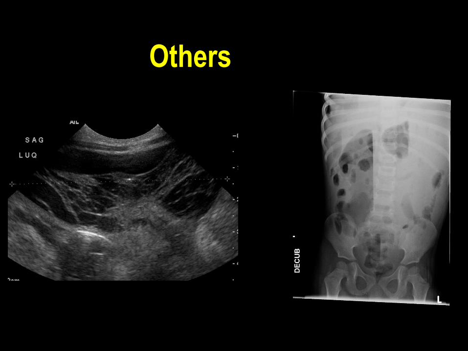
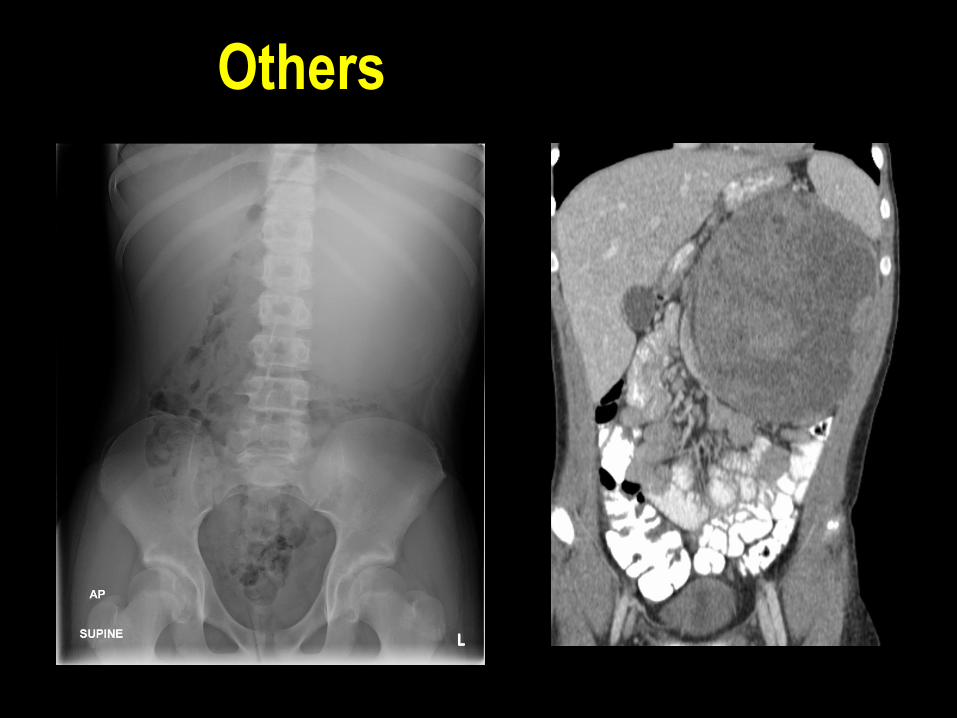
Others
Pneumoperitoneum
Acute abdomen
Calcification
Masses

Normal Gas Pattern
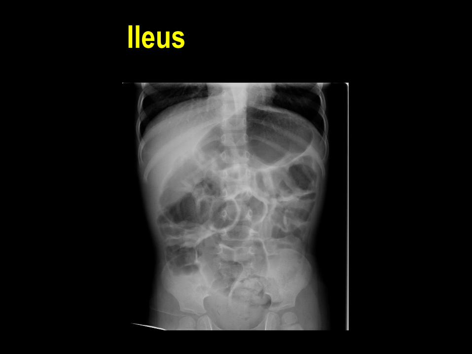
Ileus
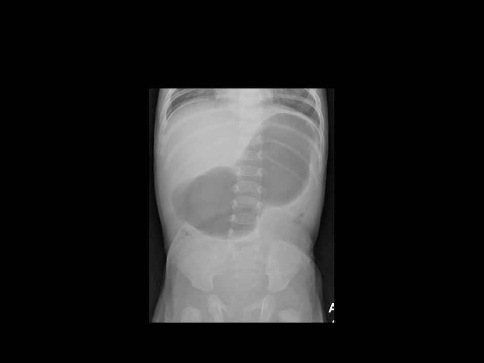


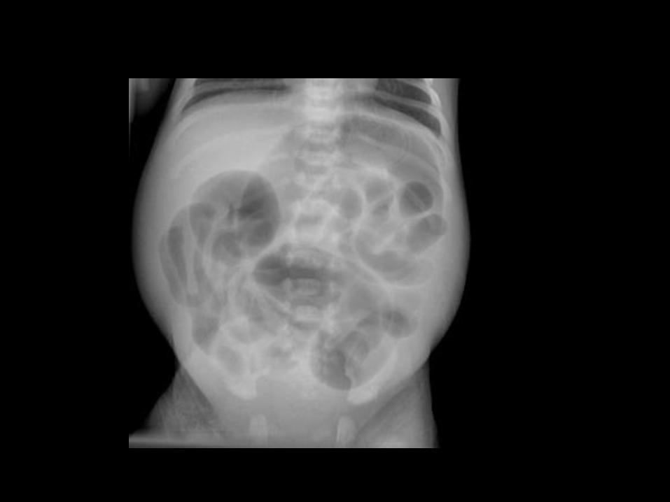
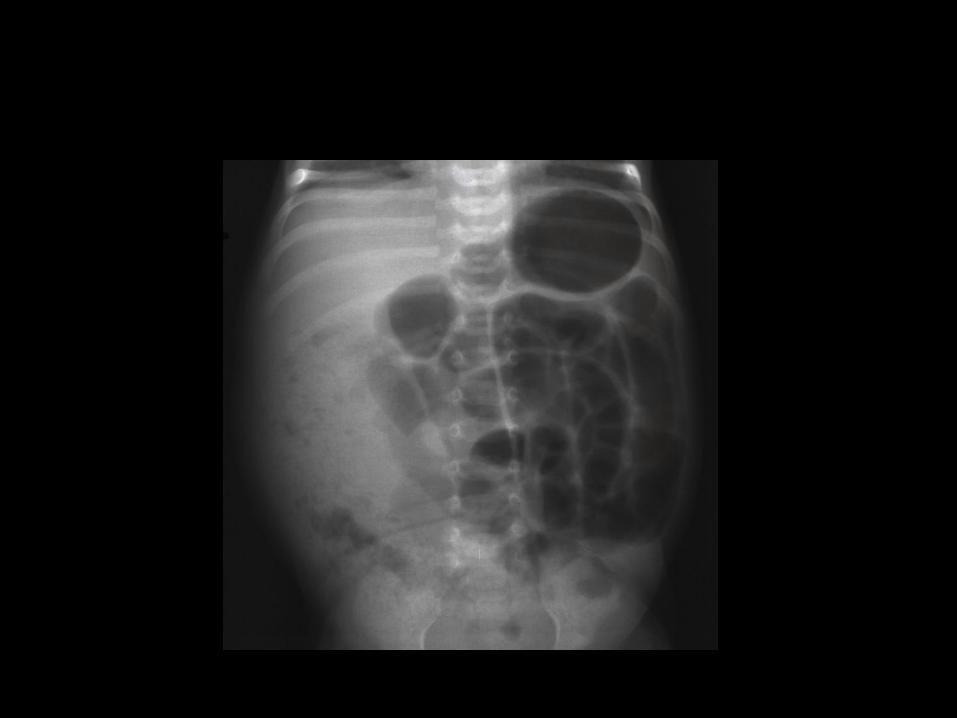
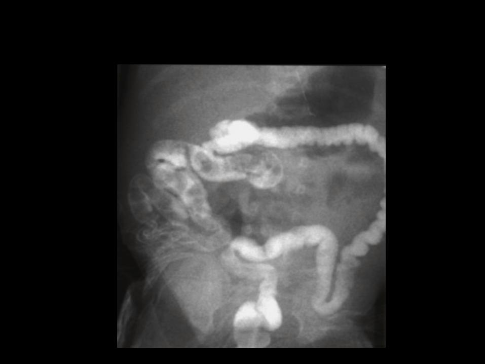
High Obstruction
• Prox to mid-ileum
• Hypoplasia/Atresia of Stomach
• Duodenal Atresia, Stenosis and Web
associated with annular pancreas (20%) ,
Down’s (30%) or part of VATER
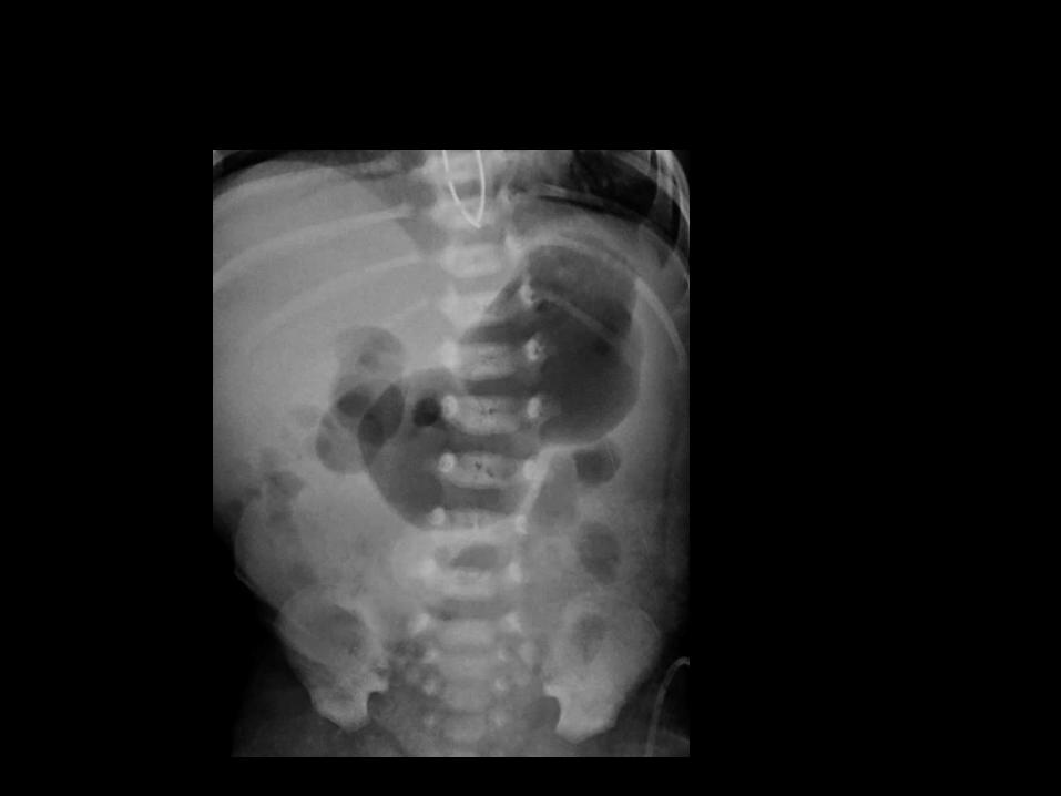
– “Double Bubble”
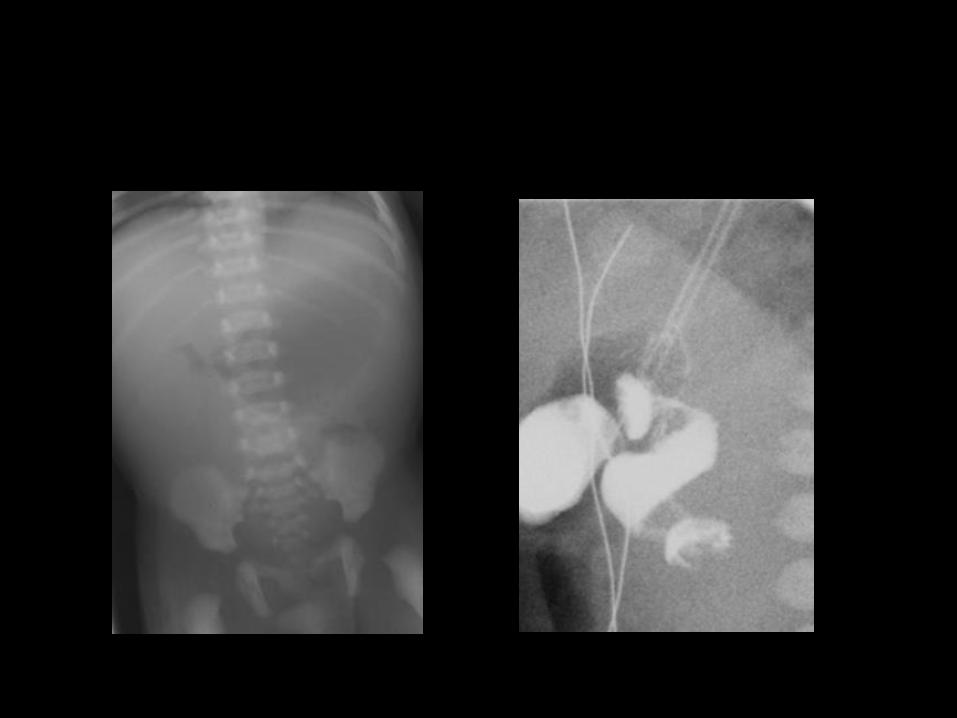
• Malrotation with Midgut Volvulus
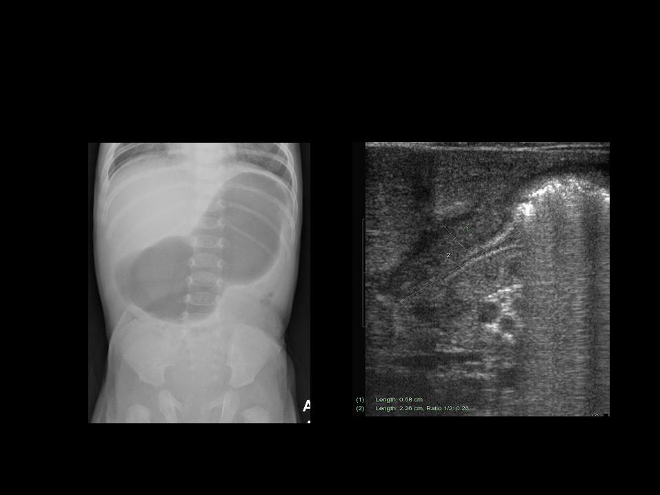
High Obstruction
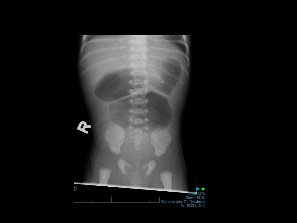
• Jejunal Atresia and Stenosis
– “Triple Bubble”
• Prox Ileal Atresia and Stenosis
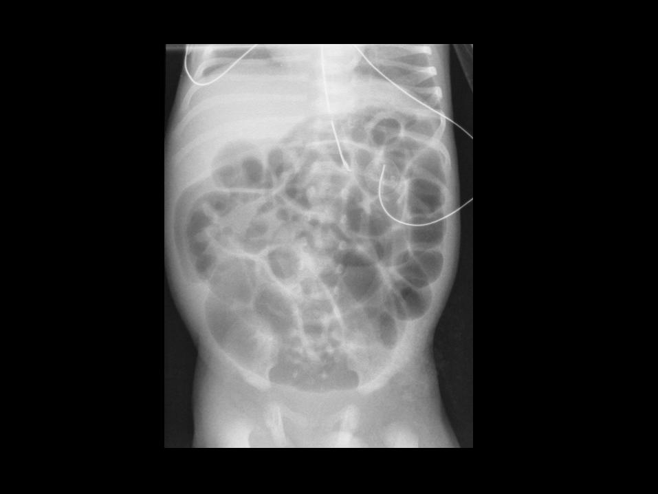

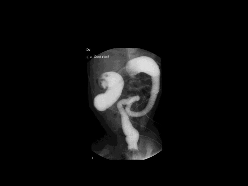
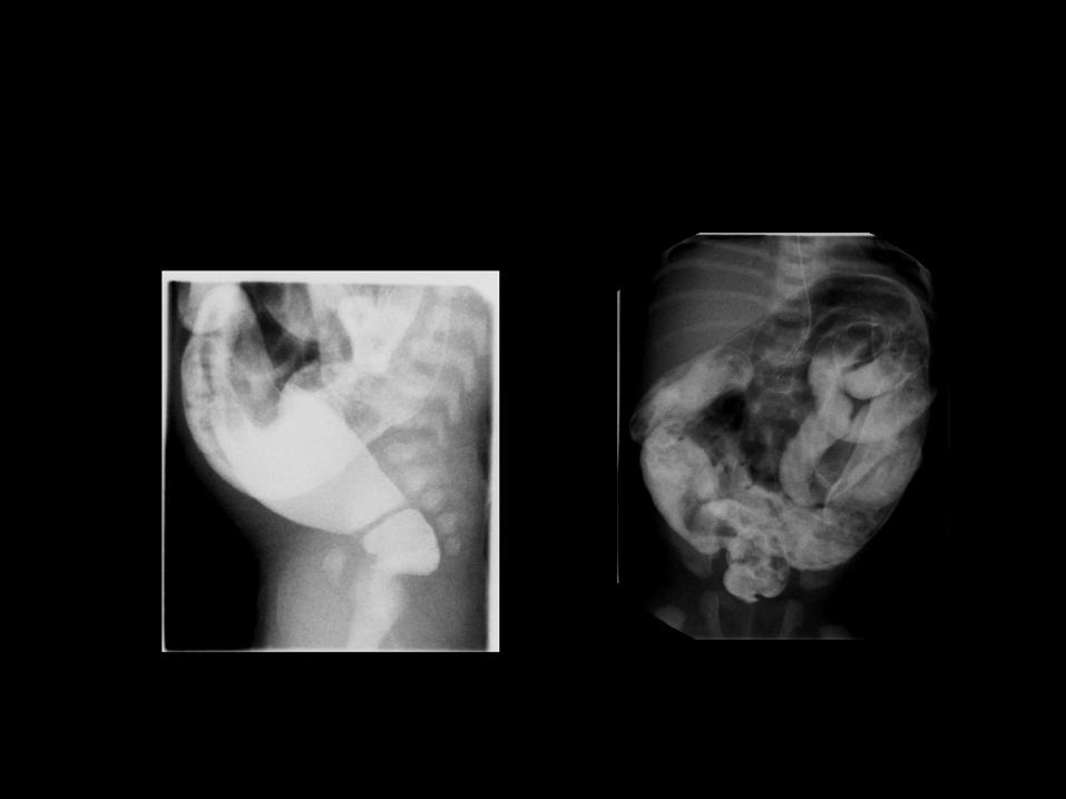

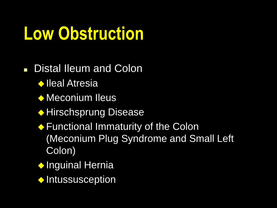
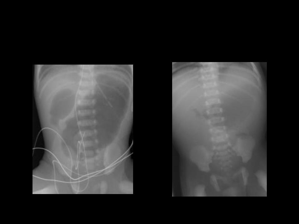
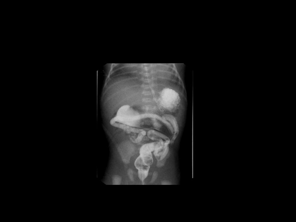
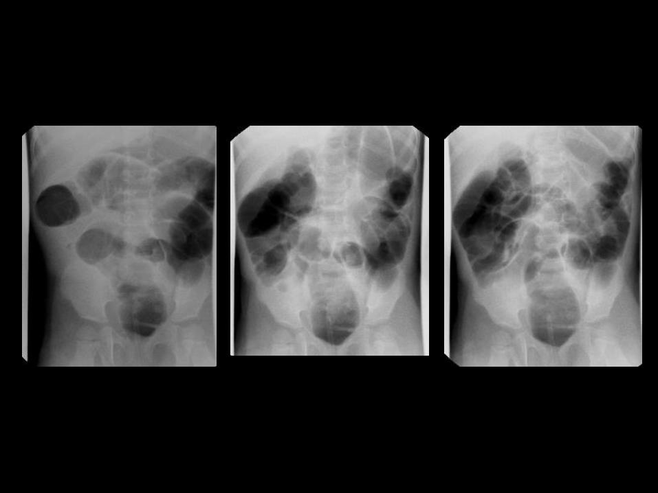
Low Obstruction
Distal Ileum and Colon
Ileal Atresia
Meconium Ileus
Hirschsprung Disease
Functional Immaturity of the Colon
(Meconium Plug Syndrome and Small Left
Colon)
Inguinal Hernia
Intussusception


Others
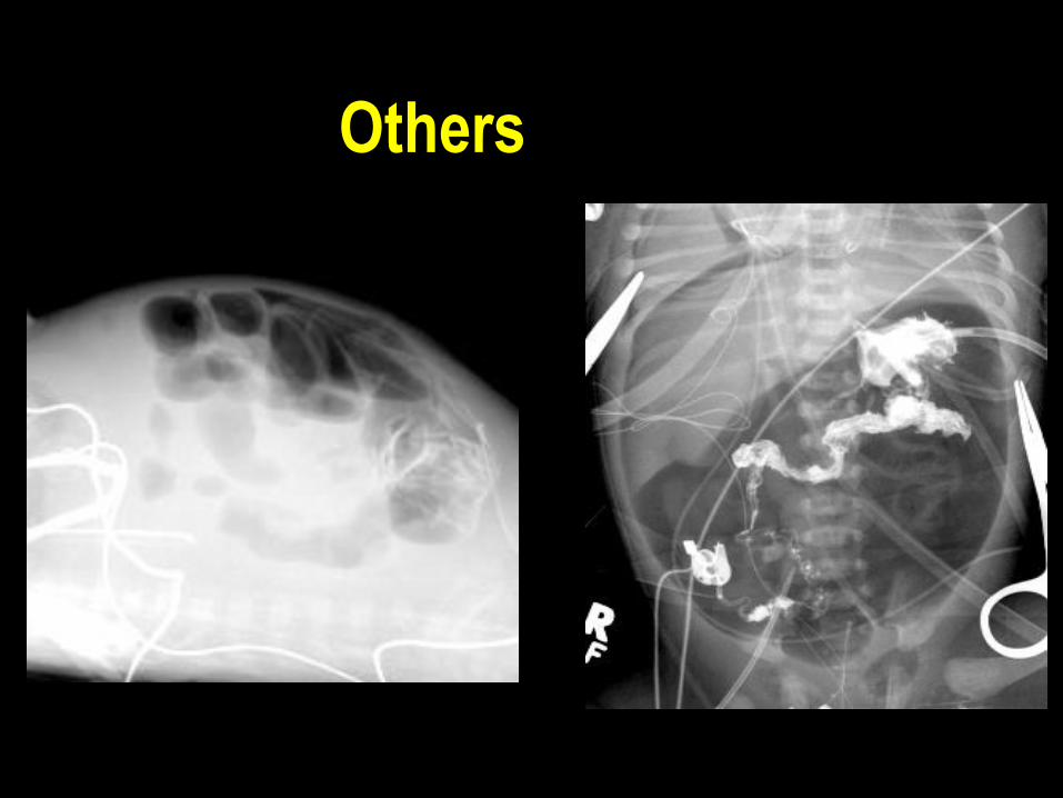
Others

Others
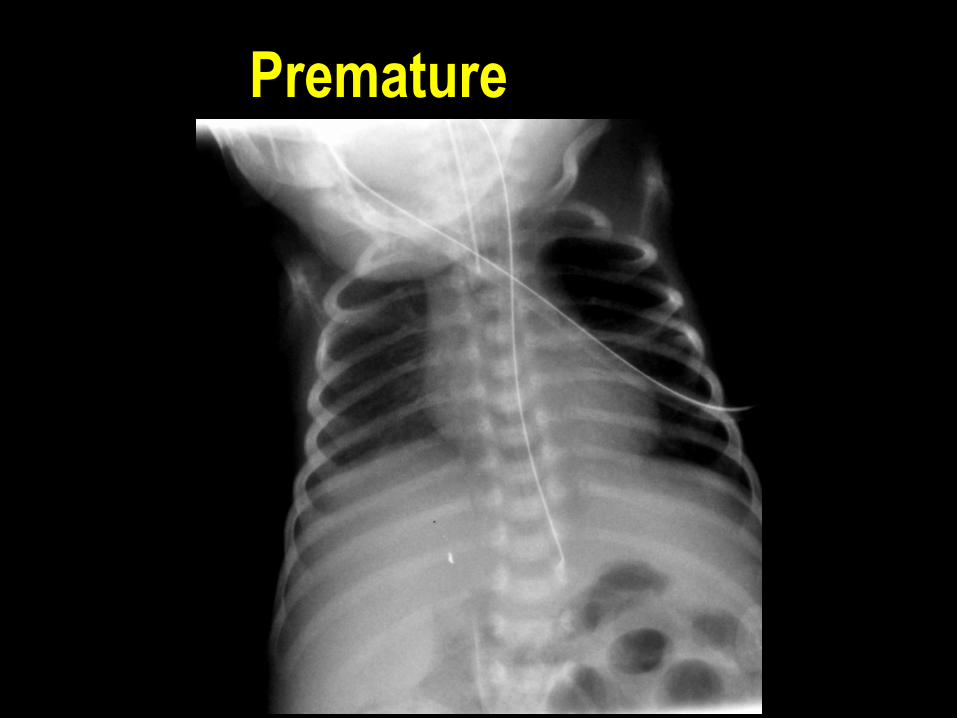
Premature

Others


HRUS & Color Doppler
Necrotizing Enterocolitis: Assessment of Bowel Viability with Color Doppler US
CDS can detect normal neonatal bowel perfusion Normative data
Bowel viability assessment by color Doppler sonography in necrotizing enterocolitis (NEC) Different perfusion patterns in NEC Absence of bowel wall perfusion by CDS is more
sensitive then AXR to detect necrotic bowel in NEC
Faingold et al, Radiology May 2005;235:587-594
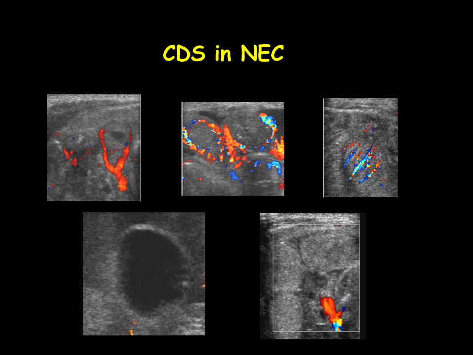
CDS in NEC
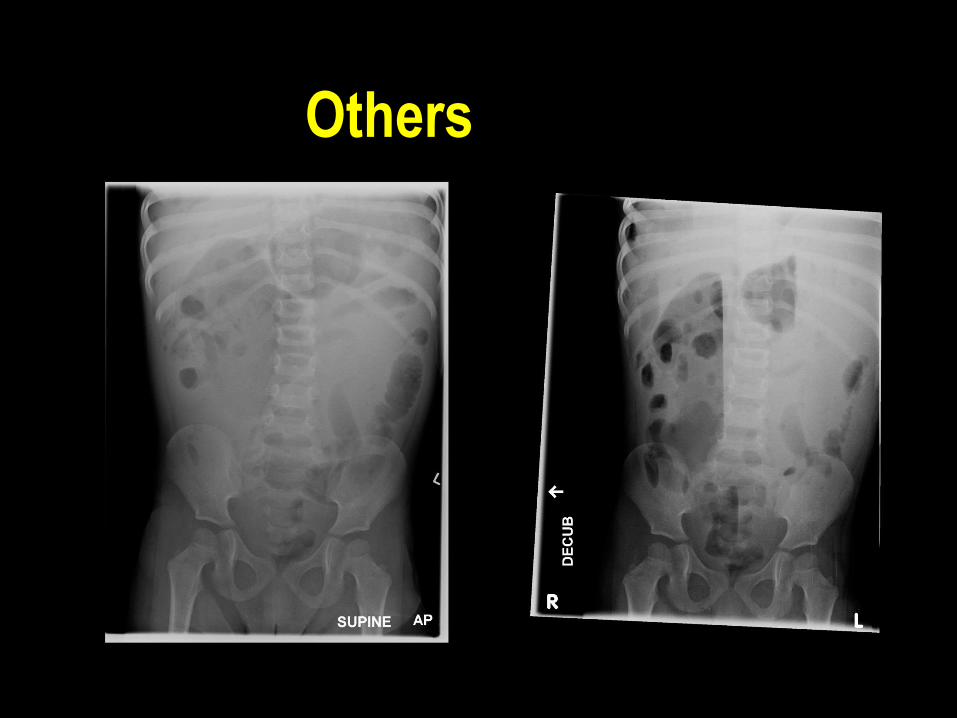
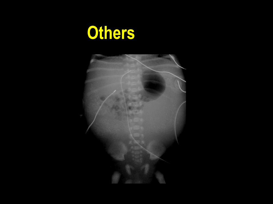
Others
Others
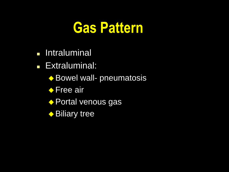
Gas Pattern
Intraluminal
Extraluminal:
Bowel wall- pneumatosis
Free air
Portal venous gas
Biliary tree
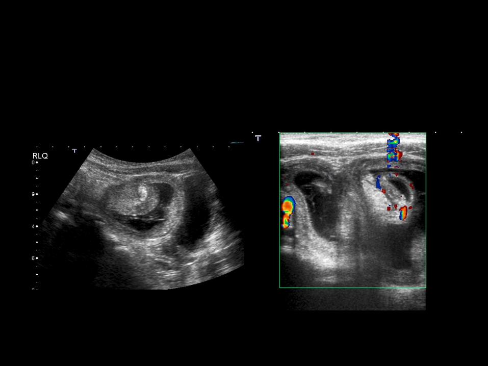
Others

Others

Cysts
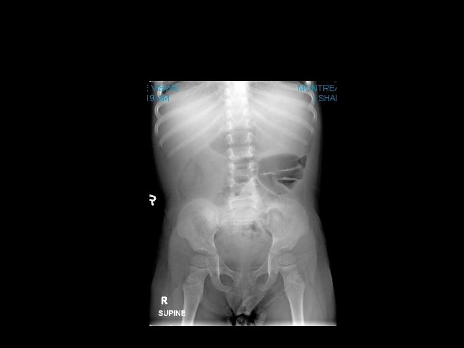
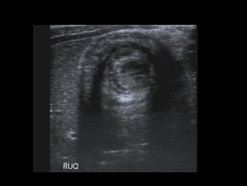
Calcifications
Liths
Stones
Tumors
Meconium peritonitis


Others

Others
Others

Others

Interventional Radiology
Non – Vascular Procedures
Biopsies and Drainages
Gastrointestinal
MSK intervention
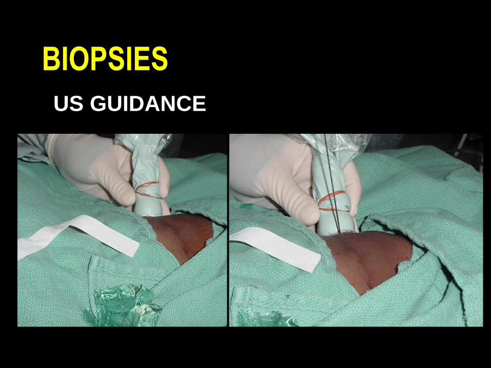
BIOPSIES
US GUIDANCE
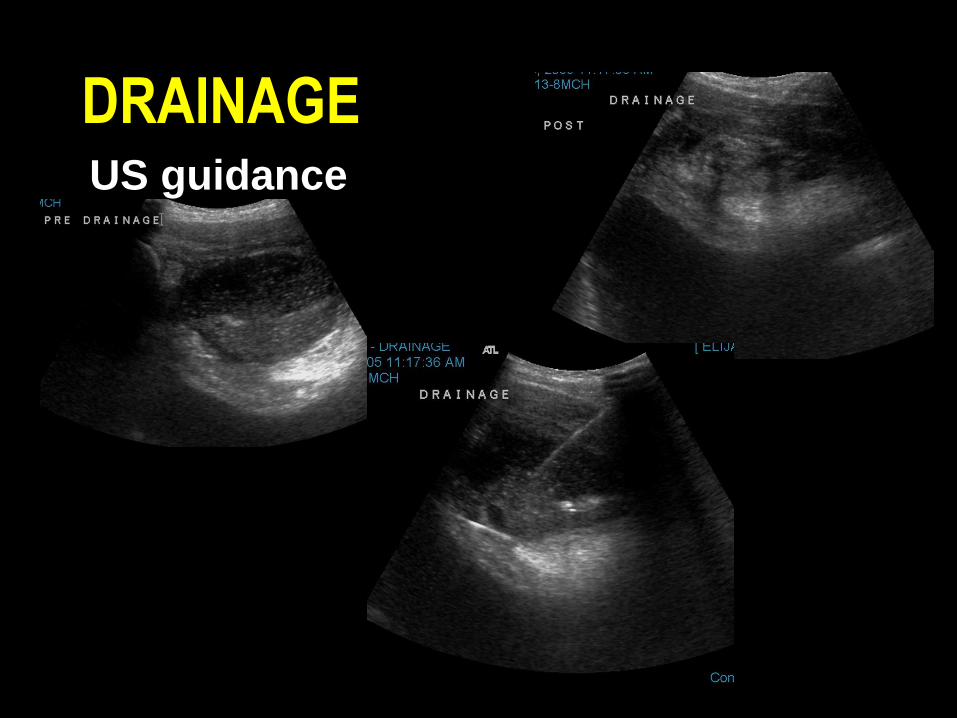
DRAINAGEUS guidance

New Technologies
PACS (Picture Archiving &communication syst.)
Availability,Improvement of Diagnosis
&care
Integration & teaching

New Horizons and Research
MRI, MRA & Functional Imaging
CTA
HRUS & Color Doppler/Perfusion
Assessment


Obrigado!
