pulmonale resultofchronicnasopharyngeal obstruction due … · atresia. he was readmitted to...
TRANSCRIPT

Arch. Dis. Childh., 1969, 44, 585.
Cor Pulmonale as a Result of Chronic NasopharyngealObstruction Due to Hypertrophied Tonsils and
AdenoidsF. J. MACARTNEY, J. PANDAY, and OLIVE SCOTT
From the Department of Paediatric Cardiology, Leeds Regional Thoracic Surgical Centre
It is important to recognize that heart failure inyoung children can be due to causes other thancongenital heart disease. A syndrome has recentlybeen recognized in children which can lead to grosscardiac failure and yet can apparently be per-manently cured simply by removing the tonsils andadenoids. It is the syndrome of cor pulmonale dueto hypoventilation resulting from chronic naso-pharyngeal obstruction. Since the first reports(Menashe, Farrehi, and Miller, 1965; Noonan,1965) others have appeared, bringing the total ofreported cases to 16 (Luke et al., 1966; Levy et al.,1967; Ainger, 1968). We present here the clinical,electrocardiographic, and radiological findings in3 patients, together with the results of cardiaccatheterization in 2 of them.
Case Reports
Case 1. This child, aged 21 years, was first admittedto another hospital when 15 months old, with a historyof marked stridor, particularly at night, a persistentpurulent nasal discharge, and a chest infection. Heimproved after treatment with antibiotics and wasdischarged after a fortnight. 2 months later he was re-admitted with a history of cough and breathlessnessfor 4 days and purulent nasal discharge. Stridor hadbeen noticed which was worse at night, and cyanosishad also been observed when he was asleep. In addition,his parents had noticed that he was drowsy in themorning and that he took a long time to become com-pletely awake. His chest infection was cured by anti-biotics, but the cyanosis at night, and morning drowsinesspersisted. He had no heart murmurs or heart failure,but x-ray of his chest showed cardiac enlargement.An electrocardiogram (ECG) showed evidence of rightatrial and right ventricular hypertrophy.He was referred for a cardiological opinion because of
the unexplained cardiomegaly. When first seen hehad signs of a moderate degree of pulmonary hyper-
Received March 28, 1969.
tension and his heart size was increased radiologically.His ECG showed marked right atrial and right ventri-cular hypertrophy. Primary pulmonary hypertensionwas considered the most likely diagnosis, but someform of cardiomyopathy was also considered. Sincehe had improved and his heart size had decreasedconsiderably by the time we saw him, he was notregarded as an urgent problem and his name was listedfor cardiac catheterization. Within 3 weeks, however,his condition again deteriorated and he was admitted tothe Cardiac Unit as an emergency.On examination he was in gross right heart failure,
with pitting oedema of the lower limbs to the level ofthe groins, marked facial oedema, moderate hepato-megaly, and shifting abdominal dullness. He wasmildly cyanosed but there was no finger clubbing.His facies were adenoidal, and he snuffled and breathedthrough his mouth all the time. At night, or wheneverhe was lying on his back, he developed marked inspira-tory stridor, increasing cyanosis, and sometimes completerespiratory obstruction. It was difficult to rouse himfrom sleep and he did not reach full consciousness for10 minutes. He frequently fell asleep during the day.His tonsils were greatly enlarged, meeting in themid-line. He had moderate sinus tachycardia, a5 cm. A wave in the venous pulse, and a slight rightventricular lift. There were no murmurs, but therewas a loud pulmonary click and the second sound wassingle and much accentuated. No added sounds wereaudible in the chest.ECG (Fig. 1) showed marked right ventricular
hypertrophy and strain, and severe right atrial hyper-trophy. The chest x-ray (Fig. 2) showed a greatlyenlarged heart, with a particularly dilated main pul-monary artery, and diffuse fluffy opacities in both lungfields. Three blood cultures were sterile, and bloodHb was 13-7 g./100 ml.,white cell count 13,200/cu.mm.,and erythrocyte sedimentation rate 1 mm./hr.He was treated with digoxin and large doses of fruse-
mide with potassium supplements, but despite this hedeteriorated, continuing to gain weight, and becomingmore somnolent and cyanosed. He was put in anoxygen tent one evening; the following morning he wasno less cyanosed and it took 20 minutes of vigorous
585
copyright. on F
ebruary 29, 2020 by guest. Protected by
http://adc.bmj.com
/A
rch Dis C
hild: first published as 10.1136/adc.44.237.585 on 1 October 1969. D
ownloaded from

Macartney, Panday, and Scottshaking to rouse him from sleep. The remarkableresemblance between this behaviour and that of a
chronic bronchitic whose respiratory centre has becomeinsensitive to a rise in arterial Pco, led to the correctdiagnosis. O. therapy was discontinued, and an
arterial blood sample taken while the child was stillasleep showed a Pco2 of 69 mm. Hg, a Po., of 65 mm. Hg,and a pH of 7 35. That such a sample could be takenat all is a measure of the depression of his centralnervous system during sleep.
Further medical treatment and training in sleepingin the prone position, which relieved respiratoryobstruction almost completely, improved his conditionsufficiently for cardiac catheterization to be performed.Right heart catheterization was performed from the rightsaphenous vein without sedation; the femoral arterywas also catheterized by an open Seldinger technique.The results (Tables I and II) showed pulmonary
hypertension, the pulmonary artery mean pressure
never dropping below 24 mm. Hg. The highest mean
value (44 mm. Hg) was the first record when the childwas asleep and breathing air, at a time when therewas considerable desaturation of the aortic blood(O2sat. = 70 5",,) and a high Pco, of 76 mm. Hg.Crying was accompanied by a reduction in the pul-monary artery pressure to 26 mm. Hg, and at the same
time, as expected, by a rise in arterial oxygen saturationand a fall in Pco2. The arterial Pco. was abnormallyhigh throughout the test and the arterial 0, saturationabnormally low except when O., was breathed. Itwas not possible to test the effect of breathing 02 whilethe child was asleep, but 1000%, 0, given while he was
crying had little effect. Intubation with an endotrachealtube was carried out but was technically difficultbecause of his short neck, large tonsils and tongue, andlax epiglottis. He required atropine 0 2 mg., andsuxamethonium 10 mg. i.v., followed by fluothane andnitrous oxide before intubation was possible. Thepatient was then allowed to recover consciousness.The figures at 100 min. (Table II) were taken justbefore he woke up, with the endotracheal tube in situ.Shortly afterwards it was coughed out.
Since these results were considered to prove thediagnosis of cor pulmonale secondary to hypoventilationdue to upper respiratory obstruction, his tonsils andadenoids were removed on May 30, 1968. At operationthe epiglottis was short, and was curled and collapsedon to the posterior wall of the pharynx. When theepiglottis was lifted up he had an excellent airway. Thetonsils were large, but no bigger than those often seenin children without stridor or obstruction of the upperairways. The tonsils and adenoids were removedwithout difficulty. Within 24 hours the child was muchimproved and was breathing well without any stridor.The surgeon thought that the respiratory obstructionhad been caused by the combination of the large broadepiglottis and the large tonsils.The child continued to make excellent progress.
When reviewed at Out-patients 1 month after hisoperation he was still mouth breathing at times but was
otherwise well. Examination of his heart was normal,
there being no evidence of pulmonary hypertension.His ECG 1 month after operation showed normal Pwaves in lead II and the R wave in V3R had fallen from21 mm. to 7 mm. (Fig. 3). A further record on October22, 5 months after operation, was normal, and by thistime the child no longer breathed through his mouth.Radiography showed a heart of almost normal size(Fig. 4).
Cardiac catheterization was repeated without anysedation on October 24. The results (Table II)showed a pulmonary artery pressure of 30/5 mm. Hg(mean pressure 12 mm. Hg); 02 saturations showedno evidence of any left-to-right shunt.
Case 2. This child, aged 20 months, had beenadmitted to hospital on numerous occasions during hislife with the diagnosis of asthmatic bronchitis and nasalobstruction. On July 27, 1967, skull x-ray after insertionof Hypaque into both nostrils showed posterior nasalobstruction due to adenoidal pads and excluded choanalatresia. He was readmitted to hospital on April 26,1968, with the same complaints of dyspnoea, nasaldischarge, and noisy breathing, and in the course ofhis stay in hospital developed severe right heart failurewhich required vigorous treatment with digitalis,diuretics, and antibiotics. At no time did he appearsomnolent.At the time of admission on May 20, he had marked
inspiratory stridor particularly in the supine position,slight cyanosis, and early finger clubbing. There wasslight hepatomegaly but no oedema. There was aslight right parasternal heave. Auscultation revealedno abnormality except for an accentuated pulmonarysecond sound. His tonsils were moderately enlarged.There was no nasal discharge.ECG showed right atrial hypertrophy and deep
symmetrical T wave inversion over the right ventricularleads, as far as V4, though on QRS voltage criteriathere was no right ventricular hypertrophy. The frontalQRS axis was -170'. The chest x-ray showedmoderate cardiac enlargement and some congestion ofthe lung fields.Two days later, cardiac catheterization (results in
Table III) was carried out without sedation. Thechild's condition was better than that of Case 1 at thetime of catheterization. The same abnormalities were
present but were not nearly so striking. The arterialPco2 was abnormally raised except during crying, andthe arterial pH correspondingly low. The admini-stration of 02 in the resting state was accompanied bya rise of arterial Pco,. The arterial 02 saturation wasabnormally low, and increased during crying. Therewas slight but definite pulmonary hypertension, whichwas increased during the administration of 02. Thepulmonary artery pressure was increased during cryingdespite the fall in arterial Pco2 and the rise in arterial02 saturation. This child was not intubated.The upper respiratory tract was investigated on
July 17, and enlarged tonsils and adenoids were found,but the epiglottis was normal. His tonsils and adenoidswere removed and he improved. Digoxin was dis-
586
copyright. on F
ebruary 29, 2020 by guest. Protected by
http://adc.bmj.com
/A
rch Dis C
hild: first published as 10.1136/adc.44.237.585 on 1 October 1969. D
ownloaded from

Cor Pulmonale as a Result of Chronic Nasopharyngeal Obstruction 587
FIG. 1. ECG before tonsillectomy in Case 1, shows right FIG. 3.-ECG 1 month after tonsillectomy in Case 1,atrial enlargement (tall peaked P waves in lead II); shows normal P waves in lead II and normal right ventri-right ventricular enlargement (tall R waves in V3R and cular activity.
and V1); T wave inversion from V3R to V5.
FIG. 2.-Chest x-ray before tonsillectomy in Case 1shows marked cardiomegaly.
continued on July 28. When seen 1 month later, therewas no evidence of heart failure and he was breathingwell through his nose with his mouth closed. Hecontinued to make satisfactory progress and there wasno recurrence of heart failure. He died after an accidenton September 10, in which his liver was ruptured.Necropsy was performed and there was no abnormalityof the heart either macroscopically or microscopically.
Case 3. This boy aged 3 years showed the samesyndrome but in a less severe form. He was admitted
FIG. 4.-Chest x-ray in Case 1, 5 months after tonsillectomy,shows marked reduction in heart size compared with Fig. 2.
to hospital for tonsillectomy and adenoidectomybecause of frequent sore throats. He had large tonsilsand cervical glands. The nursing staff reported that hehad cyanotic attacks during the night. No heartmurmurs were heard, but x-ray of his chest showed anenlarged heart (Fig. 5) and he was referred for a cardio-logical opinion. On April 7, 1967, he was mouthbreathing and had minimal cyanosis at rest. There wasprofuse purulent nasal discharge. There were nosignificant heart murmurs, but his chest x-ray showedcardiac enlargement with a large right atrium and
.1- ........::. .:
I ---; -t 7
copyright. on F
ebruary 29, 2020 by guest. Protected by
http://adc.bmj.com
/A
rch Dis C
hild: first published as 10.1136/adc.44.237.585 on 1 October 1969. D
ownloaded from
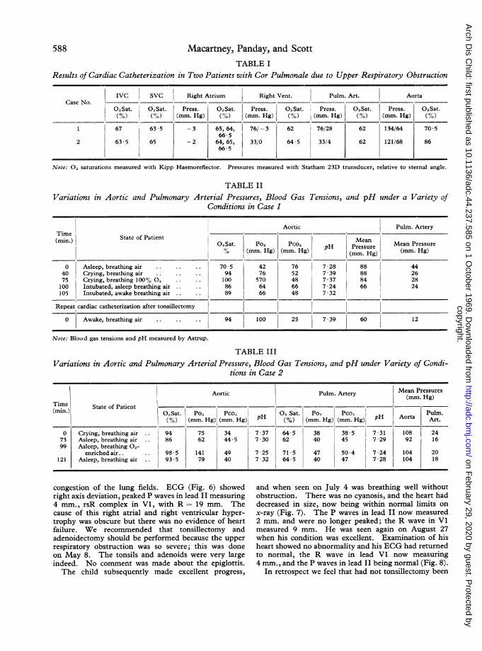
588 Macartney, Panday, and ScottTABLE I
Results of Cardiac Catheterization in Two Patients with Cor Pulmonale due to Upper Respiratory Obstruction
IVC SVC Right Atrium Right Vent. Pulm. Art. Aorta
02Sat. 02Sat. Press. 02Sat. Press. 02Sat. Press. 02Sat. Press. 02Sat.(%) (%) (mm. Hg) (%) (mm. Hg) ('%) (mm. Hg) (%) (mm. Hg) (%)
1 67 63-5 -3 65,64, 76/ -3 62 76/28 62 134/64 70566-5
2 635 65 -2 64, 65, 33/0 645 33/4 62 121/68 86665 °
Note: 02, saturations measured with Kipp Haemoreflector. Pressures measured with Statham 23D transducer, relative to sternal angle.
TABLE IIVariations in Aortic and Pulmonary Arterial Pressures, Blood Gas Tensions, and pH under a Variety of
Conditions in Case 1
Aortic Pulm. ArteryTime(mi.) State of Patient MeanO,Sat. P02 Pco2 H Pesr Mean Pressure
% (mm.Hg (mm. Hg)(mm. Hg) (mm. Hg)
0 Asleep, breathing air . .. . 70 5 42 76 7-28 88 4440 Crying, breathing air . .. . 94 76 52 7 39 88 2675 Crying, breathing 1000% 02 100 570 48 7*37 84 28100 Intubated, asleep breathing air 86 64 66 7-24 66 24105 Intubated, awake breathing air 89 66 48 7-32
Repeat cardiac catheterization after tonsillectomy
0 Awake, breathing air .94 100 25 7 39 60 12
Note: Blood gas tensions and pH measured by Astrup.
TABLE III
Variations in Aortic and Pulmonary Arterial Pressure, Blood Gas Tensions, and pH under Variety of Condi-tions in Case 2
Aortic Pulm. Artery Mean Pressures
(mimn.) tate of Patient Sat. Po Pco Sat. Po, Pco, Pulm.
(%) (mm. Hg) (mm. Hg) pH (%) (mm. Hg) (mm. Hg) pH Aorta Art.
0 Crying, breathing air .. 94 75 34 7-37 64-5 38 38 5 7-31 108 2473 Asleep, breathing air .. 86 62 44-5 7 -30 |62 |40 145 77-29 92 1699 Asleep, breathing 02-
enriched air.. .. 98 *5 141 49 7 *25 71*5 47 50*4 7 *24 104 20121 Asleep, breathing air .. 935 79 40 7 32 64 5 40 47 7 28 104 18
congestion of the lung fields. ECG (Fig. 6) showedright axis deviation, peaked P waves in lead II measuring4 mm., rsR complex in Vl, with R = 19 mm. Thecause of this right atrial and right ventricular hyper-trophy was obscure but there was no evidence of heartfailure. We recommended that tonsillectomy andadenoidectomy should be performed because the upperrespiratory obstruction was so severe; this was doneon May 8. The tonsils and adenoids were very largeindeed. No comment was made about the epiglottis.The child subsequently made excellent progress,
and when seen on July 4 was breathing well withoutobstruction. There was no cyanosis, and the heart haddecreased in size, now being within normal limits onx-ray (Fig. 7). The P waves in lead II now measured2 mm. and were no longer peaked; the R wave in Vlmeasured 9 mm. He was seen again on August 27when his condition was excellent. Examination of hisheart showed no abnormality and his ECG had returnedto normal, the R wave in lead Vl now measuring4 mm., and the P waves in lead II being normal (Fig. 8).
In retrospect we feel that had not tonsillectomy been
copyright. on F
ebruary 29, 2020 by guest. Protected by
http://adc.bmj.com
/A
rch Dis C
hild: first published as 10.1136/adc.44.237.585 on 1 October 1969. D
ownloaded from

Cor Pulmonale as a Result of Chronic Nasopharyngeal Obstruction
FIG. 5.-Chest x-ray before tonsillectomy in Case 3shows increase in transverse diameter, prominent pulmonary
artery, and fluffy hilar opacities.
FIG. 6.-ECG in Case 3 before tonsillectomy shows rightatrial hypertrophy (peaked P waves in lead II), and rightventricular enlargement (tall R wave in V3R and V1).
recommended for this boy, despite the fact that he wasthought to have a cardiac lesion, he would probablyhave progressed in the same way as Case 1, wheretonsillectomy was deferred until the cardiac lesionhad been diagnosed.
DiscussionOur initial experience in recognizing this condi-
tion was very much the same as that of others(Luke et al., 1966; Levy et al., 1967; Ainger,
FIG. 7.-Chest x-ray in Case 3, 2 months after tonsillec-tomy, shows reduction in heart size and pulmonary
vascularity.
.~~~~~~~~~~~~~~~~~~~~~~~~. . ............
....
,r,V3Rt V I V\t 4 W
M:.s; Wl 4 X
FIG. 8.-ECG in Case 3, 15 months after tonsillectomy.Record is now within normal limits.
1968). Preliminary diagnoses of cardiomyopathyor primary pulmonary hypertension were made,and only rejected when we observed the responseto administration of 02, by which time the patient'scondition had become critical. Yet, in retrospect,the clinical features of the disease are so strikingthat a firm diagnosis may be made with no moretechnical assistance than an ECG and an arterialblood sample.
589
copyright. on F
ebruary 29, 2020 by guest. Protected by
http://adc.bmj.com
/A
rch Dis C
hild: first published as 10.1136/adc.44.237.585 on 1 October 1969. D
ownloaded from

Macartney, Panday, and ScottAll 19 cases described in the literature, including
our own, had marked stridor while in the supineposition; in every case this was relieved by removalof tonsils and adenoids. Of these 19, 12 (the mostseverely affected) also showed somnolence, some-times to the point of semiconsciousness. Cyanosiswas also a feature in severe cases. In every casethere was clinical evidence of pulmonary hyper-tension. In 14 this had resulted in right heartfailure which was often considerable, and wasresistant to treatment with digitalis and diuretics.The ECG in all except one (Case 2, Menashe
et al., 1965) showed right atrial hypertrophy, andin all cases except 2 (Case 2, Luke et al., 1966; andCase 2 this series) right ventricular hypertrophy.5 of the 8 published electrocardiograms, includingthose shown here, exhibited striking deep T waveinversion over the right chest leads.
All of the 13 chest x-rays described (including our3 cases) showed cardiomegaly, usually with promi-nence of the pulmonary artery, and 9 of 13 werethought to show pulmonary oedema in the lungfields.
Cardiac catheterization has been performed in 8cases in addition to our own, and pulmonaryhypertension has been the invariable finding exceptin 2 cases (Cases 3 and 4, Luke et al., 1966).Most authors have mentioned the marked dips inpulmonary artery pressure associated with inspira-tion due to the inspiratory obstruction, and ourcases showed this. In every case there has beenarterial hypoxaemia and CO2 retention, and as onestudy in particular has shown, sleep producesprogressively greater hypoxaemia and CO2 retentiontogether with progressive increase in pulmonaryartery pressure (Menashe et al., 1965).
There is an interesting contrast between 2 of thecases described here with respect to the effects ofcrying. In the 2 cases that had cardiac catheteriza-tion, crying produced a rise in arterial Po2 and afall in arterial Pco2 but in Case 1 this was associatedwith a fall in pulmonary artery pressure, whereasin Case 2 the pulmonary artery pressure wasraised during crying. Crying in a normal childwill raise the pulmonary artery pressure by in-creasing the cardiac output. It seems that inCase 1 this effect was more than counteracted bythe pulmonary vasodilatation resultant upon theimprovement in ventilation. In Case 2, whichapproaches more nearly the normal situation,presumably the reduction of pulmonary vascularresistance occasioned by the improvement inventilation was small compared with the increasein cardiac output, so that the over-all effect uponthe pulmonary artery pressure was to raise it.
Little information is available as to the acuteeffects of administration of 02 and improving theairway on these patients. In 1 child studied byNoonan (1965) a rise in pulmonary artery pressurewas produced by administration of 100% 02, thePco2 meanwhile rising from 62 to more than100 mm. Hg. On the other hand Menashe andassociates (1965) found that in their second case 02administration caused a fall in pulmonary arterypressure, despite an increase in CO2 retention.In the case described by Levy et al. (1967), theinsertion of a nasopharyngeal tube to relieve theairway obstruction caused a fall in pulmonaryartery pressure at the same time as a rise in arterial02 saturation and a fall in arterial Pco2. Subse-quent 02 administration caused a small rise inPco2 and a fall in the pulmonary artery pressure.In the first study described here, it was not possibleto test the effect Of 02 administration during sleep,but endotracheal intubation resulted in a markedfall in mean pulmonary artery pressure from44 to 24 mm. Hg, while the arterial 02 saturationrose from 70 5 to 89%, the Pco2 fell from 76 to48 mm. Hg, and the pH rose from 7-28 to 7-32.However, the drugs given in order to assist in-tubation may themselves have affected theseresults. In the second case, intubation was notperformed, but the administration of 02-enrichedair to a point sufficient to raise arterial Po2 from62 to 141 mm. Hg resulted in a rise in meanpulmonary artery pressure from 16 to 20 mm. Hg,while arterial Pco2 rose from 44-5 to 49 mm. Hg,and pH fell from 7 * 30 to 7 * 25.The studies described here confirm that in this
syndrome pulmonary hypertension is associated witha low arterial 02 saturation and a high arterial Pco2,the classical changes of alveolar hypoventilation.They also show that relief of the upper respiratoryobstruction results in return of all these abnormalvalues towards normal, and that administration of02 results in further CO2 retention which maydepress consciousness to dangerous levels, andmay be associated with either a rise or a fall inpulmonary artery pressure. That artificially-imposed upper respiratory obstruction causesreduced ventilation and CO2 retention has beendemonstrated experimentally (Cherniack and Snidal,1956).What factors in the human are responsible for
the control of pulmonary vascular resistance ?There is little doubt that a major factor is arterialPo2, a fall in which causes pulmonary vasoconstric-tion (Euler and Liljestrand, 1946; Fishman, 1961;Enson et al., 1964). The role of arterial Pco2andpH are more controversial, but there seems to be
590
copyright. on F
ebruary 29, 2020 by guest. Protected by
http://adc.bmj.com
/A
rch Dis C
hild: first published as 10.1136/adc.44.237.585 on 1 October 1969. D
ownloaded from

Cor Pulmonale as a Result of Chronic Nasopharyngeal Obstructiona growing amount of evidence for the importantrole of pH in the control of pulmonary vesseltone, since Liljestrand first pointed out thathypoxia, by promoting lactic acid release, increaseshydrogen ion concentration which itself may causepulmonary vasoconstriction (Liljestrand, 1958).The lower the arterial 02 saturation the greater is theeffect of a change in pH, and the higher the arterialpH, the less sensitive is the pulmonary arterypressure to hypoxia (Enson et al., 1964; Harveyet al., 1967). These findings have been confirmedin the newborn calf (Rudolph and Yuan, 1966).Of particular interest in the context of this paperis a report of a study on a patient with the Pick-wickian syndrome, the mechanism of which ispresumably similar to that of cases of cor pulmonaledue to upper respiratory obstruction. Withvoluntary hyperventilation a fall in pulmonaryartery pressure was produced as well as a rise inarterial 02 saturation and arterial pH. Theadministration of 100% 02 caused no change inmean pulmonary artery pressure despite a signifi-cant increase in arterial 02 tension; and withadministration of THAM, there was a decrease inboth hydrogen ion concentration and pulmonaryartery pressure, though the 02 tension remainedessentially unchanged (Vogel and Blount, 1965).In none ofthe cases so far described of cor pulmon-
ale due to upper respiratory obstruction have thechanges in cardiac output been measured; some ofthe rise in pulmonary artery pressure induced by afall in arterial pH may be due to the resultantincrease in cardiac output rather than to an increasein pulmonary vascular resistance.
In the cases reported here, as well as in othersin the literature, there is a striking disparitybetween severity of right heart failure and severityof pulmonary hypertension. Right ventricularpressures of 200 mm. Hg systolic may be toleratedin pulmonary stenosis for some years without heartfailure supervening, yet in this syndrome it appearsthat an increase in right ventricular systolic pressureto 50 mm. Hg for a few weeks may result in rightventricular failure. This may be the result of theacuteness ofthe right ventricular pressure load, or ofdepression of myocardial performance by a combin-ation of hypoxaemia and acidaemia as Ainger(1968) has suggested, but it should be rememberedthat the incidental right ventricular pressuremeasured during right heart catheterization may below by comparison with the pressure levels reachedin the child after a night's sleep, with ever-increasinganoxia and Co2 retention.The clinical implications of the existence of this
syndrome are far reaching. Thousands of tonsil-4
lectomies are performed in this country each yearand yet this complication of tonsillar and adenoidalhypertrophy has not been recognized. Manychildren with tonsils far larger than those of thechildren described here have no respiratory obstruc-tion, and in fact are refused operation because thereis insufficient indication for it. The first childdescribed here had three sibs, all of whom hadrequired tonsillectomy for various reasons, but hewas the only one who developed this syndrome.This suggests that, in order to develop cor pulmonalein the presence of hypertrophied and infected tonsilsand adenoids, some other factor must also beinvolved. The likeliest factor for this is an abnormalreactivity of the pulmonary vasculature to hypoxiasuch as that shown in approximately 20% of normalpersons at altitude, and in some children with smallventricular septal defects (Grover et al., 1963).This mechanism has also been used to explain whynot all grossly fat people develop the Pickwickiansyndrome.A possible explanation for the recognition of this
syndrome in the past few years is that, with thetrend towards conservatism in the management ofinfected tonsils and adenoids, this syndrome hasbeen allowed to develop in children who 15 yearsago would have had a tonsillectomy.
Nevertheless, it remains quite likely that somechildren suffering from this syndrome have beenoperated on and cured without the situation beingappreciated. This happened in Case 3, the firstpatient we saw with early symptoms of this syn-drome. Clearly, the administration of opiate pre-medication to such a child is fraught with thedanger of exacerbating hypoventilation and thusproducing excessive CO2 retention and even higherpulmonary artery pressures. Furthermore, theinduction of anaesthesia is extremely likely to becomplicated by complete respiratory obstructionunless special care is taken.
Ainger (1968) has reported 2 fatalities from thissyndrome, so prompt recognition and treatment areimportant. Cardiac failure should if possible beabolished by medical treatment before operation,though if the failure proves intractable, operationmay have to be carried out as an emergency.
Training the child to sleep in the prone positionmay be as valuable as any drug therapy, and theadministration of 02, if it is given at all, should besubject to the same precautions as in an adultpatient with cor pulmonale due to obstructive air-way disease (i.e. 100% 02 should never be given and02-enriched air should only be given when constantwatch is kept for the signs and symptoms ofincreasing CO2 retention).
591
copyright. on F
ebruary 29, 2020 by guest. Protected by
http://adc.bmj.com
/A
rch Dis C
hild: first published as 10.1136/adc.44.237.585 on 1 October 1969. D
ownloaded from

592 Macartney, Panday, and ScottSummary
Three cases are reported of the syndrome of corpulmonale in children due to chronic nasopharyngealobstruction by hypertrophied tonsils and adenoids.The syndrome is characterized by: (1) Stridor inthe supine position due to nasopharyngeal obstruc-tion by hypertrophied tonsils and adenoids.(2) Somnolence. (3) Pulmonary hypertension.(4) Right heart failure. (5) Arterial hypoxia andhypercarbia. (6) Electrocardiographic changes sug-gestive of right atrial and right ventricularhypertrophy and right ventricular strain. (7)Radiographic appearances of cardiomegaly, dilata-tion of the pulmonary artery, and often pulmonaryoedema.The probable pathogenesis of the syndrome is
that pulmonary hypoventilation leads to arterialhypoxaemia and acidaemia, which in turn lead topulmonary vasoconstriction and hypertension.Cardiac failure should first be treated medically, andthe tonsils and adenoids should subsequently beremoved. Sleeping in the prone position oftenrelieves the upper respiratory obstruction. 02should be given with care since it may exacerbatethe symptoms.
We wish to express our thanks to Dr. A. P. Robertswho referred Case 1, to Dr. E. Allibone who referredCase 2, to Dr. C. S. Livingstone who referred Case 3, andto Sister N. Fitzgerald who nursed 2 of these patients.We would also like to thank Miss M. Lockwood for herwilling secretarial assistance.
REFERENcEsAinger, L. E. (1968). Large tonsils and adenoids in small children
with cor pulmonale. Brit. Heart J., 30, 356.
Cherniack, R. M., and Snidal, D. P. (1956). The effezt of obstruc-tion to breathing on the ventilatory response to CO,. j. clin.Invest., 35, 1286.
Enson, Y., Giuntini, C., Lewis, M. L., Morris, T. Q., Ferrer, M. I.,and Harvey, R. M. (1964). The influence of hydrogen ionconcentration and hypoxia on the pulmonary circulation.ibid., 43, 1146.
Euler, U. S. von, and Liljestrand, G. (1946). Observations on thepulmonary arterial blood pressure in the cat. Acta physiol.scand., 12, 301.
Fishman, A. P. (1961). Respiratory gases in the regulation of thepulmonary circulation. Physiol. Rev., 41, 214.
Grover, R. F., Vogel, J. H. K., Averill, K. H., and Blount, S. G., Jr.(1963). Pulmonary hypertension: individual and speciesvariability relative to vascular reactivity. Amer. Heart J.,66, 1.
Harvey, R. J., Enson, Y., Betti, R., Lewis, M. L., Rochester, D. F.,and Ferrer, M. I. (1967). Further observations on the effectof hydrogen ion on the pulmonary circulation. Circulation,35, 1019.
Levy, A. M., Tabakin, B. S., Hanson, J. S., and Narkewicz, R. M.(1967). Hypertrophied adenoids causing pulmonary hyper-tension and severe congestive heart failure. New Engl. J. Med.,277, 506.
Liljestrand, G. (1958). Chemical control of the distribution of thepulmonary blood flow. Acta physiol. scand., 44, 216.
Luke, M. J., Mehrizi, A., Folger, G. M., Jr., and Rowe, R. D. (1966).Chronic nasopharyngeal obstruction as a cause of cardiomegaly,cor pulmonale, and pulmonary edema. Pediatrics, 37, 762.
Menashe, V. D., Farrehi, C., and Miller, M. (1965). Hypo-ventilation and cor pulmonale due to chronic upper airwayobstruction. J. Pediat., 67, 198.
Noonan, J. A. (1965). Reversible cor pulmonale due to hyper-trophied tonsils and adenoids: studies in two cases. Amer.*ediat. Soc. Inc., May 6-8 (Abstracts), p. 48.
Rudolph, A. M., and Yuan, S. (1966). Response of the pulmonaryvasculature to hypoxia and hydrogen ion concentration changes.J. clin. Inzest., 45, 399.
Vogel, J. H. K., and Blount, S. G., Jr. (1965). The role of hydrogenion concentration in the regulation of pulmonary artery pressure.Circulation, 32, 788.
Correspondence to Dr. Olive Scott, Department ofPaediatric Cardiology, Leeds Regional Thoracic SurgeryCentre, Killingbeck Hospital, York Road, Leeds.
copyright. on F
ebruary 29, 2020 by guest. Protected by
http://adc.bmj.com
/A
rch Dis C
hild: first published as 10.1136/adc.44.237.585 on 1 October 1969. D
ownloaded from