blood gases, ph, and buffer system. introduction acid: substances that yields h ions in h 2 o. base:...
TRANSCRIPT

Blood Gases, pH, and
Buffer system
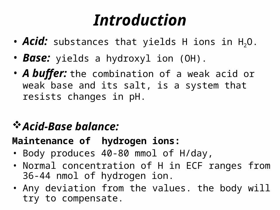
Introduction• Acid: substances that yields H ions in H2O.
• Base: yields a hydroxyl ion (OH).
• A buffer: the combination of a weak acid or weak base and its salt, is a system that resists changes in pH.
Acid-Base balance: Maintenance of hydrogen ions: • Body produces 40-80 mmol of H/day, • Normal concentration of H in ECF ranges from 36-44 nmol of
hydrogen ion. • Any deviation from the values. the body will try to compensate.
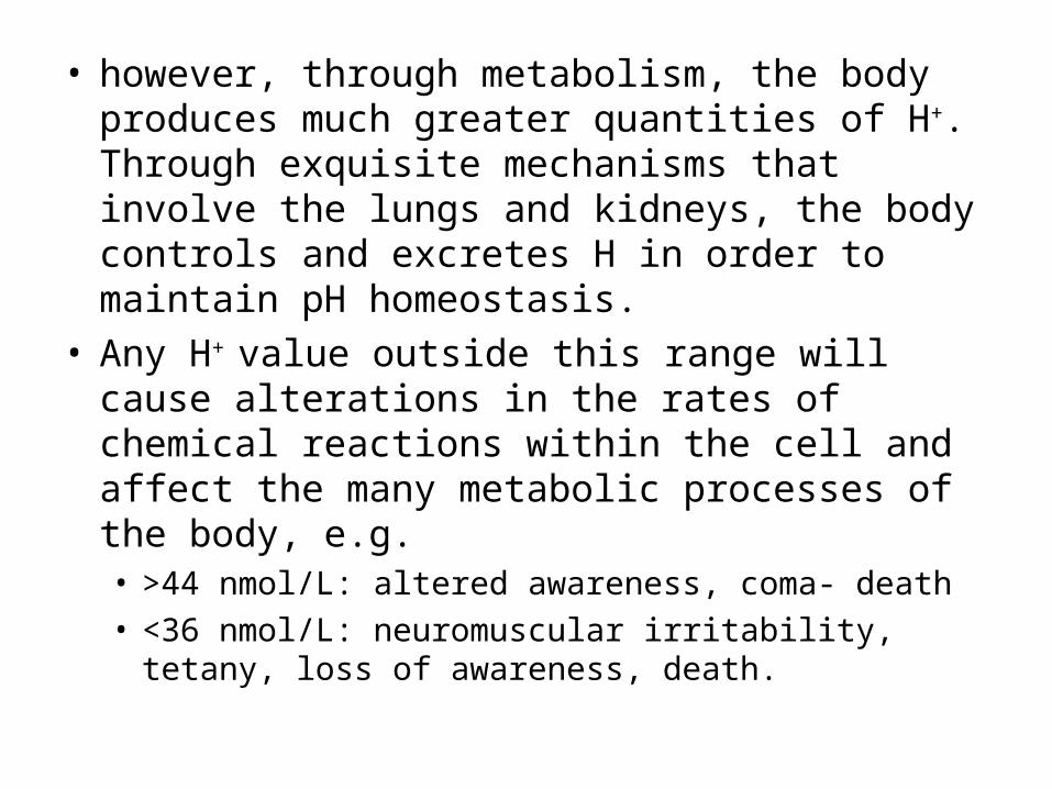
• however, through metabolism, the body produces much greater quantities of H+. Through exquisite mechanisms that involve the lungs and kidneys, the body controls and excretes H in order to maintain pH homeostasis.
• Any H+ value outside this range will cause alterations in the rates of chemical reactions within the cell and affect the many metabolic processes of the body, e.g. • >44 nmol/L: altered awareness, coma- death
• <36 nmol/L: neuromuscular irritability, tetany, loss of awareness, death.

• Reciprocal relationship in the concentration of H ions and pH
• Increase pH: decrease in H ion
• Decrease pH: increase H ions
• The reference value for arterial blood pH is 7.40 and is equivalent to an H concentration of 40 nmol/L.
• Arterial blood pH is controlled by:
1. Buffers
2. Respiratory System
3. Kidneys
Concentration of H ions and pH

Buffer System: Regulation of H+
• First line of defense to changes in H+ concentration is the buffer systems present in all body fluids. All buffers consist of a weak acid carbonic acid (H2CO3) & its salt (HCO3) bicarbonate.
• Add acid to the bicarbonate-carbonic acid system- the HCO3 combines with H from the acid to form H2CO3.
• Add a base to the system, H2CO3 combines with OH to form H2O and HCO3
• Keeps the body at the correct pH (7.35-7.45).
• Bicarbonate: carbonic acid system has low buffering capacity but still an important buffer system for 3 reasons
1. H2CO3 dissociates into CO2 & H2O allowing H+ to be eliminated as CO2 by lungs
2. Changes in PCO2 modify the ventilation rate
3. HCO3 conc. can be altered by kidney

Other systems• HPO4, H2PO4system: plays a role in plasma and red
blood cells and is involved in the exchange of sodium ion in the urine H+ filtrate.
• Proteins are capable of binding H+: Most circulating proteins have a net negative charge and are capable of binding H.
• The lungs regulate pH through retention or elimination of CO2 by changing the rate and volume of ventilation and the kidneys regulate pH by excreting acid, primarily in the ammonium ion,

Regulation of Acid-Base Balance: Lungs
• Carbon dioxide, the end product of most aerobic metabolic processes, easily diffuses out of the tissue where it is produced and into the plasma and red cells in the surrounding capillaries.
• In plasma, a small amount of CO2 is physically dissolved or combined with proteins to form carbamino compounds.
• Most of the CO2 combines with H2O to form H2CO3, which quickly dissociates into H+ and HCO3

Regulation of Acid-Base Balance: Lungs
RBC regulation• CO2 and O2 exchange, some CO2 remains in the RBC in
combination to HB (carboxyhemoglobin)• CO2 combines to water to form carbonic acid and is transported
in the blood.• Carbonic anhydrase enzymes in the RBC accelerate this process • (CO2 + H2O→H2CO3(.• The dissociation of H2CO3 causes the HCO3 concentration to
increase in the red cells and diffuse into the plasma.• To maintain electroneutrality (the same number of positively
and negatively charged ions on each side of the red cell membrane), chloride diffuses into the cell. This is known as the chloride shift. Plasma proteins and plasma buffers combine with the freed H to maintain a stable pH.


• Plasma proteins and plasma buffers combine with the freed H+ to maintain a stable pH.
• In the lungs, the process is reversed. Inspired O2 diffuses from the alveoli into the blood and is bound to hemoglobin, forming oxyhemoglobin (O2Hb).
• The H+ that was carried on the (reduced) hemoglobin in the venous blood is released to recombine with HCO3 to form H2CO3, which dissociates into H2O and CO2.
• The CO2 diffuses into the alveoli and is eliminated through ventilation.

Interrelationship of the bicarbonate and hemoglobin buffering systems.

• When the lungs do not remove CO2 at the rate of its production (as a result of decreased ventilation or disease), it accumulates in the blood, causing an increase in H+ concentration.
• If, however, CO2 removal is faster than production (hyperventilation), the H+ concentration will be decreased

Acid-Base Disorders
• Acidosis (decrease pH) → acidemia • vs. Alkalosis (increased pH) → alkalemia
• Metabolic (kidneys) or respiratory (Lung)• Inadequate elimination and excess production of CO2
in the body.• Body compensates by respiration rate and kidney.

Acidosis1. Metabolic (non-respiratory) Acidosis
Metabolic acidosis is defined as a bicarbonate level of less than 22 mEq/L with a pH of less than 7.35.
Reduce excretion of acids (Diarrhea and intestinal fistulas may cause decreased levels of base).
the treatment of metabolic acidosis is dependent upon the cause.
Causes of increased acids include:• Renal failure
• Diabetic ketoacidosis
• Anaerobic metabolism
• Starvation

2. Respiratory Acidosis: Is defined as a pH less than 7.35 with a PaCO2 greater than 45
mm Hg.Caused by hypoventilation (decrease the elimination of CO2 in
the lungs, it builds up in the blood) In plasma → increase in CO2, decrease in pH, increase in H and
HCO3
Diseases: emphysema, drugs , congestive heart failure, bronchopneumonia.
• Respiratory compensation Hyperventilation
• Renal compensationIncrease H excretion & increase reabsorption of HCO3
-
Acidosis

Signs and Symptoms of Respiratory Acidosis
Pulmonary • dyspnea • respiratory distress
Neurological • headache • Restlessness (االرق) • confusion
Cardiovascular • tachycardia

Alkalosis1. Metabolic alkalosis:
Metabolic alkalosis is defined as a bicarbonate level greater than 26 mEq/liter with a pH greater than 7.45.
An excess of base or a loss of acid within the body can cause metabolic alkalosis.
Metabolic alkalosis is one of the most difficult acid-base imbalances to treat. Bicarbonate excretion through the kidneys can be stimulated with drugs such as acetazolamide (Diamox®), but resolution of the imbalance will be slow.

Signs and Symptoms of Metabolic Alkalosis
Pulmonary • Respiratory depression
Neurological• Seizures• coma
Musculoskeletal• Weakness• muscle cramps• tetany
Gastrointestinal • Nausea• vomiting

2. Respiratory alkalosis: Respiratory alkalosis is defined as a pH greater
than 7.45 with a PaCO2 less than 35 mm Hg. Any condition that causes hyperventilation can
result in respiratory alkalosis.
These conditions include:• Increased metabolic demands, such as fever, sepsis,
pregnancy.
• Medications, such as respiratory stimulants
• Central nervous system lesions
Alkalosis
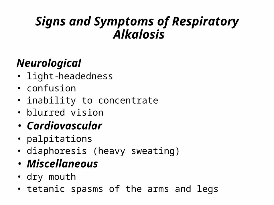
Signs and Symptoms of Respiratory Alkalosis
Neurological • light-headedness • confusion • inability to concentrate • blurred vision
• Cardiovascular• palpitations • diaphoresis (heavy sweating)
• Miscellaneous • dry mouth • tetanic spasms of the arms and legs

• Blood gases: is a measurement of how much oxygen and carbon dioxide is in your blood. It also determines the acidity (pH) of your blood.
• Blood gas measurements are used to evaluate a person's lung function and acid/base balance.
• arterial blood gas (ABG) test: measures the acidity (pH) and the levels of oxygen and carbon dioxide in the blood from an artery. This test is used to check how well your lungs are able to move oxygen into the blood and remove carbon dioxide from the blood.

Blood Gas measures • Partial pressure of oxygen (PaO2): This measures the
pressure of oxygen dissolved in the blood.• Partial pressure of carbon dioxide (PaCO2): This
measures how much carbon dioxide is dissolved in the blood.
• pH: The pH measures hydrogen ions (H+) in blood.• Bicarbonate (HCO3): Bicarbonate is a chemical
(buffer) that keeps the pH of blood from becoming too acidic or too basic.
• Oxygen saturation (O2Sat) values: Oxygen saturation measures how much of the hemoglobin in the red blood cells is carrying oxygen (O2).

Why the Test is Performed• The test is used to evaluate respiratory diseases and
conditions that affect the lungs.
• It helps determine the effectiveness of oxygen therapy.
• The test also provides information about the body's acid/base balance, which can reveal important clues about lung and kidney function and the body's general metabolic state.
• Check for severe breathing problems and lung diseases, such as asthma, cystic fibrosis.
• Find out if you need extra oxygen or help with breathing (mechanical ventilation).
Sampling
• Usually, Blood is most commonly drawn from the radial artery because it is easily accessible.
• The femoral artery is also used, especially during emergency situations or with children.
• The health care provider will insert a small needle through the skin into the artery. You can choose to have numbing medicine (anesthesia) applied to the site before the test begins.
• In some cases, blood from a vein may be used, the sample must be quickly sent to a laboratory for analysis to ensure accurate results.

• There are plastic and glass syringes used for blood gas samples. Most syringes come pre-packaged and contain a small amount of heparin, to prevent coagulation.
• Once the sample is obtained, care is taken to eliminate visible gas bubbles, as these bubbles can dissolve into the sample and cause inaccurate results.
• If a plastic blood gas syringe is used, the sample should be transported and kept at room temperature and analyzed within 30 min.
• Heparin is the only anticoagulant that is suitable for pH and blood gas determination.
• The specimen should delivered immediately to the laboratory and analyzed.
• Normal specimen stable for to 2 hours


How It Feels• Collecting blood from an artery is more painful than
collecting it from a vein because the arteries are deeper and are protected by nerves.
• Most people feel a brief, sharp pain as the needle to collect the blood sample enters the artery. If you are given a local anesthetic, you may feel nothing at all from the needle puncture, or you may feel a brief sting or pinch as the needle goes through the skin.
• You may feel more pain if the person drawing your blood has a hard time finding your artery, your artery is narrowed, or if you are very sensitive to pain.

Risks• There is very little risk when the procedure is done correctly.
• Taking blood from some people may be more difficult than from others.
• Other risks associated with this test may include:• Bleeding at the puncture site
• Blood flow problems at puncture site (rare)
• Bruising at the puncture site
• Delayed bleeding at the puncture site
• Fainting or feeling light-headed
• Hematoma (blood accumulating under the skin)
• Infection (a slight risk any time the skin is broken)

31
Error in the collection of blood gas specimen:
Syringes• Most of the plastic syringes available today are acceptable
Inexpensive, disposable, non- breakable syringe are preferable to glass syringes for cost and safety reasons.
• Sample should not be sent to the lab with the needle still on the syringe.
Vacuum tube• Vacuum collection tubes are to be avoided. • There will be dead air space after the tube filled and this will
result of decreasing of pO2 and pCO2.• The tube must be opened.

Over dilution of specimen with Heparin
• An excessive amount of heparin will dilute the specimen
• An insufficient amount of heparin will result of small fibrin clot, which are capable of disabling pH\blood gas analyzer.
• Kits with syringes containing premeasured amount of powdered heparin are available.
• CO2 will travel to plasma to air space and resulting in decrease of pCO2 and increase of pH

Blood gases (Result)
• Partial pressure of oxygen (PaO2): Greater than 80 mm Hg .
• Partial pressure of carbon dioxide (PaCO2): 35-45 mm Hg .
• pH: 7.35-7.45.
• Bicarbonate (HCO3): 23-30 mEq/L, (23-30 mmol/L).
• Oxygen saturation (O2Sat): 95%-100% .
• Remember: pCO2 >45 = acidosis, pCO2 <35 = alkalosis.
• Remember: HCO3 > 26 = alkalosis, HCO3 < 22 = acidosis.

Gas content in lungs and pulmonary and systemic circulation.

What Affects the Test• Reasons you may not be able to have the test or why the
results may not be helpful include the following:• You have a fever or an abnormally low body
temperature (hypothermia).• You have a disease that affects how much oxygen is
carried in your blood, such as severe anemia.• You smoke just before the test or breathe secondhand
smoke, carbon monoxide, or certain paint or varnish removers in closed or poorly ventilated areas.

Factors effecting the affinity of Hb for O2
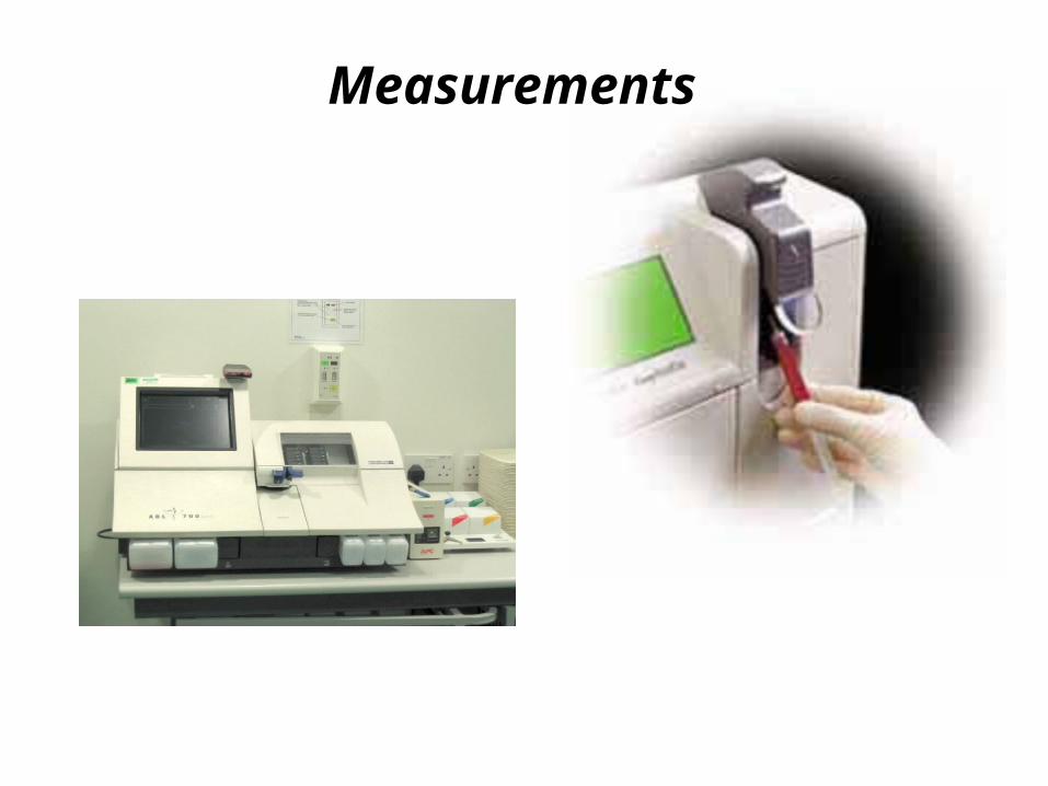
Measurements

• Determination of Oxygen Saturation.
• Designed to directly measure the various hemoglobin species.
• Each species of hemoglobin has a characteristic absorbance curve.
• The number of hemoglobin species measured will depend on the number and specific wavelengths incorporated into the instrumentation.
• For example, two-wavelength instrument systems can measure only two hemoglobin species (i.e., O2Hb and HHb).
Spectrophotometric ( co-oximeter)
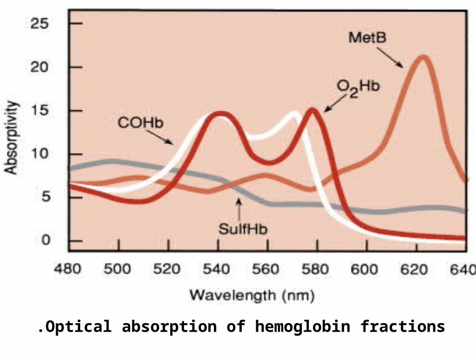
Optical absorption of hemoglobin fractions.

• Because the primary purpose of determining O2Hb is to assess oxygen transport from the lungs, it is best to stabilize the patient’s ventilation status before blood sample collection.
• An appropriate waiting period before the sample is drawn should follow changes in supplemental O2 or mechanical ventilation.
• All blood samples should be collected under anaerobic conditions and mixed immediately with heparin or other appropriate anticoagulant.
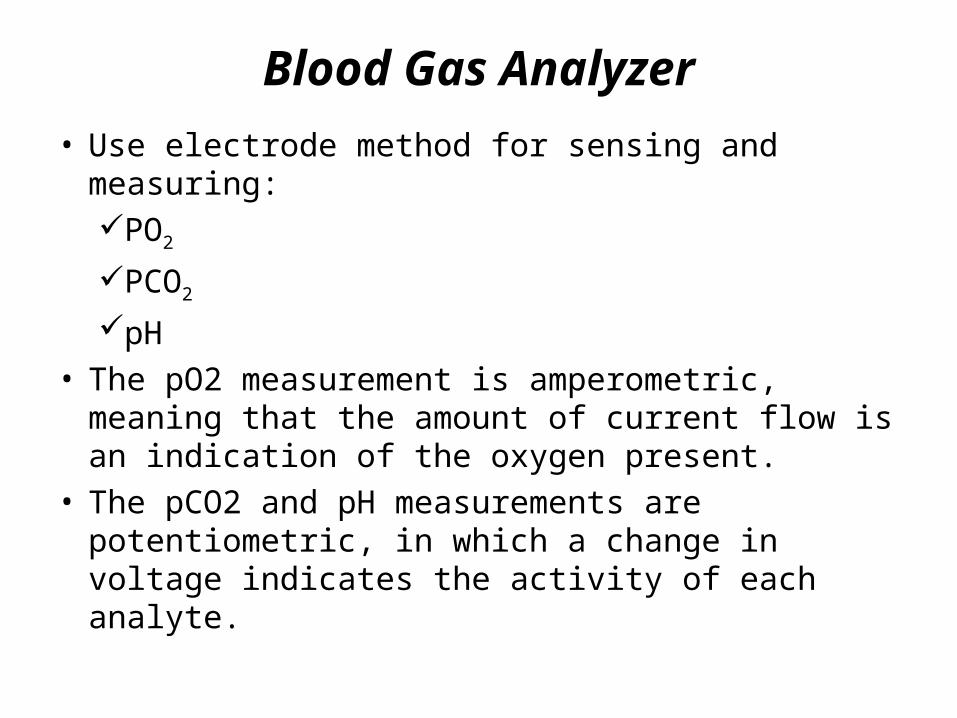
• Use electrode method for sensing and measuring:PO2
PCO2
pH
• The pO2 measurement is amperometric, meaning that the amount of current flow is an indication of the oxygen present.
• The pCO2 and pH measurements are potentiometric, in which a change in voltage indicates the activity of each analyte.
Blood Gas Analyzer
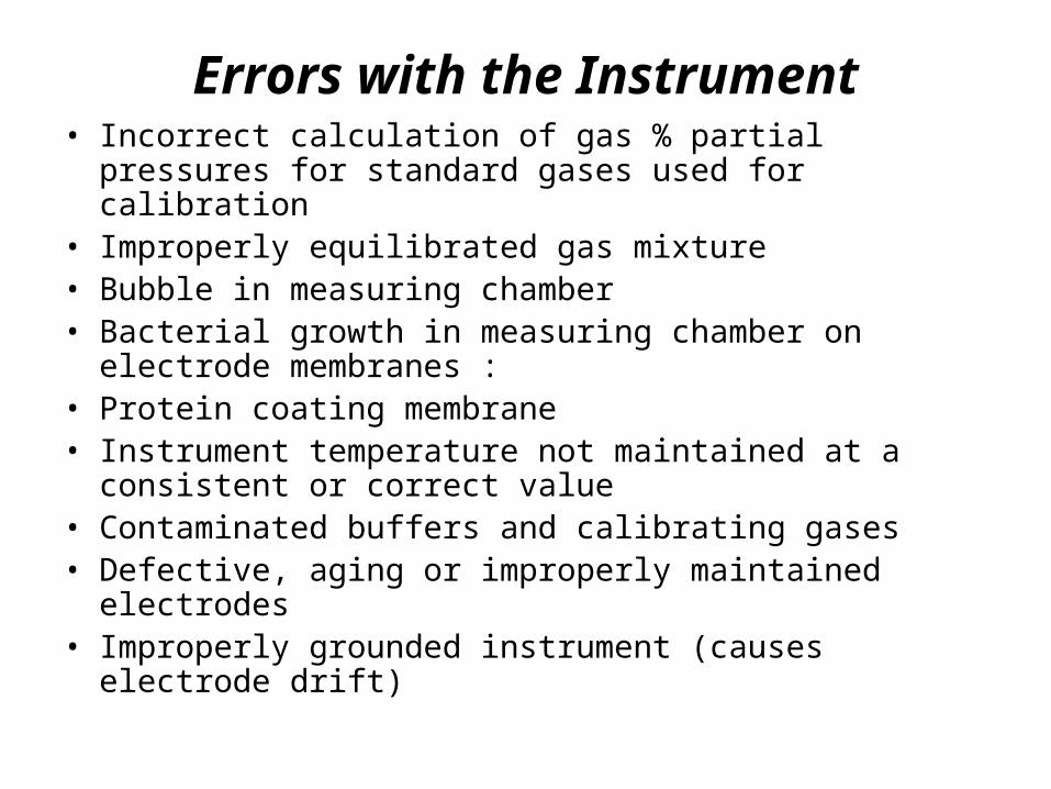
Errors with the Instrument• Incorrect calculation of gas % partial pressures for standard
gases used for calibration• Improperly equilibrated gas mixture• Bubble in measuring chamber• Bacterial growth in measuring chamber on electrode
membranes :• Protein coating membrane• Instrument temperature not maintained at a consistent or
correct value• Contaminated buffers and calibrating gases• Defective, aging or improperly maintained electrodes• Improperly grounded instrument (causes electrode drift)
Steps to an Arterial Blood Gas Interpretation
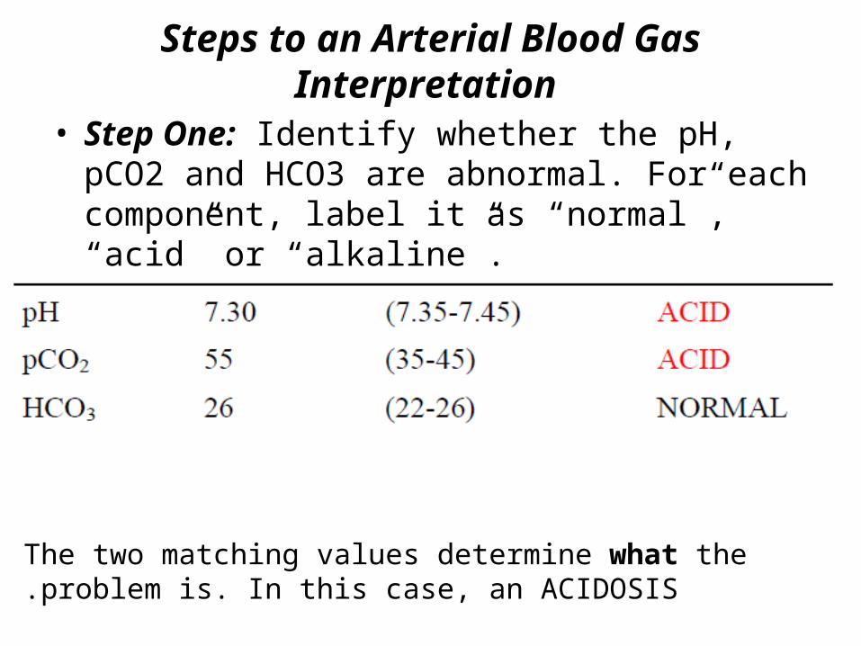
• Step One: Identify whether the pH, pCO2 and HCO3 are abnormal. For each component, label it as “normal”, “acid” or “alkaline”.
The two matching values determine what the problem is. In this case, an ACIDOSIS.

Step Two
• If the ABG results are abnormal, determine if the abnormality is due to the kidneys (metabolic) or the lungs (respiratory).
Match the two abnormalities: Respiratory (lung problem) + Acidosis = Respiratory Acidosis.

Example One: John Doe is a 55 year-old male admitted to your nursing unit with recurring bowel obstruction. He has been experiencing intractable vomiting for the last several hours despite the use of antiemetics. His arterial blood gas result is as follows:
Alkaline
Normal
Alkaline
Lungs
kidney
(MetabolicAlkalosis)

Example 2: Jane Doe is a 55-year-old female admitted to your nursing unit with sepsis. Here is her arterial blood gas result: pH 7.31, pCO2 39, HCO3 17
Acidosis
Normal
Acidosis
Lung
kidneys
(Metabolic Acidosis.)

Example 3: Jane Doe is a 19 year-old female admitted to your nursing unit with head injury. Her blood gas
• for the first time, that both the pCO2 and the HCO3 are abnormal.
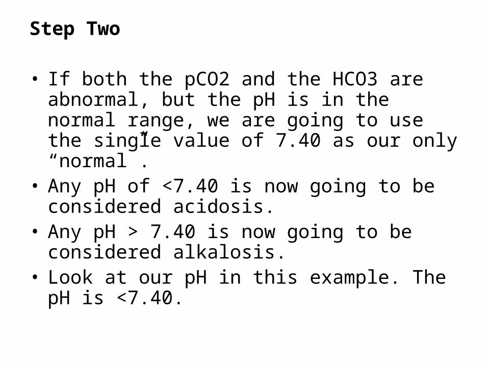
Step Two
• If both the pCO2 and the HCO3 are abnormal, but the pH is in the normal range, we are going to use the single value of 7.40 as our only “normal”.
• Any pH of <7.40 is now going to be considered acidosis.
• Any pH > 7.40 is now going to be considered alkalosis.
• Look at our pH in this example. The pH is <7.40.

• The two matching values determine what the problem is. In this case, an ACIDOSIS.
• Match the two abnormalities: Respiratory (lungs) + Acidosis = Respiratory Acidosis
