poly(a-hydroxyl acids)/hydroxyapatite porous composites
TRANSCRIPT
Poly(a-hydroxyl acids)/hydroxyapatite porous compositesfor bone-tissue engineering. I. Preparation and morphology
Ruiyun Zhang1, Peter X. Ma1,2,3
1Department of Biologic and Materials Sciences, The University of Michigan, Ann Arbor, Michigan 48109-10782Macromolecular Science and Engineering Center, The University of Michigan, Ann Arbor, Michigan 48109-10783Department of Biomedical Engineering, The University of Michigan, Ann Arbor, Michigan 48109–1078
Received 25 November 1997; accepted 16 September 1998
Abstract: Tissue engineering has shown great promise forcreating biological alternatives for implants. In this ap-proach, scaffolding plays a pivotal role. Hydroxyapatitemimics the natural bone mineral and has shown good bone-bonding properties. This paper describes the preparationand morphologies of three-dimensional porous compositesfrom poly(L-lactic acid) (PLLA) or poly(D,L-lactic acid-co-glycolic acid) (PLGA) solution and hydroxyapatite (HAP). Athermally induced phase separation technique was used tocreate the highly porous composite scaffolds for bone-tissueengineering. Freeze drying of the phase-separated polymer/HAP/solvent mixtures produced hard and tough foamswith a co-continuous structure of interconnected pores anda polymer/HAP composite skeleton. The microstructure of
the pores and the walls was controlled by varying the poly-mer concentration, HAP content, quenching temperature,polymer, and solvent utilized. The porosity increased withdecreasing polymer concentration and HAP content. Foamswith porosity as high as 95% were achieved. Pore sizes rang-ing from several microns to a few hundred microns wereobtained. The composite foams showed a significant im-provement in mechanical properties over pure polymerfoams. They are promising scaffolds for bone-tissue engi-neering. © 1999 John Wiley & Sons, Inc. J Biomed Mater Res,44, 446–455, 1999.
Key words: hydroxyapatite; polymer scaffold; tissue engi-neering; bone; composite
INTRODUCTION
Tissue engineering offers a promising new ap-proach to repair of bone fractures with bone loss, frac-tures that do not heal, and fractures due to bone tu-mors.1–3 In this approach, a porous scaffold that seedsthe cells and serves as a template for tissue regenera-tion is necessary. The design of a scaffolding materialcan significantly affect the cell seeding and growthboth in vitro and in vivo.4 Scaffolding materials shouldbe biocompatible and biodegradable. The degradationproducts should be nontoxic and must be easily ex-creted by metabolic pathways. The scaffolds should beosteoconductive so that osteoblasts attach and migrateon them, and they should be mechanically strong tomaintain their structural integrity during culture. Theideal scaffolding materials also should be easy to fab-ricate into a desired shape, and they should have acontrolled porous architecture to allow for cellgrowth, tissue regeneration, and vascularization.
Poly(a-hydroxyl acids), such as poly(L-lactic acid)
(PLLA), poly(glycolic acid) (PGA), and poly(D,L-lacticacid-co-glycolic acid) (PLGA), satisfy many of thesematerial requirements and already have been fabri-cated into scaffolds for cell transplantation and tissueengineering.4–8 One of the disadvantages of these ma-terials is that the degradation products reduce the lo-cal PH value, which, in turn, may accelerate the poly-esters’ degradation rates9 and induce an inflammatoryreaction. Another disadvantage is that the mechanicalproperties of the highly porous scaffolds made frompoly(a-hydroxyl acids) are relatively weak, which lim-its their use for bone-tissue regeneration, especially inthe in vivo implant site.
Hydroxyapatite (HAP) has been investigated as abone replacement for a long time since the materialmimics the natural bone mineral.10–13 HAP has beenstudied extensively for cell cultures and has beenfound to possess good osteoconductive proper-ties.14–17 Compact PLLA/HAP composites (with littleporosity) as bone fillers or implant materials haveshown good bone-bonding properties.18,19 By moni-toring for 24 weeks the PH value variations duringtheir incubation in phosphate-buffered saline (PBS), ithas been found that the PH value of PLLA/HAP com-posite is more stable than that of pure PLLA and pureHAP.20 The incorporation of synthetic HAP into a po-
Correspondence to: P. X. Ma, e-mail: [email protected]
© 1999 John Wiley & Sons, Inc. CCC 0021-9304/99/040446-10

rous biodegradable poly(a-hydroxyl acid) is expected,for the following reasons, to result in a promisingcomposite scaffold for bone-tissue engineering: (1) abetter cell seeding and growth environment is ex-pected because of the good osteoconductive proper-ties provided by HAP; (2) the acidic degradation by-products from polyesters may be buffered; and (3) themechanical properties may be improved.
For cell transplantation and tissue engineering, ascaffold must be fabricated into a three-dimensionalstructure with a high porosity and an appropriatepore size. Various techniques have been utilized toprepare such highly porous scaffolds.4–7,21,22 Recently,a new procedure for preparing polymer foam frompolymer solution by thermally inducing phase sepa-ration (TIPS) and subsequent sublimation of solventhas generated considerable interest.23,24 In this proce-dure, two phases—a polymer-rich phase and a poly-mer-lean phase—are formed by cooling down thepolymer solution to induce liquid–liquid phase sepa-ration. The solvent then is removed by solvent extrac-tion or sublimation in vacuo to form pores. In this pa-per, we report on the preparation and the character-istic pore morphologies of highly porous poly(a-hydroxyl acids)/HAP composite foams by solid–liquid phase separation of polymer/HAP/dioxanesuspensions.
MATERIALS AND METHODS
Materials
Poly(L-lactic acid) with an inherent viscosity of approxi-mately 1.6 was purchased from Boehringer Ingelheim (In-gelheim, Germany). Poly(D,L-lactic acid-co-glycolic aicd; 75/25) with an inherent viscosity of 0.5∼0.65 was obtained fromMedisorb Technologies International L. P. (Cincinnati,Ohio). These polymers were used without further purifica-tion. Dioxane and hydroxyapatite [Ca10(PO4)6(OH)2] wereobtained from Aldrich Chemical Company (Milwaukee,Wisconsin).
Preparation of polymer/hydroxyapatite mixture
PLLA or PLGA was weighed accurately into a flask, andthen an accurately measured amount of dioxane or dioxane/water mixture was added to the flask to make a solutionwith a desired concentration of from 1% to 7.5% (w/v). Themixture was stirred at 50°C for 2 h to obtain a homogeneouspolymer solution. HAP powder was added into the pre-pared solution to make a polymer/HAP mixture. The finalcomposition of PLLA/HAP composite foam was deter-mined by the concentration of the polymer solution andHAP content in the mixture.
Polymer/HAP composite foam fabrication
The PLLA/HAP/composite foam was prepared by solid–liquid phase separation and subsequent sublimation of sol-vent. Typically, a foam was prepared with the followingsteps: 10 mL of PLLA/HAP/dioxane mixture was put into abeaker (30 mL, prewarmed to 50°C). The beaker containingthe mixture then was rapidly transferred into a refrigeratoror a freezer at a preset temperature to solidify the solventand induce solid–liquid phase separation. The solidifiedmixture was maintained at that temperature for 2 h and thenimmersed into liquid nitrogen to deep freeze the mixture.The frozen mixture was transferred into a freeze-drying ves-sel at a temperature between −5°C and −10°C in an ice/saltbath. The samples were freeze dried at 0.5 mmHg for at least4 days to completely remove the solvent. Ninety nine per-cent of the solvent was removed in one day, and a constantsample weight was achieved within 4 days. The foamsamples then were stored in a desiccator until characteriza-tion.
Characterization
The density and porosity values of the composite foamsprepared from polymer/HAP/dioxane mixtures were mea-sured by liquid displacement, similar to a publishedmethod.25 In this method water was used as the displace-ment liquid. However, we found water was difficult to workwith because it did not penetrate very easily into the pores,probably due to the hydrophobicity of the polymers. Thusethanol was used in our procedure because it penetratedeasily into the pores and did not induce shrinkage or swell-ing as a nonsolvent of the polymers. A foam sample ofweight W was immersed in a graduated cylinder containinga known volume (V1) of ethanol. The sample was kept in theethanol for 5 min, and then a series of brief evacuation–repressurization cycles was conducted to force the ethanolinto the pores of the foam. Cycling was continued until noair bubbles were observed emerging from the foam. Thetotal volume of ethanol and the ethanol-impregnated foamthen was recorded as V2. The volume difference, (V2 − V1)was the volume of the polymer/HAP composite skeleton ofthe foam. The ethanol-impregnated foam was removed fromthe cylinder and the residual ethanol volume recorded as V3.The quantity (V1 − V3)—the volume of the ethanol held inthe foam—was determined as the void volume of the foam.Thus the total volume of the foam was: V = (V2 − V1) + (V1
− V3) = V2 − V3. The density of the foam (d) was expressedas:
d = W/(V2 − V3)
and the porosity of the foam («) was obtained by:
« = (V1 − V3)/(V2 − V3)
The porous morphologies of the composite foams werestudied with scanning electron microscopy (SEM; S-3200N,Hitachi, Japan) at 15 kV. The specimens were cut with arazor blade after being frozen in liquid nitrogen for 5 min,and then they were coated with gold using a sputter coater
447BIODEGRADABLE COMPOSITE SCAFFOLDS

(Desk-II, Denton Vacuum Inc). The gas pressure was lessthan 50 mtorr, and the current was about 40 mA. The coatingtime was 200 s.
The compressive mechanical properties of the foams weretested with an Instron 4502 mechanical tester (Instron Co.,Canton, Massachusetts). A PLLA/HAP/dioxane mixturewas cast into a circular flat-bottom Teflon vial and phaseseparated to produce circular disks (∼16 mm in diameter) formechanical testing. The top layer of the disk was removed toachieve the desired thickness (∼3 mm) and ensure a flatsurface. The aspect ratio of these mechanical testing speci-mens was chosen to match the cell-scaffolding constructsused to engineer bone tissue in our lab. A crosshead speed of0.5 mm/min was used. The compressive modulus was de-fined as the initial linear modulus. The yield strength wasdetermined from the cross point of the two tangents on thestress–strain curve around the yield point. At least fivespecimens were tested for each sample. The averages andstandard deviations were graphed. A two-tail Student’s ttest (assuming equal variances) was performed to determinethe statistical significance (p < 0.05) of the differences inmechanical properties.
RESULTS
High porosity (low density) PLLA/HAP and PLGA/HAP composite foams have been prepared by solid–liquid phase separation and subsequent sublimationof the solvent (Table I). The density increases withpolymer concentration and HAP content. In parallel,porosity decreases with increasing polymer concentra-tion and HAP content. Phase-separation temperaturedoes not show obvious effects on the porosity of thepolymer/HAP foams in the composition range stud-ied. The densities of PLGA/HAP foams are slightlyhigher than those of PLLA/HAP foams prepared from
the same polymer concentration, HAP content, andprocessing conditions.
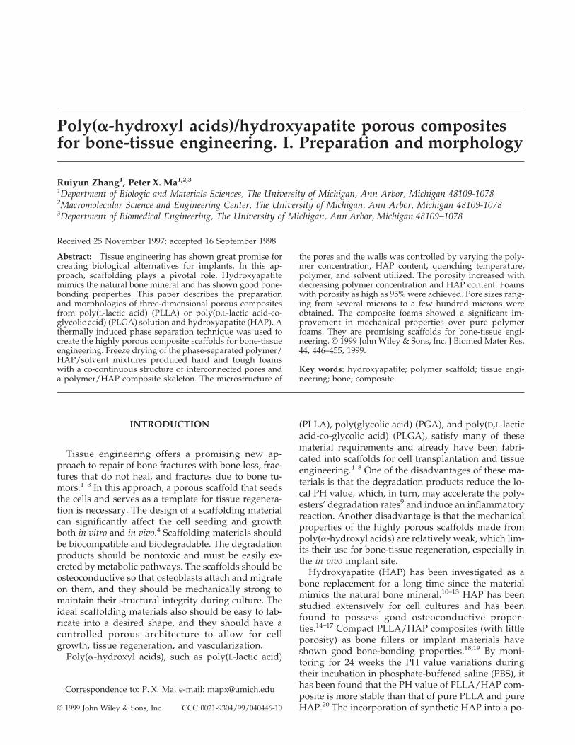
A typical SEM micrograph of the PLLA/HAP com-posite foam prepared from 2.5% (w/v) PLLA solutionwith a quenching temperature of −18°C shows co-continuous structure of interconnected irregular poresand a polymer/HAP composite skeleton [Fig. 1(a)].The irregular pores range from several microns up toabout 300 microns. The walls of the pores are com-posed of both PLLA and HAP [Fig. 1(b)]. The HAPplatelets ranging from 10 to 100 mm in size [Fig. 1(c)]are randomly distributed in the PLLA matrix.
The morphology of PLLA/HAP foam is much dif-ferent from pure PLLA foam [Fig. 1(d)] prepared withthe same procedure. The PLLA foam prepared fromsolid–liquid phase separation of the PLLA/dioxanesolution has a highly anisotropic tubular morphologywith an internal ladder-like structure. This ladder-likestructure is a characteristic morphology of the foamsformed by solid–liquid phase separation of a polymersolution.26 The channels are parallel to the direction ofsolidification (heat transfer direction). Each channelhas repeating partitions with uniform spacing perpen-dicular to the solidification direction. The diameter ofthe channels and the spacing between repeating par-titions in the channel change with cooling rate andwith the polymer concentration. The solid–liquidphase separation is attributed to the crystallization ofthe solvent. When the temperature of the polymer so-lution is lower than the freezing point (crystallizationtemperature) of the solvent, the crystallization of thesolvent takes place and the polymer phase is expelledfrom the crystallization front as ‘‘impurities.’’ A con-tinuous polymer-rich phase is formed by aggregationof polymer expelled from every single solvent crystal.
TABLE IDensities and Porosities of PLLA/HAP and PLGA/HAP Foams Prepared From
PLLA/HAP/Dioxane and PLGA/HAP/Dioxane Mixtures
PolymerConcentration Composition
QuenchingTemperature (°C)
Densityg/cm3 Porosity
2.5%(w/v) PLLA/HAP: 100/0 −18 0.045 94.8%2.5%(w/v) PLLA/HAP: 90/10 −18 0.049 93.4%2.5%(w/v) PLLA/HAP: 70/30 −18 0.060 92.5%2.5%(w/v) PLLA/HAP: 50/50 −18 0.090 89.9%2.5%(w/v) PLLA/HAP: 30/70 −18 0.120 85.1%5.0%(w/v) PLLA/HAP: 100/0 −18 0.083 92.7%5.0%(w/v) PLLA/HAP: 90/10 −18 0.086 91.7%5.0%(w/v) PLLA/HAP: 70/30 −18 0.110 91.0%5.0%(w/v) PLLA/HAP: 50/50 −18 0.144 89.2%5.0%(w/v) PLLA/HAP: 30/70 −18 0.203 86.6%2.5%(w/v) PLLA/HAP: 100/0 liquid nitrogen 0.043 92.5%2.5%(w/v) PLLA/HAP: 50/50 liquid nitrogen 0.085 88.0%2.5%(w/v) PLLA/HAP: 100/0 8 0.047 95.6%2.5%(w/v) PLLA/HAP: 50/50 8 0.090 90.9%2.5%(w/v) PLGA/HAP: 50/50 −18 0.126 87.5%5.0%(w/v) PLGA/HAP: 50/50 −18 0.151 85.7%
448 ZHANG AND MA
After solvent crystals have been sublimated, foam isformed with pores, similar to the geometry of solventcrystals. The temperature gradient along the solidifica-tion direction (from sample surface to sample center)may have led to the highly anisotropic pore structure.
When HAP is introduced into the PLLA/dioxanesolution, the crystallization of solvent is perturbed bythe existing solid HAP particles. The randomly dis-tributed HAP particles change the solvent crystalliza-tion front by impeding the crystal growth, making thecrystals of the solvent more irregular. On the otherhand, both polymer and HAP particles are expelledfrom the crystallization front, forming a PLLA/HAP-rich phase. After sublimation of the solvent, this poly-mer/HAP-rich phase forms a continuous skeleton forthe PLLA/HAP foam, and the spaces taken by solventcrystals become pores of the foam. As a result of ir-regular solvent crystal growth, the pores become ir-regular (more isotropic), and no channel structure orrepeating partitions are observed.
Good adhesion between PLLA matrix and HAP par-ticles also is observed [Fig. 1(b)]. Most of the HAPparticles bonded to PLLA matrix are on the surfaces ofthe solid walls of the pores. These HAP surfaces mayprovide a better environment for osteoblast attach-ment and growth when used as scaffolding for bonetissue engineering.
To study the effect of polymer concentration on thefoam structure, a series of PLLA/HAP compositefoams were prepared from PLLA/HAP/dioxane mix-tures, with PLLA concentration ranging from 1.0% to7.5% (w/v). The ratio of PLLA to HAP was kept atone. The composite foam made from 1.0% PLLA so-lution was composed of bonded very thin PLLA leaf-lets [Fig. 2(a,b)]. Almost all the HAP particles settled atthe bottom of the sample, presumably due to the lowviscosity of the PLLA solution. The foams preparedfrom 5.0% and 7.5% PLLA solution were very hardand tough. SEM observation shows that the porestructure of the foam from the 7.5% PLLA solution is
Figure 1. SEM micrographs of PLLA/HAP foam (PLLA/HAP: 50/50) prepared from 2.5% (w/v) PLLA/HAP/dioxanemixture (quenching temperature: −18°C), PLLA foam from 2.5% (w/v) PLLLA/dioxane solution (quenching temperature:−18°C), and HAP particles. Original magnifications: (a) PLLA/HAP, ×100; (b) PLLA/HAP, ×500; (c) HAP, ×500; (d) PLLA,×100.
449BIODEGRADABLE COMPOSITE SCAFFOLDS

almost the same as the foam from the 5.0% PLLA so-lution [Fig. 2(c–f)], and the HAP particles are uni-formly distributed. The foams from 5.0% and 7.5%PLLA solutions have morphologies slightly different
from the foam made from a 2.5% PLLA solution [Fig.1(a,b)]. The pore structure is more uniform, with poresizes ranging from about 50 to 200 microns, and thepore walls are thicker than that of foam from the 2.5%
Figure 2. SEM micrographs of PLLA/HAP foams (PLLA/HAP: 50/50) prepared from PLLA/HAP/dioxane mixtures withdifferent PLLA concentrations (quenching temperature: −18°C). Original magnifications: (a) 1.0% (w/v) PLLA/dioxane, ×100;(b) 1.0% (w/v) PLLA/Dioxane, ×500; (c) 5.0% (w/v) PLLA/dioxane, ×100; (d) 5.0% (w/v) PLLA/dioxane, ×500; (e) 7.5%(w/v) PLLA/dioxane, ×100; (f) 7.5% (w/v) PLLA/dioxane, ×500.
450 ZHANG AND MA

PLLA solution. Compared with the pore structure of afoam from the 5.0% PLLA solution, the pore size of thefoam derived from the 7.5% PLLA solution is smallerand the walls of the pores are thicker.
The effects of HAP content on the structure ofPLLA/HAP foams also have been investigated byvarying the HAP amount in the PLLA/HAP foamswhile maintaining the PLLA concentration constant.SEM observation shows that the micropore structureof the foam changes considerably with the HAP con-tent (Fig. 3). When HAP content is low, regular chan-nels and a ladder-like structure similar to those inpure PLLA foam are observed. With increasing HAPcontent, the pore structure becomes more and moreirregular. When the HAP/polymer ratio is higher than1:1 (50 wt % HAP content), the pore structure becomesso irregular that no regular channels or ladder-likestructure is observed. These results have demon-strated that the pore structure of the PLLA foam canbe modified by the incorporation of HAP.
In the preparation of polymer foam by solid–liquidphase separation from polymer solution, quenchingtemperature (cooling rate) is an effective factor in con-trolling the morphology of the foam since the crystal-line morphology of a solvent depends on the crystal-lization temperature. In this work, the effects ofquenching temperature on the structure of PLLA/HAP foam from PLLA/HAP/dioxane mixtures arestudied. The crystallization process induces twostages—nucleation and growth. Generally, a higherdegree of supercooling (at a lower temperature) in-duces a high nucleation rate and a low crystal growthrate, which leads to the formation of a large number ofsmall crystals. In contrast, a relatively lower degree ofsupercooling (at a higher temperature) includes a lownucleation rate and a high crystal growth rate, whichleads to a small number of large crystals. The freezingpoint of dioxane is about 12°C. When the temperatureof the PLLA/HAP/dioxane mixture is lower than thistemperature, crystallization of dioxane takes place.
Figure 3. SEM micrographs of PLLA/HAP foams prepared from 2.5% (w/v) PLLA/HAP/dioxane mixtures with differentHAP contents (quenching temperature: −18°C). Original magnifications: (a) PLLA/HAP: 70/30, ×100; (b) PLLA/HAP: 70/30,×500; (c) PLLA/HAP: 30/70, ×100; (d) PLLA/HAP: 30/70, ×500.
451BIODEGRADABLE COMPOSITE SCAFFOLDS

Figure 4(a,b) shows the SEM micrographs of PLLA/HAP foam formed by quenching the mixture to 8°C,which is slightly lower than the freezing point of di-oxane. At this temperature, the degree of supercoolingof the dioxane is very low, so the crystallization of thedioxane has a low nucleation and a high growth rate,which leads to large solvent crystal formation andthereby a PLLA/HAP foam with large pore sizes, upto 600 microns. The pore wall of the foam is thickerthan those prepared at lower temperatures. When thePLLA/HAP/dioxane mixture is quenched with liquidnitrogen, the microstructure of the foam formed ismuch different from that of the foams prepared athigher temperatures. In contrast to the regular ladder-like microstructure prepared from pure PLLA solutionat the same temperature, small channels with rela-tively irregular rows parallel to the solidification di-rection but few regular horizontal partitions are ob-served for the PLLA/HAP composites [Fig. 4(c,d)].The HAP particles randomly intersect the PLLA chan-
nels. The high nucleation rate and low crystal growthrate of dioxane at this quenching temperature and thegreater temperature gradient in the direction of solidi-fication may be responsible for the foam morphology.
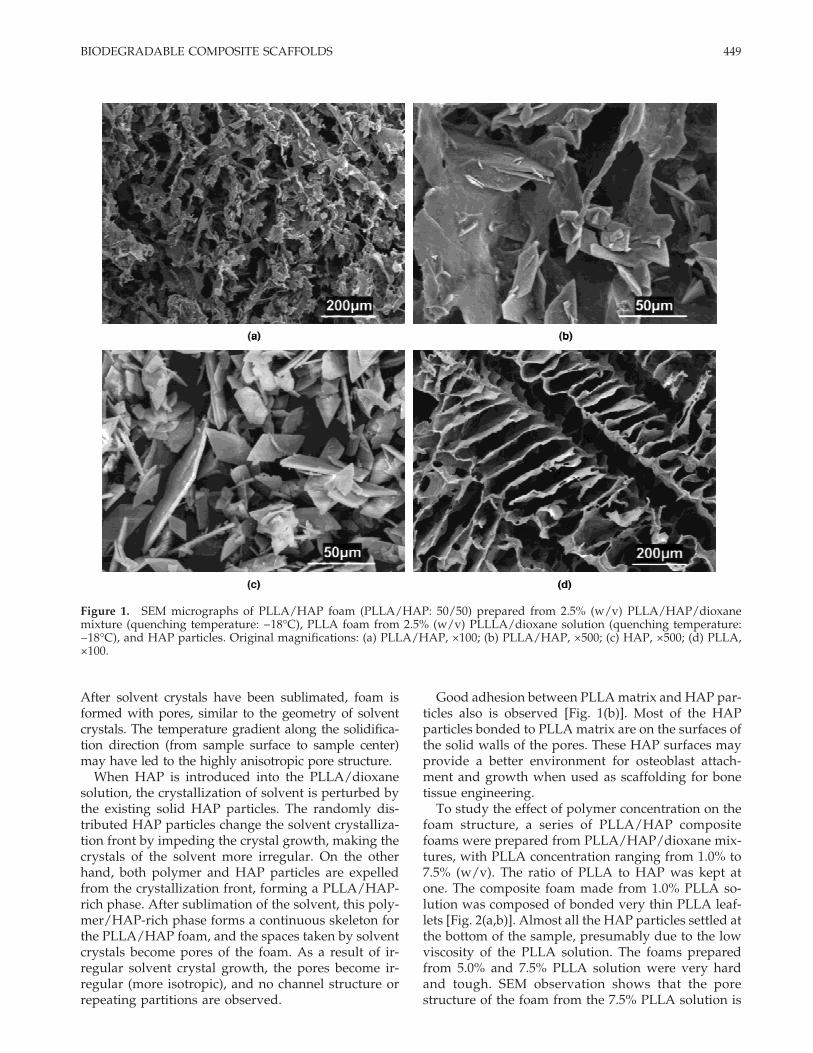
The composite foams can be prepared from HAPand poly(a-hydroxyl acids) other than PLLA with theaforementioned procedure. Figure 5 shows the micro-pore morphology of the PLGA/HAP foam producedfrom a PLGA/HAP/dioxane mixture with a PLGAconcentration of 2.5% (w/v). The microstructure of thePLGA/HAP foam is similar to that of PLLA/HAPfoam from the same polymer concentration. The poresize, ranging from 30 to 100 microns, is slightlysmaller than that of PLLA/HAP foam prepared fromthe same concentration and at the same temperature[Fig. 1(a,b)]. The densities of the PLGA/HAP foamsare higher than those of corresponding PLLA/HAPfoams. These results show that the shrinkage of thePLGA/HAP foam is higher than that of the PLLA/HAP foam. The shrinkage is attributed to the molecu-
Figure 4. SEM micrographs of PLLA/HAP foams prepared form 2.5% (w/v) PLLA/HAP/dioxane mixtures by differentquenching temperatures. Original magnifications: (a) 8°C, ×100; (b) 8°C, ×500; (c) liquid nitrogen, ×100; (d) liquid nitrogen,×500.
452 ZHANG AND MA
lar rearrangement of the polymer chains that occursduring freeze drying. The amorphous PLGA may re-arrange more easily and is less stable than the semi-crystalline PLLA during freeze drying. In any event,the shrinkage of most poly(a-hydroxyl acids)/HAPcomposite foams prepared is less than 10%.
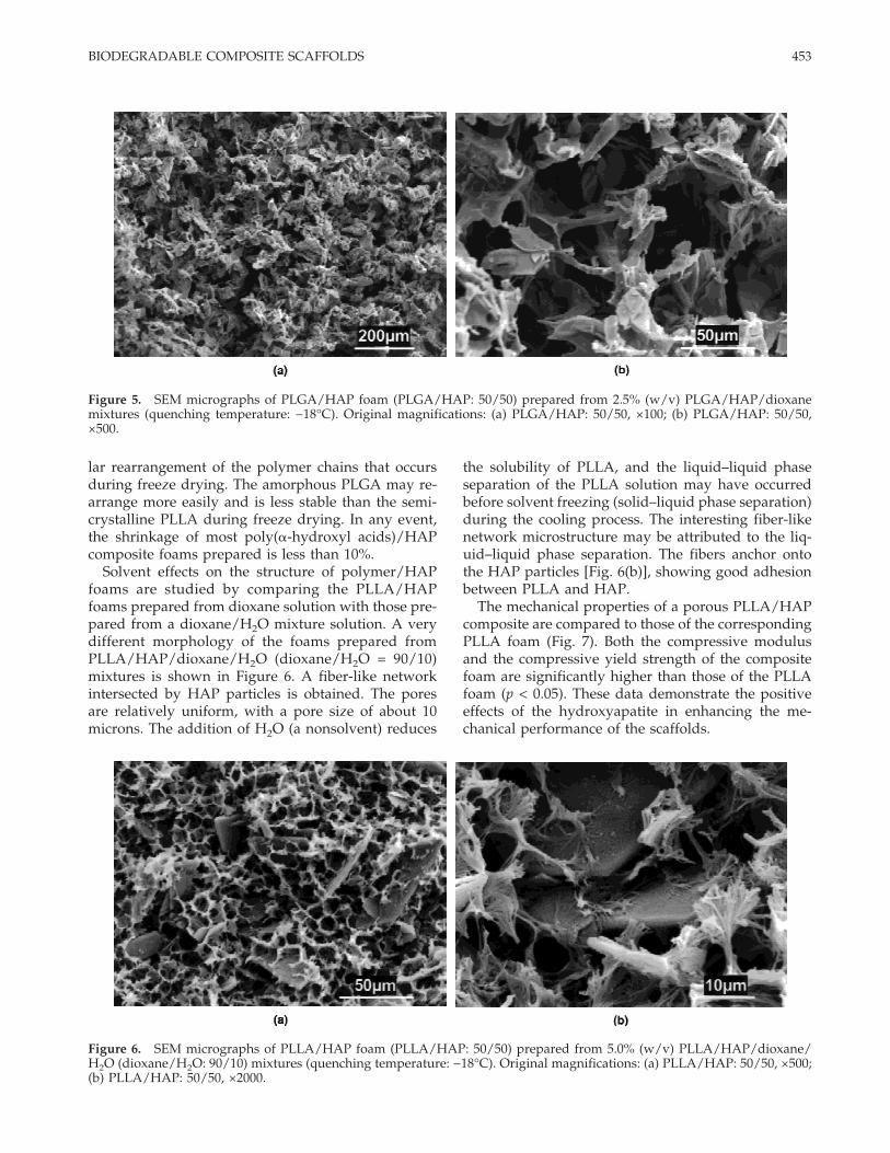
Solvent effects on the structure of polymer/HAPfoams are studied by comparing the PLLA/HAPfoams prepared from dioxane solution with those pre-pared from a dioxane/H2O mixture solution. A verydifferent morphology of the foams prepared fromPLLA/HAP/dioxane/H2O (dioxane/H2O = 90/10)mixtures is shown in Figure 6. A fiber-like networkintersected by HAP particles is obtained. The poresare relatively uniform, with a pore size of about 10microns. The addition of H2O (a nonsolvent) reduces
the solubility of PLLA, and the liquid–liquid phaseseparation of the PLLA solution may have occurredbefore solvent freezing (solid–liquid phase separation)during the cooling process. The interesting fiber-likenetwork microstructure may be attributed to the liq-uid–liquid phase separation. The fibers anchor ontothe HAP particles [Fig. 6(b)], showing good adhesionbetween PLLA and HAP.
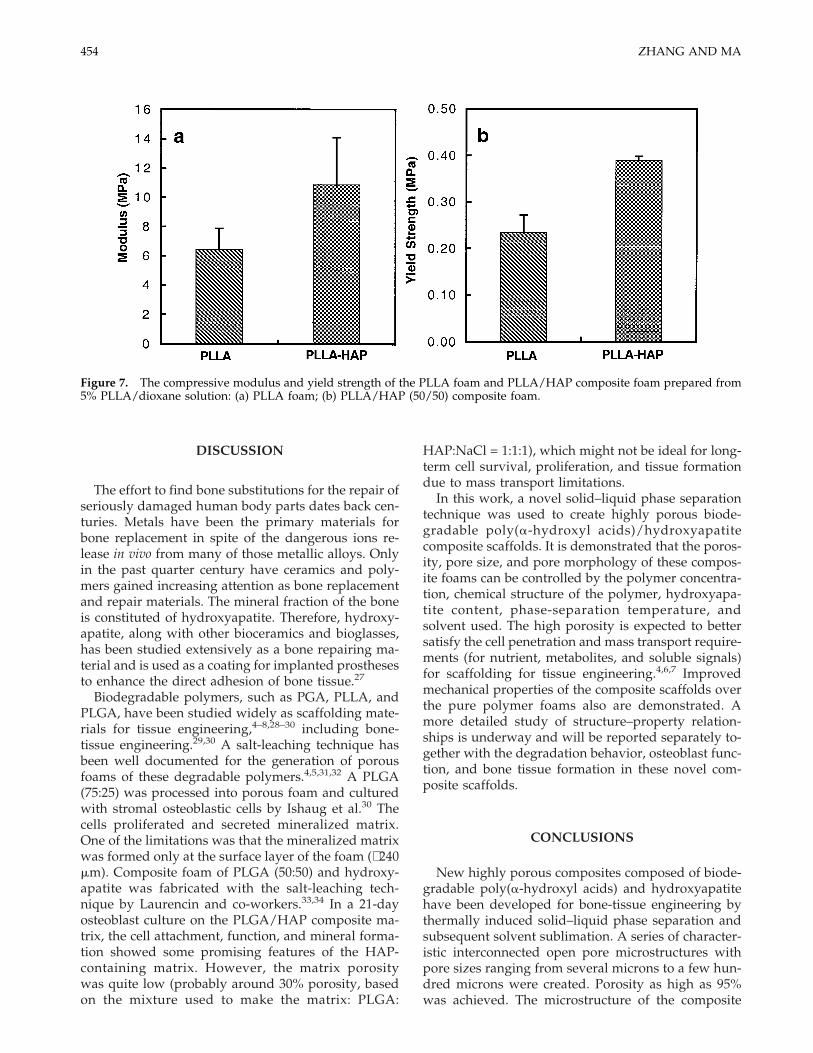
The mechanical properties of a porous PLLA/HAPcomposite are compared to those of the correspondingPLLA foam (Fig. 7). Both the compressive modulusand the compressive yield strength of the compositefoam are significantly higher than those of the PLLAfoam (p < 0.05). These data demonstrate the positiveeffects of the hydroxyapatite in enhancing the me-chanical performance of the scaffolds.
Figure 5. SEM micrographs of PLGA/HAP foam (PLGA/HAP: 50/50) prepared from 2.5% (w/v) PLGA/HAP/dioxanemixtures (quenching temperature: −18°C). Original magnifications: (a) PLGA/HAP: 50/50, ×100; (b) PLGA/HAP: 50/50,×500.
Figure 6. SEM micrographs of PLLA/HAP foam (PLLA/HAP: 50/50) prepared from 5.0% (w/v) PLLA/HAP/dioxane/H2O (dioxane/H2O: 90/10) mixtures (quenching temperature: −18°C). Original magnifications: (a) PLLA/HAP: 50/50, ×500;(b) PLLA/HAP: 50/50, ×2000.
453BIODEGRADABLE COMPOSITE SCAFFOLDS
DISCUSSION
The effort to find bone substitutions for the repair ofseriously damaged human body parts dates back cen-turies. Metals have been the primary materials forbone replacement in spite of the dangerous ions re-lease in vivo from many of those metallic alloys. Onlyin the past quarter century have ceramics and poly-mers gained increasing attention as bone replacementand repair materials. The mineral fraction of the boneis constituted of hydroxyapatite. Therefore, hydroxy-apatite, along with other bioceramics and bioglasses,has been studied extensively as a bone repairing ma-terial and is used as a coating for implanted prosthesesto enhance the direct adhesion of bone tissue.27
Biodegradable polymers, such as PGA, PLLA, andPLGA, have been studied widely as scaffolding mate-rials for tissue engineering,4–8,28–30 including bone-tissue engineering.29,30 A salt-leaching technique hasbeen well documented for the generation of porousfoams of these degradable polymers.4,5,31,32 A PLGA(75:25) was processed into porous foam and culturedwith stromal osteoblastic cells by Ishaug et al.30 Thecells proliferated and secreted mineralized matrix.One of the limitations was that the mineralized matrixwas formed only at the surface layer of the foam (∼240mm). Composite foam of PLGA (50:50) and hydroxy-apatite was fabricated with the salt-leaching tech-nique by Laurencin and co-workers.33,34 In a 21-dayosteoblast culture on the PLGA/HAP composite ma-trix, the cell attachment, function, and mineral forma-tion showed some promising features of the HAP-containing matrix. However, the matrix porositywas quite low (probably around 30% porosity, basedon the mixture used to make the matrix: PLGA:
HAP:NaCl = 1:1:1), which might not be ideal for long-term cell survival, proliferation, and tissue formationdue to mass transport limitations.
In this work, a novel solid–liquid phase separationtechnique was used to create highly porous biode-gradable poly(a-hydroxyl acids)/hydroxyapatitecomposite scaffolds. It is demonstrated that the poros-ity, pore size, and pore morphology of these compos-ite foams can be controlled by the polymer concentra-tion, chemical structure of the polymer, hydroxyapa-tite content, phase-separation temperature, andsolvent used. The high porosity is expected to bettersatisfy the cell penetration and mass transport require-ments (for nutrient, metabolites, and soluble signals)for scaffolding for tissue engineering.4,6,7 Improvedmechanical properties of the composite scaffolds overthe pure polymer foams also are demonstrated. Amore detailed study of structure–property relation-ships is underway and will be reported separately to-gether with the degradation behavior, osteoblast func-tion, and bone tissue formation in these novel com-posite scaffolds.
CONCLUSIONS
New highly porous composites composed of biode-gradable poly(a-hydroxyl acids) and hydroxyapatitehave been developed for bone-tissue engineering bythermally induced solid–liquid phase separation andsubsequent solvent sublimation. A series of character-istic interconnected open pore microstructures withpore sizes ranging from several microns to a few hun-dred microns were created. Porosity as high as 95%was achieved. The microstructure of the composite
Figure 7. The compressive modulus and yield strength of the PLLA foam and PLLA/HAP composite foam prepared from5% PLLA/dioxane solution: (a) PLLA foam; (b) PLLA/HAP (50/50) composite foam.
454 ZHANG AND MA

foams can be controlled by varying: (1) the concentra-tion of the polymer solution, (2) the content of theHAP, (3) the quenching temperature (cooling rate),and (4) the polymer and solvent utilized. The compos-ite foams showed enhanced mechanical propertiesover the pure polymer foams. The degradation behav-ior, detailed structure–property relationships, cell ad-hesion, and growth onto the composite scaffolds areunder investigation and will be reported separately.
The authors would like to thank Ming Chien for his as-sistance.
References
1. Yaszemski M, Payne R, Hayes W, Langer R, Mikos A. Evolu-tion of bone transplantation: Molecular, cellular and tissuestrategies to engineer human bone. Biomaterials 1996;17(2):175–185.
2. Crane G, Ishaug S, Mikos A. Bone tissue engineering. Nat Med1995;1(12):1322–1324.
3. Vacanti C, Vacanti J. Bone and cartilage reconstruction withtissue engineering approaches. Otolaryngol Clin N Am1994;27(1):263–276.
4. Ma PX, Langer R. Fabrication of biodegradable polymer foamsfor cell transplantation and tissue engineering. In: Yarmush M,Morgan J, editors. Tissue engineering. Totowa, New Jersey:Humana Press Inc.; 1998.
5. Mikos AG, Thorsen AJ, Czerwonka LA, Bao Y, Langer R, Win-slow DN, Vacanti JP. Preparation and characterization ofpoly(L-lactic acid) foams. Polymer 1994;35(5):1068–1077.
6. Ma PX, Langer R. Degradation, structure and properties offibrous nonwoven poly(glycolic acid) scaffolds for tissue engi-neering. In: Mikos AG, Leong KW, Yaszemski MJ, Tamada JA,Radomsky ML, editors. Polymers in medicine and pharmacy.Materials Research Society; Pittsburgh: 1995. p 99–104.
7. Ma PX, Schloo B, Mooney D, Langer R. Development of bio-mechanical properties and morphogenesis of in vitro tissue-engineered cartilage. J Biomed Mater Res 1995;29(12):1587–1595.
8. Shinoka T, Ma PX, Shum-Tim D, Breuer CK, Cusick RA, ZundG, Langer R, Vacanti JP, Mayer JE Jr. Tissue-engineered heartvalves. Autologous valve leaflet replacement study in a lambmodel. Circulation 1996;94(9):164–168.
9. Vert M, Mauduit J, Li S. Biodegradation of PLA/GA polymers:Increasing complexity. Biomaterials 1994;15(15):1209–1213.
10. Akao M, Aoki H, Kato K. Mechanical properties of sinteredhydroxyapatite for prosthetic applications. J Mater Sci 1981;16:809–812.
11. Dewith G, van Dijk HJA, Hattu N, Prijs K. Preparation, micro-structure and mechanical properties of dense polycrystallinehydroxyapatite. J Mater Sci 1981;6:1592–1598.
12. Driessen AA, Klein CP, de Groot K. Preparation and someproperties of sintered beta-whitlockite. Biomaterials 1982;3(2):113–116.
13. Best S, Bonfield W. Processing behaviour of hydroxyapatitepowders with contrasting morphology. J Mater Sci: Mater Med1994;5(8):516–521.
14. Akao M, Sakatsume M, Aoki H, Takagi T, Sasaki S. In vitromineralization in bovine tooth germ cell cultured with sinteredhydroxyapatite. J Mater Sci: Mater Med 1993;4(6):569–574.
15. Puleo DA, Holleran LA, Doremus RH, Bizios R. Osteoblastresponses to orthopedic implant materials in vitro. J BiomedMater Res 1991;25(6):711–723.
16. Klein CP, Patka P, Wolke JG, de Blieck–Hogervorst JM, deGroot K. Long-term in vivo study of plasma-sprayed coatingson titanium alloys of tetracalcium phosphate, hydroxyapatiteand alpha-tricalcium phosphate. Biomaterials 1994;15(2):146–150.
17. Jansen JA, van der Waerden JP, Wolke JG. Histologic investi-gation of the biologic behavior of different hydroxyapatiteplasma-sprayed coatings in rabbits. J Biomed Mater Res1993;27(5):603–610.
18. Higashi S, Yamamuro T, Nakamura T, Ikada Y, Hyon SH,Jamshidi K. Polymer–hydroxyapatite composites for biode-gradable bone fillers. Biomaterials 1986;7(3):183–187.
19. Flahiff CM, Blackwell AS, Hollis JM, Feldman DS. Analysis ofa biodegradable composite for bone healing. J Biomed MaterRes 1996;32(3):419–424.
20. Verheyen CCPM, Klein CPAT, Blieck–Hogervorst JMAD,Wolke JGC, van Blitterswijn CA. Evaluation of hydroxyapa-tite/poly(l-lactide) composites: Physico–chemical properties. JMater Sci: Mater Med 1993;4:58–65.
21. Mikos A, Bao Y, Cima L, Ingber D, Vacanti J, Langer R. Pre-paration of poly(glycolic acid)-bonded fiber structures forcell attachment and transplantation. J Biomed Mater Res1993;27(2):183–189.
22. Wintermantel E, Mayer J, Blum J, Eckert KL, Luscher P,Mathey M. Tissue engineering scaffolds using superstructures.Biomaterials 1996;17(2):83–91.
23. Lo H, Ponticiello MS, Leong KW. Fabrication of controlledrelease biodegradable foams by phase separation. Tiss Eng1995;1(1):15–28.
24. Schugens C, Maquet V, Grandfils C, Jerome R, Teyssie P. Poly-lactide macroporous biodegradable implants for cell transplan-tation. II. Preparation of polylactide foams by liquid–liquidphase separation. J Biomed Mater Res 1996;30(4):449–461.
25. Hsu YY, Gresser JD, Trantolo DJ, Lyons CM, GangadharamPR, Wise DL. Effect of polymer foam morphology and densityon kinetics of in vitro controlled release of isoniazid from com-pressed foam matrices. J Biomed Mater Res 1997;35(1):107–116.
26. Whinnery LL, Even WR, Beach JV, Loy DA. Engineering themacrostructure of thermally induced phase separated polysi-lane foams. J Polym Sci, Polym Chem 1996;34:1623–1627.
27. Ravaglioli A, Krajewski A. Bioceramics. London: Chapman &Hall, 1992. 422 p.
28. Shinoka T, Shum-Tim D, Ma PX, Tanel RE, Isogai N, Langer R,Vacanti JP, Mayer JE Jr. Creation of viable pulmonary arteryautografts through tissue engineering. J Thorac CardiovascSurg 1998;115(3):536–545.
29. Vacanti C, Kim W, Upton J, Vacanti M, Mooney D, Schloo B,Vacanti J. Tissue-engineered growth of bone and cartilage.Transplant Proc 1993;25:1019–1021.
30. Ishaug SL, Crane GM, Miller MJ, Yasko AW, Yaszemski MJ,Mikos AG. Bone formation by three-dimensional stromal os-teoblast culture in biodegradable polymer scaffolds. J BiomedMater Res 1997;36(1):17–28.
31. Cusick RA, Lee H, Sano K, Pollok JM, Utsunomiya H, Ma PX,Langer R, Vacanti JP. The effect of donor and recipient age onengraftment of tissue-engineered liver. J Ped Surg 1997;32(2):357–360.
32. Kim TH, Lee HM, Utsonomiya H, Ma P, Langer R, Schmidt EV,Vacanti JP. Enhanced survival of transgenic hepatocytes ex-pressing hepatocyte growth factor in hepatocyte tissue engi-neering. Transplant Proc 1997;29(1–2):858–860.
33. Laurencin CT, Attawia MA, Elgendy HE, Herbert KM. Tissue-engineered bone regeneration using degradable polymers: Theformation of mineralized matrices. Bone 1996;19(1):93S–99S.
34. Attawia MA, Herbert KM, Laurencin CT. Osteoblast-like celladherence and migration through 3-dimensional porous poly-mer matrices. Biochem Biophys Res Comm 1995;213(2):639–644.
455BIODEGRADABLE COMPOSITE SCAFFOLDS