bisphenol a glucuronidation and excretion in liver of...
TRANSCRIPT
DMD# 2004/001537
1
Bisphenol A Glucuronidation and Excretion in Liver of
Pregnant and Nonpregnant Female Rats
Hiroki Inoue, Akio Tsuruta, Satoko Kudo, Takako Ishii, Yusuke Fukushima,
Hidetomo Iwano, Hiroshi Yokota and Seiyu Kato
Department of Veterinary Physiology (H.In, A.T., S.Ku., T.I., Y.F., S.Ka.) and Veterinary
Biochemistry (H.Iw, H.Y.), School of Veterinary Medicine, Rakuno Gakuen University,
Japan
DMD Fast Forward. Published on October 4, 2004 as doi:10.1124/dmd.104.001537
Copyright 2004 by the American Society for Pharmacology and Experimental Therapeutics.
This article has not been copyedited and formatted. The final version may differ from this version.DMD Fast Forward. Published on October 4, 2004 as DOI: 10.1124/dmd.104.001537
at ASPE
T Journals on A
ugust 27, 2018dm
d.aspetjournals.orgD
ownloaded from
DMD# 2004/001537
2
Running title: Bisphenol A elimination in liver of pregnant rat
Corresponding Author: Seiyu Kato, Department of Veterinary Physiology, School of
Veterinary Medicine, Rakuno Gakuen University, Ebetsu, Hokkaido, 069-8501 Japan.
Tel. 81-11-388-4734; Fax. 81-11-387-5890;
E-mail: [email protected]
Manuscript:
Text, 28 pages
Tables, 0
Figures, 5
References, 24
Abstract, 247 words
Introduction, 440 words
Discussion, 1210 words
Abbreviations used: EHBR, Eisai hyperbilirubinemic rat; HPLC, high performance
liquid chromatography; MRP, multidrug resistance associated protein
This article has not been copyedited and formatted. The final version may differ from this version.DMD Fast Forward. Published on October 4, 2004 as DOI: 10.1124/dmd.104.001537
at ASPE
T Journals on A
ugust 27, 2018dm
d.aspetjournals.orgD
ownloaded from
DMD# 2004/001537
3
Abstract
In male rats challenged with the environmental estrogen bisphenol A, the
compound is highly glucuronidated in the liver and is excreted largely into the bile.
Given that in pregnancy the microsomal glucuronidation toward bisphenol A is
attenuated, we hypothesized that elimination of bisphenol A from the liver may be
reduced in pregnancy. This study was conducted to trace the elimination of bisphenol A
in female rats, especially in pregnancy. In Sprague-Dawley rats, 1.5 µmol bisphenol A
was perfused into the liver via the portal vein. In both the male and the nonpregnant
female the infused bisphenol A was glucuronidated, then the resultant glucuronide was
excreted mainly into the bile. In pregnant rats, however, bilious excretion of bisphenol A
glucuronide was 60% of that observed in nonpregnant rats, and venous excretion
increased reciprocally. During 1 h perfusion, total excretion of the glucuronide from the
liver of male, nonpregnant female and pregnant rats were 889.5 ± 69.6, 1256.7 ± 54.8
and 1038.8 ± 33.3 nmoles, respectively. In Eisai hyperbilirubinemic rats (EHBR),
perfusion of the liver with bisphenol A enabled us to determine that multidrug
resistance-associated protein (MRP) 2-mediating transport is the mechanism behind
excretion of the glucuronide into the bile. The expression of MRP2 has been reported to
be noticeably reduced in pregnancy. These results suggest that bisphenol A elimination
by hepatic glucuronidation is slightly less in pregnancy than in nonpregnancy and that
This article has not been copyedited and formatted. The final version may differ from this version.DMD Fast Forward. Published on October 4, 2004 as DOI: 10.1124/dmd.104.001537
at ASPE
T Journals on A
ugust 27, 2018dm
d.aspetjournals.orgD
ownloaded from
DMD# 2004/001537
4
in pregnancy more bisphenol A glucuronide is eliminated to the vein because of reduced
MRP2 expression.
This article has not been copyedited and formatted. The final version may differ from this version.DMD Fast Forward. Published on October 4, 2004 as DOI: 10.1124/dmd.104.001537
at ASPE
T Journals on A
ugust 27, 2018dm
d.aspetjournals.orgD
ownloaded from
DMD# 2004/001537
5
Introduction
Bisphenol A (2, 2-bis[4-hydroxyphenyl]propane), a compound widely used by
the chemical industry and in daily life (NTP, 1982), has been shown to act as an
estrogen on MCF-7 human breast cancer cells (Krishnan et al., 1993). In vivo,
estrogenic effects of the compound have also been reported on growth, differentiation
and c-fos protooncogene expression in the reproductive tract of female rats (Steinmetz
et al., 1998). Bisphenol A given for 7 days to pregnant CF-1 mice reduces the number of
days between vaginal opening and first vaginal estrus in offspring (Howdeshell et al.,
1999).
To elucidate the mechanisms responsible for adverse effects of bisphenol A in
the body, it is important to clarify the metabolism and disposition of the chemical
enroute to target organs such as the testis and uterus. Previously we found that in rats
bisphenol A is glucuronidated by liver microsomes and that the glucuronidation is
mediated by UGT2B1, an isoform of UDP-glucuronosyltransferase (Yokota et al., 1999).
Glucuronidation is the major metabolic pathway of the compound, as demonstrated in
primary cultures of rat hepatocytes (Pritchett et al., 2002). The resultant glucuronide
conjugate has been reported to have estrogenic activity, albeit low (Matthews et al.,
2001). After glucuronidation in the rat liver the resultant glucuronide is excreted mainly
into the bile (Inoue et al., 2001). These findings have established that bisphenol A is
This article has not been copyedited and formatted. The final version may differ from this version.DMD Fast Forward. Published on October 4, 2004 as DOI: 10.1124/dmd.104.001537
at ASPE
T Journals on A
ugust 27, 2018dm
d.aspetjournals.orgD
ownloaded from
DMD# 2004/001537
6
highly eliminated from the systemic circulation by glucuronidation during its passage
through the liver. Current understanding of the fate of bisphenol A, however, is based on
experiments on male rats. From the viewpoint of reproduction, it is essential to elucidate
the metabolism and disposition of bisphenol A in the female rat as well as the male, and
especially in pregnant rats.
Pregnancy is one of the major physiological events in which the elimination
process by glucuronidation in the liver is dramatically altered. In pregnancy,
glucuronidation activities toward bilirubin, p-nitrophenol and ethynylestradiol are
attenuated to half to one-third (Luquita et al., 2001). Bisphenol A glucuronidation is also
reduced during pregnancy in the rat liver microsomes (Matsumoto et al., 2002).
Moreover, the expression of multidrug resistance-associated protein (MRP) families
which mediate transport of chemical glucuronide is limited in pregnancy (Cao et al.,
2002). These findings led us to hypothesize that elimination of bisphenol A from the
liver may be curtailed in pregnancy. The potential public health hazards of bisphenol A
to the fetus remain unknown but if hepatic glucuronidation of the chemical is retarded
in pregnancy, then the level of exposure of the fetus is expected to increase accordingly.
The present work was conducted to elucidate the glucuronidation and elimination of
bisphenol A in pregnant and nonpregnant female rats.
This article has not been copyedited and formatted. The final version may differ from this version.DMD Fast Forward. Published on October 4, 2004 as DOI: 10.1124/dmd.104.001537
at ASPE
T Journals on A
ugust 27, 2018dm
d.aspetjournals.orgD
ownloaded from
DMD# 2004/001537
7
Materials and Methods
Chemicals. Bisphenol A was purchased from Kanto Chemical Co. (Tokyo, Japan);
bisphenol A glucuronide was obtained from Frontier Science Co. (Ishikari, Japan); and
high performance liquid chromatography (HPLC) grade acetonitrile from Labscan Ltd.
(Dublin, Ireland).
Animals. Male (330-400 g), nonpregnant female (240-280 g) and pregnant (270-340 g,
gravid day 20-21) Sprague-Dawley rats (9-11 weeks old) and male Eisai
hyperbilirubinemic rats (EHBR, 400-440g, 10 weeks old) were used. Before use, all rats
were housed under standard conditions and given food and water ad libitum. The
animals were handled according to the Laboratory Animal Control Guidelines of
Rakuno Gakuen University based on the Guide for the Care and Use of Laboratory
Animals of the U. S. National Institutes of Health.
Surgical procedure for perfusion. For perfusion study, the rats were anesthetized by
intraperitoneal injection of 60% urethane (0.3 ml/100 g body weight). Whole liver
perfusion was prepared according to the method described previously (Inoue et al.,
2001). Briefly, after anesthesia the abdomen was opened and the portal vein and
common bile duct were cannulated and the caudal vena cava was incised. Oxygenated
This article has not been copyedited and formatted. The final version may differ from this version.DMD Fast Forward. Published on October 4, 2004 as DOI: 10.1124/dmd.104.001537
at ASPE
T Journals on A
ugust 27, 2018dm
d.aspetjournals.orgD
ownloaded from
DMD# 2004/001537
8
Krebs Ringer's buffer, described below, was infused by roller pump (MP-32N, EYELA,
Tokyo, Japan) through the liver via the portal vein at a constant rate of 30 ml/min. Once
perfusion was begun, a dripping polyethylene tube was inserted into the vena cava. The
thorax was then opened and the cranial vena cava was ligated. The liver was not
excised; all experiments were performed in situ. After insertion of the polyethylene
dripping tube, each animal was euthanized by exsanguination under anesthesia.
Liver perfusion. Krebs Ringer's buffer (NaCl 115 mM, KCl 5.9 mM, MgCl2 1.2 mM,
NaH2PO4 1.2 mM, Na2SO4 1.2 mM, CaCl2 2.5 mM, NaHCO3 25 mM, glucose 10 mM)
was used in all experiments. The buffer solution was aerated by 95% O2 + 5% CO2 and
the pH was adjusted to 7.4. In accordance with the optimal dose determined by our
previous study (Inoue et al., 2001), bisphenol A was added to the substrate buffer
solution in a final concentration of 10 µM (low dose) or 50 µM (high dose), and the
buffer solutions were maintained in separate water baths at 37 °C. The liver perfusion
was carried out in a flow-through mode. Preliminary perfusion of Krebs Ringer's
solution was done for 15 min, followed by 5-min inflow of the substrate buffer solution,
then reperfusion of Krebs Ringer's solution for 55 minutes. In total, either 1.5 µM (low
dose) or 7.5 µmol (high dose) bisphenol A was infused into the liver of each rat. Once
perfusion of the substrate buffer had begun, the excreted bile and a small amount of the
This article has not been copyedited and formatted. The final version may differ from this version.DMD Fast Forward. Published on October 4, 2004 as DOI: 10.1124/dmd.104.001537
at ASPE
T Journals on A
ugust 27, 2018dm
d.aspetjournals.orgD
ownloaded from
DMD# 2004/001537
9
perfusate in the vein were collected independently at 5-min intervals for 1 hour.
HPLC analysis of reaction products. The perfusate samples were independently
centrifuged for 3 min at 9000 g, and the supernatant fraction was collected. Each bile
sampling was dissolved in distilled water at a dilution of 1:200. The supernatant and the
bile solutions were stored at –80 °C until analysis by HPLC (Tosoh, Tokyo, Japan)
according to the method described previously (Inoue et al., 2003). Briefly, the samples
were eluted with a solution of acetonitrile/H2O/acetic acid (37/63/0.1 v/v/v) at a
constant flow rate of 1 ml/min. The eluted samples were analyzed by UV 222-nm
detection using TSK-gel ODS-80Ts-reversed phase column (4.6 x 250-mm: Tosho,
Tokyo Japan). The elution peaks of bisphenol A and bisphenol A glucuronide were
noted and the concentrations compared with the standards.
Statistical analysis. Area under the curve was used for comparison of bilious and
venous excretion of the bisphenol A glucuronide in the male, nonpregnant female and
pregnant rats. Comparisons were made by either the Student's t test or analysis of
variance, and a p value of 0.05 was taken to be significant. All values are presented as
the mean ± S.E.
This article has not been copyedited and formatted. The final version may differ from this version.DMD Fast Forward. Published on October 4, 2004 as DOI: 10.1124/dmd.104.001537
at ASPE
T Journals on A
ugust 27, 2018dm
d.aspetjournals.orgD
ownloaded from
DMD# 2004/001537
10
Results
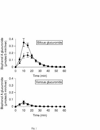
Bisphenol A glucuronidation and excretion in the liver of nonpregnant rats. On
perfusion of the liver with low-dose bisphenol A (10 µM) in Krebs Ringer's solution, in
both the male and female rats, ~100% of the substrate was absorbed in the liver. Then
about 59% of the absorbed bisphenol A was glucuronidated within the liver tissue of the
male and about 84% in the female. The resultant glucuronide that formed in the liver
was excreted mainly into the bile in both groups (Fig. 1). After 1-h perfusion, the total
amount of glucuronide excreted from the liver into the bile as well as that excreted into
the vein were significantly higher (~1.4-fold) in female rats than in male rats.
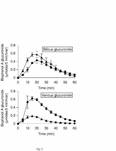
On high-dose (50 µM) of bisphenol A, 92.7% of the substrate was absorbed in
the liver of the male and 93.5% in the female. Then about 66% of the absorbed
bisphenol A was glucuronidated within the liver tissues of the male and about 91% in
the female. In the male rats, the resultant glucuronide was excreted mainly into the bile,
whereas in contrast, in the female rats, much more resultant glucuronide was excreted
into the vein (Fig. 2).
In the nonpregnant female rats, although venous excretion of the bisphenol A
glucuronide during 1-h perfusion increased slightly during anestrus, the excretory
alteration was not significant in the estrous cycle (data not shown).
This article has not been copyedited and formatted. The final version may differ from this version.DMD Fast Forward. Published on October 4, 2004 as DOI: 10.1124/dmd.104.001537
at ASPE
T Journals on A
ugust 27, 2018dm
d.aspetjournals.orgD
ownloaded from
DMD# 2004/001537
11
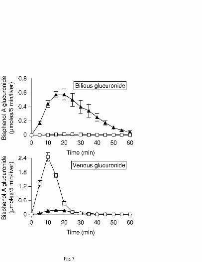
Excretion of bisphenol A glucuronide in the liver of Eisai hyperbilirubinemic rats
(EHBR). To elucidate the excretion pathway of bisphenol A glucuronide from the liver
tissues into the bile, perfusion study was made on the liver of male EHBR, which is a
rat deficient in multidrug resistance-associated protein (MRP) 2. During and after
perfusion of 50 µM bisphenol A to the EHBR liver, the bisphenol A glucuronide was
almost all excreted into the vein, indicating that MRP2 mediates bilious excretion of the
glucuornide (Fig. 3).
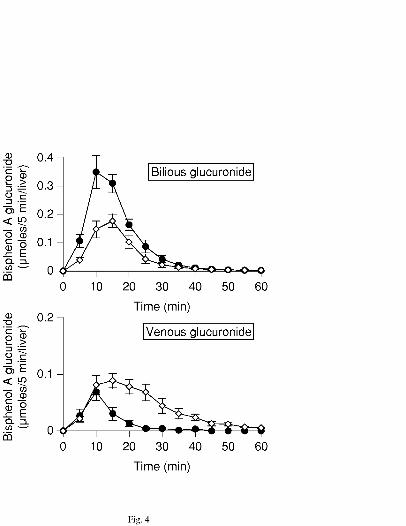
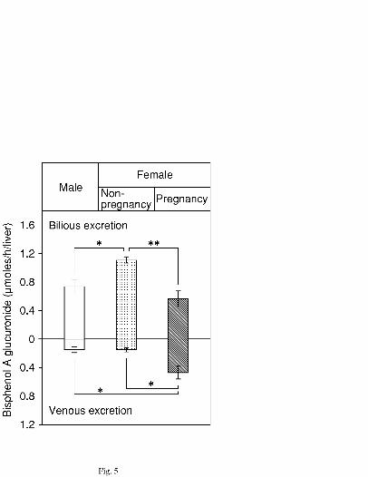
Bisphenol A glucuronidation and excretion in the liver of pregnant rats. In
pregnant rats, liver perfusion of low-dose bisphenol A (10 µM) in the Krebs Ringer's
solution resulted in about 69% of the infused substrate being glucuronidated within the
liver tissue and subsequently excreted into the bile and the vein. Bilious excretion
amounted to 54.5%, and venous excretion 45.5% (Fig. 4). During 1-h perfusion, bilious
excretion of the resulting glucuronide in pregnant rats was half of that in nonpregnant
rats. In sharp contrast, the venous excretion of the glucuronide increased 3-fold in
pregnancy (Fig. 5). The total amount of excreted bisphenol A glucuronide was slightly
lower in pregnant rats than in nonpregnant rats (p < 0.05).
This article has not been copyedited and formatted. The final version may differ from this version.DMD Fast Forward. Published on October 4, 2004 as DOI: 10.1124/dmd.104.001537
at ASPE
T Journals on A
ugust 27, 2018dm
d.aspetjournals.orgD
ownloaded from

DMD# 2004/001537
12
Discussion
This study had three main findings based on experiments on the liver of
Sprague-Dawley rats perfused with bisphenol A: First, the infused compound was
highly glucuronidated during its passage through the liver in both male and nonpregnant
female rats, then the glucuronide was excreted into the bile and vein at a higher
excretion rate in the nonpregnant female than in the male. Second, in both male and
nonpregnant female rats, the resulting glucuronidate was excreted mainly into the bile
via MRP2-mediating transport. Finally, in pregnant rats, with a slight but significant
decrease in total excretion of the glucuronide, bilious excretion of the resulting
glucuronide decreased and venous excretion increased reciprocally.
Bisphenol A glucuronidation and excretion in liver of nonpregnant female rats.
These results bear out that, in the rat liver, glucuronidation is a major pathway for the
elimination of bisphenol A. In female rats, however, total excretion of the bisphenol A
glucuronide conjugate was greater than that in male rats. This finding is in line with a
recent report that in isolated hepatocytes 100% of bisphenol A added to the medium was
metabolized into its glucuronide in female rats and 58% in male rats (Pritchet et al.,
2002). In that study, 30% of bisphenol A glucuronide/sulfate diconjugate was produced
by the hepatocytes of male rats. Such gender differences may be attributed to different
This article has not been copyedited and formatted. The final version may differ from this version.DMD Fast Forward. Published on October 4, 2004 as DOI: 10.1124/dmd.104.001537
at ASPE
T Journals on A
ugust 27, 2018dm
d.aspetjournals.orgD
ownloaded from
DMD# 2004/001537
13
isoenzymes responsible for bisphenol A metabolism. Previously we showed that the
UGT isoform UGT2B1 is involved in bisphenol A glucuronidation (Yokota et al., 1999).
In that study, 65% of microsomal bisphenol A glucuronidation in male rats was
absorbed by anti-UGT2B1 antibody, whereas 35% of the reaction was absorbed in
female rats. Other than UGT2B1, isoenzymes mediating bisphenol A glucuronidation
remain to be elucidated. Given that the UGT2B family mediates glucuronidation of
steroid hormones (Mackenzie et al., 1996; Turgeon et al., 2001), one or more steroid
UDP-glucuronosyltransferase isoenzymes other than UGT2B1 may be responsible for
catalyzing bisphenol A. Shelby et al. (2003) showed gender differences in mRNA levels
of UGT2 isoenzymes in rat liver. It is highly plausible that different isoforms of
glucuronosyltransferases are responsible for the high glucuronidation of bisphenol A in
the liver of nonpregnant female rats.
Excretion pathway of bisphenol A glucuronide from the liver. The absence of
excretion of bisphenol A glucuronide into the bile in EHBR provides unequivocal
evidence that MRP2 is the major bilious transporter for bisphenol A glucuronide.
Previously we established that in male rat liver perfused with bisphenol A at doses
ranging from 10 to 100 µM, the bilious excretion rate of the resulting glucuronide
reaches maximum at 50 µM perfusion and venous excretion of the glucuronide
This article has not been copyedited and formatted. The final version may differ from this version.DMD Fast Forward. Published on October 4, 2004 as DOI: 10.1124/dmd.104.001537
at ASPE
T Journals on A
ugust 27, 2018dm
d.aspetjournals.orgD
ownloaded from
DMD# 2004/001537
14
increases in a dose-dependent manner (Inoue et al., 2001). Similar phenomena were
demonstrated in the present experiments after liver perfusion with high-dose (50 µM)
bisphenol A in the nonpregnant female rat. These findings lead us to believe that in the
presence of over-saturation of MRP2, the compensatory sinusoidal transporting system
excretes the bisphenol A glucuronide. Thus the supposition may be made that MRP2 has
a higher affinity for bisphenol A glucuronide than the sinusoidal transporting system.
On perfusion of the liver of Eisai hyperbilirubinemic rats (EHBR) in the
present study, bilious excretion of the bisphenol A glucuronide was negligible, but
venous excretion increased dramatically. In MRP2-deficient rats, MRP3 expression is
adaptively induced and sinusoidal efflux of biliary constituents is enhanced reciprocally
(Akita et al., 2001; Cao et al., 2002; Kuroda et al., 2004). The compensating induction
of sinusoidal MRP3 has also been demonstrated under downregulation of MRP2
expression by common bile duct ligation and by lipopolysaccharide treatment (Donner
and Keppler, 2001; Soroka et al, 2001). In the light of these findings, it is plausible that
sinusoidal transporters such as MRP3 are involved in the venous excretion of bisphenol
A glucuronide. Further studies are needed to identify the sinusoidal transporters that
mediate venous excretion of the glucuronide.
Bisphenol A glucuronidation and excretion in liver of pregnant rats. Bisphenol A
This article has not been copyedited and formatted. The final version may differ from this version.DMD Fast Forward. Published on October 4, 2004 as DOI: 10.1124/dmd.104.001537
at ASPE
T Journals on A
ugust 27, 2018dm
d.aspetjournals.orgD
ownloaded from
DMD# 2004/001537
15
glucuronidation in rat liver microsomes subsides in pregnancy (Matsumoto et al., 2001).
Furthermore, in pregnancy, hepatic expression and function of MRP2 also decrease
(Cao et al., 2002). For these reasons, we hypothesized that hepatic elimination of
bisphenol A is limited in pregnancy. The present results support our hypothesis, in that
the total amount of excreted bisphenol A glucuronide during 1-h perfusion was slightly
but significantly lower in pregnant rats than in nonpregnant rats. This may indicate that
low expression of glucuronosyltransferase compromises the step of bisphenol A
glucuronidation and thus limits bisphenol A elimination in the living liver. In pregnancy,
bilious excretion of the bisphenol A glucuornide decreased and venous excretion
increased reciprocally. The venous excretion rate, however, did not increase as much as
one might expect, and the duration of excretion was prolonged. Cao et al. (2001)
showed that the expression of MRP3 also attenuates in pregnancy. The prolonged
excretion may be a result of over-saturation of MRP3. In contrast to MRP3, the
expression of MRP1, one of the sinusoidal glucuronide transporters, does not change
during pregnancy (Cao et al., 2001). These findings, together with our present results,
give rise to the view that in pregnancy low expression of MRP2 limits the transport rate
of the bisphenol A glucuronide into the bile and that sinusoidal transporting systems
such as MRP1 and 3 compensate by transporting the glucuronide into the vein.
Venous bisphenol A glucuronide excreted from the liver flows into the systemic
This article has not been copyedited and formatted. The final version may differ from this version.DMD Fast Forward. Published on October 4, 2004 as DOI: 10.1124/dmd.104.001537
at ASPE
T Journals on A
ugust 27, 2018dm
d.aspetjournals.orgD
ownloaded from
DMD# 2004/001537
16
blood circulation. Pottenger et al. (2000) showed that urinary bisphenol A glucuronide is
detectable after oral, intraperitoneal or subcutaneous administration of bisphenol A. In
the case of 1-naphthol, the kidney provides high clearance of the 1-naphthol
glucuronide (de Vries et al., 1989). By supposition, therefore, the bisphenol A
glucuronide may also be excreted into the urine and thus eliminated from the body.
Certain organs such as the lung, small intestine and placenta, however, have high
β-glucuronidase activity (Paigen, 1989; Sperker et al., 1997). In such organs, bisphenol
A glucuronide can be deconjugated to free (unconjugated) bisphenol A. In the human
also, placental β-glucuronidase activity has been demonstrated during pregnancy
(Kushari and Mukherjea, 1980). Given that the bisphenol A glucuronide remaining in
the systemic blood circulation is catalyzed by the placental β-glucuronidase, it is
plausible that bisphenol A deconjugated by β-glucuronidase would permeate the blood
of the umbilical cord. Takahashi and Oishi (2000) detected bisphenol A in rat fetuses
after maternal exposure to the compound. In the light of these findings, our present
results suggest that the risk of bisphenol A exposure to the fetus is high in spite of
preservation of bisphenol A glucuronidation in the maternal liver.
Conclusion To further elucidate the mechanism governing the detrimental effects of
endocrine disrupting chemicals on target organs, it is essential to clarify both the
This article has not been copyedited and formatted. The final version may differ from this version.DMD Fast Forward. Published on October 4, 2004 as DOI: 10.1124/dmd.104.001537
at ASPE
T Journals on A
ugust 27, 2018dm
d.aspetjournals.orgD
ownloaded from
DMD# 2004/001537
17
metabolism and elimination pathways of the chemicals during their journeys within the
body. The present study has established that, in rats, bisphenol A is highly
glucuronidated and excreted into the bile via MRP2. In pregnancy, however, because of
low expression of MRP2, bilious excretion of the resulting glucuronide decreases and
venous excretion increases reciprocally. Given that exposure of pregnant animals to
bisphenol A could adversely affect the fetus, it is critical that further work be done to
determine the fate of the venous glucuronide in its complete pathway before excretion.
This article has not been copyedited and formatted. The final version may differ from this version.DMD Fast Forward. Published on October 4, 2004 as DOI: 10.1124/dmd.104.001537
at ASPE
T Journals on A
ugust 27, 2018dm
d.aspetjournals.orgD
ownloaded from
DMD# 2004/001537
18
Acknowledgements
We wish to thank Dr. M. Tamura of Research Institute for Electronic Science,
Hokkaido University, for his technical advice and insightful discussions. We are grateful
to Dr. N. L. Kennedy for valuable suggestions.
This article has not been copyedited and formatted. The final version may differ from this version.DMD Fast Forward. Published on October 4, 2004 as DOI: 10.1124/dmd.104.001537
at ASPE
T Journals on A
ugust 27, 2018dm
d.aspetjournals.orgD
ownloaded from
DMD# 2004/001537
19
References
Akita H, Suzuki H, and Sugiyama Y (2001) Sinusoidal efflux of taurocholate is
enhanced in Mrp2-deficient rat liver. Pharm Res 18: 1119-25.
Cao J, Stieger B, Meier PJ, and Vore M (2002) Expression of rat hepatic multidrug
resistance-associated proteins and organic anion transporters in pregnancy. Am J
Physiol Gastrointest Liver Physiol 283: G757-66.
de Vries MH, Redegeld FA, Koster AS, Noordhoek J, de Haan JG, Oude Elferink RP,
and Jansen PL (1989) Hepatic, intestinal and renal transport of
1-naphthol-beta-D-glucuronide in mutant rats with hereditary-conjugated
hyperbilirubinemia. Naunyn Schmiedebergs Arch Pharmacol 340: 588-92.
Donner MG, and Keppler D (2001) Up-regulation of basolateral multidrug resistance
protein 3 (Mrp3) in cholestatic rat liver. Hepatology 34: 351-9.
Howdeshell KL, Hotchkiss AK, Thayer KA, Vandenbergh JG, and vom Saal FS (1999)
Exposure to bisphenol A advances puberty. Nature 401: 763-4.
Inoue H, Yokota H, Makino T, Yuasa A, and Kato S (2001) Bisphenol A glucuronide, a
major metabolite in rat bile after liver perfusion. Drug Metab Dispos 29:
1084-7.
Krishnan AV, Stathis P, Permuth SF, Tokes L, and Feldman D (1993) Bisphenol-A: an
estrogenic substance is released from polycarbonate flasks during autoclaving [see
This article has not been copyedited and formatted. The final version may differ from this version.DMD Fast Forward. Published on October 4, 2004 as DOI: 10.1124/dmd.104.001537
at ASPE
T Journals on A
ugust 27, 2018dm
d.aspetjournals.orgD
ownloaded from
DMD# 2004/001537
20
comments]. Endocrinology 132: 2279-86.
Kuroda M, Kobayashi Y, Tanaka Y, Itani T, Mifuji R, Araki J, Kaito M, and Adachi Y
(2004) Increased hepatic and renal expressions of multidrug resistance-associated
protein 3 in Eisai hyperbilirubinuria rats. J Gastroenterol Hepatol 19: 146-53.
Kushari J, and Mukherjea M (1980) Studies on beta-glucuronidase of the developing
human placenta. Gynecol Obstet Invest 11: 119-27.
Luquita MG, Catania VA, Pozzi EJ, Veggi LM, Hoffman T, Pellegrino JM, Ikushiro S,
Emi Y, Iyanagi T, Vore M, and Mottino AD (2001) Molecular basis of perinatal
changes in UDP-glucuronosyltransferase activity in maternal rat liver. J
Pharmacol Exp Ther 298: 49-56.
Mackenzie PI, Mojarrabi B, Meech R, and Hansen A (1996) Steroid UDP
glucuronosyltransferases: characterization and regulation. J Endocrinol 150:
S79-86.
Matsumoto J, Yokota H, and Yuasa A (2002) Developmental increases in rat hepatic
microsomal UDP-glucuronosyltransferase activities toward xenoestrogens and
decreases during pregnancy. Environ Health Perspect 110: 193-6.
Matthews JB, Twomey K, and Zacharewski TR (2001) In vitro and in vivo interactions
of bisphenol A and its metabolite, bisphenol A glucuronide, with estrogen receptors
α and β. Chem Res Toxicol 14: 149-157.
This article has not been copyedited and formatted. The final version may differ from this version.DMD Fast Forward. Published on October 4, 2004 as DOI: 10.1124/dmd.104.001537
at ASPE
T Journals on A
ugust 27, 2018dm
d.aspetjournals.orgD
ownloaded from
DMD# 2004/001537
21
NTP (1982) Carcinogenesis Bioassay of Bisphenol A (CAS No. 80-05-7) in F344 Rats
and B6C3F1 Mice (Feed Study). TR 215. Research Triangle Park, NC: National
Toxicology Program.
Paigen K (1989) Mammalian beta-glucuronidase: genetics, molecular biology, and cell
biology. Prog Nucleic Acid Res Mol Biol 37: 155-205.
Pottenger LH, Domoradzki JY, Markham DA, Hansen SC, Cagen SZ and Waechter JM,
Jr. (2000) The relative bioavailability and metabolism of bisphenol A in rats is
dependent upon the route of administration. Toxicol Sci 54: 3-18.
Pritchett JJ, Kuester RK, and Sipes IG (2002) Metabolism of bisphenol A in primary
cultured hepatocytes from mice, rats, and humans. Drug Metab Dispos 30:
1180-5.
Shelby MK, Cherrington NJ, Vansell NR, and Klaassen CD (2003) Tissue mRNA
expression of the rat UDP-glucuronosyltransferase gene family. Drug Metab
Dispos 31: 326-33.
Soroka CJ, Lee JM, Azzaroli F, and Boyer JL (2001) Cellular localization and
up-regulation of multidrug resistance-associated protein 3 in hepatocytes and
cholangiocytes during obstructive cholestasis in rat liver. Hepatology 33:
783-91.
Sperker B, Backman JT, and Kroemer HK (1997) The role of beta-glucuronidase in
This article has not been copyedited and formatted. The final version may differ from this version.DMD Fast Forward. Published on October 4, 2004 as DOI: 10.1124/dmd.104.001537
at ASPE
T Journals on A
ugust 27, 2018dm
d.aspetjournals.orgD
ownloaded from
DMD# 2004/001537
22
drug disposition and drug targeting in humans. Clin Pharmacokinet 33: 18-31.
Steinmetz R, Mitchner NA, Grant A, Allen DL, Bigsby RM, and Ben-Jonathan N (1998)
The xenoestrogen bisphenol A induces growth, differentiation, and c-fos gene
expression in the female reproductive tract. Endocrinology 139: 2741-7.
Takahashi O and Oishi S (2000) Disposition of orally administered
2,2-Bis(4-hydroxyphenyl)propane (Bisphenol A) in pregnant rats and the placental
transfer to fetuses. Environ Health Perspect 108: 931-5.
Turgeon D, Carrier JS, Levesque E, Hum DW, and Belanger A (2001) Relative
enzymatic activity, protein stability, and tissue distribution of human
steroid-metabolizing UGT2B subfamily members. Endocrinology 142: 778-87.
Yokota H, Iwano H, Endo M, Kobayashi T, Inoue H, Ikushiro S, and Yuasa A (1999)
Glucuronidation of the environmental oestrogen bisphenol A by an isoform of
UDP-glucuronosyltransferase, UGT2B1, in the rat liver. Biochem J 340: 405-9.
This article has not been copyedited and formatted. The final version may differ from this version.DMD Fast Forward. Published on October 4, 2004 as DOI: 10.1124/dmd.104.001537
at ASPE
T Journals on A
ugust 27, 2018dm
d.aspetjournals.orgD
ownloaded from
DMD# 2004/001537
23
Footnotes
This work was partially supported by Northern Advancement Center for
Science and Technology (NOASTEC) and by a Grants-in-Aid to Cooperative Research
from Rakuno Gakuen University.
Person to receive reprint requests: Seiyu Kato, Department of Veterinary Physiology,
School of Veterinary Medicine, Rakuno Gakuen University, Ebetsu, Hokkaido,
069-8501 Japan. E-mail: [email protected]
This article has not been copyedited and formatted. The final version may differ from this version.DMD Fast Forward. Published on October 4, 2004 as DOI: 10.1124/dmd.104.001537
at ASPE
T Journals on A
ugust 27, 2018dm
d.aspetjournals.orgD
ownloaded from
DMD# 2004/001537
24
Legends for Figures
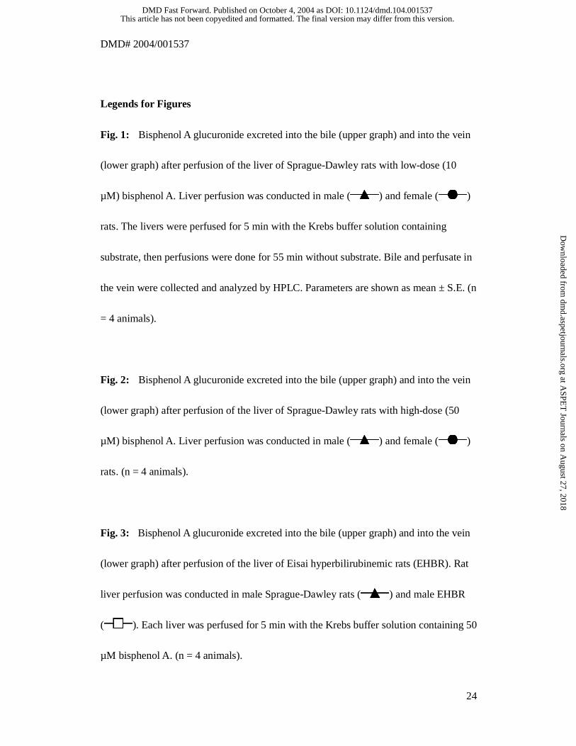
Fig. 1: Bisphenol A glucuronide excreted into the bile (upper graph) and into the vein
(lower graph) after perfusion of the liver of Sprague-Dawley rats with low-dose (10
µM) bisphenol A. Liver perfusion was conducted in male ( ) and female ( )
rats. The livers were perfused for 5 min with the Krebs buffer solution containing
substrate, then perfusions were done for 55 min without substrate. Bile and perfusate in
the vein were collected and analyzed by HPLC. Parameters are shown as mean ± S.E. (n
= 4 animals).
Fig. 2: Bisphenol A glucuronide excreted into the bile (upper graph) and into the vein
(lower graph) after perfusion of the liver of Sprague-Dawley rats with high-dose (50
µM) bisphenol A. Liver perfusion was conducted in male ( ) and female ( )
rats. (n = 4 animals).
Fig. 3: Bisphenol A glucuronide excreted into the bile (upper graph) and into the vein
(lower graph) after perfusion of the liver of Eisai hyperbilirubinemic rats (EHBR). Rat
liver perfusion was conducted in male Sprague-Dawley rats ( ) and male EHBR
( ). Each liver was perfused for 5 min with the Krebs buffer solution containing 50
µM bisphenol A. (n = 4 animals).
This article has not been copyedited and formatted. The final version may differ from this version.DMD Fast Forward. Published on October 4, 2004 as DOI: 10.1124/dmd.104.001537
at ASPE
T Journals on A
ugust 27, 2018dm
d.aspetjournals.orgD
ownloaded from
DMD# 2004/001537
25
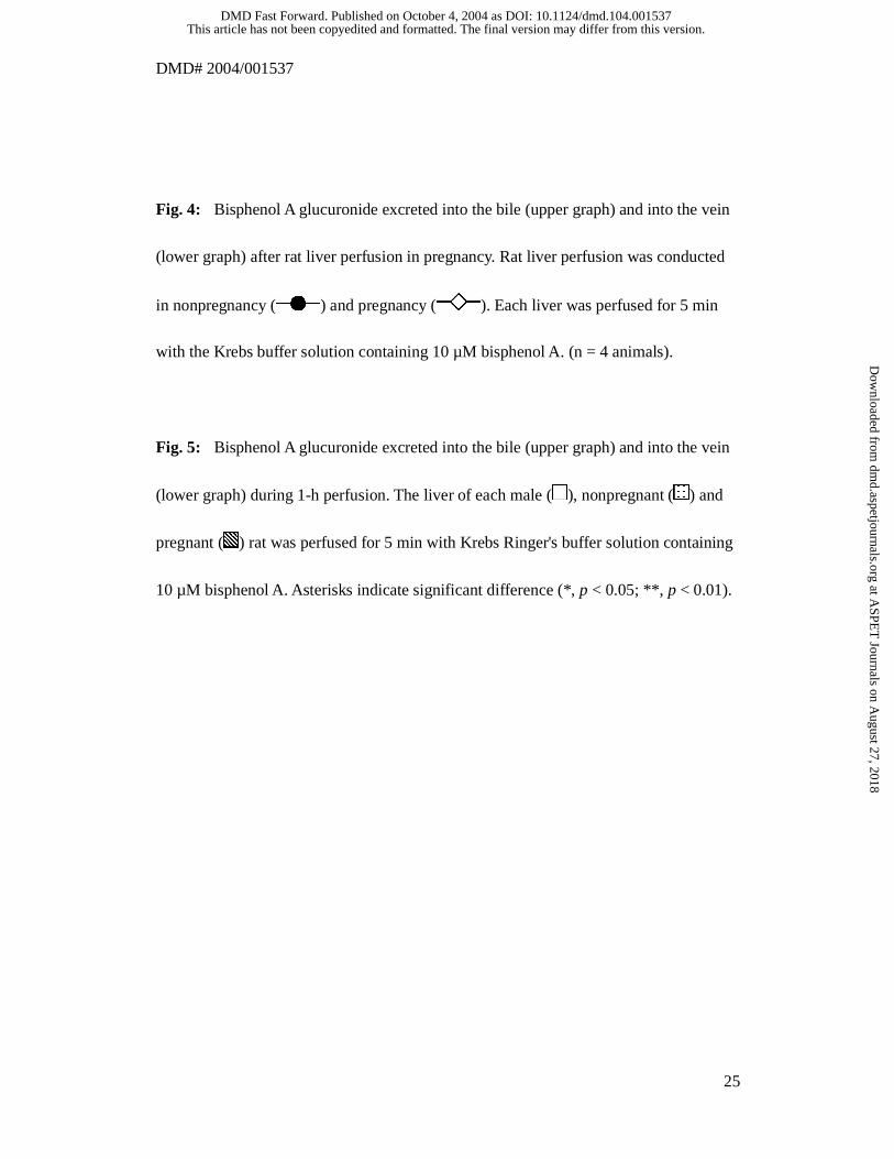
Fig. 4: Bisphenol A glucuronide excreted into the bile (upper graph) and into the vein
(lower graph) after rat liver perfusion in pregnancy. Rat liver perfusion was conducted
in nonpregnancy ( ) and pregnancy ( ). Each liver was perfused for 5 min
with the Krebs buffer solution containing 10 µM bisphenol A. (n = 4 animals).
Fig. 5: Bisphenol A glucuronide excreted into the bile (upper graph) and into the vein
(lower graph) during 1-h perfusion. The liver of each male ( ), nonpregnant ( ) and
pregnant ( ) rat was perfused for 5 min with Krebs Ringer's buffer solution containing
10 µM bisphenol A. Asterisks indicate significant difference (*, p < 0.05; **, p < 0.01).
This article has not been copyedited and formatted. The final version may differ from this version.DMD Fast Forward. Published on October 4, 2004 as DOI: 10.1124/dmd.104.001537
at ASPE
T Journals on A
ugust 27, 2018dm
d.aspetjournals.orgD
ownloaded from
This article has not been copyedited and formatted. The final version may differ from this version.DMD Fast Forward. Published on October 4, 2004 as DOI: 10.1124/dmd.104.001537
at ASPE
T Journals on A
ugust 27, 2018dm
d.aspetjournals.orgD
ownloaded from
This article has not been copyedited and formatted. The final version may differ from this version.DMD Fast Forward. Published on October 4, 2004 as DOI: 10.1124/dmd.104.001537
at ASPE
T Journals on A
ugust 27, 2018dm
d.aspetjournals.orgD
ownloaded from
This article has not been copyedited and formatted. The final version may differ from this version.DMD Fast Forward. Published on October 4, 2004 as DOI: 10.1124/dmd.104.001537
at ASPE
T Journals on A
ugust 27, 2018dm
d.aspetjournals.orgD
ownloaded from
This article has not been copyedited and formatted. The final version may differ from this version.DMD Fast Forward. Published on October 4, 2004 as DOI: 10.1124/dmd.104.001537
at ASPE
T Journals on A
ugust 27, 2018dm
d.aspetjournals.orgD
ownloaded from
This article has not been copyedited and formatted. The final version may differ from this version.DMD Fast Forward. Published on October 4, 2004 as DOI: 10.1124/dmd.104.001537
at ASPE
T Journals on A
ugust 27, 2018dm
d.aspetjournals.orgD
ownloaded from