biosolve ii
TRANSCRIPT
Safety and performance of the second-generation drug-eluting absorbable metal scaffold in patients with de-novo coronary artery lesions (BIOSOLVE-II): 6 month results of a prospective, multicentre, non-randomised, first-in-man trial
Published Online October 12, 2015 lancet

IntroductionDrug-eluting stents reduce restenosis rates compared with bare-metal stents and are the present default device for percutaneous coronary intervention Concerns have been raised about the use of drug-eluting stents, such as•The risks of delayed arterial healing,• late and very late stent thrombosis,•Hypersensitivity reactions to polymers•And accelerated in-stent formation of neoatherosclerosis.
A vessel that is permanently caged by a metal stent has some restrictions:•Compensatory vascular remodelling is prevented by the stent• Non-invasive imaging options are limited by the presence of the metal• and struts might interfere with future treatment options, including coronary bypass Surgery.These drawbacks motivated scientists to develop absorbable vascular scaffolds, which are designed to free the vessel from a permanently implanted metal stent.

Absorbable scaffold designs should ensure sufficient but temporary scaffolding with a performance similar to metal drug-eluting stents with respect to recoil, healing, and restenosis rates, followed by safe degradation and absorption, enabling restoration of vasomotion and prevention of late unfavourable effects of metal stents. So far, only two drug-eluting absorbable polymeric scaffolds have received CE-mark approval—The Absorb bioresorbable vascular scaffold system (ABSORB BVS Abbott Vascular, Santa Clara, CA) andThe DESolve bioresorbable scaff old (Elixir Medical, Sunnyvale, CA)

The absorbable metal scaffold was developed as an alternative to polymeric scaffolds. Findings from early animal studies with magnesium alloy scaffolds in porcine coronary arteries showed good biocompatibility of this material. First devices showed a good safety profile, but angiographic performance measures of these early devices were inferior to those of contemporary drug-eluting stents. BIOSOLVE-II intends to study and assess the safety and performance of this novel absorbable metal scaffold in symptomatic patients with de-novo coronary artery lesions.

MethodsStudy design and patientsThis is a prospective, multicentre, non-randomised, first-in-man trial at 13 percutaneous coronary intervention centres in Belgium, Brazil, Denmark, Germany, Singapore, Spain, Switzerland, and the Netherlands.Eligible patients were older than 18 years and younger than 80 years and had stable or unstable angina or documented silent ischaemia. A maximum of two de-novo lesions in two separate coronary arteries were allowed to be treated per patient, with a reference vessel diameter between 2.2 mm and 3.7 mm, a lesion length of 21 mm or less, and a stenosis of between 50% and 99% in diameter.

Exclusion criteria included
left ventricular ejection fraction of less than 30%, thrombus in the target vessel, severe calcification, three-vessel disease, ostial lesion, target lesion involving a side branch of more than 2.0 mm in diameter, target lesion located in or supplied by an arterial or venous bypass graft, and unsuccessful predilatation

ProceduresDREAMS 2G is a balloon-expandable sirolimus-eluting Scaffold on a rapid-exchange delivery system. The Scaffold backbone is made from an absorbable magnesium alloy with two permanent x-ray markers made from tantalum at the distal and proximal scaffold end. Degradation of the alloy includes two steps: First, the magnesium alloy is converted to hydrated magnesium oxide. Second, magnesium oxide is converted to magnesium phosphate, which is consecutively replaced by amorphous calcium phosphate. During this process, metallic magnesium is removed by diffusion from the amorphous matrix and is absorbed by the body. The surface of the scaffold backbone is completely coated with bioresorbable poly-L-lactide acid, which incorporates sirolimus

In this study Clinical follow-up was scheduled at months 1, 6, 12, 24, and 36, and imaging follow-up at 6 months in a subgroup of patients undergoing intravascular ultrasound, optical coherence tomography, and vasomotion testing. All patients were assigned to angiographic follow-up at 6 months.


OutcomesThe primary endpoint was in-segment late lumen loss at6 month follow-up.Secondary clinical endpoints were the rate of target lesion failure, defined as a composite of cardiac death, target vessel myocardial infarction, coronary artery bypass graft surgery, and clinically driven target lesion revascularisation; the rate of scaffold thrombosis; and device and procedure success

Results
Mean in-segment late lumen loss at 6 months was 0.27 mm (SD 0.37) and in-scaffold late lumen loss was 0.44 mm (0.36) (figure 2, table 2). During 6 months of vasomotion testing in 25 patients, the mean lumen diameter of 2.60 mm (SD 0.29) in the treated segment decreased to 2.49 mm (0.3) with acetylcholine (difference –0.10 mm; p=0.014) and dilated to 2.66 mm (0.33) with nitroglycerine (difference vs post-acetylcholine 0.17 mm; p<0・ 001; figure 3). For 20 (80%) patients, we noted vasodilatation or vasoconstriction after either acetylcholine or nitroglycerine administration with a threshold of change of 3.0% or more.
Target lesion failure at 6 months occurred in four patients (3%, 95% CI 1.3–8.3). •One death of unknown cause (<1%, 0.0–3.0) on day 134 postprocedure was classified as cardiac death and possible scaffold thrombosis.•One patient (<1%, 0.0–3.0) had a target vessel myocardial infarction. This patient had temporary no-reflow after scaffold implantation in the right coronary artery with clinical symptoms and electrocardiogram changes. •Two patients (2%, 95% CI 0.2–5.9) needed clinically driven target lesion revascularisation: one patient had unstable angina and a stenosis of 81% in diameter on day 84 and one had Canadian Cardiovascular Society class II angina and a stenosis of 54% in diameter at 6 month control angiography. •The decision for revascularisation was operator driven.

No definite or probable scaffold thrombosis was observed nor was any additional clinically driven target vessel revascularisation. One patient (<1%, 95% CI 0.0–3.0) died of non-cardiac causes (cancer) before 6 month follow-up.


DiscussionFindings show that DREAMS 2G improves late lumen loss compared with its precursor devices while maintaining a favourable clinical and safety profile. DREAMS 2G is built on continuous improvements of previous generations of absorbable metal scaffolds.Advantages of metal scaffolds are a good radial strength, low acute recoil, and high compliance to the vessel geometry. Furthermore, metal scaffolds can be implanted via a single-step inflation, hence providing the advantage that they can be implanted in a similar way to a permanent metal stent.
The first magnesium scaffold investigated in human coronary arteries was a bare absorbable metal scaffold tested in the PROGRESS study. This scaffold had a good safety profile, but measures of performance, such as in-scaffold late lumen loss, were not satisfactory, suggesting a need for slower scaffold absorption together with an antiproliferative drug-elution concept.
Refinement of the absorbable metal scaffold led to development of the DREAMS 1G device, with an improved alloy composition and strut geometry for better scaffolding properties and addition of a drug-polymer matrix with paclitaxel to counteract the neointimal proliferative response.

DREAMS 1G was tested in the first-in-man BIOSOLVE-I study and showed a favourable clinical safety profile and significantly improved angiographic performance measures compared with its bare absorbable metal scaffold precursor. However, BIOSOLVE-I findings still showed a substantial insegment late lumen loss of 0.52 mm (SD 0.48) and an in-scaffold loss of 0.65 mm (0.50) at 6 months.
Compared with DREAMS 1G, DREAMS 2G has a more flexible and stronger scaffold backbone design, higher bending flexibility, and higher radial force. Additionally, the drug-polymer coating has changed to sirolimus in combination with a bioresorbable poly-L-lactide acid polymer to more effectively decrease neointimal formation. The same coating is successfully used in the commercially available Orsiro drug-eluting stent.

Notably, late lumen loss is probably a moving target in absorbable scaffolds and has low relevance for prediction of long-term outcomes, because in absorbable scaffolds late lumen enlargement (beyond 1 year) was reported. Moreover, in the ABSORB cohort A trial, despite an inscaffold late lumen loss of 0.44 mm at 6 months, 5 year outcomes were excellent without any additional major adverse cardiac events beyond 6 months.
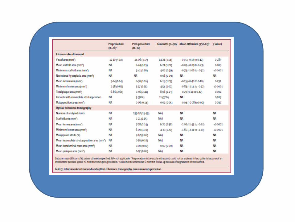
The neointimal hyperplasia area decreased from 0.30 mm in BIOSOLVE-I to only 0.08 mm in BIOSOLVE-II. Notably, the area of BIOSOLVE-II is identical to that recorded in ABSORB cohort B (0.08 mm2) and lower than that in ABSORB cohort A (0.30 mm2) and in DESolve (0.4 mm2). The discordance between late lumen loss and neointimal hyperplasia in this study might be due to decreasing radial strength in the context of the absorption process. Furthermore, intravascular ultrasound was only done in a subset of 30 patients and anatomic variations cannot be ruled out.
Post-procedure assessment of optical coherence tomography showed a similar incomplete strut apposition area for DREAMS 2G (0.16 mm) and ABSORB cohort B (0.19 mm; p=0.663). For ABSORB cohort A, 91% of the struts were apposed post procedure and DESolve had 14.45% cross sections with malapposition. At 6 months, this study could not detect any malapposed struts with optical coherence tomography because the DREAMS 2G device was fully embedded into the vessel wall. 97% of the struts were well apposed in BIOSOLVE-I and 93% were well apposed in ABSORB cohort A; 0.04% of the optical coherence tomography cross sections showed malapposition for DESolve at follow-up. No intraluminal mass was observed at any time in this study compared with intraluminal masses in 24% of patients at 6 months for ABSORB cohort B, in which intraluminal masses were more often associated with malapposed or uncovered struts.
LIMITATIONSThe present study has limitations that are inherent to a non-randomised first-in-man study. The absence of a direct comparison with other permanent stents or scaffolds restricts the interpretation of results. This study is the first-in-man experience and included patients with straightforward de-novo lesions; thus, the results cannot be generalised to other types of lesions, such as highly calcifi ed, complex, or restenotic lesions. The ideal follow-up time for absorbable scaffolds is, by contrast with permanent stents, still uncertain. They selected the 6 month follow-up timepoint for assessment of the primary endpoint in-segment late lumen loss for similarity with the earliest timepoint available in the BIOSOLVE-I trial
Conclusion
In the BIOSOLVE-II trial with nearly complete angiographic and clinical follow-up, the DREAMS 2G novel absorbable metal scaffold showed substantially improved performance measures, with a favourable safety profile up to 6 months, compared with its precursor, DREAMS 1G. No definite or probable scaffold thrombosis was observed for DREAMS 2G or any of the precursor devices, and the rates of target lesion failure and revascularisation in BIOSOLVE-II were low. Vasomotion was restored at 6 months. Hence, DREAMS 2G could be an acceptable alternative to present absorbable polymeric scaffolds.

Thank you