biomimetic mems to assist, enhance, and expand human ...gebeshuber/biomimetic_mems_8066_60.pdf ·...
TRANSCRIPT

Biomimetic MEMS to assist, enhance and expand human sensory perceptions - A survey on state-of-the art developments
Teresa Makarczuka, Tina R. Matinb, Salmah B. Karmanb,c, S. Zaleha M. Diahb, Benyamin Davajib,
Mark O. Macqueend, Jeanette Muellere, Ulrich Schmidf, Ille C. Gebeshuber*a,b
aInstitute of Applied Physics, Vienna University of Technology, Wiedner Hauptstrasse 8-10/134, 1040 Vienna, Austria;
bInstitute of Microengineering and Nanoelectronics, Universiti Kebangsaan Malaysia, 43600 UKM Bangi, Malaysia;
cDepartment of Biomedical Engineering, Faculty of Engineering, University of Malaya, 50603 Kuala Lumpur, Malaysia;
dAramis Technologies, 2 Jalan Alam Sutera 1, Bukit Jalil, 57000 Kuala Lumpur, Malaysia; etrustroom, Servitengasse 24/11, 1090 Wien, Austria;
fDepartment for Microsystems Technology, Institute of Sensor and Actuator Systems, Vienna University of Technology, Floragasse 7, 1040 Vienna, Austria
ABSTRACT
The human senses are of extraordinary value but we cannot change them even if this proves to be a disadvantage in modern times. However, we can assist, enhance and expand these senses via MEMS. Current MEMS cover the range of the human sensory system, and additionally provide data about signals that are too weak for the human sensory system (in terms of signal strength) and signal types that are not covered by the human sensory system. Biomimetics deals with knowledge transfer from biology to technology. In our interdisciplinary approach existing MEMS sensor designs shall be modified and adapted (to keep costs at bay), via biomimetic knowledge transfer of outstanding sensory perception in ‘best practice’ organisms (e.g. thermoreception, UV sensing, electromagnetic sense). The MEMS shall then be linked to the human body (mainly ex corpore to avoid ethics conflicts), to assist, enhance and expand human sensory perception. This paper gives an overview of senses in humans and animals, respective MEMS sensors that are already on the market and gives a list of possible applications of such devices including sensors that vibrate when a blind person approaches a kerb stone edge and devices that allow divers better orientation under water (echolocation, ultrasound).
Keywords: MEMS, biomimetics, sensory systems, senses, bioinspired, biomimicry
1. INTRODUCTION “What is first given to us is appearance. When combined with consciousness, it is called perception...”
(Immanuel Kant, 1724-1804)1
Immanuel Kant proposed that our knowledge of the outside world depends on our modes of perception. The human body is equipped with several senses: we can see, hear, smell, taste, feel touch and sense temperature. These senses consist of organs with specialised cells that offer receptors for certain stimuli. The receptor cells are linked to the nervous system, which is connected to the brain, where the informationis finally analysed. The human sensory systems, as well as the human perception that exists today, are the result of billions of years of evolution.
Current MicroElectroMechanical Systems (MEMS) cover the range of human sensory systems and furthermore provide data of even more signals. Not only signals that are too weak for human perceptions, but also signals, which are not covered by the human sensory system can be recognised and converted through MEMS.
*[email protected]; phone +60 13 319 8588; fax +60 38 925 0439; http://www.ille.com
Smart Sensors, Actuators, and MEMS V, edited by Ulrich Schmid, José Luis Sánchez-Rojas, Monika Leester-Schaedel, Proc. of SPIE Vol. 8066, 80661O · © 2011 SPIE
CCC code: 0277-786X/11/$18 · doi: 10.1117/12.886554
Proc. of SPIE Vol. 8066 80661O-1

Biomimetics, which is an interdisciplinary knowledge field at the crossing point of biology and technology as well as the arts2, is a promising method in the development of emerging MEMS. Applying certain principles from the biological sensory systems to technological strategies is an encouraging approach to assist, enhance and expand human sensory perception.
Essential and important questions in the field of biomimetics of the senses are: Which senses are available in nature? Which biological sensor systems exist and how are they working? Through the years of evolution the senses of organisms have developed immensely depending on their surrounding and lifestyle of the organisms. To be able to survive every organism is equipped with its specific sensory system- and indeed, in many definitions of life itself, sensing the environment and reaction to it is one of the basic properties of living entities.
The purpose of this study is to identify functions of biological sensory systems and in the second step suggest possible MEMS applications. Therefore multiple scientific databases are used to find relevant literature about extraordinary sensory systems in nature and their anatomical structures.
1.1 Biomimetics
Biomimetics aims at identifying the deep underlying principles of materials, structures and processes in living nature, and on subsequent knowledge transfer from biology to engineering and the arts. It seeks to apply certain principles from biological systems to technological strategies and innovative applications. The range of potential use for biomimetics is enormous. Nowadays it includes architecture and design, surface and materials technologies as well as sensors, medical engineering and management3.
1.2 Microelectromechanical Systems
MEMS are small, integrated devices or systems that combine electrical and mechanical components. They are composed of components between one to 100 micrometers in size. The MEMS devices themselves generally measure from 20 micrometers to one millimetre.
MEMS consist of mechanical elements, sensors, actuators, and microelectronic devices on a common silicon substrate. With all these components these systems can sense, control, and activate mechanical processes on the micro scale, and function individually or in arrays, thereby generating effects on the macro scale. Current microfabrication technology enables fabrication of large arrays of devices, which individually perform simple tasks, but in combination can accomplish complicated functions [4].
Currently, various MEMS are on the market that “sense” mechanical, electromagnetic or chemical signals equivalent to human, animal or plant senses.
2. THE SENSES 2.1 Sight
The human eye has a complex structure consisting of a transparent lens that focuses light on the retina. Rod and cone cells in the retina are the basis for conscious light perception and vision including colour differentiation and the perception of depth. The range of the electromagnetic spectrum that can be detected by the human eye comprises wavelengths between 390 and 750nm, which is called the visible spectrum5. The human eye can distinguish about 10 million colours with its trichromatic vision system6. Trichromatic colour vision means that there are three different types of photoreceptors, which are labelled according to the ordering of the wavelengths of the peaks of their spectral sensitivities. This quality of perception is adjusted to our surrounding and demands. Important characteristics of a well and extraordinary developed eye are visual acuity, contrast sensitivity, spatial resolution colour and motion perception. The focus in this chapter will be on different colour perception of specific species.
There are many species that can see frequencies that are outside the human visible spectrum. The reason lies within the different existing types and numbers of photoreceptors containing pigments of different spectral sensitivities. Consequently light from the UV- range up to the IR- range can be detected by organs of many animals. In the following sections, types of animals that can percept specific ranges of the electromagnetic spectrum are described.
The mantis shrimp possesses apposition compound eyes that contain more photoreceptor types than any other animal. These eyes are in almost continuous and independent motion and their morphological structure is quite complex.
Proc. of SPIE Vol. 8066 80661O-2

There are 16 different cell types contributing to at least three different visual modalities and twelve channel colour vision systems. These twelve narrow colour channels spread evenly through the spectrum from 300 to 700nm. With spectral half bandwidths around 20nm, these are among the sharpest spectral sensitivities in the animal world7. Another interesting fact is the mantis shrimp sensitivity in the UV range. They possess more UV photoreceptor than any other animal8. To perceive UV light it is necessary that the animals possess ocular media that do transmit UV light.
The colour perception of butterflies is of extraordinary value, too. It is an essential gift to search for nectar. Butterflies find their sources, the flowers, and make their mate choice by using their capacity for color vision9. The two compound eyes of butterflies consist of numerous anatomically identical units that are called ommatidias and are arranged in a hemisphere. Most species of butterflies possess three different types of ommatidias that contain nine photoreceptors each with maximal sensitivity in the ultraviolet (350nm), blue (450nm) and green (550nm) wavelength ranges10. This is termed “trichromatic vision” and can be found in lots of mammals. The difference to the human trichromatic vision system lies in the different ranges of wavelengths of light that stimulate the photoreceptors. Butterflies colour receptors can perceive colours in a high frequency but cannot detect lower frequencies in the range of red colour. For that reason butterflies are not interested in red flowers.
The Japanese yellow swallowtail butterfly (Papilio xuthus) has compound eyes that are furnished with six distinct classes of spectral receptors (UV λ=360nm, violet λ=400nm, blue λ=460nm, green λ=520nm, red λ=600nm, broadband λ= almost the whole human visible spectrum)11. Bumble and honey- bees possess trichromatic colour processing, too, with three types of spectral receptors in their compound eyes. Similar to most of the butterflies the sensitivity peaks of bumblebees are in the UV (λ=340nm), blue (λ=430nm) and green (λ=535nm) wavelength ranges. Honeybees are highly sensitive in the UV spectrum12. The honeybee possesses a trichromatic visual system with UV (λ=335nm), blue (λ=435 nm) and green (λ=540nm) receptors13,14. The bees use their UV receptors to detect polarized light and determine orientation for navigation15. The green receptors are used for optomotor response16 and for flight distance estimation17.
Avian eyes are characterized by the presence of oil droplets within the distal end of the inner segment of their cones, which act as cut-off filters and absorb light below their characteristic wavelength of transmission18. Together with their associated oil droplets, birds have at least three to four photoreceptors with spectral sensitivity maxima reaching from 370nm to 580nm19. Another feature that increases the complexity of colour perception is the differential distribution of oil droplets across the retina. Pigeons and several other avian species show high UV sensitivity, which they may use to view objects such as plumage or fruits reflecting UV light20. The domestic turkey (Meleagris gallopavo) retinae contain five types of rhodopsin visual pigments and five types of oil droplets in seven different types of photoreceptors.
Furthermore, UV sensitivity has also been shown for a variety of fish species like the goldfish, the rainbow trout and the roach. They have photoreceptors that can be stimulated in the UVA wavelength range of 400-320nm with maximum perception at around 360nm.
Some coral reef fish use their UV- light sensitivity for communication21. The damselfish Pomacentrus amboinensis display UV patterns on their faces and on their fins, and it has the potential for UV vision. With these gifts they can identify conspecifics (members of the same species) and other species of reef fish and communicate with them without being noticed by other fish that are not sensitive to UV light. Short-wavelength UV light is also scattered more strongly in particulate media than light of longer wavelengths22. UV signals travelling through the water are therefore attenuated faster than signals comprising longer wavelengths. Therefore, UV communication is only effective over short distances. Fish may also utilise UV in other important aspects of life, such as feeding, foraging or mate choice23.
Perception of UV light is possible for humans who went through cataract surgery. This type of surgery replaces the natural lens of the eye with a plastic lens where UV rays can pass through.
2.2 Infrared imaging and sensing – temperature sensing
Humans possess heat receptors over the entire body. These heat receptors are unspecialized free nerve endings that detect changes in temperature as well as steady state temperature. The skin of human hands presents myelinated cold receptors that exhibit a steady state response to continuous stimuli, which are sensitive to pressure and are innervated by the radial nerve24. There are several animals and insects that possess a variety of thermoreceptors, which aid them in hunting, feeding and survival. These receptors work in differently than the human ones and their sensitivities vary between species.
Infrared reception appears in snakes of the family Crotaline (the pit vipers) and the families Boidae (the boas) and Pythonidae (the pythons). They possess heat-sensitive pit organs that allow accurate and precise targeting of endothermic
Proc. of SPIE Vol. 8066 80661O-3
prey and may serve to localize cool thermal retreats25. Through the thermal information perceived by the pit organs, these snakes can detect objects by their irradiance contrast with a thermal background. Thermal information ultimately merges spatiotopically with visual information in the optic tectum26,27. The infrared and visual systems are involved in prey detection by boids and pitvipers, but the eyes are not necessarily required28. Pit organs absorb IR radiation in two major atmospheric windows: the 3-5µm range (high temperature window) and the 8-12µm range (low temperature window) which matches the IR emission of targeted prey29. They exhibit greater absorption in the 8 – 12µm range than in the 3–5µm range. The infrared radiation emitted from vertebrates with a surface temperature between 30–40°C has its maximal emission at approximately 10µm The receptors in the pit organs are able to detect very small changes in temperature. The rattlesnake (Crotaline snake) pit organ is capable of detecting a 0.003°C change in the temperature of water flowing over the pit organ. The IR- organ of boid snakes can distinguish as little as 0.026°C change in temperature24. The infrared receptors are terminal nerve masses of the trigeminal nerve arrayed in a monolayer in a membrane suspended inside each pit and irrigated by a dense capillary network30. Crotaline snakes like the rattlesnake, cottonmouth and copperhead possess two facial pits located on either side of the face between the eye and the nostril. These pits differ markedly from the boid snakes’ pits in number, location and morphology, but the electrophysiolocial responses are extremely similar. Boids like the python possess as many as 13 pairs of labial pits located on their upper and lower jaws24. The pit organ micropit and plate dimensions play an important role in the ability of the pit organs to detect infrared radiation.
Forest fire-seeking beetles (Melanophila acuminate) locate forest fires by IR – detecting pit organs in order to lay their eggs in freshly killed conifers. The larvae of the beetle can develop only in the wood of trees recently killed by a fire. Forest fires as far as 60-100 miles away can be detected by two IR detecting pit organs located on either side of their thorax near their middle legs31. The beetles detect IR radiation emitted by forest fires presumably in the 2–5µm atmospheric window with irradiances down to 60µW/cm², with a peak response at approximately 3µm. This corresponds to source temperatures between 600 and 1000°C (high temperature window) in which forest fire burns. Detection of these wavelengths at great distances is possible because the atmosphere readily transmits IR wavelengths ranging from 3-5µm32. The two pit organs are elliptical in shape and are on average 450µm long by 200µm wide and 108µm deep. Each pit organ consists of 50 to 100 densely packed dome shaped sensory organs or sensilla (12–18µm in diameter) present in the pit floor. The fire–seeking beetles’ pit organs are thermomechanical receptors that convert IR electromagnetic radiation to mechanical energy.
The wings of several butterflies play a very important role in thermoregulation like in gaining heat from the sun and protecting themselves from overheating and undesirable convection. The amount of heat can be varied by adjusting their wing position and is important to maintain the body temperature between 30 and 40°C in order to sustain their flight and feeding activity33. Thermoreceptors are located in the wings and antennae of butterflies to protect them from heat damage. Blood sucking bugs (Triatoma infestans) possess thermoreceptors that enable them to perceive the radiant heat of a warm source and estimate its temperature at a distance, when seeking homeothermic prey33.
2.3 Hearing
Hearing is the ability to perceive sound by detecting vibrations. The range of hearing for a human is 20 to 20.000 Hertz, although there is considerable variation between individuals. Sound is a sequence of pressure waves that propagate through compressible media such as air or water. The human ear has three main sections, which consist of the outer ear, the middle ear, and the inner ear. Through its complex construction it can transduce mechanical vibrations into signals of impulses that are sent to the brain (mechanoelectrical transduction)34.
There are lots of animals with different hearing abilities. Although the hearing ranges of most species overlap to a large degree, considerable variation occurs in high- and low-frequency hearing as well as in absolute sensitivity. The hearing ranges of nine species of laboratory mammals are compared with those of humans in Figure 1, which shows both the 60- and 10-dB hearing ranges for each species35. Most of the mammals have better high frequency hearing than humans, especially small species like the mouse. With 85.5kHz, its upper limit of hearing is more than two octaves higher than the 17.6kHz upper limit of humans at a sound pressure level of 60dB. The main reason is that small mammals make use of the high frequency sound localization cues which is provided by the attenuating effect of their head and furthermore by their small pinna. The pinna is the external part of the ear in humans and other mammals. Mice produce and hear sounds out of the other predators’ frequency ranges to their advantage for communication.
Proc. of SPIE Vol. 8066 80661O-4

Figure 1. The hearing ranges of laboratory animals compared with those of humans35. Thin lines indicate the range of frequencies that can be detected at 60 dB sound pressure level; thick lines indicate the range that can be detected at 10 dB. Image © 2007 by the American Association for Laboratory Animal Science. Image reproduced with permission.
Another important characteristic is the range of “good hearing”, which include the frequencies that are audible at 10 dB. For the above-named laboratory mammals these frequencies are in addition shown in Figure 1, marked with thick lines. The ranges vary in size as well as the frequencies that are encompassed. With 6.6 octaves, cats have a very broad range of good hearing. The range of good hearing is affected by the external ear or pinna, which can amplify or attenuate sound in certain frequency bands. Additionally, cats possess upright and curved ears, which direct and amplify the sounds in an excellent way. Dogs have a wide hearing range and special ears in the same way. Simultaneously to humans they move their ears towards the sound to maximize reception and consequently have a range of hearing from approximately 40 to 60.000Hz. In the wild they use their hearing capabilities to hunt and locate food36.
Sounds that are too high for us to hear are labelled as „ultrasonic“, whereas those that are too low are named „infrasonic“. Furthermore there are animals that use their fine hearing ability and its connected sound acuity for orientation. Bats for example have mastered the night skies largely by using ultrasonic echolocation to perceive their surroundings. With a hearing range between 20 and 120.000Hz they can avoid obstacles by hearing echoes and detect their insect prey37. They use different frequency modulated calls and consequently they assess the echo when the calls bounces back. The spectrum of echoes is affected by the shape of a target, by the target’s distance, by the target’s motion, by the target’s location in the transmitting and receiving beams and by interference among overlapping echoes reflected by concomitant clutter38. Dolphins and whales are further species that utilize echolocation to determine the position of objects and for communication. It enables the marine mammals to “see” in a much more complex way than it might seem. Long-distance communication of acoustic cues plays an important role for social identity in elephants. They use infrasonic calls with low fundamental frequencies less than 30Hz because of the unusual resilience of such low frequencies to attenuation39. In these mammalian calls, a wide range of acoustic characteristics typically carries information on individual identity and social meaning. In a previous research it was demonstrated that elephants respond to the playback of recorded infrasonic calls from distances of 1.2 and 2km. Furthermore, in Zimbabwe it was observed that elephants can communicate over distances up to 5km under conditions where visual and olfactory signals are not possible40.
2.4 Olfaction
Olfaction is the sense of smell. Human can recognise thousands of different smells and are able to detect odours the single molecule level. The olfactory system of humans consists of several millions of olfactory sensory neurons arrayed in a sensory epithelium located inside the nasal cavity. Each of these sensory neurons expresses one of approximately 240 odorant receptor genes. It is not a spectral sense, but rather consists of a large number of sensors with different specificities and affinities41. Humans possess approximately five to six million olfactory receptors.
Proc. of SPIE Vol. 8066 80661O-5
For many animal species the detection of odours eliciting attraction or avoidance reaction can be important for life or death. Olfaction is a dog’s primary special sense, which is said to be a thousand times more sensitive than that of humans. Dogs possess around 220 million olfactory receptors, which are ultimately connected with the highly developed olfactory lobe in the dog’s brain. They have the Jacobson’s organ, which consists of a pair of elongated, fluid-filled sacs that open into either the mouth or the nose. It is located above the roof of the mouth and behind the upper incisors (narrow-edged teeth at the front of the mouth) and is important to recognize other animals and people42.
The sense of smelling is extremely important for moths. Their olfactory organs are their two antennas, which consist of hundreds small hair (sensilla trichodea) with lots of olfactory receptors. Each pheromone olfactory neuron projects a dendrite into the hollow space of a sensillum43. In many species of moths, sex pheromones play a central role in reproduction. The females release the pheromone and the males follow the pheromone plume upwind. Male moths can locate female moths pheromones over a distance of 20km. The olfactory system in the males is remarkably sensitive and highly selective toward the structures of individual components and the overall blend composition. Also butterflies have a well-developed sense of smell. Sense receptors located in their antennae, feet, and some other parts of the body help them to find food and mates.
The olfactory system of the mouse is its best-developed and most important sense. The main olfactory epithelium of the mouse is a mosaic of 2000 populations of olfactory sensory neurons. They can detect important information about another mouse’s immune system that may aid them in mate selection, embryo implantation, prevention of inbreeding and other behaviours. They also detect specific molecules in the urine from other mice.
Also ticks have an extraordinary sense of smell, so that they can detect when the proper host draws near. Their olfactory perception is given through the so-called “Haller’s organ”, which is placed on the end segment of the first of their four pairs of legs. That organ allows the tick to detect special chemical compounds like ammoniac, butanoic acid and carbon dioxide44,45. Mosquitoes smell with their antennae too and their sense of smell is extremely sensitive to carbon dioxide levels in the air. This helps the blood-feeding insect to find its hosts. When they get closer to the host, the odours of other compounds, especially lactic acid emitted from the skin surface, allow them to find and identify the host.
A shark’s primary sense is a keen sense of smell. These marine animals possess two nostrils that are located on the underside of their snout. Water continually flows through the nostrils, giving the shark olfactory information. It can detect one drop of blood in a million drops of water and can smell blood from 0.4km away. Sharks are also attracted to the chemicals found in the guts of many species. Some sharks, like the nurse shark, possess sensory projections near the nostrils and mouth called nasal barbels. These barbells are whisker-like feelers that greatly increase their ability to sense prey46.
2.5 Vibration sensing
The human skin can sense tactile stimuli, in this case vibratory stimuli. The skin possesses mechanoreceptors that are part of the so-called “somatosensory system”, which enables humans to perceive vibrations. The sole of the foot and the back of the hand are the most sensitive parts of the skin. Trained humans can perceive an oscillation in the range from 100-200Hz of just 1µm amplitude47. There are four receptor types located in the glabrous skin that react on mechanic deformation of the skin. These receptors have different features and densities48, depending on receptor type and location49.
Spiders can distinguish slight signals between vibrations of the wind and struggles of prey with the help of their mechanoreceptors. Most spiders have hundreds of thousands of hairs on their exoskeleton that respond to various signals50. Nevertheless spiders possess more than just one source of vibration sensitivity. Of particular interest is the so-called metatarsal organ, a well-constructed lyriform organ most sensitive for vibration sensing. Besides, spiders have strain gauges (slit sensilla), airflow detectors (trichobothria) and touch receptors (tactile hairs). Possessing maybe the best vibration sensory system in the animal kingdom is very important for the species for prey capture and survival. Lots of spiders identify and locate their prey by means of vibration stimuli received through their web or another solid material such as the ground or plant structure51. Spiders are exposed to three types of vibrations that differ in regard to the spectrum of the frequencies the vibrations contain52:
• The frequency spectra of vibration generated by typical prey animals of spiders are between 400 and 900Hz.
• The frequency spectra of the background noise with low frequencies (like wind) is well below 10Hz
• Frequency components of the male vibratory courtship signals are close to 100Hz.
Proc. of SPIE Vol. 8066 80661O-6
A cockroach can streak at 70-80 times a second while rapidly changing direction to escape a predator. The cerci of the cockroach, a pair of small appendages at the end of the abdomen of arthropods, are covered with identified sensory hairs that detect air movements. A pair of long hairs at the hissing cockroach’s back helps them from not being caught or eaten. When something moves behind the cockroach, it produces a tiny breeze of air. The breeze is picked up by the cockroach through many tiny wind sensitive hairs on its cerci and consequently the cockroach jumps forward to escape. The smallest gust of hair that generates escape behaviour is 12mm/s with an acceleration of 600mm/s. Different hairs have different biases, which means that wind from certain direction moves just specific hairs53.
Sand scorpions (Smeringurus mesaensis) live on dry sand and use their sensitive vibration and chemosensory systems on the sand surface for prey capture and for finding prospective mates. Basitarsal compound slit sensilla, a unique structure present only in scorpions, and mechanosensory hairs on each leg allow scorpions to detect and locate the source of substrate vibrations produced by small arthropod prey54. Loose sand conducts compressional and surface (Rayleigh) waves at relatively low velocities (95-120m/s and 40-50m/s) compared to other natural substrates. In addition, small, constricted hairs on their pedipalps, called trichobothria, are responsive to very near-field movements and allow scorpions to precisely locate their prey during capture55.
Harbor seals are large carnivorous marine mammals, which often live in dark and turbid waters where their vibrissae, or whiskers, play an important role in orientation. A harbor seal uses its sensitive vibrissae to find food, especially in dark, deep waters or at night. Besides detecting and discriminating objects by direct touch, harbor seals use their whiskers to analyze water movements, for example those generated by prey fish or by conspecifics. Even the weak water movements left behind by objects that have passed by earlier can be sensed and followed accurately (hydrodynamic trail following)56. The harbor seals whisker possesses a specialized undulated surface structure, which effectively changes the vortex street behind the whiskers and reduces the vibrations that would otherwise be induced by the shedding of vortices from the whiskers. Vibration detection thresholds were found to decrease with frequency up to 1000Hz and remain relatively constant thereafter57.
2.6 The magnetic sense
A large variety of animals possess a magnetic sense that they often use in long-distance navigation. True navigation by animals is likely to depend on events occurring in the individual cells that detect magnetic fields. At any point on the Earth’s surface, the magnetic field can be described as a vector in three-dimensional space. The geomagnetic field is very weak and its intensity varies from 25-65µT between the magnetic equator and pole58. The direction of the resulting vector varies between being parallel and perpendicular to the Earth’s surface at the magnetic equator and poles.
The scientific literature reports on animals with separate magnetite-based magnetoreceptor cells that are specialised for magnetic field direction and intensity. It can be predicted that animals should be able to reconstruct the total field vector accurately and with high sensitivity. The map and compass’ hypothesis of Kramer states that animals must first determine their position relative to their goal (the ‘map’ step) and then set a course for the goal (the ‘compass’ step)59. Although the use of the geomagnetic field for directional information is well established experimentally, it is not known by which biophysical mechanism magnetoreception is achieved60. The magnetic sense could be detected in more than 50 animal species, comprising birds (migratory birds, pigeons, …), invertebrates (honeybee, bumblebee, butterfly, …), mollusc (snail, mussel, …), fish (trout fish, eel, ray, salmon, …), wales, sharks, sea turtles, cows, deers and salamanders, geckos, earthworms.
2.7 Electroreception
Electroreception is the ability of lots of fish to perceive electrical impulses. Their electrical sense is used for electrical communication61, for passive electrical sensing of prey62, and for active electrolocation63. Active electrolocation is the sensing of objects in the environment as distortions in the electric field generated by a fish’s own electric organ discharge (EOD). This perception is very important for fish living in murky waters with low visibility.
Based on the voltage of the emitted discharge, electric fish are commonly classified as weakly electric or strongly electric. The electric organs generating either low or high voltage or high current pulses are modified from muscle cells or from branched nerve endings. The fish can produce voltage ranges from a few Volts to over 700 Volts. Electric organ cells, also called electrocytes, maintain a standing electromotive force between inside and outside by ion pumps, like ordinary cells. They can be discharged by brief signals under brain control64. The strongly electric torpedo ray (Torpediniformes) generates up to 50V and 1kW of electricity from large, paired, kidney-shaped electric organs. These strong EODs are used for both defence and to subdue prey65.
Proc. of SPIE Vol. 8066 80661O-7

In contrast, skates possess small, paired electric organs within the tail, which emit intermittent weak EODs of variable amplitude (tens of millivolts)66. These weak EODs are used in intraspecific communication67. Active electroperception provides information about target distance, target conductivity and target direction. Sensing the weak bioelectric fields generated by other animals is called passive electroreception. It is a specialized sense found in a large range of aquatic vertebrates primarily designed for the detection of weak bioelectric fields produced by the muscle contractions of other aquatic organism. Electroreceptive sensory organs can be broadly categorised into two distinct classes, which are ampullary (DC detectors) and tuberous (AC detectors). Primarily they are based upon the cellular morphology of the receptor organs and secondarily on their respective frequency tuning characteristics68. Ampullary receptors are broadly tuned to low frequency fields (0.1–25Hz), while tuberous receptors are tuned to higher frequency fields from 50Hz to over 2kHz69. The ampullary electroreceptors detect animate and inanimate electric fields, by measuring minute changes in potential between the water at the skin surface and the basal surface of the receptor cells. The “ampullae of Lorenzini” are complicated specialized skin sense organs characteristic of sharks and rays. They consist of electroreceptor cells that are connected to the seawater by pores on their snouts and other zones of the head. Other fish species like polypterids, eels, catfish and lungfish possess electrosensory tuberous organs.
3. MEMS SENSORS Current MEMS cover a wide range of the human sensory system, and additionally provide data about signals that are too weak for the human sensory system (in terms of signal strength, X1 in Figure 2) and signal types that are not covered by the human sensory system (X2 in Figure 2).
Figure 2. Functional regions of smart MEMS sensors compared to the human sensory system70.
Bascially, three types of signals are sensed by MEMS: chemical signals, mechanical signals and electromagnetic signals. Current MEMS sensors or sensor systems sense light (infrared - IR, visible, ultraviolet - UV), motion, temperature, magnetic fields, gravity, humidity, vibration, pressure, electrical fields, sound, stretch, motion, position, molecules including toxins, nutrients and pheromones. They can even sense metabolic milieus such as glucose level, oxygen level or osmolality and signal molecules such as hormones, neurotransmitters and cytokines. MEMS systems can sense airflows, altitude and blood pressure. There are Differential Mobility Detectors based on radio frequency modulated IMS devices that can “smell” certain chemicals in concentrations of just 100 ppb, fall detection sensor systems, pedometers, deformation monitors (ShapeAccellArrays), crash detectors and vital sign monitors. All such devices can be envisaged to assist, enhance and expand human sensory capabilities.
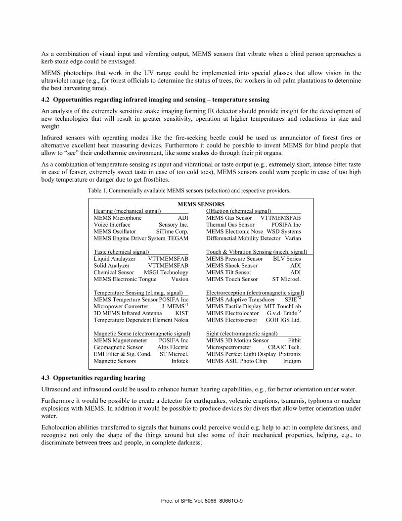
There are various ways to organise MEMS sensors. Table 1 shows commercially available MEMS sensors and respective providers. In Tables 2-4 MEMS sensors are organized according to the signal they measure, and grouped similarly to section 2, with “Health Monitoring” as additional group.
4. MEMS-BASED SENSORY OPPORTUNITES 4.1 Opportunities regarding sight
A possible application would be to create special glasses that allow vision in the UV spectrum. Such glasses could help to determine the status of plants and identify the best harvesting time.
Proc. of SPIE Vol. 8066 80661O-8
As a combination of visual input and vibrating output, MEMS sensors that vibrate when a blind person approaches a kerb stone edge could be envisaged.
MEMS photochips that work in the UV range could be implemented into special glasses that allow vision in the ultraviolet range (e.g., for forest officials to determine the status of trees, for workers in oil palm plantations to determine the best harvesting time).
4.2 Opportunities regarding infrared imaging and sensing – temperature sensing
An analysis of the extremely sensitive snake imaging forming IR detector should provide insight for the development of new technologies that will result in greater sensitivity, operation at higher temperatures and reductions in size and weight.
Infrared sensors with operating modes like the fire-seeking beetle could be used as annunciator of forest fires or alternative excellent heat measuring devices. Furthermore it could be possible to invent MEMS for blind people that allow to “see” their endothermic environment, like some snakes do through their pit organs.
As a combination of temperature sensing as input and vibrational or taste output (e.g., extremely short, intense bitter taste in case of feaver, extremely sweet taste in case of too cold toes), MEMS sensors could warn people in case of too high body temperature or danger due to get frostbites.
Table 1. Commercially available MEMS sensors (selection) and respective providers.
4.3 Opportunities regarding hearing
Ultrasound and infrasound could be used to enhance human hearing capabilities, e.g., for better orientation under water.
Furthermore it would be possible to create a detector for earthquakes, volcanic eruptions, tsunamis, typhoons or nuclear explosions with MEMS. In addition it would be possible to produce devices for divers that allow better orientation under water.
Echolocation abilities transferred to signals that humans could perceive would e.g. help to act in complete darkness, and recognise not only the shape of the things around but also some of their mechanical properties, helping, e.g., to discriminate between trees and people, in complete darkness.
MEMS SENSORS Hearing (mechanical signal) Olfaction (chemical signal) MEMS Microphone ADI MEMS Gas Sensor VTTMEMSFAB Voice Interface Sensory Inc. Thermal Gas Sensor POSIFA Inc MEMS Oscillator SiTime Corp. MEMS Electronic Nose WSD Systems MEMS Engine Driver System TEGAM Differenctial Mobility Detector Varian Taste (chemical signal) Touch & Vibration Sensing (mech. signal) Liquid Analayzer VTTMEMSFAB MEMS Pressure Sensor BLV Series Solid Analyzer VTTMEMSFAB MEMS Shock Sensor ADI Chemical Sensor MSGI Technology MEMS Tilt Sensor ADI MEMS Electronic Tongue Vusion MEMS Touch Sensor ST Microel. Temperature Sensing (el.mag. signal) Electroreception (electromagnetic signal) MEMS Temperture Sensor POSIFA Inc MEMS Adaptive Transducer SPIE72 Micropower Converter J. MEMS71 MEMS Tactile Display MIT TouchLab 3D MEMS Infrared Antenna KIST MEMS Electrolocator G.v.d. Emde73 Temperature Dependent Element Nokia MEMS Electrosensor GOH IGS Ltd. Magnetic Sense (electromagnetic signal) Sight (electromagnetic signal) MEMS Magnetometer POSIFA Inc MEMS 3D Motion Sensor Fitbit Geomagnetic Sensor Alps Electric Microspectrometer CRAIC Tech. EMI Filter & Sig. Cond. ST Microel. MEMS Perfect Light Display Pixtronix Magnetic Sensors Infotek MEMS ASIC Photo Chip Iridigm
Proc. of SPIE Vol. 8066 80661O-9

4.4 Opportunities regarding olfaction
MEMS could be installed in refrigerators to signal foul and mouldy food. Moreover, detectors could be produced, which can distinguish between clean and dirty clothes. In addition it would be possible to create detectors that tell humans when rooms should be aerated.
MEMS olfaction for disease detection: Ancient doctors used to smell the urine of their patients, which characteristically changes its smell in certain diseases. Dogs can smell prostate cancer and diabetes. Some specifically trained dogs even can smell hypoglycemia. Identification of the signals and transfer to MEMS technology seems highly promising.
MEMS olfaction for smooth gatherings: Upcoming discomfort, fear or aggression of others in gatherings of people could be detected via a system of MEMS sensors monitoring body language, chemicals, air quality, temperature, etc. – such applications could be used for individual and social communication (e.g., intercultural communication), in cooperation as well as in meetings (e.g., social, business, political, scientific), but also to avoid mass hysteria / panic in public space, stadiums, after catastrophes etc.
MEMS detectors monitoring physiological parameters in healthy and health-impaired persons could act as early warning devices. When directly attached to the body, they could even activate other MEMS that would start acting (e.g., insulin pumps in people with diabetes).
Table 2. MEMS sensors on the market and in research stage concerning touch and vibration sensing, sight and hearing.
Touch & Vibration sensing Sight Hearing − GPS based navigation system − MEMS 3-Axis Digital Output
Acceleration Sensor − MEMS Accelerometer − MEMS Digital Compass − MEMS FOG-based inertial
sensor − MEMS Integrated Dual-Axis
Gyroscope − MEMS Pressure Sensor for
AUV − MEMS Pressure Measurement
System − MEMS Pressure Sensor − MEMS Pressure Transducer − MEMS Real-time Oscillator − MEMS Rotation Sensor − MEMS Shock Sensor − MEMS Surface Force Sensor − MEMS Tilt Sensor − MEMS Touch Sensor − MEMS Vibration Analysis − MEMS Wireless Sensory
System − MEMS-based Control Device
for User Interface − MEMS-sensors in Ballistic
Munitions − Piezoresistive MEMS pressure
sensor − Polymer-based MEMS multi-
modal sensory skin
− 3D Motion MEMS Sensor − Beast X-3 MEMS Gyro System − Electrically Tunable Fabry-
Pérot Filters for near-UV, visible, near-IR
− High Intensity Air Cooled UV Light System w/configurable Curing Length (in MOEMS Package)
− High Intensity Water Cooled UV Light System w/configurable Curing Length (in MOEMS Package)
− Mask Aligner System − MEMS Interferometer − Micromirror Spatial Light
Modulator (SLM) for fs Pulse Shaping in UV
− Nanodevice Products - Voxdot Nanotaggants
− Nanophotonics & Electrooptical Products - LEDs
− Nanophotonics & Electrooptical Products - Photovoltaics
− Nanophotonics & Electrooptical Products - Polymer- OLEDs
− PerfectLight Display Technology
− Photochip in Digital Camera − UV-visible-NIR
Microspectrophotometer
− Analog MEMS Microphone
− CMOS MEMS Microphones
− Digital MEMS Microphone
− MEMS Abnormal Sounds and Vibrations Detector
− MEMS Bionic Vector Hydrophone
− MEMS Cochlear Implant
− MEMS Engine Driver System
− MEMS Hearing Aid Device
− MEMS Micro-acoustic Components
− MEMS Speaker − MEMS Voice Interface − MEMS-based Hair
Acoustic Flow Sensors − MEMS-based Pulse
Generator and Current Source
− Silicon MEMS Oscillator
Proc. of SPIE Vol. 8066 80661O-10

Table 3. MEMS sensors on the market and in research stage concerning health monitoring, smelling and taste.
Health Monitoring Smelling (Olfaction) Taste
− Drug Delivery System − MEMS Blood Presure Monitor − MEMS Glucose Monitor and
Insulin Pump
− MEMS Gas Sensor − MEMS Gas Analayzer − MEMS Thermal Conductivity
Gas Sensor − MEMS GC Gas Sensor − Differential Mobility Detector
(DMD) − MEMS electronic nose
− MEMS Electronic Tongue
− MEMS Liquid Analayzer
− MEMS Solid Analyzer − MESMEMS Chemical
Sensor
Table 4. MEMS sensors on the market and in research stage concerning the magnetic sense, electroreception and temperature sensing.
Magnetic Sense Electroreception Temperature Sensing − EMI Filtering & Signal
Conditioning − Linear MEMS Magnetometer − Magnetic Latching MEMS-
based RF Switch − MEMS Digital Compass − MEMS Geomagnetic Sensor − MEMS Magnetic Sensors − MEMS Magnetometer − MEMS Silicon Magnetic
Sensors − MEMS with Magnetorestrictive
Materials − MEMS-based Magnetic Coils − Scalar MEMS magnetometer
− MEMS Adaptive Transducer − MEMS Brain Computer
Interface − MEMS Electrolocator − MEMS Electroreceptor − MEMS Electrosensory Systems − MEMS Neural Control − MEMS Neural Control − MEMS Tactile Displays
− 3D MEMS IR Antenna − MEMS Microbolometer − MEMS Micropower
Converter − MEMS Temperature
Dependent Element − MEMS Temperture
Sensor − MEMS Thermal
Actuator − MEMS Thermoelectric
Device
4.5 Opportunities regarding vibration Sensing
The detection of weak or strong water movements could be used to get more knowledge about water currents near the coastline in order to avoid people drifting into deep water levels. In earthquake prone areas, MEMS seizmographs attached to the body, sending information about increased seizmic activity to the wearers, e.g. by a certain tactile signal, would be helpful for early measures.
4.6 Opportunities regarding the magnetic sense
MEMS with magnetic sensors can be used for mobile navigation and enable advanced location-based services. In mobile phones or cars they could be utilized as digital compasses. Furthermore they indicate the direction in which a person or vehicle is moving, even when GPS is not available. MEMS compasses signalling the direction to e.g., North or “home” to the human body, e.g., in form of tactile stimuli, could act as “built-in” compass that can be used in highly adverse conditions (bad waether, no more light available, snowstorm, deep shade, deep in the jungle, on expeditions, etc.).
Proc. of SPIE Vol. 8066 80661O-11

4.7 Opportunities regarding electroreception
Electrosensing and -location could be used in devices for target location and orientation (special cameras) in muddy waters, blood and other places with special conditions like high temperatures or pressures. Furthermore these devices could determine parameters like material properties, thickness and material defects. MEMS electrolocators that transfer the signal, e.g. to auditory stimuli, could help divers to locate objects in extreme situations.
5. SUMMARY AND OUTLOOK This paper gives an overview of senses in organisms, describes MEMS that detect similar signals, and presents a list of MEMS based on existing technology that could be used to assist, enhance and expand human sensory capabilities, opening new possibilities for MEMS-based sensory opportunities. Stroble and co-workers74 as well as Jordan and co-workers75 provide additional reading on senses in nature and biomimetic sensors.
In the next step, the respective bandwidths of the natural and technologically available sensors will be collected. Subsequently, a push pull analysis will be performed and the market needs concerning MEMS to assist, enhance and expand human sensory capabilities will be investigated in relation to the available technological potential (at reasonable cost): On the market the consumers usually ‘pull’ the goods or information - they create a demand, while the offerers ‘push’ their products toward the consumers – they create a supply (Fig. 3). In this respect two different aspects are of particular interest: 1. Where does current MEMS technology excel (available solutions)? And 2. Where do natural sensor systems excel in general (what is easily replicable by ‘off the shelf’ systems)?
Figure 3. The push-pull analysis relates market demands and MEMS potential. X1 denotes sugnals that are too weak for
the human sensory system (in terms of strength), and X2 denotes signal types that are not covered by the human sensory system. Pull: There is pull from the market, defining what the customers require regarding assistance, enhancement and expansion of human senses. Push: In this area the available solutions or the technological potential for the creation of solution are assessed.
Subsequently, selection and process definition follow: With regard to the defined pull the corresponding methods of animated nature will be predefined and corresponding systems in human technology will be outlined. This is particularly important, as in later stages the specifications of key components will be required. To apply best practice process schemes with the support of animated nature, it is of paramount importance that key processes in the determined sensory areas are defined. This will be undertaken with a special focus on existing standard technology and sensors (MEMS). The processes will deal with standard components, as the costs will be kept as low as possible. The basis prepared via such research can act as incubator to attract seed capital for further research and development.
Proc. of SPIE Vol. 8066 80661O-12

ACKNOWLEDGEMENTS
The National University of Malaysia funded part of this work with its leading-edge research project scheme ‘Arus Perdana’, and the Austrian Society for the Advancement of Plant Sciences funded part of this work via the Biomimetics Pilot Project ‘BioScreen’. Profs. F. Aumayr, H. Störi and G. Badurek from the Vienna University of Technology are acknowledged for enabling ICG three years of research in the inspiring environment in Malaysia.
REFERENCES
[1] Kant, I. [2] Bar-Cohen, Y., [Biomimetics: Biologically inspired technologies], Taylor & Francis, CRC Press, (2005). [3] Gebeshuber, I. C., Gruber, P. and Drack, M., “A gaze into the crystal ball - biomimetics in the year 2059,” Proc.
IMechE Part C: J. Mech. Eng. Sci. 223(C12), 50st Anniversary Issue, 2899-2918 (2009). [4] Lyshevski, S. E., [MEMS and NEMS: Systems, Devices, and Structures], CRC Press, (2002). [5] Starr, C., [Biology: Concepts and Applications], Thomson Brooks/Cole (2005). [6] Judd, D. B. and Wyszecki, G., [Color in Business, Science and Industry], Wiley Series in Pure and Applied
Optics (3rd edition), New York, Wiley-Interscience, 388 (1975). [7] Marshall, J., Cronin, T. W. and Kleinlogel, S., “Stomatopod eye structure and function: A review,” Arthropod
Structure & Development 36, 420-448 (2007). [8] Osorio, D., Marshall, N. J. and Cronin, T. W., “Stomatopod photoreceptor spectral tuning as an adaptation for
colour constancy in water,” Vision Research 37, 3299-3309 (1997). [9] Arikawa, K., “Spectral organization of the eye of a butterfly, Papilio,” Journal of Comparative Physiology A:
Neuroethology, Sensory, Neural, and Behavioral Physiology 189(11), 791-800 (2003). [10] Kelber, A. and Pfaff, M., “True colour vision in the orchard butterfly, Papilio aegus,” Naturwissenschaften 86,
221-224 (1999). [11] Menzel, R. and Backhaus, W. G. K., “Color vision of honeybees: phenomena and physiological mechanisms,”
Facets of Vision, Springer, Berlin & Heidelberg, 281-297 (1989). [12] Frisch, K., “Der Farbensinn und Formensinn der Biene,“ Zool. J. Physiol. 37, 41-44 (1914). [13] Menzel, R. and Blakers, M., “Colour receptors in the bee eye - Morphology and spectral sensitivity,” Journal of
Comparative Physiology A 108, 11-33 (1976). [14] Autrum, H. and Zwehl, V., “Die spektrale Empfindlichkeit einzelner Sehzellen des Bienenauges,“ Zeitschrift
für vergleichende Physiologie 48, 357-384 (1964). [15] Menzel, R. and Snyder, A. W., “Polarized light detection in the bee, Apis mellifera,” Journal of Comparative
Physiology A 88, 247-270 (1974). [16] Kaiser, W., “The spectral sensitivity of the honeybee’s optomotor walking response,” Journal of Comparative
Physiology A 90, 405-408 (1974). [17] Chittka, L. and Tautz, J., “The spectral input to honeybee visual odometry,” Journal of Experimental Biology
206, 2393-2397 (2003). [18] Emmerton, J. and Remy, M., “The pigeons sensitivity to ultraviolet and ‘visible’ light,” Experientia 39, 1161-
1163 (1983). [19] Cohen, D. M. and Goldsmith, T. H., “Four spectral classes of cone in the retina of birds,” Journal of
Comparative Physiology A 159, 473-479 (1986). [20] Burkhardt, D., “UV vision: a bird’s eye view of feathers,” Journal of Comparative Physiology A 164, 787-796
(1989). [21] McFarland, W. N. and Loew, E. R., “Ultraviolet visual pigments in marine fishes of the family Pomacentridae,”
Vision Research 34, 1393-1396 (1994). [22] Jerlov, N. G., [Marine optics], Elsevier Scientific, Amsterdam, 231 (1976). [23] Archer, S. N. and Lythgoe, J. N., “The visual pigment basis for cone polymorphism in the guppy, Poecilia
reticulate,” Vision Research 30, 225-233 (1990). [24] Barrett, R., Maderson, P. F. A. and Meszler, R. M., “The pit organs of snakes,” Biology of Reptilia 2, Academic
Press, London, 277-314 (1970).
Proc. of SPIE Vol. 8066 80661O-13

[25] Grace, M. S., Woodward, O. M., Church, D. R. and Calisch, G., “Prey targeting by the infrared-imaging snake python: effects of experimental and congenital visual deprivation,” Behavioural Brain Research 119, 23-31 (2001).
[26] Ebert, J. and Westhoff, G., “Behavioural examination of the infrared sensitivity of rattlesnakes (Crotalus atrox),” Journal of Comparative Physiology A 192, 941-947 (2006).
[27] Hartline, P. H., Kass, L. and Loop, M. S., “Merging of modalities in the optic tectum: infrared and visual integration in rattlesnakes,” Science 199, 1225-1229 (1978).
[28] Kardong, K. and Mackessey, G., “The strike behavior of a congenitally blind rattlesnake,” Journal of Herpetology 25, 208-211 (1991).
[29] Grace, M. S., Church, D. R., Kelly, C. T., Lynn, W. and Cooper, T. M., “The python pit organ: imaging and immunocytochemichal analysis of an extremely sensitive natural infrared detector,” Biosensory & Bioelectronics 14, 53-59 (1999).
[30] Amemiya, F., Nakano, M., Goris, R. C., Kadota, T., Atobe, Y., Funakoshi, K., Hibiya, K. and Kishida, R., “Microvasculature of crotaline snake pit organs: possible function as a heat exchange mechanism,” Anatomy Record 254, 107-115 (1999).
[31] Evans, W. G., “Infrared receptors in Melanophila acuminate DeGeer,” Nature 202, 211 (1964). [32] Schmitz, H. and Beckmann, H., “The photomechanic infrared receptor for the detection of forest fires in the
beetle Melanphila cuminata,” Journal of Comparative Physiology A 182, 647-657 (1998). [33] Schmitz, H., “Thermal characterization of butterfly wings - 1. Absorption in relation to different colour, surface
structure and basking type,” Journal of Thermal Biology 19, 403-412 (1994). [34] Rattay, F., Gebeshuber, I. C. and Gitter, A. H., “The mammalian auditory hair cell: a simple electric circuit
model,” Journal of the Acoustical Society of America 103(3), 1558-1565 (1998). [35] Heffner, H. E and Heffner, R. S, “Hearing ranges of laboratory animals,” Journal of the American Association
for Laboratory Animal Science 46(1), 11-13 (2007). [36] hypertextbook.com/facts/2003/TimCondon.shtml [37] Griffin, D. R., [Listening in the dark: the acoustic orientation of bats and men], Yale University Press, New
Haven, (1958). [38] Simmons, J. A., Saillant, P. A., Wotton, J. M., Haresign, T., Ferragamo, M. J. and Moss, C. F, “Composition of
biosonar images for target recognition by echolocating bats,” Neural Networks 8(7/8), 1239-1261(1995). [39] McComb, K., Reby, D. and Baker L., “Long-distance communication of acoustic cues to social identity in
African elephants,” Animal Behaviour 65, 317-329 (2003). [40] Martin, R. B., “Aspects of elephant social organization,” Rhodesia Sci.News 12, 184-187 (1978). [41] Keller, A. and Vosshall, L. B., “Human olfactory psychophysics,” Current Biology 14, 875-878 (2004). [42] Correa, J. E., “The dog’s sense of smell,” Alabama Cooperative Extension System UNP-0066 (2005). [43] Kaissling, K. E. and Thorson, J., “Receptors for neurotransmitters, hormones and pheromones in insects,”
Biomedical Press, Amsterdam, 261-282 (1980). [44] Steullet, P. and Guerin, P. M., “Identification of vertebrate volatiles stimulating olfactory receptors on tarsus I
of the tick Amblyomma variegatum Fabricius (Ixodidae),“ Journal of Comparative Physiology A 174(1), 39-47 (1994).
[45] Nuttal, G. H., Cooper, W. F., Arthur, D. R., Warburton, C., Robinson, L. E., [Ticks, a monograph of the Ixodoidea Part 2: Bibliography of the Ixodoidea], Cambridge University Press, 82 (1915).
[46] www.elasmodiver.com/shark_senses.htm [47] Schmidt, R. F. and Lang, F., [Physiologie des Menschen], Springer Medizin Verlag, Heidelberg, 307 (2007). [48] Johansson, R. S. and Vallbo, A. B., “Tactile sensibility in the human hand: Relative and absolute densities of
four types of mechanoreceptive units in glabrous skin,” Journal of Physiology 286, 283-300 (1979). [49] Oey, H. and Mellert, V., “Vibration thresholds and equal vibration levels at the human fingertip and palm,” ICA
Kyoto, Japan, 1-4 (2004). [50] Barth, F. G., “Spider mechanoreceptors,” Current Opinion in Neurobiology 14, 415-422 (2004). [51] Barth, F. G., [A spider’s world: senses and behavior], Springer Verlag, Berlin & Heidelberg, 227 (2002). [52] Barth, F. G., “Vibrationssinn und vibratorische Umwelt von Spinnen,“ Naturwissenschaften 73 (9), 519-530
(1986). [53] Manning, A. and Dawkins, M. S., [An introduction to animal behavior], Cambridge University Press
Cambridge, 7-8 (1998).
Proc. of SPIE Vol. 8066 80661O-14

[54] Brownell, P. H., “Compressional and surface waves in sand: used by desert scorpions to locate prey,” Science 197, 479-482 (1977).
[55] Hoffmann, C., “Bau und Funktion der Trichobothrien von Euscorpius carpathicus L.,“ Zeitschrift für Vergleichende Physiologie 54, 290-352 (1967).
[56] Hanke, W., Witte, M., Miersch, L., Brede, M., Oeffner, J., Michael M, Hanke, F., Leder, A. and Dehnhardt, G., “Harbor seal vibrissa morphology suppresses vortex-induced vibrations,“ Journal of Experimental Biology 213(15), 2665-2672 (2010).
[57] Mills, F. H. J. and Renouf, D., “Determination of the vibration sensitivity of harbour seal Phoca vitulina (L.) vibrissae,” Journal of Experimental Marine Biology and Ecology 100, 3-9 (1986).
[58] Maeda, K., Henbest, K. B., Cintolesi, F., Kuprov, I., Rodgers, C. T., Liddell, P. A., Gust, D., Timmel, C. R. and Hore, P. J., “Chemical compass model of avian magnetoreception,” Nature 453, 387-390 (2008).
[59] Kramer, G., “Wird die Sonnenhöhe bei der Heimfindeorientierung verwertet?“ J. Ornithol. 94, 201-219 (1953) [60] Walker, M. M., Todd, E. D. and Kirschvink, J. L., “The magnetic sense and its use in long-distance navigation
by animals,” Current Opinion in Neurobiology 12, 735-744 (2002). [61] Moller, P., [Electric fishes: history and behavior. Fish and Fisheries], Pitcher TJ. Chapman & Hall, London,
120-124 (1995). [62] Hopkins, C. D., “Neuroethology of electrolocation,” Journal of Comparative Physiology A 173(6), 689-695
(1993). [63] Helligenberg, W., [Neural nets in electric fish], Computional Neuroscience Series, MIT Press, Cambridge,
(1991). [64] Bullock, T. H., Hopkins, C. D., Popper, A. N. and Fay, R. R., [Electroreception], Springer Handbook of
Auditory Research 21, 472 (2005). [65] Belbenoit, P., “Fine analysis of predatory and defensive motor events in Torpedo marmorata (Pisces),” Journal
of Experimental Biology 121, 197-226 (1986). [66] Bennett, M. V. L., [Electric organs] Fish Physiology 5, Academic Press, New York, 347-349 (1971). [67] Bratton, B. O. and Ayers, J. L., “Observations on the electric organ discharge of two skate species
(Chondrichthyes, Rajidae) and its relationship to behavior,” Environmental Biology of fishes 20, 241-254 (1987).
[68] Szabo, T., “Anatomy of the specialized lateral line organs of electroreception,” Handbook of Sensory Physiology III/3, Springer-Verlag, New York, 147-200 (1974).
[69] New, J. G., “The evolution of vertebrate electrosensorysystems,” Brain Behav. Evol. 50, 244-252 (1997). [70] Karman, S. B., Macqueen, M. O., Matin, T. R., Diah, S. M., Mueller, J., Yunas, J., Makarczuk, T. and
Gebeshuber, I. C., “On the way to the bionic man: A novel approach to MEMS based on biological sensory systems,” submitted to Adv. Mat. Tech.
[71] Mitcheson, P. D., Green, T. C., Yeatman, E. M. and Holmes, A. S., “Architectures for vibration-driven micropower generators,” Journal of Microelectromechanical Systems 13(3), 429-440 (2004)
[72] Shahinpoor, M., “Potential applications of electroactive polymer sensors and actuators in MEMS technologies,” Proc. SPIE 4234, 203-214 (2001)
[73] v. d. Emde, G., “Biomimetic sensors: Active electrolocation of weakly electric fish as a model for active sensing in technical systems,” Journal of Bionic Engineering 4(2), 85-90 (2007).
[74] Stroble, J. K., Stone, R. B. and Watkins, S. E., “An overview of biomimetic sensor technology,” Sensor Review 29(2), 112-119 (2009).
[75] Jordan, K., Calderone, D., Rubin, A. and Wickenden, A. E., [A review of biological communication mechanisms applicable to small autonomous systems], Army Research Lab Report, ARL-TR-5340, 1-95 (2010).
Proc. of SPIE Vol. 8066 80661O-15